PEG介导法制备拟南芥原生质体及瞬时表达方法
拟南芥原生质体制备
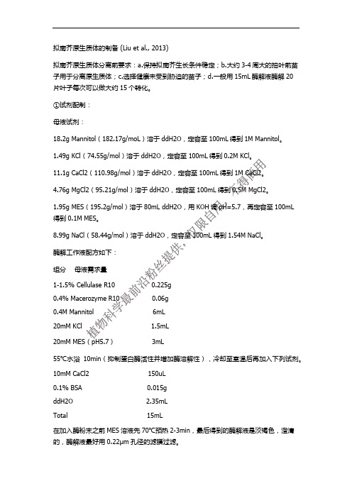
拟南芥原生质体的制备 (Liu et al., 2013)拟南芥原生质体分离前要求:a.保持拟南芥生长条件稳定;b.大约3-4周大的抽叶前苗子用于分离原生质体;c.选择健康未受到胁迫的苗子;d.一般用15mL酶解液酶解20片叶子每次可以做大约15个转化。
①试剂配制:母液试剂:18.2g Mannitol(182.17g/moL)溶于ddH2O,定容至100mL得到1M Mannitol。
1.49g KCl(74.55g/mol)溶于ddH2O,定容至100mL得到0.2M KCl。
11.1g CaCl2(110.98g/mol)溶于ddH2O,定容至100mL得到1M CaCl2。
4.76g MgCl2(95.21g/mol)溶于ddH2O,定容至100mL得到0.5M MgCl2。
1.95g MES(195.2g/mol)溶于80mL ddH2O,用KOH调pH=5.7,再定容至100mL得到0.1M MES。
8.99g NaCl(58.44g/mol)溶于ddH2O,定容至100mL得到1.54M NaCl。
酶解工作液配方如下:组分母液需求量1-1.5% Cellulase R10 0.225g0.4% Macerozyme R10 0.06g0.4M Mannitol 6mL20mM KCl 1.5mL20mM MES(pH5.7) 3mL55℃水浴10min(抑制蛋白酶活性并增加酶溶解性),冷却至室温后再加入下列试剂。
10mM CaCl2 150uL0.1% BSA 0.015gddH2O 2.35mLTotal 15mL在加入酶粉末之前MES溶液先70℃预热2-3min,最后得到的酶解液是淡褐色,澄清的,酶解液最好用0.22μm孔径的滤膜过滤。
W5 Solution配方:组分母液需求量154mM NaCl 10mL125mM CaCl2 12.5mL5mM KCl 2.5mL2mM MES(pH5.7)2mLddH2O 73mLTotal 100mLPEG40%(m/V):组分母液需求量PEG4000 8g0.2M Mannitol 4mL0.1M CaCl2 2mLddH2O 6mLTotal to 20mLPEG溶液在转染前1h配置好,确保PEG能够充分溶解。
原生质体制备

拟南芥原生质体制备转化方法选取开花前生长良好情况的拟南芥(Columbia型)叶片,用刀片切成0.5-1 mm宽的叶条;浸于配置好的酶解液中(每5-10 mL酶解液约10-20片叶子);暗箱抽真空,30 min;转移至室温,继续黑暗酶解3-5 h;当酶解液变绿时轻轻摇晃培养皿促使原生质体释放;加入等量的W5溶液稀释酶解液;用60-100目筛子(W5溶液润湿)过滤酶解液;将滤液转移至30 mL eppendorf管,500-700 g离心1-2 min,弃上清;加入10 mL预冷的W5溶液轻柔重悬原生质体;放于冰上静置30 min;保持室温在23℃以下;500 g离心8-10 min,弃上清;加入1 mL MMG溶液重悬原生质体(使其终浓度约在2×10⁵个/mL);即得到原生质体溶液。
用移液器吸取100 L的以上原生质体溶液于1.5 mL eppendorf管,加入10L DNA(10-20 g的质粒DNA),轻柔混合后,加入110 L PEG溶液,轻柔拍打eppendorf管使其混合完全;室温下诱导转化5-15 min;加入400-440 L W5溶液稀释转化混合液,轻柔颠倒,使之混合完好以终止转化反应;500 g离心2 min,弃上清;加入1 mL W5溶液悬浮清洗,500 g 离心2 min,弃上清;再用1 mL WI溶液轻柔重悬原生质体;室温(20-25℃)下,诱导载体表达18小时以上。
相关溶液配制PEG4000溶液(现用现配)组分用量PEG4000 1 g1 M CaCl20.25 mL0.8甘露醇0.625 mL水0.75 mL纤维素酶解液试剂15 mL酶液体系1-1.5 % Cellulase R10 (YaKult Honsha) 0.225 g 纤维素酶0.2-0.4 % Mecerozyme R10 (YaKult Honsha) 0.045 g 果胶酶0.4 M 甘露醇 1.09 g甘露醇干粉20 mM KCl 1 mL 0.3 M KCl20 mM MES, pH5.7 1 mL 0.3 M MES,pH5.7加水10 mL55℃水浴加热10 min,冷却至室温后加入以下试剂10 mM CaCl2 1 mL 0.15 M CaCl20.1% BSA 1 mL 1.5 % BSA5 mM β-巯基乙醇1mL 75 mM β-巯基乙醇用0.45 m滤膜过滤后即可使用。
玉米和拟南芥的原生质体制备及瞬时表达体系的研究

玉米和拟南芥的原生质体制备及瞬时表达体系的研究作者:赵苏州卢运明张占路赵杨敏王磊来源:《安徽农业科学》2014年第12期摘要[目的]优化玉米和拟南芥原生质体的制备条件及瞬时表达体系。
[方法]以不同时期玉米叶片和拟南芥为材料分离原生质体,通过对不同酶浓度,解离时间和渗透压的研究,优化最佳分离原生质体体系;原生质体再经PEG介导的转化,通过GFP基因表达百分比鉴定原生质体活性和转化效率。
[结果]玉米和拟南芥原生质分离的最佳条件为:纤维素酶1.5%,离析酶0.5%;拟南芥酶解4 h,甘露醇浓度0.4 mol/L;玉米酶解6 h,甘露醇浓度0.45~0.50 mol/L。
最佳生长时期为:拟南芥6~8片叶;玉米二片叶。
用去内毒素质粒经PEG(玉米30% PEG,拟南芥40% PEG)诱导转化置于黑暗下培养,原生质体活性和转化效率较高,原生质体中可以观察到明显的GFP荧光。
[结论]玉米和拟南芥原生质体的制备受酶浓度,解离时间和渗透压的影响,在原生质体转化过程中,质粒DNA纯度,PEG浓度等是影响转化效率的关键因素,与拟南芥原生质体相比玉米原生质体要稳定性差一些。
关键词玉米;原生质体;拟南芥中图分类号S513;Q819文献标识码A文章编号0517-6611(2014)12-03479-04基金项目国家转基因生物新品种培育重大专项(2011ZX08003-002)。
作者简介赵苏州(1988-),男,山东临沂人,硕士研究生,研究方向:植物转录因子。
*通讯作者,研究员,博士,从事植物基因沉默研究。
植物原生质体是指植物细胞通过机械或酶解等特殊方法脱去细胞壁后,留下的裸露、具有生活力、被细胞膜所包围的原生质团。
原生质体由于没有细胞壁,可相对容易摄取外源DNA、质粒、病毒颗粒等外源遗传物质,是进行遗传转化的理想受体;同时,原生质体也是获得细胞无性系和选育突变体的优良起始材料;还可通过诱导形成杂种细胞,为创造新杂种开辟了途径。
PEG介导拟南芥叶片原生质体瞬时表达方法

PEG介导拟南芥叶片原生质体瞬时表达方法1.B5培养基上萌发拟南芥种子,待根长至1-3厘米时即可移栽到土里,温室培养,光照12h/12h,150μE。
2.准备好一个90mm培养皿,称1.82克D-甘露醇于20ml双蒸水中。
培养皿的盖子用来切叶片。
3.取4周后未抽台前的叶片,约90片。
切成1mm宽的长条,置于甘露醇溶液中。
可以一边切一边从植株上取。
4. 配酶解液,100ml三角瓶,15ml酶解体系。
5. 将步骤3中细条捞出,置于酶解液中。
黑暗,23℃,40-50rpm酶解3小时。
6. 酶解液过100-200目的筛子,将过滤后的绿色混合物置于15ml离心管(直径约1cm)中,均分为两管。
4℃,60 g,15min,brake 设为4-5。
7. 弃上清,沉淀用冰冷的W5溶液轻柔洗涤,每管4ml。
4℃,100 g,1min,brake 设为4-5。
8. 弃上清,沉淀用冰冷的W5溶液轻柔洗涤,每管4ml。
冰上放置30min。
9. 23℃,100 g,1min,brake 设为4-5。
弃上清,每管沉淀用0.5ml MaMg重悬。
(本步骤及以下操作均在23℃。
)10. 取约10-20ug 质粒于1.5ml EP管中,加100ul 步骤9中的原生质体。
用200ul 枪头(剪去前端)轻柔混匀。
11. 加入110ul PEG/Ca 溶液,轻柔混匀。
放置20-30min。
12. 加入0.44ml W5 溶液,来回颠倒混匀。
23℃,100 g,1min,brake 设为4-5。
13. 弃上清,加100ul W5,混匀。
加900ul W5,混匀。
14. 上述混合液体置于六孔板内,23℃,弱光,孵育6-18小时。
15. 荧光观察,观察之前100g,常温,离心2分钟,终体积控制在50ul左右。
Solution RecipesEnzyme solution1ml 15%cellulase R10 (RS is too strong)1ml 4.5%macerozyme R10 (Yakult Honsha, Tokyo, Japan)1.09 g mannitol1ml 0.3M KCl1ml 0.3M MES, pH 5.7Heat the enzyme solution at 55oC for 10 min (to inactivate proteases and enhance enzymesolubility) and cool it to room temperature before adding1ml 0.15M CaCl21ml 0.75mM β-mercaptoethanol1ml 1.5% BSAPEG solution (40%, v/v)1 g PEG4000 (Fluka, #81240) **Very Important!!0.75 ml H2O0.625 ml 0.8 M mannitol0.25 ml 1M CaCl2W 51000 ml154 mM NaCl 9.0 g125 mM CaCl2 18.4 g5 mM KCl 0.37 g5 mM glucose 0.9 g0.03% MES 0.3 gpH to 5.8 with KOHautoclave 20 minutes in 125 bottlesMaMg solution100 ml15 mM MgCl2 1.5 ml 1M MgCl20.1% MES 0.1 g0.4 M mannitol 7.3 gpH to 5.6 with KOHautoclave 20 minutes in 125 ml bottlesReferences1. Sheen, J. 2002, A transient expression assay using Arabidopsis mesophyll protoplasts./sheenweb/2. Doelling & Pikaard. 1993, Transient expression in Arabidopsis thaliana protoplasts derived from rapidlyestablished cell suspension cultures. Plant Cell Reports 12: 241-244Enzyme solutionPrepare 20 mM MES (pH 5.7) containing 1.5% (wt/vol) cellulase R10, 0.4% (wt/vol) macerozyme R10,0.4Mmannitol and 20mMKCl.Warm the solution at 55 ℃for 10 min to inactivate DNAse and proteases and enhance enzyme solubility. Cool it to room temperature (25 ℃) and add 10mM CaCl2, 1–5 mM β-mercaptoethanol (ptional) and 0.1% BSA. ▲CRITICAL :Addition of 1–5 mM β-mercaptoethanol is optional, and its use should bedetermined according to the experimental purpose. ▲CRITICAL Before the enzyme powder is added, the MES solution is preheated at 70 ℃for 3–5 min. The final enzyme solution should be clear light brown. Filter the final enzyme solution through a 0.45µmsyringe filter device into a Petri dish (100x 25mm2 for 10 ml enzyme solution).▲CRITICAL The enzyme solution should be prepared fresh.WI solutionPrepare 4 mM MES (pH 5.7) containing 0.5 M mannitol and 20 mM KCl. The prepared WI solution can be stored at room temperature (22–25 ℃).W5 solutionPrepare 2mM MES (pH 5.7) containing 154mM NaCl, 125mM CaCl2 and 5 mM KCl. The prepared W5 solution can be stored at room temperature.MMG solutionPrepare 4 mM MES (pH 5.7) containing 0.4 M mannitol and 15mMMgCl2. The preparedMMG solution can be stored at room temperature. PEG–calcium transfection solution Prepare 20–40% (wt/vol) PEG4000 in ddH2O containing 0.2 M mannitol and 100 mM CaCl2. ▲CRITICAL Prepare PEG solution at least 1 h before transfectionto completely dissolve PEG. The PEG solution can be stored at room temperature and used within 5 d. However, freshly prepared PEG solution gives relatively better protoplast transfection efficiency. Do not autoclave PEG solution.Protoplast lysis bufferPrepare 2.5 mM Tris–phosphate (pH 7.8) containing 1 mM DTT, 2 mM DACTAA, 10% (vol/vol) glycerol and 1% (vol/vol) Triton X-100. The lysis buffer should be prepared fresh.MUG substrate mix for GUS assayPrepare 10 mM Tris–HCl (pH 8) containing 1 mM MUG and 2 mM MgCl2. The prepared GUS assay substrate can be stored at –20 ℃PRO CEDUREPlant growth _ Timing 3–4 weeks生长3-4周1| Grow Arabidopsis plants on either Metro-Mix 360 or Jiffy7 soil in a greenhouse or anenvironment-controlled chamber with a relatively short photoperiod (10–13 h light at 23 ℃/11–14 h dark at 20℃) under low light (50–75 µE m–2 s–1) and 40–65% relative humidity.较短的光照时间(10-13小时光照23℃/11-14小时黑暗20℃) 40-65%的相对湿度.▲CRITICAL STEPCol-0 and Ler have been extensively tested in our lab. In general, Arabidopsis plants are very sensitiveto all kinds of environmental changes (e.g., drought, flooding, extreme temperature and constant mechanical perturbation). Try to maintain aconstant environment as much as possible.注意:经过我们实验室的反复验证,总的来说拟南芥对各种环境变化非常敏感(如干旱,水淹,极端温度和不停地机械性干扰)。
拟南芥原生质体的提取和转化

拟南芥原生质体的提取和转化(1)取4周后抽薹前的叶片,切成1mm宽的长条,置于甘露醇溶液(称1.82g D-甘露醇于20ml双蒸水中);共需叶片约90片;(2)将步骤1中细条捞出,置于酶解液中;避光,23℃,40-50rpm摇床上酶解3小时;(3)酶解液过100-200目的筛子,收集滤液,置于15ml离心管中,均分为两管;于4℃,60g,离心15min;(4)原生质体用冰冷W5溶液轻柔洗涤,每管4ml;4℃,100g,离心1min;(5)弃上清,沉淀用冰冷的W5溶液轻柔悬浮,每管4ml;冰上放置30min;(6)23℃,100 g离心1min;弃上清,每管沉淀用0.5ml MaMg重悬;以下操作均在23℃下进行:(7)取约10-20ug 质粒于1.5ml EP管中,加100ul 步骤6中的原生质体;用200ul 枪头(剪去前端)轻柔混匀;(8)加入110ul PEG/Ca 溶液,轻柔混匀;放置20-30min;(9)加入0.44ml W5 溶液,来回颠倒混匀;23℃,100 g,1min,brake 设为4-5;(10)弃上清,加100ul W5,混匀;加900ul W5,混匀;(11)上述混合液体置于六孔板内,23℃,避光,孵育6-18小时。
(1)酶解液:cellulose R10 15%Macerozyme R10 0.3%Mannitol 1.09gKCl 0.3MMES 0.3M调节pH值到5.7,55℃加热10min,冷却到室温再加入下列溶液CaCl20.15Mβ-巯基乙醇0.75mM(2)PEG溶液(40%,v/v)PEG4000 1g0.8M Mannitol 0.625ml1M CaCl2 0.25m(3)W5溶液154mM NaCl 9.0g125mM CaCl218.4g5mM KCl 0.37g5mM glucose 0.9g0.03%MES 0.3g用KOH调pH至5.8,定容至1000ml,高压灭菌(4)MaMg溶液1M MgCl20.5ml0.1%MES 0.1g0.4M Mannitol7.3g用KOH调pH至5.8,定容至1000ml,高压灭菌。
棉花叶肉原生质体分离及目标基因瞬时表达体系的建立
作物学报 ACTA AGRONOMICA SINICA 2014, 40(2): 231-239http:///ISSN 0496-3490; CODEN TSHPA9E-mail: xbzw@本研究由国家自然科学基金项目(31171590)资助。
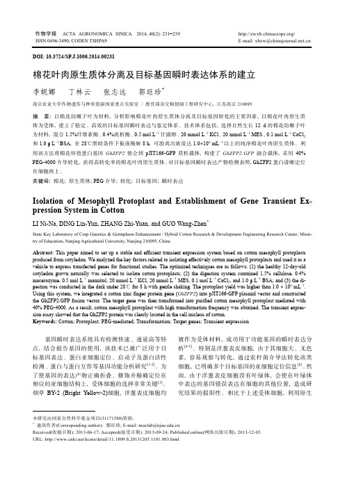
*通讯作者(Corresponding author): 郭旺珍, E-mail: moelab@Received(收稿日期): 2013-06-17; Accepted(接受日期): 2013-09-24; Published online(网络出版日期): 2013-12-05. URL: http:///kcms/detail/11.1809.S.20131205.1101.003.htmlDOI: 10.3724/SP.J.1006.2014.00231棉花叶肉原生质体分离及目标基因瞬时表达体系的建立李妮娜 丁林云 张志远 郭旺珍*南京农业大学作物遗传与种质创新国家重点实验室 / 教育部杂交棉创制工程研究中心, 江苏南京210095摘 要: 以棉花幼嫩子叶为材料, 分析影响棉花叶肉原生质体分离及目标基因转化的主要因素, 以棉花叶肉原生质体为受体, 建立了稳定、高效的目标基因瞬时表达与鉴定体系。
技术体系包括, 选择自然生长12 d 的棉花幼嫩子叶为材料, 混合1.5%纤维素酶、0.4%离析酶、0.5 mol L -1甘露醇、20 mmol L -1 KCl 、20 mmol L -1 MES 、0.1 mol L -1 CaCl 2和1.0 g L -1 BSA, 在28℃黑暗条件下振荡酶解8 h, 可游离出浓度达1.0×10⁶ mL -1以上的纯净棉花叶肉原生质体。
利用该方法将棉花锌指蛋白基因GhZFP2整合到pJIT166-GFP 质粒载体, 构建了GhZFP2:GFP 融合载体, 采用40% PEG-4000介导转化, 获得高转化率的棉花叶肉原生质体。
大豆原生质体瞬时表达体系的建立及大豆隐花色素的亚细胞定位

中文摘要大豆原生质体瞬时表达体系的建立及大豆隐花色素的亚细胞定位隐花色素是植物体内的一类重要的蓝光受体,能够介导光信号调控植物的去黄化、开花、避荫反应等多种生长发育过程。
隐花色素在模式植物拟南芥中得到了较好的研究,而在大豆等主要作物中的研究较少。
原生质体瞬时表达体系能够快速、高效地实现目标蛋白的亚细胞定位分析。
本文希望利用大豆原生质瞬时表达体系对大豆隐花色素在黑暗和蓝光下的亚细胞定位进行分析,为大豆隐花色素的功能预测提供信息。
由于尚无大豆原生质体转化方法的报道,本文尝试利用酶解法裂解大豆叶片细胞壁获得了原生质体,并用PEG介导的方法成功将外源载体转入大豆叶片原生质体并表达。
进一步对酶解液组成、PEG分子量、叶片时期、质粒浓度和转化时间等影响因素进行分析,确定了大豆叶片原生质体提取和转化的最佳条件,建立了高效的大豆叶片原生质体瞬时转化体系,转化效率达50 %以上。
通过该体系将大豆7个隐花色素(GmCRYs)转入大豆叶片原生质体并观察了其在黑暗和蓝光下的亚细胞定位情况。
具体结果如下:1)最佳叶片时期:出土10天的真叶或刚展开的第一片三出复叶。
2)最佳裂解酶成分:1.0 %纤维素酶,0.2~0.4 %果胶酶。
3)最佳PEG分子量:4000。
4)最佳质粒浓度:大于或等于1 μg/μL。
5)最佳转化时间:15分钟。
6)大豆隐花色素的亚细胞定位:GmCRY1a、GmCRY1b、GmCRY1c和GmCRY1d在黑暗下分布于细胞核和细胞质中,见蓝光后迅速在细胞质中形成类似光小体的结构。
GmCRY2a、GmCRY2b和GmCRY2c持续定位在细胞核中,见蓝光后没有观察到明显的光小体结构形成。
关键词:大豆,隐花色素,原生质体,亚细胞定位AbstractEstablishment of the Protoplasts Transient Gene Expression System and the Subcellular Localization of Cryptochromes in SoybeanCryptochromes are a kind of blue light receptors which mediate the regulation of growth and development in plants by mediating various light responses including de-etiolation of seedlings, flowering time, shade avoidance response, and so on. Many secret of cryptochromes have been revealed in Arabidopsis, however, it is little known about the cryptochromes in soybean. The transient gene expression system using protoplasts has provided a rapid and highly efficient way for the subcellular localization.We want to investigate the subcellular localization of soybean cryptochromes in darkness and under blue light using soybean protoplasts and provide information on the function of cryptochromes in soybean. There is little report about soybean protoplasts transformation, however, we have successfully isolated the protoplasts by enzymatic digestion of mesophyll cells from soybean and transformed the expression vector to the protoplasts for subsequent transient expression, which is mediated by PEG. Moreover, we optimized the main influencing factors including the suitable enzyme digestion system, the molecular weight of PEG, the different growth stages of leaves, the plasmids concentrations and the incubation time, and developed an efficient transient gene expression system using soybean mesophyll protoplasts whose transformation efficiency is over 50 %. We studied the subcellular localization of cryptochromes in soybean in darkness and under blue light using this system. The main results are showed as follows:1)The best growth stages of leaves: the 10-day-old unifoliolates afteremergence and the first trifoliolates which is just unfolding.2)The best enzyme digestion system: Celluase 1.0 %, Pectolase Y-230.2~0.4 %.3)The best molecular weight of PEG: 4000.4)The best plasmids concentrations: greater than or equal to 1 μg/μL.5)The best incubation time: 15 min.6)The subcellular localization of cryptochromes in soybean: The GmCRY1a、GmCRY1b、GmCRY1c and GmCRY1d are located in the nucleus and thecytoplasm of cells in darkness,and formed photobody-like structure incytoplasm while transferred to blue light. The GmCRY2a、GmCRY2b andGmCRY2c are always present in the nucleus, and it is difficult to observe thephotobody formation of the GmCRY2a、GmCRY2b and GmCRY2c.Keywords:Soybean, Cryptochromes, Protoplast, Subcellular localization中英文对照表英文缩写英文全称中文CRY Cryptochrome 隐花色素PHY Phytochrome 光敏色素Phot Phototropin 向光素PEG polyethylene glycol 聚乙二醇FAD Flavin Adenine Dinucleotide 黄素腺嘌呤二核苷酸SPA Suppressor of Phytochrome A 光敏色素A抑制子COP Constitutive Photomorphogenic 持续性光形态建成CIB cryptochrome-interactingbasic-helix-loop-helix 隐花色素互作bHLH转录因子BIC Blue-light Inhibitor of Cryptochromes 隐花色素的蓝光抑制子YFP Yellow Fluorescent Protein 黄色荧光蛋白DAE Days After Emergence 出土后天数目录第1章 绪论 (1)1.1 隐花色素 (1)1.1.1 隐花色素的发现 (1)1.1.2 隐花色素的结构 (2)1.1.3 隐花色素的功能 (3)1.1.4 植物隐花色素的光激活及光信号传递机制 (8)1.2 植物原生质体的基因瞬时表达体系 (12)1.2.1 原生质体的分离 (12)1.2.2 原生质体的转化 (13)1.2.3 原生质体的培养 (14)1.3 研究目的与意义 (15)第2章 材料与方法 (16)2.1 试验材料 (16)2.2 主要试剂的配制 (17)2.3 大豆原生质体瞬时表达体系的建立 (19)2.3.1 pA7-YFP质粒的扩繁与提取 (19)2.3.2 大豆叶片原生质体的提取 (22)2.3.3 大豆叶片原生质体的转化 (23)2.4 大豆隐花色素的亚细胞定位观察 (26)2.4.1 大豆隐花色素进化树及蛋白序列分析 (26)2.4.2 pA7-GmCRYs-YFP载体的构建 (26)2.4.3 pA7-GmCRYs-YFP载体转化大豆叶片原生质体 (29)2.4.4 大豆隐花色素亚细胞定位的观察 (29)第3章 结果与分析 (30)3.1 大豆原生质体的提取与转化 (30)3.1.1 大豆叶片裂解酶浓度的选择 (30)3.1.2 PEG分子量对大豆叶片原生质转化的影响 (31)3.1.3 叶片生长时期对大豆叶片原生质体化的影响 (31)3.1.4 质粒浓度对大豆叶片原生质转化的影响 (32)3.1.5 PEG处理时间对大豆叶片原生质转化的影响 (33)3.2 大豆隐花色素色素的序列分析及亚细胞定位 (33)3.2.1 隐花色素的蛋白序列比对和进化树分析 (33)3.2.2 大豆隐花色素(GmCRYs)的亚细胞定位 (35)第4章 讨论 (39)4.1 大豆叶片原生质体的转化 (39)4.2 大豆隐花色素的亚细胞定位 (40)第5章 结论 (43)参考文献 (44)作者简介 (53)致 谢 (54)第1章绪论1.1 隐花色素植物不能像动物一样移动,面对时刻变化的周边环境,它们只能通过调控自身生长来适应环境。
peg介导的原生质体转化原理

peg介导的原生质体转化原理
PEG介导的原生质体转化,是一种重要的生物技术手段,被广泛应用于基因工程、农业、医药等领域。
PEG介导转化的原理是利用高浓度的聚乙二醇(PEG)和电脉冲处理,使目标细胞可被外源性DNA转化为原生质体,进而实现基因转移。
PEG介导转化的过程分为两个主要步骤。
第一步是将目标细胞与外源性DNA混合,并加入高浓度的PEG。
PEG具有极强的渗透作用,能够破坏细胞膜结构,使DNA能够进入细胞内。
同时,PEG也能够保护DNA不被细胞内核酸酶降解,增加DNA转化的成功率。
第二步是利用电脉冲处理,以破坏细胞膜并形成孔道,使PEG和DNA能够进入细胞质。
这些孔道在电脉冲结束后自行修复,从而保证细胞的完整性。
此时,外源性DNA已经被转化为原生质体,可以整合进目标细胞的基因组中,产生新的表型。
PEG介导转化的优点是操作简单、成本低廉、转化效率高、适用范围广等,因此被广泛应用于生物学研究、基因工程、生物制药等领域。
但是,PEG介导转化也存在一些缺点,如转化后的细胞可能会受到伤害、转化效率受到转化细胞类型、外源性DNA大小和浓度等因素的影响,需要针对具体情况进行优化。
一种基于农杆菌介导的拟南芥瞬时转化技术优化

一种基于农杆菌介导的拟南芥瞬时转化技术优化郭勇;王玉成;王智博【摘要】利用根癌农杆菌(Agrobacterium tumefaciens)介导的拟南芥瞬时转化体系影响因素来确定最佳转化条件.以生长15日龄的拟南芥幼苗为试验材料,以转化pCAMBIA1301空载体的根癌农杆菌EHA105为目的菌株进行瞬时转化.研究了吐温20、菌液OD值、乙酰丁香酮(AS)及转化时间等对拟南芥瞬时转化效率的影响.结果表明:以体积分数为0.05%的吐温、菌液OD600值为1.0和120 μmol/L 的AS侵染拟南芥2.5 h后,再共培养72 h,能够得到高瞬时转化效果.【期刊名称】《东北林业大学学报》【年(卷),期】2016(044)006【总页数】5页(P41-44,83)【关键词】拟南芥;GUS基因;瞬时表达;实时定量;农杆菌介导转化【作者】郭勇;王玉成;王智博【作者单位】林木遗传育种国家重点实验室(东北林业大学),哈尔滨,150040;林木遗传育种国家重点实验室(东北林业大学),哈尔滨,150040;林木遗传育种国家重点实验室(东北林业大学),哈尔滨,150040【正文语种】中文【中图分类】Q786拟南芥是重要的模式植物,很多植物基因的功能都是通过基因转入拟南芥中进行研究,而转基因又分稳定表达与瞬时表达两种方式[1],稳定表达需要的培养、鉴定时间较长,而与之相比,瞬时表达具有简单、快捷、周期短、准确等优点[2],并且表达效率较稳定,转化率高。
当需要在短时间内进行基因功能的分析或者蛋白间的互作以及蛋白与基因间互作等的研究时,瞬时表达的方法可作为一种高效的手段[3]。
基于瞬时转化技术使基因在宿主体内瞬时表达,是一种快速的研究基因表达、蛋白质亚细胞定位及基因间互作的一种重要手段,与传统的转基因相比,瞬时表达不需要整合到染色体上,而且瞬时表达还不受基因的位置效应和基因沉默的影响,也不会产生可遗传的子代,生物安全性高[4]。
PEG介导玉米叶肉细胞原生质体瞬时基因转化体系的应用研究

PEG介导玉米叶肉细胞原生质体瞬时基因转化体系的应用研究目录摘要 (I)Abstract ..................................................................................................... I II 缩略词表.................................................................................................... V 1 文献综述. (1)1.1原生质体瞬时表达系统的研究简史 (1)1.1.1 原生质体在植物细胞生理生化特性中的研究 (1)1.1.2 原生质体瞬时转化方法的研究 (1)1.1.3 原生质体筛选基因表达调控中的启动子 (1)1.1.4 原生质体在蛋白翻译水平的研究 (2)1.2 叶肉细胞原生质体瞬时表达系统的优劣性 (2)1.3 原生质体在玉米中的应用 (3)1.3.1 玉米叶肉细胞原生质体的应用 (3)1.3.2玉米愈伤组织再生原生质体在玉米转基因中的应用 (4)1.4 sRNAs介导基因沉默 (5)1.4.1 sRNAs简介 (5)1.4.2 miRNA 与siRNA (6)1.4.3 amiRNA技术 (7)2引言 (8)3 材料与方法 (10)3.1 实验材料 (10)3.1.1 植物材料与培养条件 (10)3.1.2 培养植物所需要注意到的事项 (11)3.2主要生化试剂和仪器 (11)3.3 常用溶液配方 (11)3.4 实验步骤 (11)3.4.1原生质体分离(所用溶液都见附录B) (11)3.4.2 原生质体的收集 (11)3.4.3显微镜观察和计数 (12)3.4.4 PEG转染 (12)3.4.5 原生质体的培养 (13)3.5 ZmMYC2-2的克隆 (13)3.5.1玉米B73的DNA提取(CTAB法) (13)3.5.2 ZmMYC2-2的PCR扩增及质粒构建 (14)3.6 ZmMYC2-2的amiRNA克隆 (16)3.6.1用WMD 网站设计ZmMYC2的amiRNA (16)3.6.2在候选amiRNA的列表中选择amiRNA (16)3.6.3 Overlapping-PCR方法克隆amiRNA (17)3.7 Q-RT-PCR鉴定表达量 (19)3.8 高效液相色谱检测激素和次生代谢物的含量 (22)4 结论与讨论 (23)4.1 亚细胞定位分析 (23)4.2 过表达ZmMYC2-2后相关基因在转录水平的表达分析 (23)4.3过表达ZmMYC2-2后丁布类抗虫次生代谢物的含量测定 (27)4.4 amiRNA对ZmMYC2-2的沉默效率分析 (28)4.5全文总结 (33)参考文献 (34)附录A 主要生化试剂和仪器 (44)附录B 原生质体分离液配方 (47)致谢 (50)个人简介 (51)1文献综述1.1原生质体瞬时表达系统的研究简史1.1.1 原生质体在植物细胞生理生化特性中的研究Cocking等人早在50年以前就首次发表了一篇关于描述植物原生质体分离方法的文章。
拟南芥原生质转化
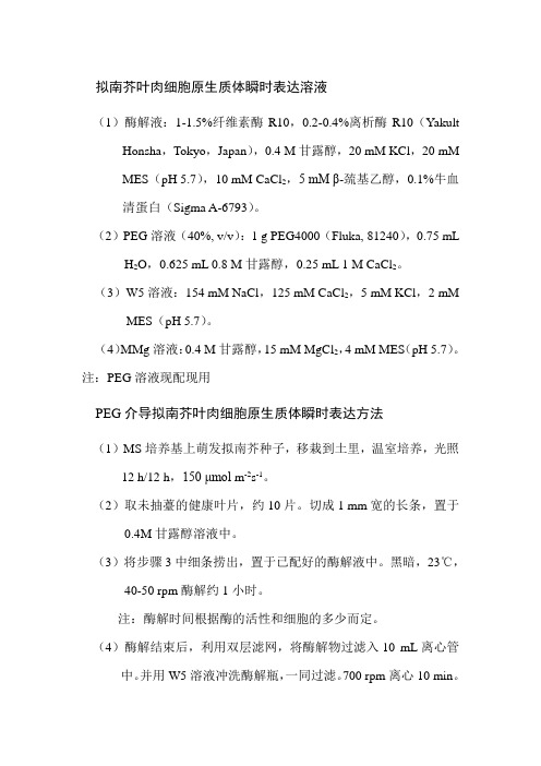
拟南芥叶肉细胞原生质体瞬时表达溶液(1)酶解液:1-1.5%纤维素酶R10,0.2-0.4%离析酶R10(Yakult Honsha,Tokyo,Japan),0.4 M甘露醇,20 mM KCl,20 mMMES(pH 5.7),10 mM CaCl2,5 mM β-巯基乙醇,0.1%牛血清蛋白(Sigma A-6793)。
(2)PEG溶液(40%, v/v):1 g PEG4000(Fluka, 81240),0.75 mL H2O,0.625 mL 0.8 M甘露醇,0.25 mL 1 M CaCl2。
(3)W5溶液:154 mM NaCl,125 mM CaCl2,5 mM KCl,2 mM MES(pH 5.7)。
(4)MMg溶液:0.4 M甘露醇,15 mM MgCl2,4 mM MES(pH 5.7)。
注:PEG溶液现配现用PEG介导拟南芥叶肉细胞原生质体瞬时表达方法(1)MS培养基上萌发拟南芥种子,移栽到土里,温室培养,光照12 h/12 h,150 μmol m-2s-1。
(2)取未抽薹的健康叶片,约10片。
切成1 mm宽的长条,置于0.4M甘露醇溶液中。
(3)将步骤3中细条捞出,置于已配好的酶解液中。
黑暗,23℃,40-50 rpm酶解约1小时。
注:酶解时间根据酶的活性和细胞的多少而定。
(4)酶解结束后,利用双层滤网,将酶解物过滤入10 mL离心管中。
并用W5溶液冲洗酶解瓶,一同过滤。
700 rpm离心10 min。
(5)弃上清,沉淀用冰冷的W5溶液轻柔洗涤,每管4 mL。
100 g,1 min。
(6)弃上清,沉淀用冰冷的W5溶液轻柔洗涤,每管4 mL。
冰上放置30 min,使原生质体恢复。
(7)23℃,100 g,1 min。
弃上清,每管沉淀用0.5 mL MMg重悬。
本步骤及以下操作均在23℃(8)取约10-20 μg质粒于1.5 mL EP管中,加100 μL步骤9中的原生质体。
拟南芥叶肉原生质体瞬时表达分析方法

Transient Expression in Arabidopsis Mesophyll Protoplasts We share this protocol freely with laboratories conducting basic research. A simple registration form is required to help us keep a record of the users of this protocol and for future communication. For commercial purposes, please contact Corporate Sponsored Research & Licensing Office at MGH (617-428-0200)For citing this protocol:Yoo, S.D., Cho, Y.H., and Sheen, J. 2007. Arabidopsis mesophyll protoplasts: A versatile cell system for transient gene expression analysis. Nature Protocols, 2: 1565-1575.Sheen, J. 2002, A transient expression assay using Arabidopsis mesophyll protoplasts./sheenweb/The protocol has been streamlined and can be applied to different types of plant materials. The growth condition of the plants seems most critical for experimental reproducibility. Each lab may need to work out the best plant growth conditions based on the available facilities and local supplies, including water, soil, air and light bulbs. Lower light (50-75µmole m-2 s-1) and shorter photoperiod (<13 hr) are more desirable to prolong vegetative growth. Clean air, water and soil are essential!! No need for excess nutrient or seedling transplant (sow your cold stratified seeds directly on wet soil). The quality of DNA and PEG is critical. The protocol is simple and the approach is powerful. However, it only awards success to the scientists who are willing to take the time needed to master the assay with patience and faith. Potential wounding and stress problems could be minimized during the experimental process to avoid high background. The protoplasts generated using this protocol have been used to study hormone, sugar, stress and defense responses using reporter genes that show similar responses in intact plants. Real-time RT-PCR is routinely used to confirm that the endogenous gene response is similar to that of the corresponding reporter gene. Good luck with your experiments!Jen Sheen, Dept of Molecular Biology, Simches Research Center 7624E, 185 Cambridge Street, MGH, Boston, MA 02114Updated on 9/26/97, 2/28/98, 9/11/00, 12/16/00, 4/6/01, 9/1/01, 9/29/01, 10/3/01, 4/21/02,5/27/02, 10/10/03, 12/23/03, 3/22/04, 4/14/09, 9/9/09, 11/15/09, 6/17/10, 5/22/11, 9/16/2012 References: Lee, H. et al. 2011. Nature 473: 376-379.Boudsocq, M., Willmann, M. et al. 2010. Nature 464: 418-422.Fujii, H., Chinnusamy, V., et al. 2009. Nature 462: 660-664.Müller, B. & Sheen, J. 2008, Nature 543: 1094-1097Shan, L., He, P. et al. 2008, Cell Host & Microbe 4: 17-27Yoo, S.D. et al. 2008, Nature 451: 789-795Baena-González, E., Rolland, F. et al. 2007, Nature 448: 938-942Yoo, S.D., Cho, Y.H., and Sheen, J. 2007. Nature Protocols 2: 1565-1575He, P., Shan, L. et al. 2006. Cell 125: 563-575Yanagisawa, S., Yoo, S.D., & Sheen, J. 2003, Nature 425: 521-525Asai, T., Tena, G. et al. 2002, Nature 415: 977-983Sheen, J. 2001, Plant Physiol 127:1466-1475Hwang, I. & Sheen, J. 2001, Nature 413: 383-389Kovtun, Y. et al., 2000, PNAS 97: 2940-2945Kovtun, Y. et al., 1998, Nature 395: 716-720.Yanagisawa, S. & Sheen, J. 1998, Plant Cell 10: 75-89.Sheen, J. PNAS 98: 975-980.Chiu, W.L. et al., 1996, Curr Biol 6: 325-330.Sheen, J. 1996, Science 274: 1900-1902.Jang, J.C. & Sheen, J. 1994, Plant Cell 6:1665-1679.Abel, S. & Theologis, S. 1994, Plant J 5: 421-427Sheen, J. 1993. EMBO J. 12: 3497-3505.Schäffner, A.R. & Sheen, J. 1992. Plant J. 2: 221-232.Schäffner, A.R. & Sheen, J. 1991. Plant Cell 3: 997-1012.Sheen, J. 1991. Plant Cell. 3: 225-245.Sheen, J. 1990. Plant Cell 2: 1027-1038.Damm, B., Schmidt, R., and Willmitzer, L. 1989, MGG 217: 6-12Negrutiu, I. et al., 1987, Plant Mol Biol 8: 363-373A Transient Expression Assay Using Arabidopsis Mesophyll ProtoplastsA. Protoplast IsolationPlant Materials:BE plants grown on the B5 mediumGreenhouse-grown BE, Col, L er and C24 plants and most mutants are fineUse well expanded leaves from 3-4 weeks old plants (the second and/or third/fourth pair, 1-2 cm) before flowering. It’s OK to use plants that are 1-3 weeks old but more plants are needed. Suggested plant growth condition: low light (50-75 µmole m-2 s-1), short photoperiod (12-13h), 22-25o C, water as needed (no need to add nutrient solution when grown in soil), good/fresh air flow (very important). Please check the local water and soil quality or contamination. Protoplast Isolation Procedure:Cut 0.5-1 mm leaf strips with fresh razor blades without wounding. This is perhaps the most tedious part for most people. Piling leaves for more efficient cutting is optional. However, I consider it easier and more efficient than peeling the lower epidermis of the leaves one by one. It takes some practice. Some labs use a tape to peel the epidermal layer and it seems to work well. A large preparation yields around 107 protoplasts/g fresh weight (about 100 to 150 leaves digested in 40-60 ml of enzyme solution). For a practice and most experiments, 10-20 leavesdigested in 5-10 ml cellulase/macerozyme solution will give 0.5 -1 x 10⁶ protoplasts that are enough for more than 50-100 samples (1-2 x 10⁴ protoplasts per sample). Please note that it is not necessary to use 106 protoplasts per sample for gene expression analysis as commonly recommended in other protoplast protocols. The experiments can be easily scaled up or down as long as the recommended DNA/protoplast ratio is followed (see below). To observe subcellular localization or protein-protein interactions by BiFC of tagged proteins, 103 -104 cells are more than enough. For promoter analysis, 100-1000 cells are sufficient, depending on the promoter strength and reporter gene used. For protein expression and co-immunoprecipitation, 104 -105 protoplasts are needed. Around 105 protoplasts are needed for RNA isolation.Submerge the leaf strips in enzyme solution in a Petri dish, and put it into to a vacuum desiccator. Apply vacuum infiltration for 5-30 min. Continue the digestion for another 60 to 90 min with gentle shaking (40 rpm on a platform shaker) or digest up to 3 h without shaking in the dark (depending on the experimental goals and desirable responses). This step needs to be tested empirically for your own assay. For soil-grown plants, do not digest the leaves for overnight. The usual prolonged incubation of leaves for 12-18 h in the dark for protoplast isolation is stressful and might eliminate physiological responses of leaf cells. However, the stress might be potentially important for the dedifferentiation and regeneration processes when sterile plants grown on culture medium are used. The enzyme solution should turn green which indicates the release of round protoplasts (check under microscope, the size of Arabidopsis mesophyll protoplasts is around 15 to 50 µm). Release the protoplasts by shaking at 80 rpm for 1-5 min. No intention is made to release protoplasts 100%. Be gentle with the protoplasts!Filter the enzyme solution containing protoplasts with a 35-75 µm nylon mesh. Spin at 100x g to pellet the protoplasts in a round-bottomed tube for 1-2 min (speed 3-4 with an IEC clinical centrifuge or <1000 rpm on a bench top centrifuge). Higher speed or the addition of CaCl2 (50 mM) may be used if the protoplast recovery is poor. The pelleted protoplasts should be resuspended easily by gentle shaking. Wash protoplasts once in cold washing/incubation (WI) solution for electroporation or W5 solution for PEG transfection, and resuspend protoplasts in the same solution at 2 x 10⁵/ml. Keep the protoplasts on ice (30 min) in WI or W5 solution unless cold responses will be studied. For some experiments, protoplasts could be kept at room temperature before use (Please test). Although the protoplasts can be kept on ice for at least 24 h, freshly prepared protoplasts should be used for the study of regulated gene expression, signal transduction, and protein trafficking, processing and localization. We use these protoplasts to study leaf cell responses to sugars, auxin, ABA, cytokinin, ethylene, GA, heat, cold, EtOH,H2O2, calcium, elicitors and peptides. The responses in protoplasts are similar to those observed in intact plant leaves. These protoplasts are also a good source for the isolation of intact nuclei and chloroplasts. If W5 solution is used, spin down protoplasts (speed 3 for 1 min) and resuspend in MMg solution (2 x 10⁵/ml) before PEG transfection. For FACS (fluorescence-activated cell sorting) with protoplasts, it is better to use W5 instead of WI because the low solubility of mannitol.B. PEG TransfectionAll steps are carried out at 23o CAdd 10 µl DNA (10-20 µg of plasmid DNA of 5 kb in size. Please test down to 2-4 µg)Add 100 µl protopolasts to a microfuge tube (2 x 10⁴ protoplasts), mix wellAdd 110 µl of PEG/Ca solution, mix well (handle 6 samples each time)Incubate at 23o C for 3-30 min (please test)Dilute with 0.44 ml W5 solution, mix wellSpin at speed 3 (1 Krpm) in a Clinical centrifuge for 1 min, remove PEGResuspend protoplasts gently, dilute in 100 µl, add to 1 ml WI or W5 (6-well plates)This procedure can be scaled up or down depending on the experimental needsDue to the “transient” nature of the experiments, it is not necessary to perform experiments under sterile conditions. The addition of Amp (50 µg/ml) can prevent bacterial growth if it is necessary.The system is most suitable for the study of early events in diverse signal transduction pathways, gene regulation, and cell death (Asai et al., Plant Cell 12:1823-1835, 2000).The use of carrier DNA is unnecessary.Use 6-well tissue culture dishes (Falcon 3046) for protoplast incubation. We now also use 12-well (0.5 ml) or 24-well (0.25 ml) plates for large experiments (with 50-200 samples).The dish can be coated with 5% calf serum for 1 sec before use to prevent the sticking of the protoplasts to the plastic.The protoplast incubation time is 2-6 h for RNA analysis and 2-16 h for enzyme activity analysis and protein labeling or immunoblot analysis. About 100-1000 protoplasts are sufficient for reporter enzyme assays, 104-105 protoplasts are required for protein labeling & immunoprecipitation or immunoblot analysis, and 105-6 protoplasts for RNA analysis.These protoplasts can be cultured for cell wall regeneration and cell cycle initiation with proper medium and plant hormones (Damm, B. et al., MGG 217: 6-12. 1989).PEG transformation efficiency is 50-90% based on GFP expression. If your protoplasts are healthy (from proper leaf materials. Please test), most protoplasts remain intact. Electroporation efficiency is 10-30% (depending on plant conditions). More than 50% protopolasts can be killed by electroporation. However conditions could be adjusted to reduce killing (Please test). Protoplasts produced from different plant species and tissues or growth conditions may show different electroporation tolerance. For instance, etiolated or greening maize mesophyll protoplasts tolerate electroporation extremely well.Harvest protoplasts by centrifugation at 100x g for 1-2 min. Remove the supernatant. Freeze and store samples at -80o C until ready for analysis.Add 100 µl of hypotonic buffer (10 mM Tris, pH 8 and 2 mM MgCl2) or LUC lysis buffer (Promega), vortex vigorously for 2 sec to lyse the protoplasts. (The LUC lysis buffer contains 1% Triton X-100. Thus, gentle vortex is sufficient.)A fast and economical xylenes extraction protocol is used for CAT assay (Seed and Sheen, 1988, Gene 67, 271-277; Sheen, in the supplement 9.6.5 of the Current Protocols in MolecularBiology, Ausubel eds). Heating the cell extract at 65o C for 10 min in the presence of 5 mM EDTA might eliminate potential inhibitors for CAT assay (It seems to be useful for Columbia but not C24). We got good CAT activity without heat treatment. We use a Promega kit for LUC assay with a luminometer or a plate reader. The GUS assay has been described by Jefferson, R. (Add the cell extract into 10-100 µl of 1 mM MUG, incubate for 30-90 min at 37o C, add 0.2-0.9 ml 0.2 M Na2CO3 to stop the reaction, and measure the fluorescence of MU).C. SolutionsEnzyme solution1-1.5 % cellulase R10 (RS is too strong) (0.1-0.15 g /10 ml)0.2-0.4% macerozyme R10 (Yakult Honsha, Tokyo, Japan) (0.02-0.04 g/10 ml)0.4 M mannitol (0.8M mannitol stock)20 mM KCl (2M KCl stock)20 mM MES, pH 5.7 (0.2 M stock)Heat the enzyme solution at 55o C for 10 min (to inactivate proteases and enhance enzyme solubility) and cool it to room temperature before adding10 mM CaCl2 (1 M stock)5 mM β-mercaptoethanol (optional)0.1% BSA (Sigma A-6793 or A-7906) (optional) (10% stock, sterile)The enzyme solution is light brown but clear (passed through a 0.45 µm filter).PEG solution (40%, v/v)4 g PEG4000(Fluka, #81240 can be purchased from Sigma since 2009) **VERY Important!!3 ml H2O2.5 ml 0.8 M mannitol1 ml 1M Ca(NO3)2 or CaCl2 (no difference)Washing and incubation solution (WI)0.5 M mannitol,4 mM MES, pH 5.720 mM KClW5 solution154 mM NaCl (5M stock)125 mM CaCl25 mM KCl2 mM MES (pH 5.7) (no glucose since we use glucose as a signal)MMg solution0.4 M mannitol15 mM MgCl2 (1M stock)4 mM MES (pH 5.7)D. Electroporation40 ug plasmid DNA4-6 x 10⁴ protoplasts/300 ul of 0.3 M mannitol/4 mM MES, pH 5.7/150 mM KCl300-400 V/cm5 msec, 200 uF, 1-2 pulsesE. Enzymes and nylon filtersThere is no "catalogo number"Here is what we ordered.They call it "DESCRIPTION OF GOODS"CELLULASE"ONOZUKA"R-10 (100g) (for Arabidopsis and dicot leaves) CELLULASE"ONOZUKA"RS (100g) (for maize and monocot leaves)MACEROZYME R-10 (100g) (for both dicot and monocot leaves)The Cellulase and macerozyme are purchased fromYakult Pharmaceutical IND. CO., LTD.Shinbashi MCV Building5-13-5 Shinbashi Minato-KuTokyo, JapanTel 03-5470-8911 (international call 81-3-5470-8911)Fax 03-5470-8921 (international fax 81-3-5470-8921)The purchasing process can take up to a few weeks.You can pay for express mail delivery, which takes 3-7 days.You can send e-mail (yakultph@) or call (81-3-5470-8911) to make an order request. If you don’t speak Japanese (I do not), please be very patient and insist on asking for someone who can speak English with you to make the order. Speak slowly and politely is the key!! HIROTO WATANABEDEPUTY GENERAL MANAGERorHaruhiko KanaiDeputy Manager of Technical Department(Note that the name of the manager may change over the years)Yakult Pharmaceutical Ind Co LTD5-13-5 Shinbashi Minato-Ku Tokyo Japan 105-0004FAX 81-3-5470-8921Email yakultph@URL: hppt://www.yakult.co.jp/ypi/You may order the re-packaged Yakult enzymes from other companies (Google search), but they are in smaller package (10g) and much more expensive.The Nylon filters (35-75 µm) can be purchased from Carolina Biological SuppliesF. Troubleshooting TipsA list of factors that can be systematically tested if problems occur.Arabidopsis accessionsArabidopsis growth condition is accession-dependent (e.g., flowering time differences). Since leaves before bolting are used, Col and L er plants will perform better under shorter photoperiod (10-12 h) and lower light.Seed quality (age, growth condition)Plates (B5 medium)SoilGrowth condition (temperature, humidity, light intensity, photoperiod, water, nutrient) Plants, Enzymes & DNALeaf ageLeaf development & morphologyEnzyme solution (enzyme quality, heat treatment)Digestion timeReporter genesPEGMannitolW5Plasmid DNA quality (We use the more economical CsCl gradient for plasmid DNApurification. Make sure you remove the salt)Protoplast/DNA ratioProtoplast qualityResting timeResponse timeIncubation time (for gene expression or protein labeling)Culture platesExperiment size/duration (Very ambitious experiments tend to fail more often)Stimuli application (timing, concentration, duration, etc.). We always test themextensively to cover a broad range of signal dosage or concentrations when a new signal is applied for the first time.Arabidopsis plants are very sensitive to any kind of environmental changes. So, be sensitive to your plants’ needs and behaviors.。
拟南芥原生质体转化
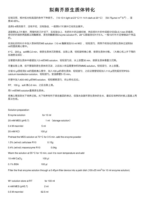
拟南芥原⽣质体转化在短⽇照,相对低光和⾼湿的条件下种苗⼦。
(10-13 h light at 23°C/11-14 h dark at 20°C)(50-75µmol m-2 S-2),湿度40-65%。
选择3-4周的苗⼦,没有开花,没有胁迫,⼀般第5-7⽚真叶已经完全展开。
选取第5,6,7⽚真叶,⽤锋利的⼑⽚切下,在实验台上,先把叶⽚的边缘切掉,然后将叶⽚的中间部分切成0.5-1mm 的细条,将切好的细条两⾯都沾到酶解液,浸泡到酶解液Enzyme solution中。
28°C⿊暗消化叶⽚5 h。
⼀般10⽚叶⽚⾜够做20个样品的。
向消化好的叶⽚中加⼊等体积的W5 solution(10 ml 酶解液加10 ml W5),轻轻混匀,⽤筛⼦将消化的原⽣质体过滤到50ml的圆底离⼼管中。
4°C,200 g,soft离⼼2 min,使原⽣质体沉到管底,去除上清,轻轻旋转离⼼管,使原⽣质体分散。
(⼤离⼼机上升下降的加速度设成5).沿管壁向原⽣质体中缓缓加⼊5 ml的W5 solution,轻轻混匀后,冰上放置30 min,使原⽣质体靠重⼒沉降。
尽量去除上清,但不要碰到原⽣质体的沉淀,之后加⼊样品需要体积的MMG solution。
轻轻混匀,冰上放置。
分装10 µl质粒到2 ml的圆底离⼼管中,加⼊100 µl的原⽣质体,轻轻拨匀,之后沿管壁轻轻加⼊110 µl预先配好的PEG-calcium transfection solution,轻轻拨匀。
室温静置5-15 min。
向管中加⼊400-440 µl的W5 solution,轻轻颠倒混匀,终⽌转化反应。
RT,100 g,soft 离⼼2 min,之后去除上清。
⽤1 ml的W5 solution重悬原⽣质体。
将离⼼管放到光下培养过夜。
光下培养有利于某些基因的表达,但强光会破坏原⽣质体的⽣长,最后在培养的时候上⾯盖上两层卫⽣纸。
一种适用于多种植物的农杆菌介导基因瞬时表达的方法
随着科学技术的不断发展,人类对植物基因研究的需求也日益增加。
而农杆菌介导的基因瞬时表达方法则成为了植物基因研究领域的热门话题之一。
本文将围绕这一主题展开讨论。
一、农杆菌介导的基因瞬时表达方法简介农杆菌介导的基因瞬时表达方法是一种将外源基因导入植物细胞中并在短时间内表达的技术。
其原理是利用土壤中常见的植物致病菌——农杆菌(Agrobacterium tumefaciens)来传递外源基因,使其经由农杆菌转座子插入到植物细胞的基因组中,从而实现目的基因的表达。
二、农杆菌介导的基因瞬时表达方法的优势1. 高效性:农杆菌介导的基因瞬时表达方法能够在短时间内使目的基因得到高效表达,为研究人员提供了更快捷、高效的研究手段。
2. 适用范围广:农杆菌介导的基因瞬时表达方法不仅适用于单一植物种类,还适用于多种植物,包括但不限于拟南芥、烟草、水稻等。
3. 可操作性强:相比于稳定转化方法,农杆菌介导的基因瞬时表达方法不需要等待植株成熟,而是直接在植物叶片上进行操作,省时省力。
三、农杆菌介导的基因瞬时表达方法的研究现状目前,科研人员对农杆菌介导的基因瞬时表达方法进行了大量探索与研究,不断优化该技术并拓展其应用范围。
通过改变农杆菌株系、转染条件、辅助基因等因素,有效提高了基因转化效率,并使其更适用于不同植物。
四、农杆菌介导的基因瞬时表达方法的应用前景随着生命科学领域的不断发展,对植物基因研究的需求逐渐增加,而农杆菌介导的基因瞬时表达方法正是满足这一需求的重要手段。
未来,该方法有望在植物遗传改良、抗病株培育、蛋白表达等方面发挥更为广泛的作用。
五、结语农杆菌介导的基因瞬时表达方法作为一种重要的植物基因研究技术,其独特的优势和潜在的应用前景吸引着越来越多的研究人员投入到相关领域的研究中。
随着技术的不断完善和发展,相信农杆菌介导的基因瞬时表达方法一定会为植物基因研究领域注入新的活力,为人类农业生产和生态环境的改善带来新的希望。
农杆菌介导的基因瞬时表达方法在植物基因研究领域中具有广阔的应用前景,其高效性和适用范围广的特点使其成为研究人员们研究植物基因功能和调控的重要工具。
拟南芥原生质体制备转化操作

采用基因枪轰击洋葱表皮观察了 GFP 的瞬时表达。
主要试剂
高渗培养液(MS 无机盐、40g/L 甘露醇),高渗固体培养基(MS 无机盐、40g/L 甘露醇、0.7% agar),无水乙醇,无菌去离子水,12.5 M CaCl2, 0.1 M 亚精胺,等渗培养基(MS 培养基)
主要设备
高速离心机,1.5 ml Ep 管,涡旋器,超净台,基因枪,气瓶,激光共聚焦显微镜
10 5 mM β-Mercaptoethanol
1ml 75mM β-Mercaptoethanol 母液
11 用 0.45μm 滤膜过滤后使用
过滤
2. PEG4000 溶液(一次配置可以保存五天,但是最好现用现配,每个样品需 100μl PEG4000 溶液,可根据实验样品量调整溶液 配置总量)
实验材料
洋葱内表皮
实验步骤
1. 瞬时表达载体构建
2. 基因枪轰击洋葱表皮观察 GFP 瞬时表达 1) 实验材料准备
撕取洋葱内表皮,置于高渗培养液(MS 无机盐、40g/L 甘露醇)中,室温 100 rpm 处理 4 hours;然后转入高渗固体培养基(MS 无机盐、40g/L 甘露醇、0.7% agar)。
1.09g 干粉
4 20mM KCl
1 ml 0.3 M KCl 母液
5 20mM MES,pH5.7
1 ml 0.3 M MES,pH5.7 母液
6
加入 10ml 水
7 55℃水浴加热 10 分钟,冷却至室温后加入以下试剂
8 10mM CaCl
1 ml 0.15M CaCl2
9 0.1﹪ BSA
1 ml 1.5﹪BSA(4℃保存)
5. WI 溶液
WI(200ml)
PEG介导法制备拟南芥原生质体及瞬时表达方法
采用PEG介导拟南芥叶片原生质体瞬时表达法。
具体方法如下:
1)在MS培养基上用无菌牙签点种拟南芥种子,萌发后待根长至1-3厘米(2周左右),移栽到营养土:蛭石为1:1的培养土中,置于培养箱,温度22℃,光照16h,湿度70%,培养2-3周;
2)配制酶解液,选10ml酶解体系,置于90mm培养皿中;
3)在长势良好且未抽薹的拟南芥植株上选取鲜嫩叶片10-15片,用洁净的手术刀在培养皿的盖子上将叶片切成1mm宽的细条;
4)将切好的细条均匀铺在含酶解液的培养皿中,可以一边切一边从植株上取;
5)将酶解液和细叶条真空抽滤30 min;
6)将培养皿取出,静置在黑暗中,23℃,酶解3小时;
7)酶解液过100-200目的筛子,将过滤后的绿色混合物置于2.0 ml离心管中,常温,100 g离心1 min;
8)弃上清,沉淀用冰冷的W5溶液轻柔洗涤,4℃,100 g,离心1min;
9)弃上清,沉淀用冰冷的W5溶液轻柔洗涤,冰上放置30min;
10)23℃,100 g,离心1min,弃上清,每管沉淀用0.5ml MaMg重悬(本步骤及以下操作均在23℃);
11)取约10-20ug质粒于2.0 ml EP管中,加100ul 制备好的原生质体,用剪去前端的200ul 枪头轻柔混匀;
12)加入110ul PEG/Ca溶液,轻柔混匀,23℃静置5-30min;
13)加入800 ul W5 溶液,轻柔的颠倒混匀,23℃,100 g,离心1min;
14)弃上清,加100ul W5,混匀,再次再加100ul W5,混匀;
15)上述混合液体置于恒温加热器中,23℃,避光,孵育6-18小时。
16)荧光观察,观察之前轻柔混匀,吸取观察物所用的枪头必须剪去前端。
peg介导原生质体的方法
peg介导原生质体的方法PEG(聚乙二醇)介导的原生质体(或叫质粒转化)是一种常用的实验方法,在基因工程和分子生物学实验中应用广泛。
PEG介导的质粒转化可以将外源DNA或质粒DNA导入到细胞膜内,进而实现外源DNA的定向插入到细胞基因组中。
本文将对PEG介导的原生质体方法方面的基本定义、优缺点、实验步骤和应用等方面作简要介绍。
一、定义质粒转化(Transformation)指将外源DNA导入到真核细胞或原核细胞内,使之产生遗传变异并表现出相应的遗传特性的过程。
PEG介导的原生质体实验是将目标DNA结合PEG 后直接加入原生质体内使之进行DNA转移的一种有效实验方法。
二、优缺点1.优点① PEG介导的质粒转化操作简单,不需要特殊设备;② PEG介导的质粒转化法对不同种类的细胞具有普适性,并能够同时转化一定量的细胞;③ PEG介导的质粒转化法可用于导入不同长度的DNA分子和DNA/RNA杂交分子;④ 经PEG介导的原生质体方法转化后的细胞繁殖时间快,生长期短;⑤ PEG介导的质粒转化法所需材料价格相对较低,实验费用较低。
2.缺点① 质粒转化法的成功率有一定的限制,繁殖时间短的细胞转化效率相对较低;② 所产生的转化体可能会出现末梢复制(Efficiency)和端口关闭(Productivity)的问题;③ 质粒载体的大小和复杂性会对转化效率产生影响;④ 转化体的有效性依赖于目标细胞内对目标DNA的自然摄取作用。
目标细胞表面分子的特异性决定了PEG介导原生质体方法的成功率。
三、实验步骤1.制作质粒制备质粒是PEG介导质粒转化实验中的重要一环。
制作质粒的前提是先有合适的质粒载体,常用的载体有pUC18、pGEM等。
2、制备细胞要进行PEG介导的原生质体实验,需要制备合适的细胞,以接受外源DNA。
常用的细胞有大肠杆菌、酵母菌等。
3、PEG介导转化① 质粒和目标细胞混合,其中细胞数目的控制非常重要。
通常,500μL-1000μL液体用100μg(或5 pmol)DNA和约50μL PEG(50%溶液)混合即可。
拟南芥原生质体制备原理

拟南芥原生质体制备原理拟南芥(Arabidopsis thaliana)是一种被广泛用于植物生物学研究的模式植物,拥有许多有益的特性,如小型体型、短生命周期和丰富的基因信息。
其中,原生质体(Protoplast)是拟南芥研究中广泛应用的一种实验材料,因其不包含细胞壁,使得研究人员可以更方便地通过转化等技术来分析和操作细胞。
本文将主要介绍拟南芥原生质体的制备原理,包括其基本步骤和关键操作。
一、拟南芥原生质体制备的基本步骤1.1 组织样品的准备首先,需要准备包含优良品种的拟南芥植株的新鲜叶片、茎段等组织样品作为原始材料。
这些组织样品应该是健康、无病虫害的,并且在生长状态良好的阶段。
要注意,不同部位的组织对原生质体制备的效率可能存在差异,因此可以根据具体实验目的选择合适的组织样品。
1.2 组织样品的消化将准备好的组织样品切碎,并加入含有合适浓度的细胞壁分解酶的酶解缓冲液中。
酶解缓冲液的选择应根据具体实验设计和预期结果来确定。
一般来说,含有高效的纤维素酶和果胶酶的酶解缓冲液可以更好地分解细胞壁,从而得到高质量的原生质体。
然后,将组织样品放置在酶解缓冲液中,在适当的温度下进行酶解。
酶解的时间和温度可以根据实验需求来确定,常见的条件为25-30℃,酶解时间约为3-6小时。
1.3 原生质体的分离和纯化经过酶解后,将酶解液通过筛网或离心等方法进行过滤,从而得到含有原生质体的悬浮液。
为了去除杂质和细胞残片,可以将悬浮液进行一次或多次的离心,得到较为纯净的原生质体沉淀。
最后,将原生质体沉淀用含有适当浓度的蔗糖等质量调节液进行浓缩和纯化,以获得高质量的原生质体。
二、拟南芥原生质体制备的关键操作2.1 细胞壁分解酶的选择细胞壁分解酶是原生质体制备中非常关键的一步,选择适当的酶解酶组合可以确保高效的细胞壁降解。
一般来说,搭配使用纤维素酶和果胶酶可以较好地完成细胞壁降解。
此外,还可以根据实验需求添加其他辅助酶,如蛋白酶和胰蛋白酶等,以进一步提高酶解效果。
- 1、下载文档前请自行甄别文档内容的完整性,平台不提供额外的编辑、内容补充、找答案等附加服务。
- 2、"仅部分预览"的文档,不可在线预览部分如存在完整性等问题,可反馈申请退款(可完整预览的文档不适用该条件!)。
- 3、如文档侵犯您的权益,请联系客服反馈,我们会尽快为您处理(人工客服工作时间:9:00-18:30)。
采用PEG介导拟南芥叶片原生质体瞬时表达法。
具体方法如下:
1)在MS培养基上用无菌牙签点种拟南芥种子,萌发后待根长至1-3厘米(2周左右),移栽到营养土:蛭石为1:1的培养土中,置于培养箱,温度22℃,光照16h,湿度70%,培养2-3周;
2)配制酶解液,选10ml酶解体系,置于90mm培养皿中;
3)在长势良好且未抽薹的拟南芥植株上选取鲜嫩叶片10-15片,用洁净的手术刀在培养皿的盖子上将叶片切成1mm宽的细条;
4)将切好的细条均匀铺在含酶解液的培养皿中,可以一边切一边从植株上取;
5)将酶解液和细叶条真空抽滤30 min;
6)将培养皿取出,静置在黑暗中,23℃,酶解3小时;
7)酶解液过100-200目的筛子,将过滤后的绿色混合物置于2.0 ml离心管中,常温,100 g离心1 min;
8)弃上清,沉淀用冰冷的W5溶液轻柔洗涤,4℃,100 g,离心1min;
9)弃上清,沉淀用冰冷的W5溶液轻柔洗涤,冰上放置30min;
10)23℃,100 g,离心1min,弃上清,每管沉淀用0.5ml MaMg重悬(本步骤及以下操作均在23℃);
11)取约10-20ug质粒于2.0 ml EP管中,加100ul 制备好的原生质体,用剪去前端的200ul 枪头轻柔混匀;
12)加入110ul PEG/Ca溶液,轻柔混匀,23℃静置5-30min;
13)加入800 ul W5 溶液,轻柔的颠倒混匀,23℃,100 g,离心1min;
14)弃上清,加100ul W5,混匀,再次再加100ul W5,混匀;
15)上述混合液体置于恒温加热器中,23℃,避光,孵育6-18小时。
16)荧光观察,观察之前轻柔混匀,吸取观察物所用的枪头必须剪去前端。
