2014年IES腹腔镜下腹股沟疝修补术指南
腹腔镜下经腹腹膜前腹股沟疝修补术
5. 死亡冠
为异常的闭孔动 脉 跨过耻骨梳韧带 与腹壁下动脉和 闭孔动脉相连
6. 危险三角
位于输精管和精索血管之间 有髂外动静、脉通过。
7. 疼痛三角
位于精索血管的外侧、髂耻束的下方 有生殖股神经的生殖支和股支、股神经、股外侧 皮神经穿过
手术操作要点
1.戳孔选择
脐孔,患侧 腹直肌外侧平 脐水平和对侧 腹直肌外侧脐 下水平
2. 髂耻束
腹横筋膜增厚的部分
起于髂前上棘,止于 耻骨上支 全程伴行于腹股沟韧 带的深面 开放式手术中较难看 到
3. 肌耻骨孔
内界:腹直肌的 外缘 外界:髂腰肌 上界:腹横筋膜 和腹内斜肌 下界:骨盆的骨 性边缘。
4. 腹壁下动脉
进入腹膜前间隙的 重要标志 直疝和斜疝的定位
禁忌症:
不能耐受全麻患者、腹腔内感染、腹膜炎
相对禁忌症:
下腹部腹腔粘连、腹腔积液、凝血功能障碍 、嵌顿疝等
并发症:
包括疝手术并发症、腹腔镜手术并发症及进腹 手术并发症。具体有疝复发、血肿(血清肿)、
阴囊气肿、神经感觉异常及小肠粘连、梗阻等。
腔镜视野下腹股沟区的主要解剖结构
1.腹膜皱襞
脐正中韧带:脐尿管 闭塞后残留的痕迹 脐内侧韧带:闭塞的 脐动脉表面腹膜皱襞 脐外侧韧带:腹壁下 动脉表面腹膜皱襞
1997年我国开展无张力疝修补术。
近年来,省内的部分三级医院相继开展腹腔镜下腹
股沟疝修补术,至今已经发展成为成熟的术式。
我院自2010年7月至今已成功完成120余例腹腔镜经
腹腔镜疝修补PPT课件

编辑版ppt
17
我们的工作
2015年4月以来我们科已经完成LIHR8例 手术,(双侧腹股沟疝1例,骑跨疝1例, 斜疝6例,直疝2例),术中发现对侧隐匿疝 3例,平均住院时间4.5天,一例出现血清 肿,2月后自行吸收,电话随访目前无复发。
编辑版ppt
18
弱区域。 符合力学原理,有效缓冲腹腔内压力的冲
击。
编辑版ppt
16
优势和劣势
优势:1、穿刺代替切口。2、微创切割。3、 神经痛发生率低。4、术后恢复快。5、复 发率低。6、可行疝囊超高位结扎。7、可 同时行双侧腹股沟疝的诊断和治疗。8、住 院时间短。
劣势:1、学习曲线长。2、设备昂贵,费 用高。
在我国部分大型疝中心LIRH手术已经占主 导地位,手术量超过每年1000例。
编辑版ppt
3
历史和现状
2014年《欧洲成人腹股沟疝治疗指南》将 开放的Lichtenstien手术和LIHR列为原发 性腹股沟疝的首选治疗方案,并强调LIHR 比开放手术需要更长的学习曲线。
2014年我国《成人腹股沟疝治疗指南》同 样将Lichtenstien手术和LIHR建议为原发 性腹股沟疝的首选治疗方案
编辑版ppt
4
应用解剖
皱襞与陷窝
编辑版ppt
5
应用解剖
腹膜前间隙:壁层腹膜和腹横筋膜前层之 间的间隙。
Retzius间隙:即耻骨膀胱间隙,位于正中线,前 方是腹直肌、腹横筋膜、耻骨,后方是膀胱。上 达脐平面,下达盆底肌,外至腹壁下动脉,基本 为无血管区。
Bogros间隙:位于腹壁下动脉外侧。
编辑版ppt
6
应用解剖
髂耻束:伴行于腹股沟韧带深面,是腹横 筋膜增厚的部分,起于髂嵴内侧和髂前上 棘,止于耻骨上支。
腹腔镜下经腹腹膜前腹股沟疝修补术资料讲解共24页

腹腔镜下经腹腹膜前腹股沟疝修补术
资料讲解
21、没有人陪你走一辈子,所以你要 适应孤 独,没 有人会 帮你一 辈子, 所以你 要奋斗 一生。 22、当眼泪流尽的时候,留下的应该 是坚强 。 23、要改变命运,首先改变自己。
24、勇气很有理由被当作人类德性之 首,因 为这种 德性保 证了所 有其余 的德性 。--温 斯顿. 丘吉尔 。 25、梯子的梯阶从来不是用来搁脚的 ,它只 是让人 们的脚 放上一 段时间 ,以便 让别一 只脚能 够再往 上登。
66、节制使快乐增加并使享受加强。 ——德 谟克利 特 67、今天应做的事没有做,明天再早也 是耽误 了。——裴斯 泰洛齐 68、决定一个人的一生,以及整个命运 的,只 是一瞬 之间。 ——歌 德 69、懒人无法享受休息之乐。——拉布 克 70、浪费时间是一桩大罪过。——卢梭
腹腔镜下腹股沟疝修补术

随着腹腔镜技术的发展,腹腔镜下腹股沟疝修补术逐渐成为主流术式。该术式具 有创伤小、恢复快、并发症少等优点,被广泛应用于临床。
腹腔镜下腹股沟疝修补术的优势
微创
腹腔镜手术采用小孔操作,避免了传统手术的大切口,减少了手术创伤和出血量,有利于 术后恢复。
视野清晰
腹腔镜下手术视野清晰,可以放大手术视野,便于医生精细操作,减少误伤周围组织的风 险。
复发预防
遵医嘱进行复发预防措施,如使用疝气带等 。
心理支持
保持积极心态,参加康复训练和支持小组, 提高自我管理能力。
生活质量评估
症状改善
评估术后疼痛、肿胀等症状是否得到改善,以及改善程度。
活动能力
评估患者术后活动能力恢复情况,如行走、站立、日常生活自理等。
心理状态
关注患者术后心理状态变化,及时发现并处理焦虑、抑郁等情绪问题。
或咳嗽等患者。
临床表现
腹股沟疝的主要症状为腹股沟区 可复性包块,站立或咳嗽时包块 突出,平卧或按压后可消失。若 发生嵌顿,可出现疼痛、恶心、
呕吐等症状。
腹腔镜下腹股沟疝修补术的发展
传统手术方法
传统的腹股沟疝修补术多采用开放手术,包括张力修补和无张力修补两种。张力 修补术因术后疼痛明显、恢复时间长等缺点已逐渐被淘汰;无张力修补术采用人 工材料进行修补,术后疼痛较轻,但存在感染、复发等并发症的风险。
异常情况,应及时就医。
活动恢复
根据医生建议,逐步恢复日 常活动。避免剧烈运动和提 重物,以免腹压增加。
饮食调整
保持饮食清淡,多吃蔬菜水 果,保持大便通畅。避免辛 辣、刺激性食物。
随访管理
定期随访
术后需定期到医院进行随访,以评估恢复情 况,及时发现并处理潜在问题。
腹腔镜下腹股沟疝修补术的

复发率低
腹腔镜下腹股沟疝修补术的临床效果显著,复发率较低。 长期观察表明,该手术方法能够有效地提高患者的生活质量。
06
腹腔镜下腹股沟疝修补 术的案例分享与讨论
案例一:老年患者的治疗
总结词
安全、有效的治疗方式
详细描述
腹腔镜下腹股沟疝修补术对于老年患者来说是一种安全、有效的治疗方式。相较于传统手 术方式,腹腔镜手术具有术后疼痛轻、恢复快、并发症少等优点,同时也能够减轻患者及 家属的心理负担。
腹腔镜下腹股沟疝修 补术的
汇报人: 日期:
目录
• 引言 • 腹腔镜下腹股沟疝修补术的术前准备 • 腹腔镜下腹股沟疝修补术的操作流程 • 腹腔镜下腹股沟疝修补术的术后护理 • 腹腔镜下腹股沟疝修补术的临床效果与展
望 • 腹腔镜下腹股沟疝修补术的案例分享与讨
论
01
引言
疝的概述
01
02
03
定义
疝是指体内器官或组织通 过先天或后天形成的薄弱 点,从正常位置进入邻近 或远离部位的现象。
02 03
详细描述
双侧疝是腹股沟疝中较为严重的一种类型,腹腔镜下腹股 沟疝修补术可以同时处理两侧的疝,具有高效、安全的特 点。同时,该手术方式还可以减少手术时间和住院时间, 降低患者的经济负担。
参考来源
相关文献报道
THANKS FOR WATCHING
感谢您的观看
医生的准备
医生应全面了解患者 的病史和身体状况, 以便制定最佳的手术 方案。
医生需要与患者进行 充分的沟通,以了解 其需求和期望。
医生需要熟练掌握腹 腔镜技术,以便在手 术中应对各种复杂情 况。
手术器械的准备
腹腔镜设备
包括摄像头、光源、气腹机等 。
腹腔镜下腹股沟疝修补术
腹腔镜下腹股沟疝修补术在医学领域的不断创新和发展中,腹腔镜下腹股沟疝修补术作为一项重要的外科技术,已经在解决腹股沟疝引起的不适和并发症方面取得了显著的成就。
腹股沟疝作为常见的外科问题,对患者的生活质量和健康状况产生了明显的影响。
传统的开放手术方法虽然有效,但腹腔镜下腹股沟疝修补术通过其微创、快速康复和较少并发症的优势,逐渐成了治疗腹股沟疝的首选方法之一。
1.腹股沟疝的发病机制腹股沟疝是指腹腔内的脏器或组织通过腹股沟区域的弱点或缺陷膨出到皮肤下的现象,其发病机制涉及腹腔内的压力变化、腹股沟区域的解剖结构以及个体的遗传因素等多种因素。
腹股沟区域是腹部与大腿相交的部位,在这个区域有一些天然开口用于腹部血管和腹股沟淋巴结的通道,包括腹股沟韧带、腹股沟环以及肌肉组织等。
然而,由于生理或环境的因素,这些开口可能会变得松弛或扩大,造成一个腹腔内的“门户”,导致腹腔内的组织或脏器穿过这些缺陷,形成腹股沟疝。
腹股沟疝通常在腹部压力增加时出现,比如咳嗽、打喷嚏、举重、便秘等情况下,患者会感觉到一块突出的肿块,一般位于腹股沟区域,在躺下或轻轻按压时消失,但在腹压增加时又会重新出现,有时还会伴随不适、疼痛或压迫感。
另外,遗传因素也在腹股沟疝的发病中起到一定作用,有些人天生就具有腹股沟区域的解剖结构上的薄弱点,特别是家族中有腹股沟疝病史的人极易受到遗传因素的影响。
目前,临床上将腹股沟疝分为两种类型:直股疝和斜股疝。
直股疝发生在腹股沟区域的前壁,脏器通过腹股沟环内侧部的薄弱点膨出;而斜股疝则发生在腹股沟区域的侧壁,脏器穿过腹股沟环外侧的弱点。
1.腹股沟疝的治疗措施当涉及腹股沟疝的治疗,医疗专业人士会根据患者症状、疾病严重程度以及个人的医疗历史来制定个性化的治疗计划。
对于小型、无症状或轻微不适的腹股沟疝,医生会建议进行观察和保守治疗,包括避免过重的体力劳动、避免用力过度、保持正常体重、避免便秘等,通常适用于疝囊没有扭转、脏器没有卡住以及没有明显疼痛的情况,患者需要注意疝囊的大小变化,以及任何疼痛或不适的迹象。
腹股沟疝修补方法

腹股沟疝修补方法
腹股沟疝修补方法有以下几种:
1. 传统开放手术修补:通过在腹股沟区域进行切口,将腹股沟疝囊复位,并用缝线将腹股沟肌肉修补。
这种方法是传统的修补方式,但术后恢复时间较长。
2. 腹腔镜手术修补:通过在腹部进行小切口,插入腹腔镜和其他外科工具,修补腹股沟疝。
腹腔镜手术具有创伤小、恢复时间短的优点。
3. 不张力修补(tension-free repair):在腹股沟区域放置一块特殊的网状支架(mesh),以加固腹股沟区域,避免因肌肉张力引起的疝复发。
这种方法可以通过开放手术或腹腔镜手术进行。
4. 对复杂腹股沟疝的修补:对于复杂或难治的腹股沟疝,可能需要较大的手术切口和更复杂的修补方式,如修补可能涉及到附睾或输精管的腹股沟疝。
需要根据患者的具体情况和医生的建议选择适当的修补方法。
手术前需要进行评估和必要的准备工作,手术后还需要注意伤口护理和术后恢复。
腹腔镜腹股沟疝修补术课件
5.术中可探查是否有隐匿疝,并得到及时的治疗。 Find and treat mutiple unexpected and concealed hernia 6.治疗双侧疝、复合疝与复发疝具有一定的优势。 Ideally suitable for relapse hernia、bilateral hernia and
strangulated hernia 4.腹腔镜手术后严重粘连者。severe Post-
laparoscopic operation adhesion 5.复杂滑动疝。complicated sliding hernia 6.合并妊娠者。combined with pregnancy
南京医科大学附属南京第一医院
(3)腹膜内补片植入术疝修补术( intraperitoneal onlay mesh IPOM)
南京医科大学附属南京第一医院
•文档仅供参考,不能作为科学依据,请勿模仿;如有不当之处,请联系网站或本人删除。
LIHR手术的合理性
1.符合病因学说,腹横筋膜重建。
Rebuild the transverse fascia
南京医科大学附属南京第一医院
•文档仅供参考,不能作为科学依据,请勿模仿;如有不当之处,请联系网站或本人删除。
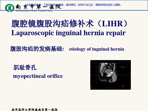
肌耻骨孔 myopectineal orifice
南京医科大学附属南京第一医院
•文档仅供参考,不能作为科学依据,请勿模仿;如有不当之处,请联系网站或本人删除。
腹股沟疝修补的目标—完整覆盖肌耻骨 孔
平。 Allowed clear visualization of all preperitoneal fascial planes
高清手术|腹腔镜腹股沟疝修补(TAPP七步法)

高清手术|腹腔镜腹股沟疝修补(TAPP七步法)
导读
腹股沟疝是外科常见疾病之一,目前治疗成人腹股沟疝的唯一可靠方法是手术治疗,传统开放式无张力疝修补术是治疗成人腹股沟疝的手术方式之一,但其术后并发症多见,创伤大,复发率高,术后出现慢性疼痛的几率较高,且异物感较为明显。
近几年来,随着医疗设备的不断更新、发展,腹腔镜技术为治疗腹股沟疝提供了新方法,用腹腔镜经腹腹膜前补片植入术(TAPP)治疗成人腹股沟疝,具有创伤小、疼痛轻、恢复快、复发率低等优势,逐步成为治疗成人腹股沟疝的主要术式。
患者介绍
男性,66岁,身高171 cm,体重65 kg
主诉:发现右腹股沟可复性肿块2月余
体检:右腹股沟区肿块5*5 cm,可回纳
诊断:右侧腹股沟斜疝
手术视频
同济大学附属杨浦医院普外科
术者:李健
助手:李奇
手术核心步骤
1. 戳卡的安置
2. 腹膜的切开
3. 疝囊的处理
4. 腹膜前间隙的游离
5. 补片的覆盖
6. 补片的固定
7. 腹膜的关闭。
腹腔镜下腹股沟疝修补术的临床应用和选择
盘盍( 电王版 )08 5 第 2 第 1 CiJ e iAd il a n E c v ei ) a 20 .02N . 20 年 月 卷 期 h Hra bo n lS  ̄ lt f vr0 . v 08V1 .o1 n n m aW lu ( emi sn M c
to e s e ad n h r ame t f n u n l e i b tsmu tn o s o e ain h sg i e o u a i .T e am r v r y r g r ig t ete t n g i a r a u i l e u p rt a an d p p lr y h i oi hn a o t o i s d s t v l a e t e o t o fl p r s o i i h n o sh r i i e a d t o l ain , ft s t y wa o e au t h u c me o a a o c p c smu a e u e na w t r g r o c mpi t s h u h c o r c re c ae,a d lt ic mfr.M e h d A rto p c ie a ay i wa d f 4 ai ns o e ae e urn ert n ae ds o o t to s er s e t n lss s ma e o 6 p t t p r td v 2 e s h n o sy fr b l trl h r i u i g te lp r s o i e h i u e w e c o e 0 4 a d D c mb r i mu a e u l o i ea e a s h a a o c p c tc nq e b t e n O t b r 2 0 n e e e a n n 2 0 .C mp iain e e r gsee n l w u a ef r d u i g ac mb n t n o u sin ar n 05 o l t sw r e i rd a d a f l — p w sp r me s o i ai f e t n i a d c o t oo o n o q o e
成人腹股沟疝诊疗指南(2014 年版)

成人腹股沟疝诊疗指南(2014 年版)(转载)发表者:江志鹏24人已访问“中华医学会外科学分会疝和腹壁外科学组”与“中国医师协会外科医师分会疝和腹壁外科医师委员会”虽隶属不同,但目标一致,即致力于提高我国疝和腹壁外科的诊疗水平。
为此,两者互相协作,从2013 年着手准备,今年初组织修订,共同讨论,最终完成《成人腹股沟疝诊疗指南(2014 版)》。
(以下简称为“指南”)。
需要说明的是“指南”的前身为《成人腹股沟疝诊疗指南(2012 版)》[1],本次修订依据国内外近年有关学科的进展和我国的国情,增添了一些条款,还增加了“指南”中的部分附件(腹股沟疝的常规修补方法和腔镜修补方法),目的在于强调腹股沟疝外科治疗的专业化和规范化,提高我国疝外科的治疗水平。
1. 定义腹股沟疝是指发生在腹股沟区域的腹外疝,即在腹股沟区域腹壁存在缺损,有突向体表的疝囊结构,腹腔内的器官或组织可通过先天的或后天形成的腹壁缺损进入疝囊。
典型的腹股沟疝具有疝环、疝囊、疝内容物和疝被盖等结构。
依据解剖学上的“肌耻骨孔”概念,腹股沟疝包括斜疝、直疝、股疝及较为罕见的股血管前、外侧疝等。
2. 病因和病理生理2.1 病因2.1.1 鞘状突未闭是腹股沟疝发生的先天性因素。
2.1.2 腹腔内压力腹内压和瞬间的腹内压变化是产生腹外疝的动力。
2.1.3 腹壁局部薄弱各种引起腹股沟区域腹壁的组织胶原代谢或成份改变所致的腹壁薄弱与腹股沟疝的发病有关。
2.1.4 其它遗传因素,长期吸烟,肥胖,下腹部低位切口等可能与腹股沟疝的发生有关。
2.2 病理生理当腹腔内器官或组织进入疝囊后,由于疝环的存在,可压迫疝内容物,形成嵌顿疝。
若为肠管时,可造成肠管的机械性梗阻而产生一系列临床表现和病理生理变化。
随着受压时间延长,肠管出现水肿、渗出和被嵌顿肠管发生血运障碍,若未及时治疗,可导致疝内容物坏死,穿孔,产生严重的腹膜炎,甚至危及生命。
3. 分类与分型对腹股沟疝分类与分型之目的有三个方面: 准确的描述病情; 选择适宜的治疗方案; 比较及评价不同方法的治疗效果。
腹腔镜腹股沟疝修复手术操作指南(最全版)
腹腔镜腹股沟疝修复手术操作指南(最全版)介绍腹股沟疝是指腹腔内脏器官通过腹股沟区域的腹股沟环进入阴囊或区域的异常现象。
腹腔镜手术是一种常用的治疗方法,本指南将介绍腹腔镜腹股沟疝修复手术的操作步骤和注意事项。
操作步骤1. 患者准备- 麻醉在手术开始前,根据患者的情况选择适当的麻醉方式。
- 体位将患者摆放在仰卧位,双下肢自然分开。
- 术区准备对手术区域进行消毒和覆盖无菌巾。
2. 手术入路- 选择手术入路根据患者的情况和疝修复的需要,选择合适的手术入路,如经脐入路、经腹股沟入路等。
- 创建工作通道在选定的手术入路上建立工作通道,为器械和相机的进入提供便利。
3. 腹腔探查- 引入相机将相机导入腹腔,进行腹腔探查和评估疝的大小和类型。
- 处理肠管如有需要,对暴露的肠管进行处理,并将其移至适当位置。
4. 疝修复- 膝撑法或张力法根据疝的类型和大小,选择膝撑法或张力法进行疝修复。
在腹膜和腹股沟环之间植入网片,并固定在适当位置。
- 注意事项在进行疝修复时,需要注意保护神经、血管和脏器,确保手术安全和效果。
5. 结束手术- 清点器械结束手术前,清点手术器械,确保无遗漏。
- 切口处理对手术切口进行处理和缝合。
注意事项- 术前评估患者的疝类型、大小和手术适应症。
- 手术中注意保护神经、血管和脏器,避免损伤。
- 术后给予患者适当的护理和指导。
以上是腹腔镜腹股沟疝修复手术操作指南的简要介绍,详细的操作步骤和注意事项请参考相关专业文献和指南。
(注意:本文档为指南性内容,仅供参考,具体操作请在专业人员的指导下进行。
)。
腹腔镜腹股沟疝修补术

汇报人:XX
• 腹股沟疝概述 • 腹腔镜腹股沟疝修补术原理及优势 • 术前准备与评估 • 手术步骤与技巧 • 术后管理与并发症防治 • 康复指导与随访计划
01
腹股沟疝概述
定义与分类
腹股沟疝定义
腹股沟疝是指腹腔内脏器通过腹 股沟区的缺损向体表突出所形成 的包块,俗称“疝气”。
发生率降低。
复发率低
腹腔镜手术能够更彻底 地处理疝囊和修补缺损
,从而降低复发率。
适应症与禁忌症
适应症
适用于各种类型的腹股沟疝,包括直疝、斜疝和股疝等。
禁忌症
严重心肺功能不全、凝血功能障碍、腹腔内广泛粘连等患者 不宜进行腹腔镜手术。
03
术前准备与评估
患者教育与心理支持
术前宣教
向患者详细解释腹腔镜腹股沟疝修补术的目的、方法、预期效果及可能的风险, 确保患者充分理解并同意手术。
康复指导与随访计划 康复指导与随访计划
• 饮食禁忌:患者应避免进食辛辣、油腻、生冷等刺激性食 物,以及容易引起腹胀的食物,如豆类、碳酸饮料等。
康复指导与随访计划 康复指导与随访计划
术后早期活动
术后患者应尽早下床活动,促进肠道蠕 动和血液循环,预防深静脉血栓的形成 。
VS
适度锻炼
随着术后恢复,患者可进行适度的锻炼, 如散步、慢跑、瑜伽等,以增强身体免疫 力和促进伤口愈合。
其他并发症防治
密切关注患者术后病情变化,及时发现并处理出血、神经损伤等并 发症。
06
康复指导与随访计划
康复指导与随访计划 康复指导与随访计划
术后初期饮食
术后24小时内,患者应以流食或半流食为主,如稀饭、米汤、面条等,避免进食过硬、过热及刺激性 食物。
李金斯坦修补方法
成人腹股沟疝诊疗指南(2014年版,附件)中华医学会外科学分会疝和腹壁外科学组中国医师协会外科医师分会疝和腹壁外科医师委员会8.附件8.1 常规腹股沟疝修补方法1)李金斯坦修补方法李金斯坦平片修补方法是美国Lichtenstein医生在上世纪80年代所建立的用人工材料进行腹股沟疝无张力修补的方法(Tension-Free Hernioplasty)。
这一方法彻底改变了以往的组织对组织的张力缝合技术,并以术后复发少,疼痛轻,恢复快,而广为接受和使用。
(拍下此图,扫一扫,可下载至手机)1)切口:斜切口,从内环上方(髂前上棘至耻骨结节连线中点上方1cm)至耻骨结节,长约6cm-7cm2)按层切开皮肤、皮下组织、浅筋膜和深筋膜3)牵开切口,显露腹外斜肌腱膜和外环口4)打开腹外斜肌腱膜(长约6cm-7cm)5)向两侧充分的游离腹外斜肌腱膜:内侧至腹直肌外侧缘;外侧至腹股沟韧带6)紧贴外环口内上,游离精索,可发现此处的无血管层面,可用电刀进行分离7)完全游离精索:外上至内环,内下达耻骨结节(可不切断提睾肌)8)寻找疝囊:斜疝疝囊位于精索的内侧上方,且被精索内筋膜包绕,故与精索紧密相连9)若斜疝较小,在腹股沟管内,尚未进入阴囊,可紧贴疝囊做分离11)若斜疝较大,进入阴囊,可在腹股沟管的中、上部显露疝囊,近疝囊颈位置切开10)完全游离疝囊,然后将其内翻回纳12)回纳疝内容物后完全横断疝囊,近端缝合结扎,远端确切止血后,旷置17)若直疝疝囊颈宽大可间断褥式缝合数针,关闭缺损13)游离精索后,在其内上方检查无疝囊后,探查直疝三角,由于病人平卧,直疝疝囊向上凸起并不明显,嘱病人咳嗽,更易于发现14)提起疝囊,从外下方游离至疝囊颈部,用电刀在疝囊颈切开腹横筋膜18)在切口外上处腹外斜肌腱膜下方,用手指做钝性分离,为补片放置准备充足的空间15)切开一周腹横筋膜,准备回纳疝囊16)将直疝疝囊内翻回纳腹腔19A)网片(约6-7cm×12-13cm)头端呈圆弧型,尾端呈燕尾状,剪开后形成上、下两片,上片较宽,下片较窄19B)也可以用自粘式免缝合补片(分为左侧和右侧两种型号),在靠近腹股沟韧带一侧可修剪掉一小部分20A)将精索穿行于平片的上、下片之间,尾部呈燕尾状交叉、覆盖,缝合为新内环20B)自粘式免缝合补片, 先将带勾爪一面折叠(方法如图)20C)提起精索,将补片方置精索下方,带有蓝色标志一端朝向耻骨结节20D)打开补片一侧, 套住精索,按压两层,成为新的内环21A)平片放置在腹股沟管的后壁,其尾部放入腹外斜肌腱膜下方,充分展平,平片内侧须超过耻骨结节1-1.5cm,内上方覆盖直疝三角,内侧缘超过腹内斜肌、腹横肌缘(联合腱)2cm,下方覆盖腹股沟韧带21C)补片向外上展平,置于腹外斜肌腱膜下方间隙21D)按紧补片可自粘于腹股沟管的后壁22A)按层次分别依次间断缝合:腹外斜肌腱膜、皮下组织和皮肤诸层。
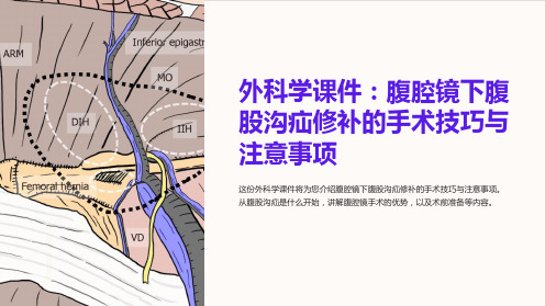
《外科学课件:腹腔镜下腹股沟疝修补的手术技巧与注意事项》
术中体位
在腹腔镜下腹股沟疝修补手术中,患者被放置在仰卧位,头稍微高于躯干。 这个体位有助于手术器械的插入和操作。
腹股沟解剖结构
了解腹股沟区域的解剖结构对于腹腔镜下腹股沟疝修补手术至关重要。重要 的结构包括靠近腹股沟区域的血管、神经和腹股沟环。
手术器械准备
腹腔镜下腹股沟疝修补手术需要一系列特殊的器械,包括腹腔镜、各种取向 器具和缝合材料。确保手术器械准备充分,以保证手术的顺利进行。
无张力修补技术
无张力修补技术是一种通过使用特殊的缝合技术,减少张力,在不增加肌肉 层压力的情况下修复腹股沟区域的方法。
腹腔镜下乳状突气囊注水法
腹腔镜下乳状突气囊注水法是一种在手术中使用乳状突气囊,并通过注入水来扩大操作空间的技术。
注意事项:手术操作中常见错误及其避免 方法
手术操作中常见的错误包括器械选择不当、操作技巧不准确等。为了避免这些错误,需要充分的培训和经验, 并且严格遵守手术规范。
腹腔镜手术的优势
腹腔镜手术相比传统开放手术具有许多优势,包括小切口、更少的术后疼痛、 更快的康复时间和更小的创伤。
术前准备工作
术前准备工作包括了解患者的病史、进行体格检查、进行必要的检查和测试, 并给患者提供详细的术前指导。
麻醉
腹腔镜下腹股沟疝修补手术通常使用全身麻醉。这种麻醉方法可以确保患者 在手术过程中没有疼痛感。
皮下普通缝合技术
皮下普通缝合技术是一种通过将组织逐层缝合,修复腹股沟区域的缺陷的方 法。
肌肉下普通缝合技术
肌肉下普通缝合技术是一种通过将肌肉层逐层缝合,修复腹股沟区域的缺陷 的方法。
绕钉技术
绕钉技术是一种使用特殊的针线穿刺腹股沟区域,并通过绕钉来修复缺陷的 方法。
绕线钉技术
腹腔镜下疝气修补术
腹腔镜下疝气修补术
P第a1g2页e 12
术前访视
1. 术前禁食禁饮;特殊情况请听从医师指导。 2. 术前一天,做好个人卫生工作。 3. 保持正常饮食,营养均衡。 4. 手术当日,取下身上佩戴饰品。
腹腔镜下疝气修补术
P第a1g3页e 13
术前准备
1. 麻醉方式:全麻 2. 病人准备:开放静脉通道,必要时导尿 3. 手术体位:建立气腹,帮助医生有足够空间观察操作,患者通
腹腔镜下疝气修补术
P第a1g6页e 16
巡回护士
腹腔镜下疝气修补术
P第a1g7页e 17
手术配合
1. 提前洗手,检验腹腔镜器械完整性,准备好穿刺器,与巡
回护士清中
注意气腹机上显示压力、流
量,观察腹部膨隆情况。
3. 两侧建立操作孔 。
4. 将疝囊完全游离后还纳入腹
临床表现
1. 病人在站立、咳嗽、用力时,在腹股沟部有肿块隆起,平卧 位或用手推挤可消失。 2. 斜疝包块呈梨形,可进入阴囊。 3. 疝还纳后,用指压内环,肿块不出现,外环口扩大,可及咳 嗽击感。 4. 疝嵌顿后有肠梗阻表现或不能还纳。 5. 应与腹股沟直疝,股疝,交通性鞘膜积液等判别。
腹腔镜下疝气修补术
术后护理
包扎 整理衣裤
腹腔镜下疝气修补术
检验皮肤
搬运护送
P第a2g1页e 21
注意事项
腹腔镜疝修补术是一项新型手术,病人及家眷缺 乏了解,所以要做好病人心理护理
器械护士应熟悉手术过程,以利配合手术,缩短 手术时间,降低CO2气腹用量,严格查对手术用 物
腹腔镜下疝气修补术
做好腔镜器械等清洗与保养
P第a2g2页e 22
常为头低脚高位15-30度,CO2维持压力10-15mmHg
- 1、下载文档前请自行甄别文档内容的完整性,平台不提供额外的编辑、内容补充、找答案等附加服务。
- 2、"仅部分预览"的文档,不可在线预览部分如存在完整性等问题,可反馈申请退款(可完整预览的文档不适用该条件!)。
- 3、如文档侵犯您的权益,请联系客服反馈,我们会尽快为您处理(人工客服工作时间:9:00-18:30)。
Chapter 1: Perioperative management: evidencefor antibiotic and thromboembolic prophylaxisin endoscopic/laparoscopic inguinal hernia surgery?Chapter 2: Technical key points in TAPP repairWhich is the safest and most effective method ofestablishing pneumoperitoneum and obtaining access tothe abdominal cavity?Level1BIn thin patients (BMI\27), the direct trocar insertion is asafe alternative to the Veress needle technique (strongerevidence).GradeCThe direct trocar insertion (DTI) can be used in order toestablish pneumoperitoneum as a safe alternative toVeress needle, Hasson approach or optical trocar, ifpatient’s risk factors are considered and the surgeon isappropriately trained (new recommendation).What kind of trocars should be used?Is there any relation between the trocar type and riskof injury and/or trocar hernias?Level2BUse of 10-mm trocars or larger may predispose to hernias,especially in the umbilical region or in the obliqueabdominal wall (Stronger evidence). GradeBFascial defects of 10 mm or bigger should be closed(Stronger evidence).Is clinical examination efficient enough?What is the role of TAPP and other techniques inreliable assessment? GradeBA thorough closure of peritoneal incision or biggerperitoneal tears should be achieved (Stronger evidence).Chapter 3: Technical key points in TEPHow should a large direct sac be handled?Level4Alternatively to fixation of the extended fascia transversalisto Copper’s ligament the direct inguinal hernia defect canbe closed by a pre-tied suture loop (new statement).GradeDAs alternative the primary closure of direct inguinal herniadefects with a pre-tied suture loop can be used (newrecommendation).How should a large indirect sac be handled?Level3Transection of a large indirect sac does not lead tosignificant differences in postoperative pain, length ofhospital stay and recurrence, but to a significant higherseroma rate (new statement).GradeCA large indirect sac may be ligated proximally and divideddistally without the risk of a higher postoperative pain andrecurrence rate, but with an increased postoperativeseroma rate (new recommendation).Should a drain be used after a TEP repair? Shouldseromas be aspirated?Level3Drain after TEP significantly reduces the incidence ofseroma formation with increasing the risk of infection orrecurrence (new statement).GradeCA closed-suction drain can be used to reduce the risk ofseroma formation without increased risk of infection(new recommendation).Has extraperitoneal local anesthetic treatment duringTEP a positive effect on postoperative pain? New(added) questionLevel 1AExtraperitonealbupivancaine treatment during endoscopicTEP inguinal hernioplasty is not more efficaciousfor thereduction of pain than placebo.GradeAExtraperitoneal bupivacaine treatment during endoscopicTEP inguinal hernia repair for the reduction ofpostoperative pain should not be performed.Chapter 4: TEP versus TAPP: which is better?Level1ATAPP has a longer hospital stay compared to TEP (new).Level1BPotentially serious adverse events are rare after both TAPPand TEP (stronger evidence).TAPP has a longer operation time compared to TEP (new).Level2CTEP has more intra-operative and postoperative surgicalcomplication rate compared to TAPP (new).GradeABoth techniques are acceptable treatment options foringuinal hernia repair and there is sufficient data toconclude that both TAPP and TEP are effective methodsof laparoscopic inguinal hernia repair (strongerevidence).Chapter 5: Endoscopic/laparoscopic surgeryin complicated hernias: feasibility, risks, and benefitLevel3TEP inguinal-scrotal hernia repair remains an advantageousapproach during the difficult scrotal hernia that requires‘‘conversion’’ to an open repair, because the pre-peritonealdissection performed laparoscopically allows for reductionof the hernia and optimal mesh placement once the herniarepair has been converted and is performed from theanterior approach (new).GradeCTEP approach for the large, difficult scrotal hernia mayserve as an adjunct to dissection and definition of the preperitonealspace allowing for easier hernia and meshplacement once the case is ‘‘converted’’ to open repair(new).Level3Laparoscopic hernia repair for incarcerated inguinal herniahas been successfully and safely performed in the pediatricpopulation (new). GradeCLaparoscopic hernia repair for incarcerated inguinal herniamay be successfully and safely performed in the pediatricpopulation by surgeons with laparoscopic expertise (new).Level4Women are at increased risk of having an occultsynchronous femoral hernia (New).GradeCWhen performing inguinal hernia repair in women, extraeffort should be undertaken to reveal and treat occultsynchronous femoral hernia (New).Chapter 6: Mesh size and recurrenceChapter 7: Heavy or light weight mesh in TAPPand TEP—functional outcome and quality of lifeLevel 1AThe statistical significance that lighter meshes with largerpores results in improvement of quality of life is notconsistent in recently published meta-analyses. Subsetanalysis revealed no higher risk of recurrence after usinglightweight meshes in laparoscopic inguinal herniarepair (New).Level2BThe middle- and long-term results of prospective studies inmen do notsupport the hypothesis that bilateral inguinalhernia repair with alloplastic meshprosthesis causesmale infertility or decreasing the sperm motility (New).GradeBA monofilament implant with a pore size of at least1.0–1.5 mm (usually meaning low-weight) consisting of aminimum tensile strength in all directions (includingsubsequent tearing force) of 16 N/cm appeared to be mostadvantageous;however, this assumption mainlysummarizes personal and published clinical and experimental experiences (stronger evidence).The application of large porepolypropylene meshes inendoscopic hernia repair is harmless concerningazoospermia and should therefore further used (New).Chapter 8: Slitting or not slitting of mesh—does itinfluence outcome?Level1Cutting a slit in the mesh to allow the structures of thefunicel to pass does not compromise testicular perfusionand testicular volume (New).GradeBBased on available evidence we recommend not to cut a slitin the mesh although cutting does not compromise testisperfusion (New).Chapter 9: Mesh fixation modalities: is therean association with acute or chronic pain?Level1AFixation and non-fixation of the mesh in TEP areassociated with equally risk of postoperative pain orrecurrence (New).Level1BFibrin glue fixation is associated with less chronic painthan stapling. GradeAIf TEP technique is used, non-fixation has to be consideredin all types of inguinal hernias except large direct defects(MIII, EHS classification) (strongerrecommendation).GradeBIn case of TAPP repair non-fixation should be considered intypes LI, II, and MI, II hernias (EHS classification).For fixation, fibrin glue should be considered to minimizethe risk of acute postoperative pain (modifiedrecommendations).Chapter 10: Risk factors and prevention of acuteand chronic pain in TAPP and TEPLevel1AThere is no difference of chronic pain after TEP and TAPP(stronger evidence).Fixation and non fixation of the mesh in TEP are associatedwith equally risk of postoperative pain (see chapter‘‘Fixation’’) (new).Level1BFibrin glue fixation is associated with less chronic painthan stapling (see chapter ‘‘Fixation’’) (new).Level2AAge below median (40–50 years) is a risk factor for acutepain (stronger evidence).Age below median (40–50 years) is a risk factor for chronic pain (stronger evidence).Severe acute postoperative pain is a risk factor for chronicpain (stronger evidence).GradeAIf TEP technique is used non fixation has to be consideredin all types of inguinal hernias except large defects (L III,MIII; EHS classification; see chapter ‘‘Fixation’’) (new).GradeBIn case of TAPP repair non fixation should be considered intypes LI, LII, MI, MII hernias (EHS classification, see Chapter ‘‘Fixation’’) (new).Chapter 11: Urogenital complications associatedwith TAPP and TEPLevel2BInguinal hernia repair with mesh is not associated with anincreased risk of, or clinically important risk for, maleinfertility. (new).GradeBGroin hernia repair using mesh techniques may continue tobe performed without major concern about the risk formale infertility. (new).Chapter 12: Intraperitoneal onlay mesh (IPOM)for inguinal hernia repair—still a therapeutic option?Chapter 13: Role for open preperitoneal meshplacement in the era ofendo/laparoscopic inguinalhernia repairLevel1BMinimally invasive open approaches (i.e., Kugel) mayoffer a cost advantage over laparoscopic approaches.(new).Chapter 14: Single port surgery or reduced portsin endoscopic/laparoscopic hernia repair (New chapter)Level2BSingle port laparoscopic hernia repair is a safe and feasiblealternative to traditional multiport technique although hasnot been showed to be superior or more effective.Single port laparoscopic hernia repair may offer a bettercosmetic outcome and patient’s satisfaction.Single port laparoscopic hernia repair has no increased risk compared with standard multiport technique.Homemade ports, as an alternative to commerciallyavailable ports, provides a feasible and safe alternatives GradeBSingle port laparoscopic inguinal hernia repair is safe andfeasible alternative options to conventional laparoscopy inselected cases but further RCTs are needed.Both TAPP and TEP can be performed with equal results inselected cases. Chapter 15: Convalescence after hernia surgery (Newchapter)Is post-surgeryphysical strain related to groin herniarecurrence?Level1BThere is no evidence for an increase in recurrence risk dueto physical strain (including heavy lifting) after groinhernia surgery irrespective of the method ofsurgery.Level 3 Immediate return to work (within 1–3 days) is notassociated with hernia recurrence.Immediate resumption of activity of daily living (ADL)(within 1–3 days) is not associated with herniarecurrence.Short convalescence is not associated with a higherrecurrence risk, and some studies even show an inverserelation GradeBPatients should be actively assured that physical activity ofany kind does not jeopardize the stability of groin herniarepair.Patients should be encouraged to resume work and ADLafter 1 day.What are the limiting factors for the resumption ofwork and physical activities after groin hernia repair?StatementsLevel2APain is an important limiting factor for the resumption ofwork and physical activities after groin hernia repair.Level 3 Patients’ attitude toward convalescence is heavily influenced by their surgeons’recommendation.Return to work is heavily influenced by the type of sickleavecompensation.GradeCEffective pain control is a prerequisite of early return towork and ADL. GradeBPatients should be counseled with regard to availability andside effects of analgesics.What period of physical inactivity, if any, is recommendedafter groin hernia repair?Level1BNo specific period of physical inactivity is required aftergroin hernia repair. GradeB The patient’s individual wish after counseling is to berespected andfacilitated, e.g., by generous analgesicsprescription; however, extended periods of sick-leave areusually not necessary and should not be supportedIn which way, if any, does convalescence pertain to thechoice of surgical procedure?Level1APostoperative pain is less pronounced after endoscopic ascompared to openhernia repair.Endoscopy hernia surgery is associated with shortervocational downtime and earlier resumption of ADL ascompared to open hernia repair.GradeBWith respect to convalescence, endoscopic hernia repair ispreferable over open techniques.Chapter 16: Sportsman hernia—diagnosisand treatmentLevel2BCT scan has high accuracy in detecting posterior walldeficiency (PWD. (new)Level1BSurgery (endoscopic placement of retropubic mesh) ismore efficient than conservative therapy for the treatment of sportsman’s hernia. (stronger evidence).In Sportsman’s hernia the re sults of surgical repair to theposterior inguinal wall are excellent. (strongerevidence).For conservative treatment the use of radiofrequency denervation of both ilio-inguinal nerve and inguinalligament in the treatment of refractory Sportsman’sHernia is safe and efficacious at least in the short term,and is superior to anesthetic/steroid injection. (new).GradeAEndoscopic placement of retropubic mesh must beconsidered a seriousoption for Sportsman hernia.(stronger evidence).For conservative treatment of refractory Sportsman’shernia, radiofrequency denervation of both ilio-inguinalnerve and inguinal ligament must be considered, in theshort term, an alternative toanesthetic/steroid injection.(new).Chapter 17: Evidence based training for endoscopic/laparoscopic hernia repair (New chapter)Level1ASimulation training improves trainee satisfaction, traineeknowledge, time and process measure of skills,behaviors, compared to no training and tonon-simulationtraining.Level1AComputer simulation and box trainers improve operativeperformance.Box training is as effective as computer simulation andresults in higher learner satisfactionLevel1BCognitive training plus mastery learning on box trainersimproves patient outcomeLevel2BGOALS-GH is an objective and valid measure of skillsrequired to perform LIHR (TAPP and TEP).Training on fresh frozen cadaver has higher face validity than training on a VR trainer.GradeAA simulation trainer should be available to all learners toimprove operative performance.At the current time, box trainers are preferred overcomputer-assisted simulation for inguinal hernia repair.GradeBA proficiency-based curriculum for the available trainertool should be established to improve patient outcomes.A validated assessment tool should be used to assessproficiency.Chapter 18: Costs in endoscopic/laparoscopic and openhernia surgery (New chapter)Level1AWhen using disposable trocars and instruments direct costs(hospital) are higher for laparoscopic inguinal herniarepair.Total costs (hospital and societal) are lower forlaparoscopic inguinal hernia repair compared to open.Operation time is a cost-relevant factor.Time for anesthesia is a cost-relevant factor.Experience andquality of performance are cost-relevantfactors.Simulator-training may improvequality of performance.Level2CHernia surgery is cost-effective. It may be superior to‘‘watchful waiting’’ in the long paroscopic hernia surgery offers a higher cost-utilitycompared to open.Hospitals costs for laparoscopic hernia repair may besimilar or lower compared to open but there is a largevariation in cost per QALY generated by individualproviders.In hospitals with a high case load costs are lower.GradeANon-disposable trocars and instruments must be considered.Non-fixation techniques should be considered. Use of no orindigenous balloon must be considered. Operative performance and education of the surgeons mustbe improved.To shorten the learning curve of traineesurgeons, simulatortraining should be introduced. GradeB In hernia disease surgery might be superior to ‘‘watchful waiting’’.From the point of cost-utility laparoscopic inguinal herniarepair may be considered.Toenhance the case load centralization of hernia surgeryshould be considered.。
