结直肠肿瘤组织miR-215表达及临床意义
microRNA在结肠癌组织的表达及其临床意义
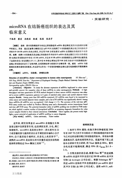
N:远端正常结肠组织;T:结肠癌组织上升的pm一面RNA 图3 定量PcR检测发生结肠癌时p咒-miRNA的变化情况
real.time qRT—PCR晒say.The potential biomarker utibty 0f miRNA profiling w鹪revealed by PAM analy-
sis,which applied a modest number of dysregulated llliRNAs硒Pmmising出agnostic biomarke瑙for c010n
miR_22l、miR一222、miR.223 miR.224。所有结果均以元 ±s用柱状图表示,数据用student t—t检验进行比较分 析,表示8P<O.05,表示oP<0.ol。
3.Northem blot:根据定量PCR实验结果,我们随 机挑选了miR-21,做Northem blot实验,证明IIliR-21 在发生结肠癌时上调(图2)。研究结果与RT-PCR的 结果一致。
miR—lOb、miR—16、miR.17-3p、miR-17-5 p、miR一18a、llliR—
19a、miR_20a、miR-21、miR以、miR-25、miR-27a、miR-
3l、miR一34a、miR.92、miR-93、miR一103、miR一106a、miR— 106b、miR—107、miR.126宰、miR一128a、miR—128b、miR一 132、miR一141、miR-142.3p、miR一142-5p、miR一146b、miR一 148a、miR.155、miR一18la、miR—181b、miR—188、miR- 189、miR一196a、miR—199a、miR.199b、miR.200a、miR一 200b、miR一200c、miR-203、miR-205、miR-2lO、miR-219、
miR—21在大肠癌中表达及临床意义

miR—21在大肠癌中表达及临床意义大肠癌是临床最常见的恶性肿瘤之一,在全世界范围内,大肠癌的发病率男女均处于恶性肿瘤的第3位,在我国,随着生活水平的不断提高,饮食习惯的改变,大肠癌的发病率呈上升态势,排在恶性肿瘤和致死因素的第4位,大肠癌是我国肿瘤患者死亡的主要原因之一。
微小RNA是一种真核生物内源性小分子单链RNA,微小RNA分子通过翻译抑制或者其他形式的调节机制抑制靶基因的表达。
目前,已发现多种微小RNA在大肠癌组织及大肠癌细胞系中异常表达,我们应用实时定量反转录聚合酶联反应技术,在大肠癌组织和对应正常大肠黏膜中进行miR-21的表达水平检测,并结合术后病理资料,分析miR-21与大肠癌临床病理因素的相关性,以其为大肠癌诊断和抗转移治疗提供新的线索。
资料与方法2010年6月~2011年5月收治进展期大肠癌患者64例,手术切除的肿瘤组织,全部患者均经病理证实为进展期大肠癌,术前均未行放化疗,所以患者均取得知情同意,同时收集距癌灶>5cm正常肠黏膜组织作为正常对照。
试剂与仪器:微小RNA提取试剂盒及聚合RNA加尾试剂盒均为美国安边公司产品,逆转录试剂盒、实时荧光定量聚合酶联反应试剂盒为日本塔克拉公司产品,实时定量聚合酶联反应仪购自美国寇伯特公司。
方法:总RNA抽提取10mg大肠癌组织及对照正常黏膜组织,按照使用Ambion公司的微小RNA提取试剂说明书步骤提取总RNA。
然后对RNA加尾,由于miRNAs是长约22核苷酸的短小片段RNA,常规聚合酶联反应无法设计出上下游引物。
本实验中我们先对所有RNA的3’末端加聚苷腺酸尾巴,对其人为地增加长度。
反转录过程中,所使用的引物3’端为一人为设计的序列,具体步骤参照Ambion公司的聚苷腺酸加尾试剂盒说明书。
逆转录及荧光定量聚合酶联反应时,以u6管家基因做为内参,每个标本设3个复孔。
取总RNA为模板,按照逆转录试剂盒说明书配反应体系,逆转录出互补DNA;然后按照实时荧光定量聚合酶联反应试剂盒说明书。
miR-21、miR-31在大肠癌和癌旁正常组 织中的表达和临床意义
分类号:R656.9 密级:一般U D C :617.55 编号:2010212451广州医学院硕士学位论文miR-21、miR-31在大肠癌及癌旁正常组织中的表达及临床意义研究生:宋韬导师:胡伟民主任医师申请学位级别:医学硕士年级:二零一零级学科专业:外科学研究方向:结直肠癌综合治疗论文提交日期:2013年5月论文答辩日期:2013年5月学位类型:临床医学专业学位学位授予单位:广州医学院答辩委员会主席:评议人:2013年5月广州医科大学硕士研究生学位论文miR-21、miR-31在大肠癌及癌旁正常组织中的表达及临床意义Expressions of miR-21、miR-31 in colorectal cancer and cancer-adjacent colorectal tissues and theirclinical significances研究生:宋韬导师:胡伟民学号:2010212451 学科专业:外科学年级:2010级院系:研究生院广州医学院附属肿瘤医院二零一三年五月目录缩略词表 (1)中文摘要 (2)英文摘要 (5)前言 (9)材料和方法 (12)结果 (21)讨论 (39)结论 (49)参考文献 (50)附图 (53)综述 (56)致谢 (68)学位论文原创性声明 (69)学位论文知识产权权属声明 (69)关于学位论文使用授权的说明 (69)缩略词表缩略词英文名中文名miRNA MicroRNA 微小核糖核酸RT-qPCR Real-time quantitative PolymeraseChain Reaction 实时荧光定量聚合酶链式反应AJCC American Joint Committee onCancer美国癌症联合会MSI-H high-frequency microsatelliteinstability高频率微卫星不稳定性WHO World Health Organization 世界卫生组织RISC RNA-induced silencing complex 核糖核酸诱导沉默复合物PDCD4 Programmed Cell Death 4 程序性细胞死亡因子4 OD Optical Density 光密度1.49 groups of colorectal cancer samples PCR results: miR-21 with the results of 45cases, miR-31 with the results of 44 cases,both have results in 41 cases.2.45 cases with positive results of miR-21: The ΔCT mean of Cancer tissues was-0.68±4.24, the ΔC T mean of cancer-adjacent normal tissues was 4.28±3.30,the median fold change was 15.53.The miR-21 expression level is increased significantly in colorectal cancer tissues compared with cancer-adjacent normal tissues(P<0.05).44 cases with positive results of miR-31: The ΔCT mean of Cancer tissues was-0.45±2.46, the ΔCT mean of cancer-adjacent normal tissues was 1.53±3.81,the median fold change was 2.38.The miR-31 expression level is increased significantly in colorectal cancer tissues compared with cancer-adjacent normal tissues(P<0.05).25 cases with positive results of miR-21 in stageⅠand stage ⅡCRC samples:19(76%) cases showed high-expression,6(24%) cases showed low-expression, the median fold change was 2.83. The miR-21 expression level is increased significantly in stageⅠand stage Ⅱ CRC tissues compared with cancer-adjacent normal tissues (P<0.05).24 cases with positive results of miR-31 in stageⅠand stage ⅡCRC samples:13(54%) cases showed high-expression,11(46%) cases showed low-expression, the median fold change was 1.05.The expression level of miR-31 in stageⅠand stage ⅡCRC tissues had no statistically significant differences compared with cancer-adjacent normal tissues (P>0.05). The two genes expressed in Ⅲ and stage Ⅳ colorectal cancer were up-regulated(P<0.05).3.45 cases with positive results of miR-21: Serosal non-invasion 15 cases, themedian fold change was 3.63,the mean rank was 17.20; Serosal invasion 30 cases, the median fold change was 66.95,the mean rank was 25.90.No lymph node metastasis 26 cases, the median fold change was 2.91,the mean rank was 16.96;Lymph node metastasis 19 cases, the median fold change was 279.82,the mean rank was 31.26. Ⅰstage 13 cases, the median fold change was 2.98,the mean rankwas 14.92; Ⅱstage 12 cases, the median fold change was 2.83,the mean rank was17.25; Ⅲstage 14 cases, the median fold change was 148.42,the mean rank was29.64; Ⅳstage 6 cases, the median fold change was 873.40,the mean rank was36.50.44 cases with positive results of miR-31: Serosal non-invasion 13 cases, themedian fold change was 0.77,the mean rank was 14.88; Serosal invasion 31 cases, the median fold change was 4.11,the mean rank was 25.69.No lymph node metastasis 25 cases, the median fold change was 1.01,the mean rank was 16.36;Lymph node metastasis 19 cases, the median fold change was 9.65,the mean rank was 30.58. Ⅰstage 11 cases, the median fold change was 0.77,the mean rank was14.59; Ⅱstage 13 cases, the median fold change was 1.13,the mean rank was18.19; Ⅲstage 14 cases, the median fold change was 5.77,the mean rank was26.79; Ⅳstage 6 cases, the median fold change was 71.20,the mean rank was36.33.The expression levels of miR-21、miR-31 in CRC has significant positive correlation with serosal invasion、lymph node metastasis and the clinical stage(P<0.05). There was no relation to the expression levels of miR-21 and miR-31 in sex、age、tumor location、tumor size、general type、histological type、pathological grade and the number of metastatic lymph nodes(P>0.05).4.Both have results in 41 cases:high-expression of miR-21 34(83%)cases,low-expression of miR-21 7(17%)cases; high-expression of miR-31 27(66%)cases, low-expression of miR-31 14(34%)cases; combined high- expression 25(61%)cases. The expression levels of miR-21 and miR-31 in CRC were positively correlated(R=0.483, P<0.05). There is no significant difference between the positive rate of both high-expression(P>0.05).Conclusion:1. The expression levels of miR-21 and miR-31 in CRC were up-regulated comparedto cancer-adjacent normal tissues.The expression levels of miR-21 in stageⅠandstage ⅡCRC is increased significantly compared with cancer-adjacent normal tissues. The expression level of miR-31 in stageⅠand stage Ⅱ CRC tissues had no statistically significant differences compared with cancer-adjacent normal tissues.The two genes expressed in Ⅲand stage Ⅳcolorectal cancer were up-regulated.2. The expression levels of miR-21 and miR-31 in CRC has significant positivecorrelation with serosal invasion、lymph node metastasis and the clinical stage.There was no relation to the expression levels of miR-21 and miR-31 in sex、age、tumor location、tumor size、general type、histological type、pathological grade and the number of metastatic lymph nodes.3. The expression levels of miR-21 and miR-31 in CRC were positively correlated.There is no significant difference between the positive rate of both high-expression.Key words:colorectal cancer;miR-21; miR-31;RT-qPCR;clinicopathologic feature(图5)mir-21、mir-31△CT值的箱式图(CT值与该样本起始拷贝数的对数存在线性关系,起始拷贝数越多,CT值越小)。
miRNA-21在结直肠癌中的表达研究
a d j a c e n t n o n - t u mo r t i s s u e s . To e v a l u a t e t h e c o r r e l a t i o n o f c l i n i c p a t h o l o g i c f a c t o r s wi t h mi RNA- 2 1 l e v e l i n c o l o r e c t a l
( 吉林大学第二 4 1 )
摘要 : 目 的 检 测 Mi c r o R NA - 2 1 ( mi RNA 一 2 1 ) 在正常人和结直肠癌患者血清 , 以 及 结 直 肠 癌 患 者 癌 组 织 和 癌 旁 组
织 中的 表 达 , 探 讨 其 与结 直 肠 癌 临 床 病 理 参 数 的关 系 。方 法 采用实时荧光定量 P C R ( R T — q P C R) 技 术 检 测 正 常 人 和
c a n c e r . Me t h o d s Re a l — t i me RT— P CR wa s u s e d t O d e t e c t t h e e x p r e s s i o n o f mi RNA一 2 1 i n s e r u m o f n o r ma l h u ma n a n d
miRNA-21在结直肠癌中的表达研究

miRNA-21在结直肠癌中的表达研究邱实;孟凡旭;刘念;刘林林【摘要】目的检测MicroRNA-21(miRNA-21)在正常人和结直肠癌患者血清,以及结直肠癌患者癌组织和癌旁组织中的表达,探讨其与结直肠癌临床病理参数的关系.方法采用实时荧光定量PCR(RT-qPCR)技术检测正常人和结直肠癌患者血清,以及结直肠癌患者癌组织和癌旁组织中的表达,分析miRNA-21的表达与结直肠癌临床病理参数的关系.结果 miRNA-21在结直肠癌患者血清中表达水平显著高于正常人血清(P<0.05);结直肠癌组织中miRNA-21表达水平明显高于癌旁组织;中晚期(Ⅲ期和Ⅳ期)结直肠癌组织中miRNA-21表达水平明显高于早期(Ⅰ期和Ⅱ期)结直肠癌(P<0.05);低分化肿瘤组织中miRNA-21表达水平明显高于中高分化者;有淋巴结转移的结直肠癌组织中miRNA-21表达水平明显高于无淋巴结转移者.结论miRNA-21在结直肠癌患者血清及肿瘤组织中呈高表达,可能与结直肠癌的发生发展有关.【期刊名称】《中国实验诊断学》【年(卷),期】2014(018)001【总页数】3页(P91-93)【关键词】微小RNAs;结直肠肿瘤;聚合酶链反应【作者】邱实;孟凡旭;刘念;刘林林【作者单位】吉林大学第二医院,放疗科,吉林,长春130041;吉林大学第二医院,放疗科,吉林,长春130041;吉林大学第二医院,放疗科,吉林,长春130041;吉林大学第二医院,放疗科,吉林,长春130041【正文语种】中文【中图分类】R735.3结直肠癌(colorectal cancer,CRC)是胃肠道中常见的恶性肿瘤,随着近些年我国人口老龄化加剧、饮食习惯和生活方式的改变,结直肠癌的发病率和死亡率较前明显上升[1]。
结直肠癌的早期筛查与诊断越来越受人们的关注。
microRNA (miRNA)是由Lee等于1993年发现的一类新的长度为19-25的内源性非编码RNA分子,参与了发育、细胞增殖、分化及凋亡等一系列重要生命过程。
Mir 对应癌症

根据书中列举的115种miRNA的分类,家族中其他未列举的不做介绍Mir-15家族:Mir-15a,mir-16:慢性淋巴细胞白血病mir-195:新生儿心脏异常Hsa-Mir-146a:骨关节炎软骨Hsa-Mir-150:胃癌Hsa-Mir-155:白血病Mir-181a:抑制肿瘤Hsa-Mir-223:抑制肝癌、白血病Hsa-Mir-101:存在于脊椎动物中Mir-10家族:Hsa-Mir-125a-5-p:促进非小细胞肺癌细胞侵袭Hsa-Mir-10a:慢性,急性骨髓性白血病Hsa-Mir-10b:促进肿瘤细胞生长Hsa-Mir-126:内皮细胞Hsa-Mir-143:参与心脏形成Hsa-Mir-145:抑制直肠癌Hsa-Mir-148a:阻止骨关节炎软骨退化Mir-221家族:Hsa-Mir-221:肝癌Hsa-Mir-222:表达胚胎干细胞Hsa-Mir-373:非小细胞肺癌低表达Hsa-Mir-122:丙肝Hsa-Mir-133a:肌肉组织Hsa-Mir-9:乳腺癌Hsa-Mir-26a:抑制恶性肿瘤Hsa-Mir-31:乳腺癌Hsa-Mir-124:胃癌Hsa-Mir-1:心肌梗死Hsa-Mir-142家族Hsa-Mir-142-3p:白血病Hsa-Mir-142-5p:白血病Hsa-Mir-138:头颈部鳞状细胞癌Hsa-Mir-187:心肌梗死Hsa-Mir-192家族Hsa-Mir-192:胃癌Hsa-Mir-215:胃癌Hsa-Mir-204:头颈部鳞状细胞癌Hsa-Mir-214:子宫颈癌,胰腺癌Hsa-Mir-216:胰腺癌Hsa-Mir-22:乳腺癌,前列腺癌Hsa-Mir-32:前列腺癌Hsa-Mir-103家族Hsa-Mir-103:甲状腺癌Hsa-Mir-107:糖尿病Hsa-Mir-105:结直肠癌Hsa-Mir-17家族Hsa-Mir-106a:胃癌Hsa-Mir-106b:胶质瘤Hsa-Mir-17-3p:慢性骨髓性白血病Hsa-Mir-17-5p:非霍奇金淋巴瘤Hsa-Mir-18a:非霍奇金淋巴瘤Hsa-Mir-20a:非霍奇金淋巴瘤Hsa-Mir-199a-3p:宫颈癌Hsa-Mir-128:胶质瘤Hsa-Mir-129-5p:人类成骨肉瘤细胞系Hsa-Mir-132家族Hsa-Mir-132:食管癌Hsa-Mir-212:肝癌Hsa-Mir-134:肝癌Hsa-Mir-137:直肠癌,鳞状细胞癌,黑色素瘤Hsa-Mir-8家族Hsa-Mir-141:胰腺癌Hsa-Mir-429:结直肠癌Hsa-Mir-200a:子宫内膜腺癌Hsa-Mir-200b: 肺腺癌Hsa-Mir-144:不同肿瘤细胞的共同标识Hsa-Mir-153:非小细胞肺癌Hsa-Mir-194:结肠癌Hsa-Mir-196b:非小细胞肺癌Hsa-Mir-33a:代谢紊乱和心血管疾病Hsa-Mir-375:糖尿病Hsa-Mir-19家族:Hsa-Mir-19a:1型脊髓小脑的共济失调,乳腺癌Hsa-Mir-19b:类风湿性关节炎Hsa-Mir-25家族Hsa-Mir-92a:白血病标志物Hsa-Mir-92b:肝癌Hsa-Let-7家族Hsa-Let-7a:乳腺癌Hsa-Let-7b:恶性间皮瘤,淋巴瘤Hsa-Let-7c:肝癌Hsa-Let-7d:卵巢癌Hsa-Let-7e:恶性间皮瘤Hsa-Let-7f:糖尿病Hsa-Let-7g:肝癌Hsa-Let-7i:淋巴瘤Hsa-Mir-21:胃癌Hsa-Mir-34家族Hsa-Mir-34a:乳腺癌Hsa-Mir-34b: 非小细胞肺癌Hsa-Mir-34c-5p:非小细胞肺癌Hsa-Mir-127家族Hsa-Mir-127-3p:弥漫性大B细胞淋巴瘤Hsa-Mir-127-5p:肝癌Hsa-Mir-96:膀胱尿路上皮癌Hsa-Mir-182:肺鳞癌Hsa-Mir-183:乳腺癌Hsa-Mir-29家族Hsa-Mir-29a:白血病Hsa-Mir-29b:白血病Hsa-Mir-29c:白血病Hsa-Mir-140-5p:肝癌Hsa-Mir-193a-5p:膀胱癌Hsa-Mir-219-3p:类风湿性关节炎Hsa-Mir-28家族Hsa-Mir-28-3p:早产Hsa-Mir-151-3p:结核性脑膜炎Hsa-Mir-151-5p: 结核性脑膜炎Hsa-Mir-337-5p:胃癌Hsa-Mir-374b:刺激血管细胞形成纤维增值性病变Hsa-Mir-376b:肝癌Hsa-Mir-380:非小细胞肺癌Hsa-Mir-448:乙肝Hsa-Mir-453:Hsa-Mir-335:胃癌Hsa-Mir-135家族Hsa-Mir-135a:Hsa-Mir-135b:Hsa-Mir-148b:非小细胞肺癌Hsa-Mir-205:乳腺癌,前列腺癌,膀胱癌,肺癌Hsa-Mir-208家族Hsa-Mir-208a:急性心肌梗死Hsa-Mir-208b:急性心肌梗死Hsa-Mir-210:心肌梗死,肾上腺皮质癌Hsa-Mir-218:结肠癌Hsa-Mir-224:胰腺导管癌Hsa-Mir-302a:Hsa-Mir-345:Hsa-Mir-371家族Hsa-Mir-371-3p:婴幼儿血管瘤Hsa-Mir-371-5p:婴幼儿血管瘤Hsa-Mir-424:宫颈癌Hsa-Mir-483-5p:肝癌。
慢病毒介导miR-215过表达对克罗恩病的影响及作用机制

・论著・慢病毒介导miR-215过表达对克罗恩病的影响及作用机制侯静芳杨华赵世民王进海【摘要】目的研究慢病毒介导miR215过表达对克罗恩病(CD)小鼠的影响及其作用机制。
方法48只BALB/c小鼠随机分成空白组(A组)12只,模型组36只。
模型组经2,4,6-三硝基苯磺酸(TNBS)造模成功后的30只小鼠随机分成TNBS组(B组)、Lenti-Scramble(C组)、Lenti-miR-215(D组),每组各10只。
每日观察小鼠体质量、粪便性状、隐血便血,计算小鼠疾病活动指数(DAI)积分;ELISA试剂盒检测血清白细胞介素8(IL-8)JL-10#髓过氧化物酶(MPO)的水平;HE染色观察结肠组织病理变化;Realtime PCR检测结肠组织miR-215mRNA和Beclin1mRNA的水平;Western blotting检测结肠组织Beclin1、肿瘤坏死因子p(TNF p)和IL1*蛋白的水平。
结果与A组比较,B、C、D组小鼠DAI评分显著升高,miR-215mRNA的表达水平显著降低(P<0.05),B、C组IL8、MPO、TNF&和IL1*表达水平显著升高,IL-0、Beclin1蛋白表达水平显著降低(P<0.05)。
D组小鼠DAI评分、IL8、MPO、TNF&和IL1*蛋白水平均显著低于B、C组(P<0.05),而IL-10、miR-215mRNA、Beclin1mRNA、Beclin1蛋白的表达水平显著高于B、C组(P<0.05)。
结论miR215过表达对于CD小鼠有一定的保护作用,该作用与Beclin1介导的自噬通道有关。
【关键词】克罗恩病;miR-215;Beclin1DOI:10.3969#.issn.1673-534X.2019.05.009Effects and mechanism of lentivirus-mediated over-expression of miR-215on Crohn's disease HOUJingfcrng.Health Science Center,XL a n J i aotong University,Q!an710000,China-YANG Hua.Cadre Ward,Department o f Senirr Digestive Medicine,Xi an No.1Hospital,Xi an710002,China-ZHAO Shimin.Department of Digestive Medicine,Xi an Daxing Hospital,Xi an710016,China-WANG Jinhai.Department of Digestive Medicine,the Second Af f iliated Hospital of Xi'anJiaotong University,Xi'an710004,China【Abstract!Objective This paper attempts to investigate the effect of lentivirus mediated miR-215over-expression on CD mice and its mechanism.M ethods48BALB/c mice were randomly divided intothe blank group(Group A with12mice)and the model group(36mice).After successful TNBSmodeling,the model group was further randomly divided into the TNBS group(Group B),the lenti-scramblegroup(GroupC)%andthelenti-miR-2!5group(GroupD)%!0ineachgroup.TheDAIscore wascalculatedbyobservingthebodyweight%fecalcharacters%andoccultbloodandstoolbloodofthemiceeveryday.TheserumIL-8%IL-!0%and KPO weredetectedbyELISA.Thepathologicalchangesof colon tissues were observed by HE staining.The real-time PCR was performed to detect the miR-215and Beclin1mRNA in colon tissues.The Beclinl,TNF-&,and IL-1proteins in colon tissues weredetected by Western blo t ing.Results ComparedwithGroupA%theDAIscoresofmiceinGroupsB%作者单位=710000西安交通大学医学部(侯静芳,现就职于西安市第一医院干部病房老年消化内科)710002西安市第一医院干部病房老年消化内科(杨华)710016西安市大兴医院消化内科(赵世民);710004西安交通大学第二附属医院消化内科(王进海)通信作者:王进海,Email:j inhaiwang@C,and D were significantly increased,and the expression of miR-215mRNA was significantly decreased (P<0.05).Compared with Group A,the IL-8,MPO,TNF-&,and IL-1in Groups B and C were significantly increased,while the IL-10and Beclin1were significantly decreased(P<0.05).The DAI score,IL-8,MPO,TNF-&protein,and IL-1protein in Group D were significantly lower than those in GroupBandC(P<0.05)whereas7heexpressionsofIL-10miR-215mRNA Beclin1mRNA and Beclin1protein were significantly higher than those in Groups B and C(P<0.05).Conclusion The over-expression of miR-215has a protective e f ect on CD mice whichisrelatedtobeclin1-mediated autophagypathway*【Keywords】Crohns disease;miR-215;Beclin1克罗恩病(CD)以非特异性的炎性损伤、肉芽肿、结肠局灶性为主要病变特征切。
结直肠肿瘤组织miR-215表达及临床意义
2 01 6 Vl 0 1 . 3 No. 3 7
临床 医药文献杂志
J o u ma l o f Cl i n i c a l Me d i c a l 7 3 6 7
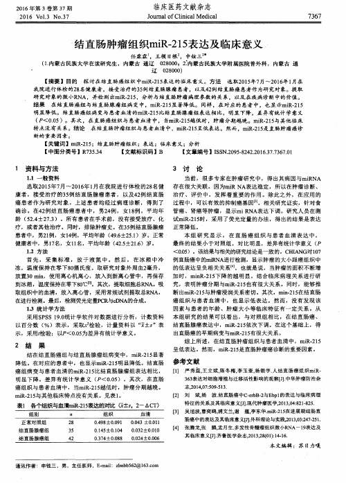
结直肠肿瘤组织mi R 一 2 1 5 表达及 临床意义
研 究对 象的微 小R N A,并检 测 d  ̄ mi R . 2 1 5 ,分析与结直肠肿 瘤病 理参数的关 系,以及在疾病诊 断 中的价值。 结果 在 结直肠癌 组与结 直肠腺 瘤组病 变 中,mi R 2 1 5 显 著降低 。同样 ,在对应 的患者 中,也显 - , P , mi R. 2 1 5 明显 降低 。结直肠癌组病 变与患者血 清的mi R . 2 1 5 比结直肠腺瘤组表达相 比 ,明显下 降,差异 有统计学意义 ( P<0 . 0 5)。其 次 ,在直肠癌组 织与 患者血 清中 ,当mi R . 2 1 5 越低 时 ,肿瘤分期越 晚。mi R . 2 1 5 与其他 临床 特点没有 关系。结论 在 结直肠肿 瘤组织与患者血清 中,mi R . 2 1 5 呈低表达。然 而,mi R - 2 1 5 是直肠肿瘤癌诊
到冰箱 ,温度保持在零T8 o ' c [ 2 ] 。其次,提取细胞总R N A 。吸 究,表 明肿瘤 分期与mi R. 2 1 5 也有很大关系 。同时,能够推 断出mi R - 2 1 5 与肿瘤浸润关系密切。其次,mi n . 2 1 5 在结直肠 取组织 中的血清 ,放入离心管 ,采用常
1 . 2 方 法
本 组 研 究 显 示 ,在 直 肠癌 组织 与 患者 血清 表达 中 , 最终 的结 果小于对 照组 ,对 比明显,差异有统计学意义 ( P <O . 0 5 )。该结果与相关的研究结论是一致的 。C HL N G对l 0 7 的低表达呈负相关 关系【 4 ] 。也就是说 ,当肿瘤 的面积 不断增
miR-21、miR-155、miR-210的临床表达意义及其与淋巴瘤病理特征的相关性分析
2 2 6 3 6 1
【 摘要】 目的 分析 m i R 一 2 1 、 m i R 一 1 5 5 、 m i R 一 2 1 0的临床表达意义及其 与淋 巴瘤病理特征 的相关 性。 方法 选取
2 0 1 4年 l 2月一 2 0 1 6年 1 2 月 南 通市 肿瘤 医院 收治 的 6 2 例 经 病理 学 证实 的淋 巴瘤 患 者作 为研 究对 象 ,分 别对 淋
[ Ab s t r a c t ] Ob j e c t i v e T o i n v e s t i g a t e t h e c l i n i c a l s i g n i i f c a n c e o f mi R一 2 1 , mi R 一 1 5 5 a n d mi R一 2 1 0 a n d t h e i r c o r r e l a t i o n
F ENG Xi a o y u n Y A N L i n g l i n g
De p a r t me n t o f L a b o r a t o r y , Na n t o n g T u mo u r Ho s p i t a l , J i a n g s u P r o v i n c e , Na n t o n g 2 2 6 3 6 1 ,C h i n a
结直肠癌中miR-21表达与临床病理因素及预后的关系
E x pr e s s i o n of m i R-21 i n c ol o r e c t a l c a n c e r a n d i t s cI i ni cO p a t h OI Ogi c s i gn i f i c a n c e
D e p a r t me n t o f C o l o r e c t a l a n d An a l S u r g e r y. t h e F i f s t A f il f i a t e d H o s p i t a l o f We n z h o u Me d i c a l U n i v e r s i t y. W e n z h o u 3 2 5 0 0 0.Ch i n a
结直肠癌预后 影响 因素 。 结果
结直肠癌 组织 中 mi R 一 2 1的相对表达量 明显高于其 癌旁正常黏膜 组织 ( P <O . 0 5 o结直 肠癌组织
mi R 一 2 1 表达水 平与肿瘤直径 、 淋 巴结转移 、 临床分期有 关( 均P <O . 0 5) ; 与性别 、 远处转移 、 分化程 度、 浸 润深度和肿瘤位 置无关 ( 均 P>O 0 5 o远 处转移 、 分 化程度 、 mi R 一 2 1表达水平 是影响患者预 后的独立危险 因素【 均P <O . 0 5) , 其R R值及 9 5 %C 1 分 别为 5 . 0 5 3 ( 1 . 3 9 5 ~ 5 . 7 8 7 ) 、 3 . 9 9 8 ( 1 . 3 9 5 — 1 8 . 3 0 5) 、 3 . 8 3 7 ( 1 . 2 6 6 ~ 1 1 . 6 3 1 o 结论 【关键词 】 结直肠癌 mi R 一 2 1 预后 mi R 一 2 1与结直肠癌患者预后和疾 病进展 密切相 关。 Co x比例风险 回归模型
胃癌组织miR-215表达变化及其与患者预后的关系
选择 胃癌患者 5 5例 , 取手术切除的 胃癌组织及
其 配对 的癌旁正常组织 , 采用 q R T — P C R法 检测 m i R - 2 1 5 表达 , 并 分析其 与患者临床 病理参数 及预后 的关 系。结果
胃癌组织 m i R . 2 1 5的相对表达量 明显 高于癌旁 正常组 织 ( P< 0 . 0 5 ) , 且其 表达 与 胃癌 组织分 化程 度 、 淋 巴结 转
究 发现 , 在 多 种 肿 瘤 组 织 中 可 检 测 到 异 常 表 达 的
2 1 5表达 变化 , 并 分 析其 表 达 与 患者 临 床 病 理 参 数 及 预后 的关 系 。现分析 结果 并报告 如下 。
1 资 料 与方法
1 . 1 临 床资料
选 择 同期 在 我 院手 术 治疗 的 胃癌
山东医药 2 0 1 7年第 5 7卷箍 墨
胃癌 组 织 m i R 一 2 1 5表 达 变 化 及 其 与 患 者 预后 的关 系
刁卓 , 刘晓超 , 唐 玉虎 , 范 莉静 ( 汉 中市 中心 医院 , 陕西 汉 中 71 5在 胃癌 发生 、 发展 中的作用 。方法
究 经 医院伦 理委 员会 批准 , 患者 均知 情 同意 。
1 . 2 m i R - 2 1 5表 达 检 测 采 用 q R T - P C R 法 。 取
手术切除的胃癌组织及癌旁正常组织 , T R I z o l 法提取
( S u p p l 4 ): ¥ 7 2 1 一 ¥ 7 2 8 .
胃癌 组织 中 m i R 一 2 1 5表 达 上 调 , 可 能 具 有 癌 基 因作 用 。2 0 1 0年 1— 4月 , 我们 观 察 了 胃癌 组 织 m i R 一
miR-324-5p和miR-215-5p在多发性骨髓瘤中的表达及预后分析
miR-324 -5p 和miR-215 -5p 在多发性骨髓瘤 中的表达及预后分析**基金项目:辽宁省自然科学基金资助项目(173****1001)△通信作者:艾丽梅,教授,E-mail :*****************宋翠薇I,艾丽梅仏'锦州医科大学抚顺市中心医院研究生培养基地(辽宁抚顺113006); 2锦州医科大学附属第一医院血液科(辽宁锦州 121000)【摘要】 目的 检测多发性骨髓瘤(MM )患者血清中miR - 324 -5p 和miR -215 -5p 的表达水平,研究其表达水平和预后的关系 方法 收集60例MM 患者血清标本,为初治组(20例)、巩固治疗组(20例)、复发难治组(20例);20例健康人血清标本(健康对照组),实时荧光定量PCR 检测血清中miR -324 -5p 和 miR-215 -5p 的表达水平。
结果 与健康对照组比较,MM 患者血清中miR-215 -5p 表达水平下降(P< 0. 008 3),初治组和难治复发组患者血清miR -324 -5p 表达水平下降(P <0.008 3),巩固治疗组与健康对照组血清miR -324 -5p 表达水平差异无统计学意义(P >0. ()08 3);巩固治疗组血清miR -324 -5p 表达水平高于初治组及难治复发组(P <0.008 3);巩固治疗组血清miR-215 -5p 表达水平高于初治组(P V 0. 008 3),巩固治疗和难治复发患者间差异无统计学意义(P>0. 008 3);初治组与复发难治组之间两种miRNAs 表达水平差异无统计学意义(P>0. 008 3)? miR -324 -5p 和miR -215 -5p 表达水平与p 2微球蛋白和红细胞体积 分布宽度呈负相关,与血红蛋白呈正相关 初治MM 患者血清miR -324 -5p 和miR-215 -5p 高表达组无 进展生存期高于低表达组,差异有统计学意义(P<0. 05).,结论 miR -324 -5p 和miR-215 -5p 在MM 血清中表达水平降低,miR -324 -5p 和miR -215 -5p 可能与疾病密切相关,其低表达可能提示预后不良【关键词】miR -324 -5p ; miR -215-5p ;多发性骨腌瘤;预后【中图分类号】R733.3;R730.7【文献标志码】ADOI : 10. 13820/j. cnki. gdyx. 20200279Expression and prognosis analysis of miR - 324 - 5p and miR - 215 - 5p in multiple myeloma. SONG Cui 一wei , Al Li - mei. Department of Postgraduate Hematology , Fiishun Central Hospital , Jinzhou Medical University , Fush- un 113006, Liaoning , ChinaCorresponding author : Al Li - mei. E 一 mail : alm 121001 @ 163. com[Abstract ] Objective To evaluate the serum expression levels of miR - 324 - 5p and miR -215 — 5p in multiplemyeloma ( MM) , and then study the correlations between expression levels and prognosis of MM. Methods The serumsamples of 60 MM patients and 20 healthy controls were collected. The MM patients were divided into three groups , the newly diagnosed group ( n =20) , consolidation group ( n =20) and relapse or refractory group ( n = 20). The serum ex pression levels of miR -324 - 5p and miR -215 - 5p were assessed by Real - time quantitative PCR. Results Compared with healthy controls , the expression levels of miR -215 — 5p were down - regulated in MM patients , the expression levels of miR -324 -5p between consolidation treatment group and healthy controls had no significant differenee, and the ex pression levels of initially treated and refractory or relapse patients were significantly lower than those in healthy controls. The expression level of miR — 324 - 5p in consolidation treatment group was higher than those in initially treated and re fractory or relapse patients. The expression level of miR -215 - 5p in consolidation treatment patients was higher than that of initially treated patients ; and have no difference with refractory or relapse patients. The expression levels of miR -324 - 5p and miR -215 - 5p had no significant difference between the newly diagnosed and relapsed or refractory MM pa tients. And the expression levels of miR — 324 -5p and miR -215 -5p were negatively correlated with 02 niicroglobulinand red blood cell volume distribution width , and were positively correlated with hemoglobin. Survival analysis of the new ly diagnosed MM patients showed that patients with low expression levels of miR - 324 - 5p and miR -215 - 5p had lower disease - free survival than those with high expression. Conclusions The serum expression levels of miR - 324 - 5p and miR -215 -5p are clown — regulated in MM. The two miRNAs may be closely related to the disease and the low serum ex-pression levels of miR-324—5p and miR—215-5p may indicate poor prognosis.[Key words]miR-324—5p;miR-215—5p;multiple myeloma;prognosis多发性骨髓瘤(multiple myeloma,MM)是一种浆细胞恶性增殖性疾病,发病占血液系统肿瘤的第2位,仅次于非霍奇金淋巴瘤,约占13%[1]0随着对其生物学、病理学和新的治疗方案的研究,患者的生存率和生活质量得到了显著提高。
《2024年直肠粘膜刷检细胞miRNA-182-5p在结肠癌诊断价值的临床研究》范文

《直肠粘膜刷检细胞miRNA-182-5p在结肠癌诊断价值的临床研究》篇一一、引言随着医疗科技的不断进步,对于疾病的早期诊断与治疗手段也在日益精细和全面。
尤其是结肠癌这类常见的恶性肿瘤,如何早期诊断与准确治疗已经成为医疗领域的焦点问题。
近年的研究显示,通过研究机体细胞内miRNA的表达模式可以实现对疾病的诊断。
其中,直肠粘膜刷检细胞miRNA-182-5p在结肠癌的诊断中具有重要价值。
本文旨在探讨这一指标在结肠癌诊断中的实际价值。
二、方法本研究采用直肠粘膜刷检法收集患者细胞样本,并运用高通量测序技术对样本中的miRNA-182-5p进行检测。
同时,我们选取了健康人群作为对照组,对两组的miRNA-182-5p表达水平进行对比分析。
三、结果1. 直肠粘膜刷检细胞miRNA-182-5p的表达情况通过对患者和健康人群的样本进行检测,我们发现结肠癌患者直肠粘膜刷检细胞中miRNA-182-5p的表达水平明显高于健康人群。
这一结果提示我们,miRNA-182-5p的检测可能成为结肠癌早期诊断的一个重要指标。
2. miRNA-182-5p与结肠癌病情的关系我们对不同分期、不同病情的结肠癌患者进行了分析,发现miRNA-182-5p的表达水平与病情的严重程度密切相关。
病情越严重,miRNA-182-5p的表达水平越高。
这表明miRNA-182-5p的检测不仅可用于早期诊断,也可用于评估患者的病情严重程度。
3. miRNA-182-5p的诊断价值通过对比分析,我们发现miRNA-182-5p的检测在结肠癌的诊断中具有较高的敏感性和特异性。
其诊断的准确率明显高于传统的诊断方法,如肠镜、病理检查等。
这表明miRNA-182-5p的检测具有较高的临床应用价值。
四、讨论本研究表明,直肠粘膜刷检细胞miRNA-182-5p在结肠癌的诊断中具有重要价值。
其表达水平与结肠癌的病情严重程度密切相关,且其诊断的敏感性和特异性均较高。
《2024年直肠粘膜刷检细胞miRNA-182-5p在结肠癌诊断价值的临床研究》范文

《直肠粘膜刷检细胞miRNA-182-5p在结肠癌诊断价值的临床研究》篇一一、引言结肠癌是一种常见的消化道恶性肿瘤,其发病率逐年上升,严重威胁人们的生命健康。
早期诊断和及时治疗是提高结肠癌患者生存率的关键。
然而,传统的结肠癌诊断方法如结肠镜检查和病理组织学检查,存在一定程度的侵入性和不便性,因此寻找一种无创、简便、高效的诊断方法显得尤为重要。
近年来,随着分子生物学技术的发展,microRNA(miRNA)在肿瘤诊断和治疗中的价值逐渐受到关注。
本研究旨在探讨直肠粘膜刷检细胞中miRNA-182-5p在结肠癌诊断中的价值。
二、研究方法1. 研究对象本研究纳入经结肠镜检查和病理组织学检查确诊的结肠癌患者及健康体检者。
所有研究对象均签署知情同意书,并经医院伦理委员会批准。
2. 样本采集与处理采用直肠粘膜刷检法采集研究对象的样本,提取细胞中的RNA,并进行miRNA-182-5p的检测。
3. 实验方法运用实时荧光定量PCR技术检测miRNA-182-5p的表达水平,并分析其与结肠癌诊断的相关性。
三、结果1. miRNA-182-5p在结肠癌患者与健康体检者中的表达差异本研究发现,miRNA-182-5p在结肠癌患者中的表达水平显著高于健康体检者(P<0.05)。
这表明miRNA-182-5p的表达水平与结肠癌的发生密切相关。
2. miRNA-182-5p在结肠癌诊断中的价值通过对miRNA-182-5p的表达水平进行诊断分析,我们发现其对于结肠癌的诊断具有较高的敏感性和特异性。
在早期结肠癌诊断中,miRNA-182-5p的检测有助于提高诊断的准确率。
3. miRNA-182-5p与其他临床指标的关联性本研究还发现,miRNA-182-5p的表达水平与结肠癌患者的肿瘤大小、淋巴结转移等临床指标具有一定的关联性。
肿瘤越大、淋巴结转移越多的患者,其miRNA-182-5p的表达水平往往越高。
四、讨论本研究表明,直肠粘膜刷检细胞中miRNA-182-5p的表达水平与结肠癌的发生、发展密切相关,对于结肠癌的诊断具有一定的价值。
Micro

Micro摘要】目的:探讨micro RNA-215(mi R-215)在结直肠癌Lovo细胞系中的表达并观察对结直肠癌细胞系增殖凋亡能力的影响。
方法:结直肠癌细胞系Lovo与正常结直肠细胞系CCD-18Co分别行细胞培养,Real-time PCR检测mi R-215的表达差异;转染mi R-215表达质粒(mi R-215 mimics)、mi R-215抑制质粒(mi R-215 inhibitor)于Lovo细胞,筛选出稳定表达的细胞株,并Real-time PCR检测mi R-215的表达,MTT流式细胞仪检测mi R-215对Lovo细胞增殖和细胞凋亡的影响。
结果:mi R-215在结直肠癌细胞系Lovo中的表达与正常结直肠细胞系相比均明显增高(P<0.01)。
稳定转染后,与NC组相比,转染mi R-215拟似物后mi R-215相对表达量明显升高(t=6.528,P<0.05),MTT检测Lovo细胞增殖活力下降(t=3.472,P<0.05),细胞凋亡比例明显升高(t=5.704,P<0.05);而转染mi R-215抑制物后mi R-215相对表达量显著下降(t=6.324,P<0.05),Lovo细胞增殖活力却明显上升(t=3.331,P<0.05),细胞凋亡比例显著降低(t=2.300,P<0.05)。
结论:mi R-215在结直肠癌细胞中呈低表达,抑制mi R-215表达可促进Lovo细胞的增殖,降低细胞凋亡比例。
【关键词】结直肠癌;微小RNA;增殖;细胞凋亡【中图分类号】R73.3 【文献标识码】A 【文章编号】2095-1752(2016)28-0131-03结直肠癌是世界上最常见的消化道恶性肿瘤之一,近几年其发病率和死亡率明显升高[1-2]。
随着对结直肠癌研究加深,已经明确结直肠癌的发生、发展是一个多步骤、多因素参与的过程,这个过程中伴随着基因水平和表观遗传水平改变的累积[3-4]。
检测结直肠癌组织中miR-21,miR-106a,miR-143的表达水平

检测结直肠癌组织中miR-21,miR-106a,miR-143的表达水平【摘要】目的探讨良性结肠腺瘤与结肠腺癌组织中Hsa-miR-21-5p,Hsa-miR-143-3p,Hsa-miR-106a-5p表达水平的差异对临床诊断结肠腺瘤及癌变可能的意义。
方法利用荧光实时定量PCR(qRT-PCR)检测结肠腺瘤、结肠癌患者组织及瘤旁组织中Hsa-miR-143-3p,Hsa-miR-21-5p,Hsa-miR-106a-5p的表达水平,并分析三者的表达对结肠腺瘤或结肠癌的诊断意义。
结果结肠腺瘤组织中Hsa-miR-21-5p,Hsa-miR-106a-5p的表达水平较瘤旁组织显著增高(p<0.05),Hsa-miR-143-3p的表达水平显著降低(p<0.05);经病理鉴定癌变的组织中Hsa-miR-21-5p表达水平显著高于良性瘤组织,差异有统计学意义(p<0.05),而Hsa-miR-106a-5p的表达水平与良性瘤组织无统计学差异(p>0.05),Hsa-miR-143-3p的表达水平低于良性瘤组织,差异有统计学意义。
结论 Hsa-miR-21-5p,Hsa-miR-106a-5p,Hsa-miR-143-3p的表达水平对临床诊断结肠腺瘤及是否发生癌变有指导意义。
【关键词】结肠腺瘤;结肠腺癌; miRNA; Hsa-miR-21-5p; Hsa-miR-106a-5p; Hsa-miR-143-3pThe expression and significance Hsa-miR-21-5p, Hsa-miR-106a-5p, Hsa-miR-143-3p in colorectal adenomas【Abstract】 Objective Detecting the expression of Hsa-miR-21-5p, Hsa-miR-106a-5p, Hsa-miR-143-3p in colorectal adenomas and colon adenocarcinoma, to investigate their clinical diagnosis significance in colon adenoma and colon adenocarcinoma. Methods Fluorescence quantitative real-time PCR (qRT-PCR) assay were used to detect the expressions of Hsa-miR-143-3p, Hsa-miR-21-5p, Hsa-miR-106a-5p in colorectal adenoma tissues and Adenomatous adjacent tissues, further reveal their roles during colon adenoma or colon cancer diagnosis on the basis of corresponding pathological diagnosis. Results Hsa-miR-21-5p , Hsa-miR-106a-5p expression levels in colorectal adenomas than tumor adjacent tissues was significantly higher (p<0.05), the expression level of Hsa-miR-143-3p significantly lower (p<0.05); the expression Hsa-miR-21-5p was significantly higher than benign tumor tissue, the difference was statistically significant (p<0.05), but Hsa-miR-106a-5p was not (p>0.05), Hsa-miR-143-3p expression level lower than benign tumor tissue, the difference was statistically significant. Conclusion It is meaningful to detect the expression of Hsa-miR-21-5p,Hsa-miR-106a-5p, Hsa-miR-143-3p for clinical diagnosis of colorectal adenoma and whether cancerous instructive.【Key words】 Colorectal adenomas; Colorectal adenocarcinoma; miRNA; Hsa-miR-21-5p; Hsa-miR-106a-5p; Hsa-miR-143-3p结肠癌是发病率较高的消化系统恶性肿瘤,其5年生存率很低,结肠腺瘤尤其是进展期结肠腺瘤是结肠癌的主要癌前病变,早发现早诊断是提高患者生存率的关键。
结直肠癌中miR-21表达与临床病理因素及预后的关系
结直肠癌中miR-21表达与临床病理因素及预后的关系任约翰;叶星照;厉金雷;陈积贤【摘要】目的探讨结直肠癌组织中miR-21表达与临床病理因素及预后的关系.方法取59例结直肠癌组织标本及癌旁正常黏膜组织标本,采用茎环逆转录实时荧光定量PCR方法检测结直肠癌组织及癌旁正常黏膜组织中miR-21的相对表达量,并分析癌组织中miR-21表达与临床病理因素的关系.用Kaplan-meier法计算生存率,采用log-rank检验、Cox比例风险回归模型分析结直肠癌预后影响因素.结果结直肠癌组织中miR-21的相对表达量明显高于其癌旁正常黏膜组织(P<0.05).结直肠癌组织miR-21表达水平与肿瘤直径、淋巴结转移、临床分期有关(均P<0.05);与性别、远处转移、分化程度、浸润深度和肿瘤位置无关(均P >0.05).远处转移、分化程度、miR-21表达水平是影响患者预后的独立危险因素(均P<0.05),其RR值及95%CI分别为5.053(1.395~5.787)、3.998(1.395~18.305)、3.837(1.266~11.631).结论 miR-21与结直肠癌患者预后和疾病进展密切相关.%Objective To investigate the expression of miR-21 in colorectal cancer and its association with clinicopathological characteristics and the prognosis of patients.Methods The expression of miR-21 in 59 specimens of colorectal cancer and matched non-tumor adjacent tissue specimens was examined with stem-loop real-time RT-PCR.The relationship between the expression of miR-21 and clinicopathologic features of colorectal carcinoma was analyzed.Kaplan-Meier survival curves,log-rank test and multivariate Cox regression were used to analyze the factors related to prognosis of colorectal cancer.Results The expression level of miR-21 was significantly higher in colorectal cancer tissue than that in the matchednormal colorectal mucosa (2.698 ± 1.973 vs.0.653± 0.213,P<0.05).The expression of miR-21 was positively correlated with tumor size,lymph node metastasis and clinical stage (P<0.05),but not with sex,distant metastasis,cell differentiation,depth of tumor infiltration and location (P >0.05).Kaplan-Meier univariate analysis showed that distant metastasis,cell differentiation,depth of tumor infiltration and miR-21 was the prognostic factor for colorectal cancer (P<0.05).The Cox regression analysis showed that distant metastasis,cell differentiation and miR-21 was an independent prognostic risk factor of colorectal cancer(P<0.05),RR value and 95%CIwas 5.053 (1.395-5.787),3.998 (1.395-18.305) and 3.837 (1.266-11.631).Conclusion The results indicate that miR-21 serves as a molecular marker for disease progression and prognosis of colorectal cancer.【期刊名称】《浙江医学》【年(卷),期】2017(039)018【总页数】4页(P1544-1547)【关键词】结直肠癌;miR-21;预后;Cox比例风险回归模型【作者】任约翰;叶星照;厉金雷;陈积贤【作者单位】325000 温州医科大学附属第一医院结直肠肛门外科;325000 温州医科大学附属第一医院结直肠肛门外科;325000 温州医科大学附属第一医院结直肠肛门外科;温州医科大学附属第三医院肿瘤外科【正文语种】中文结直肠癌是世界上最常见的恶性肿瘤之一,全球每年约有120万人新发结直肠癌,术后5年生存率为60%~70%[1],严重危害人类的健康。
结直肠癌中miR-21的表达及其临床病理学意义

结直肠癌中miR-21的表达及其临床病理学意义摘要】目的:分析结直肠癌组织中miR-21的表达及其临床病理学意义。
方法:选取我院2012年1月-2012年12月间手术切除的结直肠癌标本30例作为研究对象,通过采用TaqMan实时定量RT-PCR法对该组标本中的结直肠癌组织miR-21的表达进行检测,并与其癌旁正常组织miR-21的表达进行对比观察,从而探讨结直肠癌临床病理因素与miR-21的表达之间的关系。
结果:通过与对比癌旁正常组织miR-21的表达进行对比观察发现,结直肠癌组织miR-21的表达明显要高很多,差异具有统计学意义(P<0.05)。
结直肠癌组织miR-21的表达与患者的年龄、性别、肿瘤部位等情况没有较大的关联,而与其淋巴结转移、肿瘤病理学分级、及术后生存率三个方面有着紧密的关系。
结论:结直肠癌组织miR-21的表达要高于正常组织,并且它可能与大肠癌的发生及肿瘤的生长有着密切的联系。
【关键词】结直肠癌 miR-21 临床意义【中图分类号】R730.2 【文献标识码】A 【文章编号】1672-5085(2014)12-0082-01MiR - 21 expression in colorectal cancer and its clinical pathological significance【Abstract】 objective: to analysis of miR - 21 expression in colorectal cancer tissues and its clinical pathological significance. Methods: from January 2012 to December 2012 surgical resection of colorectal cancer specimens from 30 cases as the research object, by adopting the TaqMan real-time quantitative rt-pcr method to this group of specimens of colorectal cancer tissue miR - 21 expression, and normal tissue adjacent to carcinoma with miR - comparing the expression of 21 observation, which discuss clinicopathological factors of colorectal cancer with the relationship between the expression of miR - 21. Results: by contrast with the normal tissue adjacent to carcinoma miR - comparing the expression of 21 observation found that miR - 21 expression of colorectal cancer tissues has much higher, the difference is statistically significant (P < 0.05). The expression of colorectal cancer tissue miR - 21 with the patient's age, gender, tumor location, and so on and so forth no larger correlation, and lymph node metastasis, tumor pathology classification, and postoperative survival rate three aspects have close relation. Conclusion: the expression of colorectal cancer tissue miR - 21 than in normal tissue, and it may be related to the occurrence of colorectal cancer and tumor growth is closely related.【Key words】Colorectal cancer miR-21 Clinical significance随着科学技术水平和医疗水平的进步与发展,对于病理的研究也更加深入。
结直肠癌中miR-21的表达及其临床病理学意义
结直肠癌中miR-21的表达及其临床病理学意义鲁华东;刘忠臣;潘超;吕彦;殷平【期刊名称】《临床与实验病理学杂志》【年(卷),期】2013(29)5【摘要】目的探讨结直肠癌组织中miR-21的表达及其临床病理学意义.方法采用TaqMan实时定量RT-PCR法检测43例结直肠癌组织及其癌旁正常组织中miR-21的表达,分析miR-21的表达与结直肠癌临床病理因素之间的关系.结果miR-21在结直肠癌组织中的表达明显高于癌旁正常组织(P<0.05),结直肠癌组织中miR-21的表达与肿瘤病理学分级、淋巴结转移及术后生存率密切相关(P<0.05),与患者性别、年龄、肿瘤部位等无明显相关性(P>0.05).结论 miR-21在结直肠癌中表达升高,miR-21高表达同结直肠癌病理学分级、淋巴结转移及术后生存率密切相关,miR-21可能在大肠癌的发生、发展过程中发挥重要作用.%Purpose To investigate the miR-21 expression in cancerous tissues from patients with colorectal carcinoma ( CRC ) and to evaluate its dinicopathologic significance. Methods TaqMan real-time quantitative RT- PCR was used to detect the miR-21 expression in cancerous tissues and normal tissues from 43 patients with colorectal carcinoma and to observe its relationship with dinicopathologic parameters. Results There was a significantly higher expression of miR-21 in CRC tissues in comparison with adjacent normal colon tissues ( P <0. 05 ). High miR-21 expression was associated with dinicopathologic features, such as tumor grade, lymph node metastasis and patient survival ( P <0. 05 ). However, there was no relationshipbetween miR-21 expression and other clinicopathologi-cal parameters including sex, age and tumor site. Conclusion The expression of miR-21 was up-regulated in colorectal carcinoma. High miR-21 expression correlates with tumor grade, lymph node metastasis and patient survival, which suggests that mir-21 may play an important role in the development and progression of colorectal carcinoma.【总页数】4页(P516-519)【作者】鲁华东;刘忠臣;潘超;吕彦;殷平【作者单位】厦门大学附属中山医院病理科,厦门,361004;厦门大学附属中山医院胃肠外科,厦门,361004;厦门大学附属中山医院病理科,厦门,361004;厦门大学附属中山医院病理科,厦门,361004;厦门大学附属中山医院病理科,厦门,361004【正文语种】中文【中图分类】R735【相关文献】1.COX-2和VEGF在结直肠癌组织中的表达及其临床病理学意义 [J], 乌日尼勒图;德力格尔图;2.接头蛋白 DOK2在结直肠癌中的表达及临床病理学意义 [J], 翟立广;陈国荣3.COX-2和VEGF在结直肠癌组织中的表达及其临床病理学意义 [J], 乌日尼勒图;德力格尔图4.miR-21和miR-106a在乳腺癌组织中的表达及临床病理学意义 [J], 白建平;卢建跃;孙新增;张霆5.结直肠癌组织中APC-7的表达及其临床病理学意义 [J], 崔瑛;朴杰;皮永浩;朴光华因版权原因,仅展示原文概要,查看原文内容请购买。
- 1、下载文档前请自行甄别文档内容的完整性,平台不提供额外的编辑、内容补充、找答案等附加服务。
- 2、"仅部分预览"的文档,不可在线预览部分如存在完整性等问题,可反馈申请退款(可完整预览的文档不适用该条件!)。
- 3、如文档侵犯您的权益,请联系客服反馈,我们会尽快为您处理(人工客服工作时间:9:00-18:30)。
研 究对 象的微 小R N A,并检 测 d  ̄ mi R . 2 1 5 ,分析与结直肠肿 瘤病 理参数的关 系,以及在疾病诊 断 中的价值。 结果 在 结直肠癌 组与结 直肠腺 瘤组病 变 中,mi R 2 1 5 显 著降低 。同样 ,在对应 的患者 中,也显 - , P , mi R. 2 1 5 明显 降低 。结直肠癌组病 变与患者血 清的mi R . 2 1 5 比结直肠腺瘤组表达相 比 ,明显下 降,差异 有统计学意义 ( P<0 . 0 5)。其 次 ,在直肠癌组 织与 患者血 清中 ,当mi R . 2 1 5 越低 时 ,肿瘤分期越 晚。mi R . 2 1 5 与其他 临床 特点没有 关系。结论 在 结直肠肿 瘤组织与患者血清 中,mi R . 2 1 5 呈低表达。然 而,mi R - 2 1 5 是直肠肿瘤癌诊
在进行检测。最后,检测荧光定IP C R -  ̄c D N A的合成 。 癌组织 与患者 血清 中 ,也 显示低表 达 。然而 ,没有发现 该
因素 与患者 的年龄 、肿瘤 大小等 临床特 征有一 定关系 。从 1 . 3 统计学方法 采用 S P S S 1 9 . 0 统计学 软件 对数据进 行分 析,计数 资料 本组研 究 的结果可 以看 出,与对 照组相 比 ,在 结直肠癌 、
到冰箱 ,温度保持在零T8 o ' c [ 2 ] 。其次,提取细胞总R N A 。吸 究,表 明肿瘤 分期与mi R. 2 1 5 也有很大关系 。同时,能够推 断出mi R - 2 1 5 与肿瘤浸润关系密切。其次,mi n . 2 1 5 在结直肠 取组织 中的血清 ,放入离心管 ,采用常规试剂提取总R NA,
以百分数 ( %)表示 ,采取, c 检 验 ,计量 资料 以 “ ± ”表 结直肠腺癌表 达中 ,mi R 2 1 5 依次下调 。在这个基础 上,得 示 ,采用t 检验 ;以P<0 . 0 5 为差异有 统计 学意义。 出直肠癌的早期病变与mi R . 2 1 5 有很 大关 系。
1 资料与方法
1 . 1一般资料
3 讨
论
当前 ,很多专 家在肿瘤 研究 中,得 出其 病因与mi R NA
选 取2 0 1 5  ̄ I z 7 月~2 0 1 6 年1 月在我 院进行 体检 的2 8 名 健 存在很大 关联 。因为mi R NA表达稳定 ,所 以在肿瘤 诊断 、 康 者 ,接 受治疗 的3 5 例 结直肠腺 瘤患者 ,以及4 2 例结直 肠 治疗 、评价 中 ,发 挥着重 要 的作用 。除此之外 ,在应 用 的 癌 患者作 为研 究对 象 。上 述患者 均经 过病理 诊断 ,得到 了 过程 中,可 以有 效的抑制癌基 因口 ] 。相 关研 究证实 ,针对食 确 诊 。在4 2 例 结直肠 癌 患者 中,男2 4 例 ,女 1 8 例 。平均 年 管癌 、肾癌等 肿瘤 ,显示mi R NA表达下调 。研 究人员在测
2 0 1 6年 第 3卷 第 3 7期
2 01 6 Vl 0 1 . 3 No. 3 7
临床 医药文献杂志
J o u ma l o f Cl i n i c a l Me d i c a l 7 3 6 7
结直肠肿瘤组织mi R 一 2 1 5 表达及 临床意义
任 霖 霖 。王模 日根 , 申铉 三 ( 1 . 内蒙古民族大学在读研究生 ,内蒙古 通辽 0 2 8 0 0 0 l 2 : 内蒙古 民族大学附属医院普外科 ,内蒙古 通
‘
辽
0 2 8 0 0 0 )
【 摘要 】目的 探 讨在结 直肠癌组 织 中mi R. 2 1 5 表达 的临床意义 。方 法 选取2 0 1 5 年7 月~2 0 l 6 年1 月在 我院进行体检 的2 8 名健 康者 ,接 受治疗的3 5 例结 直肠腺瘤 患者 ,v X Y L 4 2 例 结直肠癌患者作为研 究对 象。提取
2 结
果
综上所述 ,在 结直肠肿瘤组织与 患者血清 中,mi R . 2 1 5 呈低表达 。然而,mi R - 2 1 5 是直肠肿瘤癌诊 断的重要 因素 。
断的重要 因素 。
【 关键词 】mi R . 2 1 5 ;结直肠肿瘤组 织;表达 ;临床意 义;分析 【 中图分类号 】R 7 3 5 . 3 4 【 文献标识码 】B 【 文章编号 】I S S N. 2 0 9 5 . 8 2 4 2 . 2 0 1 6 . 3 7 . 7 3 6 7 . 0 1
4 2 . 5 ±2 1 . 6 )岁。
首 先 ,采 集标 准 ,放 于 液 氮 中 。然 后 ,在 冰 箱 中冷 例 直肠癌中的mi I { NA 进行检测 ,显示肿瘤 的大小跟癌 组织 中
冻 ,温 度保持在 零下8 0 摄 氏度。取研 究对 象外周血2 毫升 ,
放置3 0 mi n ,使用离心机离心 ,放入到新离心管 中,再保存 加时 ,mi n R. 2 1 5 - F降 的越 明显 。结 合临床病理 关系进 行研
健康者中,男1 7 名,女l 1 名,平均年龄
1 . 2 方 法
本 组 研 究 显 示 ,在 直 肠癌 组织 与 患者 血清 表达 中 , 最终 的结 果小于对 照组 ,对 比明显,差异有统计学意义 ( P <O . 0 5 )。该结果与相关的研究结论是一致的 。C HL N G对l 0 7 的低表达呈负相关 关系【 4 ] 。也就是说 ,当肿瘤 的面积 不断增
龄 ( 5 2 . 4 ±2 7 . 3 )。所 有患者在手术前 ,没有接受放疗 、化 试mi R. 2 1 5 时 ,采用 了荧光定量的办法 得 出的结果是表达
疗 ,或者其他治疗 。同时,排 除肿瘤史 。在 3 5 例 结直肠腺瘤 正常降低 。
患者中,男2 1 例 ,女l 4 例,平均年龄 ( 4 9 . 6 ±2 5 . 1 )岁。正常
