2010_欧洲肿瘤医学会:软组织肉瘤
临床医学肿瘤学试题含答案
临床医学肿瘤学试题含答案1、良性与恶性软组织肿瘤之比为()。
A、200:1B、50:1C、10:1D、5:1E、1:1答案:A:绝大多数软组织肿瘤为良性,而软组织肉瘤则相对少见,两者比例约为200:1。
2、下列那一项不是骨肉瘤的病变特点?()A、肿瘤质地柔软,无砂粒感B、起源于干骺端,呈偏心性C、X线上可见Codman三角D、是由前成骨细胞发生的E、可有肿瘤性新生骨答案:A:骨肉瘤是最常见的原发骨恶性肿瘤,病变多位于生长快速的长骨干骺端,瘤细胞直接形成骨样组织或未成熟骨。
肿瘤质地软,其中仅掺杂以少量砂砾样骨板。
X线表现可有不同的形态,可见Codman三角3、下列不属于经典型霍奇金淋巴瘤的是()。
A、富于淋巴细胞的经典霍奇金淋巴瘤B、结节硬化型C、结节性淋巴细胞为主型霍奇金淋巴瘤D、淋巴细胞衰减型E、混合细胞型答案:C:霍奇金淋巴瘤的病理分类:2000年WHO将霍奇金淋巴瘤分为结节性淋巴细胞为主型霍奇金淋巴瘤和经典型霍奇金淋巴瘤,后者分为:① 结节硬化型(1级和2级);②混合细胞型;③淋巴细胞衰减型;④富于淋巴细胞的经典霍奇金淋巴瘤。
4、有关胃癌的CT检查不正确的是()。
A、当病变与邻近脏器间脂肪层消失,提示有脏器受侵的可能B、早期胃癌CT扫描主要表现为胃壁局限性增厚,表面可不光滑,增强扫描病变不强化C、当肿瘤浸透浆膜层时,CT表现为浆膜面不光滑,周围脂肪间隙内有点、条状影D、进展期胃癌主要表现为胃壁局限性或弥漫性增厚,可见向腔内或腔外突出的肿块,也可伴有溃疡,增强扫描有不同程度强化E、当病变与邻近脏器间脂肪间隙清晰存在则为脏器无侵犯的可靠表现答案:B:早期胃癌CT扫描主要表现为胃壁局限性增厚,表面可不光滑,增强扫描病变可有强化。
5、在恶性肿瘤患者中,在病程的不同时期需要作放射治疗的大约占()。
A、90%B、30%C、70%D、50%E、100%答案:C:放射治疗是恶性肿瘤治疗的三大手段之一,据统计,约有60%~70%的癌症患者需要接受放疗。
肉瘤的名词解释

肉瘤的名词解释肉瘤是一种常见的肿瘤类型,它属于恶性肿瘤的一种。
与良性肿瘤相比,肉瘤生长迅速,侵袭性强,有较高的复发和转移风险。
肉瘤可以发生在身体的各个部位,包括骨骼、软组织以及内脏器官等。
尽管肉瘤的治疗一直是医学领域的研究焦点之一,但至今仍然面临很大的挑战。
首先,肉瘤可以分为多种亚型,每一种亚型的生长方式、组织学特征以及临床表现都有所不同。
最常见的肉瘤类型包括骨肉瘤、软组织肉瘤和脂肪肉瘤等。
骨肉瘤主要发生在骨骼中,如股骨或胫骨等,而软组织肉瘤则发生在身体的软组织部位,如肌肉、皮下组织等。
脂肪肉瘤则以良性脂肪瘤恶变而来,通常发生在脂肪组织丰富的区域,如大腿、手臂等。
此外,肉瘤还可分为其他亚型,如纤维肉瘤、血管肉瘤以及肉瘤样肉芽肿等。
其次,肉瘤的起因尚不清楚。
虽然遗传学、环境因素以及某些病理变化与肉瘤的发生有关,但具体的致病机制尚未完全阐明。
研究表明,肉瘤的发生可能与基因突变、染色体异常以及细胞增殖调控失衡相关。
此外,一些生活习惯和环境因素,如吸烟、暴露于致癌物质、放射线等也可能增加肉瘤的风险。
另外,肉瘤的诊断通常需要通过组织病理学检查来确诊。
临床上,医生会根据患者的症状、体征以及影像学检查结果来初步判断是否存在肿瘤。
然后,需要取得病灶组织进行活检,进一步明确肿瘤的性质。
病理学检查可以观察细胞类型、组织结构以及异常变化,从而确定肿瘤是否为肉瘤以及其亚型。
肉瘤的治疗主要包括手术切除、放疗和化疗等措施。
对于早期发现的肉瘤,手术切除通常是首选的治疗方法。
手术的目的是尽可能将肿瘤完整地切除,并且保持周围组织的正常功能。
然而,由于肉瘤的侵袭性和复发率较高,手术可能不能完全根治肿瘤,因此通常需要辅助的放疗和化疗。
放疗是利用高能射线杀死肿瘤细胞或控制其生长的治疗方法。
通过精确计算剂量和照射方向,放疗可以精确地破坏肿瘤细胞,减少肿瘤复发和转移的风险。
化疗则是通过使用抗肿瘤药物来杀死肿瘤细胞。
化疗通常被用于术前或术后治疗,以减小肿瘤的体积或预防转移。
软组织肉瘤亚型及亚型定义
软组织肉瘤是一种罕见且多样化的间叶性癌症,起源于间充质组织,即来源于胚胎中胚层的非上皮源性组织。
根据世界卫生组织(WHO)自2020年以来的最新分类,软组织肉瘤包括50多种不同的组织亚型。
以下是一些常见的软组织肉瘤亚型及其定义:
滑膜肉瘤:滑膜肉瘤是一种含有滑膜成分的软组织肉瘤,其特点是在滑膜组织中形成肉瘤样的肿瘤细胞。
胃肠间质瘤:胃肠间质瘤是一种起源于胃肠道的软组织肉瘤,其特点是在胃肠道中形成肉瘤样的肿瘤细胞。
恶性周围神经鞘瘤:恶性周围神经鞘瘤是一种起源于周围神经系统的软组织肉瘤,其特点是在周围神经系统中形成肉瘤样的肿瘤细胞。
多形性脂肪肉瘤:多形性脂肪肉瘤是一种起源于脂肪组织的软组织肉瘤,其特点是在脂肪组织中形成肉瘤样的肿瘤细胞。
多形性横纹肌肉瘤:多形性横纹肌肉瘤是一种起源于横纹肌肉的软组织肉瘤,其特点是在横纹肌肉中形成肉瘤样的肿瘤细胞。
未分类的多形性肉瘤:未分类的多形性肉瘤是一种无法明确分类的软组织肉瘤,其特点是在体内的某个部位形成肉瘤样的肿瘤细胞。
以上只是一部分常见的软组织肉瘤亚型,实际上,软组织肉瘤的亚型种类非常多,每种亚型都有其独特的特性和治疗方法。
对于具体的某一种亚型,可能需要专业医生的进一步诊断和治疗。
软组织肉瘤ppt课件

软组织肉瘤
1
内容
定义及统计学资料 诊断 分级及分期 治疗 预后及随访
2
定义
软组织肿瘤是除骨骼、淋巴造血组织和神经组织以外的所有非上皮性组 织,包括纤维组织、脂肪组织、平滑肌组织、横纹肌组织、脉管组织以 及各种实质脏器支持组织的肿瘤。 根据肿瘤生物学潜能,WHO分为四个类型: ①良性。绝大多数不复发,即使复发也为非破坏性,局部完整切除几乎 可治愈,罕见转移。 ②中间性局部侵袭性。常复发,可伴局部浸润和破坏,但几无转移,如 韧带样纤维瘤、非典型脂肪性肿瘤/高分化脂肪肉瘤、卡波西型血管内 皮细胞瘤等。 ③中间性偶见转移性。肿瘤呈侵袭性生长,远处转移概率<2%,见于丛 状椎纤维组织细胞肿瘤、血管瘤样纤维组织细胞瘤、孤立性纤维瘤(大部 分为血管外皮瘤)、炎性肌纤维母细胞性肿瘤等。 ④恶性 又称为软组织肉瘤
IFOS为基础方案Vs. 不含IFOS方案
以IFOS为基础的方案在治疗反应上显著好于不含IFOS 的方案, 但1年生存率没有显著性差异;
EORTC 62012 研究
AI方案较ADM显著提高了有效率和PFS, 但在OS上还是 没有明显获益.
25
26
27
其他化疗策略
Gemcitabine为基础的方案
12
分期 AJCC
13
治疗(四肢、躯干及头颈部)
Ⅰ期肿瘤(T1a~2b,N0,M0,G1),手术是主要治疗方式。≤5cm 的病灶,切缘大于1cm或深筋膜完整,术后局部复发的可能性很 小,不再需要其他治疗。切缘≤1cm时,可考虑术后放疗或再次 手术。不能手术的Ⅰ期肿瘤可行放疗。
14
治疗(四肢、躯干及头颈部)
氮烯咪胺(Dacarbazine)
软组织肉瘤诊治中的常见问题
软组织肉瘤诊治中的常见问题1、肉瘤是什么?医学上所说的肉瘤是指间叶组织来源的恶性肿瘤,仅占所有恶性肿瘤的1%.通常包括皮下纤维组织、脂肪、平滑肌、横纹肌、脉管、间皮、滑膜、骨、软骨等组织的恶性肿瘤。
2、肉瘤是恶性的吗?肉瘤和癌有什么区别?肉瘤和癌都是一种恶性肿瘤,它们的区别在于肉瘤和癌的来源不同,肉瘤是间叶组织来源的恶性肿瘤,而癌是指上皮来源的恶性肿瘤。
肉瘤又进一步分为骨来源的肉瘤和软组织来源的肉瘤。
而癌相对于肉瘤来说更多见,常见的癌有大家熟悉的肺癌、胃癌、食管癌、肝癌、胰腺癌、肠癌、肾癌、膀胱癌、前列腺癌、甲状腺癌、乳腺癌等。
因此,可以简单地认为肉瘤就是四肢肌肉骨骼的恶性肿瘤,而癌是器官内脏的恶性肿瘤。
另外还有一种混合型的癌肉瘤,这种肿瘤相对少见,但恶性度更高。
3、什么人容易得肉瘤?肉瘤相对于癌有其发病的特点。
而骨的肉瘤和软组织的肉瘤又有所不同。
骨的肉瘤好发于年轻人,这是它和其他恶性肿瘤相比一个最大的特点,这个特点也决定了它的预后不好。
骨的肉瘤中最常见的骨肉瘤好发于男性,男性患者约是女性患者的1.5-2倍,发病年龄多在10-30岁之间,尤以10-15岁为发病高峰。
尤文肉瘤也是好发于男性的肿瘤,发病年龄也较低,约90%的病例在5-25岁间发病,10-20岁间的发病率最高。
软骨肉瘤是另一种原发骨的恶性肿瘤,它也好发于男性,男性患者也约是女性患者的1.5-2倍,但其很少发病于20岁之前,因此和骨肉瘤和尤文肉瘤不同,它是一种成年型的肿瘤。
软组织的肉瘤中最常见的是恶性纤维组织细胞瘤、滑膜肉瘤、脂肪肉瘤和横纹肌肉瘤。
它们的发病年龄要远高于骨的肉瘤,据统计,在软组织肉瘤的患者中,30-70岁的患者占到70%,其中又以50-60岁人群最多,尤其是恶性纤维组织细胞瘤和脂肪肉瘤均好发于40岁以上人群。
所以,在肉瘤的诊断中,年龄是一个很重要的因素。
目前尚未发现与肉瘤发病明确相关的生活因素,但外伤和射线可能和肉瘤的发病相关。
软骨肉肿瘤分级标准

软骨肉肿瘤分级标准软骨肉肿瘤(Chondrosarcoma)是一种罕见但具有一定恶性潜力的肿瘤,主要影响骨骼系统中的软骨组织。
为了确定肿瘤的严重程度和预后,医学界制定了软骨肉肿瘤的分级标准。
这些标准包括肿瘤的组织特征、分化程度和亚型等因素,从而帮助医生进行治疗方案选择和疾病预后评估。
软骨肉瘤的分级标准常根据肿瘤细胞的核染色质形态来进行分级。
一般情况下,软骨肉肿瘤分为三个级别:Grade I、Grade II和Grade III。
Grade I是最低级别,也是最好的预后。
在这一级别中,肿瘤细胞的核染色质呈现正常形态,胞质也较多。
这种分级下的软骨肉肿瘤生长缓慢,患者拥有较高的生存几率。
Grade II是中等级别,肿瘤细胞的核染色质形态和胞质较Grade I级别更为异质化,且边缘不太规则。
这类肿瘤生长相对较快,恶性程度也有所增加。
患者的预后相对于Grade I级别的软骨肉瘤要差一些。
Grade III是最高级别,也是最恶性的等级。
在这一级别中,肿瘤细胞核染色质明显异常,胞质较少。
这类软骨肉肿瘤的生长迅速,病情较为严重,患者的生存率明显降低。
此外,软骨肉肿瘤还可以根据亚型进行进一步的细分。
常见的亚型有致密、结节性、浆液性和混合性等。
这些亚型在分级标准中是重要的参考因素之一,因为它们与肿瘤的生长方式、侵袭性和疾病预后密切相关。
软骨肉肿瘤的分级标准对于实施最佳的治疗方案和预测患者的预后具有重要意义。
根据肿瘤的等级和亚型,医生可以选择手术切除、放疗、化疗等治疗方法,以期达到最佳的治疗效果。
同时,这些标准也使医生能够更准确地预测患者的预后,通过合理规划随访和治疗计划,提高患者的生存率和生活质量。
总之,软骨肉肿瘤的分级标准是医学界根据肿瘤生物学特征所制定的一套评估和分类系统。
通过对肿瘤细胞的核染色质形态、分化程度和亚型等因素的评估,医生可以更好地了解肿瘤的恶性程度,并制定最合适的治疗方案和随访计划,从而提高患者的治疗效果和生存质量。
WHO软组织肿瘤分类(第5版),2020
WHO软组织肿瘤分类(第5版),2020展开全文转自:王绍武星海医品脂肪细胞肿瘤(Adipocytic tumours)ICD-O编码肿瘤名称-------------------------------------------良性(Benign)8850/0 脂肪瘤 NOS (Lipoma NOS)8856/0 肌内脂肪瘤 (Intramuscular lipoma)软骨样脂肪瘤 (Chondrolipoma)脂肪瘤病 (Lipomatosis)弥漫性脂肪瘤病 (Diffuse lipomatosis)多发对称性脂肪瘤病(Multiple symmetrical lipomatosis)盆腔脂肪瘤病 (Pelvic lipomatosis)类固醇脂肪瘤病(Steroid lipomatosis)艾滋病毒脂肪代谢障碍(HIV lipodystrophy)神经脂肪瘤病(Lipomatosis of nerve)8881/0 脂肪母细胞瘤(Lipoblastomatosis)局限性(脂肪母细胞瘤)(Localized (lipoblastoma))弥漫性(脂肪母细胞瘤)(Diffuse (lipoblastomatosis))8861/0 血管脂肪瘤NOS (Angiolipoma NOS)细胞性血管脂肪瘤(Cellular angiolipoma)8890/0 肌脂肪瘤(Myolipoma)8862/0 软骨样脂肪瘤(Chondroid lipoma)8857/0 梭形细胞脂肪瘤(Spindle cell lipoma)8857/0 非典型梭形细胞/多形性脂肪瘤(Atypical spindle cell/pleomorphic lipomatous tumour Hibernoma)8880/0 冬眠瘤(Hibernoma)-------------------------------------------中间性(局部侵袭性)Intermediate (locally aggressive)8850/1 非典型性脂肪瘤性肿瘤(Atypical lipomatous tumour)-------------------------------------------恶性(Malignant)8851/3 脂肪肉瘤,高分化,NOS (Liposarcoma, well-differentiated, NOS)8851/3 脂瘤样脂肪肉瘤(Lipoma-like liposarcoma)8851/3 炎性脂肪肉瘤(Inflammatory liposarcoma)8851/3 硬化性脂肪肉瘤(Sclerosing liposarcoma)8858/3 去分化脂肪肉瘤(Dedifferentiated liposarcoma)8852/3 黏液样脂肪肉瘤(Myxoid liposarcoma)8854/3 多形性脂肪肉瘤(Pleomorphic liposarcoma)上皮样脂肪肉瘤(Epithelioid liposarcoma)8859/3 粘液样多形性脂肪肉瘤(Myxoid pleomorphic liposarcoma)-------------------------------------------成纤维细胞/肌成纤维细胞性肿瘤(Fibroblastic and myofibroblastic tumours)良性(Benign)-------------------------------------------8828/0 结节性筋膜炎 (Nodular fascitis)血管内筋膜炎 (Intravascular fasciitis)颅筋膜炎 (Cranial fasciitis)8828/0 增生性筋膜炎 (Proliferative fascitis)8828/0 增生性肌炎 (Proliferative myositis)骨化性肌炎和指趾纤维骨性假瘤 (Myositis ossificans and fibro-osseous pseudotumour of digits)缺血性筋膜炎 (lschaemic fascilitis)8820/0 弹力纤维瘤 (Elastofibroma)8992/0 婴儿纤维性错构瘤 (Fibrous hamartoma of infancy)结肠纤维瘤病 (Fibromatosis colli)幼年性玻璃样变纤维瘤病 (Juvenile hyaline fibromatosis)包涵体纤维瘤病 (Inclusion body fibromatosis)8813/0 腱鞘纤维瘤 (Fibroma of tendon sheath)8810/0 增生性成纤维细胞瘤 (Desmoplastic fibroblastoma)8825/0 肌成纤维细胞瘤 (Myofibroblastoma)8816/0 钙化性腱膜纤维瘤(Calcifying aponeurotic fibroma) EWSRI-SMAD3阳性纤维母细胞瘤(新出现)(EWSR1-SMAD3-positive fibroblastic tumour(emerging))8826/0 血管肌成纤维细胞瘤 (Angiomyofibroblastoma)9160/0 富细胞血管纤维瘤 (Celular angiofibroma)9160/0 血管纤维瘤NOS (Angiofibroma NOS)8810/0 项型纤维瘤 (Nuchal fibroma)8811/0 肢端纤维粘液瘤 (Acral fibromyxoma)8810/0 Gardner纤维瘤 (Gardner fibroma)-------------------------------------------中间性(局部侵袭性)Intermediate (locally aggressive)8815/0 孤立性纤维性肿瘤,良性(Solitary fibrous tumour, benign)8813/1 掌/跖纤维瘤病 (Palmar/plantar-type fibromatosis)8821/1 韧带样型纤维瘤病 (Desmoid-type fibromatosis)8821/1 腹外硬纤维瘤 (Extra-abdominal desmoid)8822/1 腹部纤维瘤病 (Abdominal fibromatosis)8851/1 脂肪纤维瘤病 (Lipofibromatosis)8834/1 巨细胞成纤维细胞瘤 (Giant cell fibroblastoma)-------------------------------------------中间性(偶有转移性)Intermediate(rarely metastasizing)8832/1 隆突性皮肤纤维肉瘤NOS (Dermatofibrosarcoma protuberans NOS)8833/1 色素性隆突性皮肤纤维肉瘤(Pigmented dermatofibrosarcoma protuberans)8832/3 纤维肉瘤性隆突性皮肤纤维肉瘤 (Dermatofibrosarcoma protuberans, fibrosarcomatous)黏液性隆突性皮肤纤维肉瘤(Myxoid dermatofibrosarcoma protuberans) 隆突性皮肤纤维肉瘤伴肌样分化(Dermatofibrosarcoma protuberans with myoid differentiation) 斑块样隆突性皮肤纤维肉瘤(Plaque-like dermatofibrosarcoma protuberans)8815/1 孤立性纤维性肿瘤NOS (Solitary fibrous tumour NOS) 脂肪形成(脂肪瘤性) 孤立性纤维性肿瘤(Fat-forming(lipomatous) solitary fibrous tumour)富巨细胞性孤立性纤维性肿瘤(Giant cell-rich solitary fibrous tumour)8825/1 炎性肌成纤维细胞性肿瘤(Inflammatory myofibroblastic tumour)上皮样炎性肌成纤维母细胞肉瘤(Epithelioid inflammatory myofibroblastic sarcoma)8825/3 肌纤维母细胞肉瘤 (Myofibroblastic sarcoma)8810/1 CD34阳性浅表成纤维细胞瘤(Superficial CD34-positive fibroblastic tumour)8811/1 黏液炎性成纤维细胞肉瘤(Myxoinflammatory fibroblastic sarcoma)8814/3 婴儿纤维肉瘤 (Infantile fibrosarcoma)-------------------------------------------恶性(Malignant)8815/3 孤立性纤维性肿瘤,恶性(Solitary fibrous tumour, malignant)8810/3 纤维肉瘤NOS (Fibrosarcoma NOS)8811/3 黏液性纤维肉瘤 (Myxofibrosarcoma)上皮样黏液性纤维肉瘤 (Epithelioid myxofibrosarcoma)8840/3 低度恶性纤维黏液样肉瘤 (Low-grade fibromyxoid sarcoma)8840/3 硬化性上皮样纤维肉瘤(Sclerosing epithelioid fibrosarcoma)所谓的纤维组织细胞性肿瘤(So-called fibrohistiocytic tumours)良性(Benign)9252/0 腱鞘巨细胞肿瘤NOS (Tenosynovial giant cell tumour NOS)9252/1 腱鞘巨细胞肿瘤,弥漫型(Tenosynovial giant cell tumour, diffuse)8831/0 深部良性纤维组织细胞瘤(Deep benign fibrous histiocytoma)---------------------------------------------------中间性(偶有转移性)Intermediate(rarely metastasizing)8835/1 丛状纤维组织细胞瘤(Plexiform fibrohistiocytic tumour)9251/1 软组织巨细胞瘤 (Giant cell tumour of soft parts NOS) --------------------------------------------恶性(Malignant)9252/3 恶性腱鞘巨细胞瘤 (Malignant tenosynovial giant cell tumour)血管性肿瘤 (Vascular tumours)良性(Benign)---------------------------------------------9120/0 血管瘤NOS (Haemangioma NOS)9132/0 肌内血管瘤 (Intramuscular haemangioma)9123/0 动静脉血管瘤 (Arteriovenous haemangioma)9122/0 静脉型血管瘤 (Venous haemangioma)9125/0 上皮样血管瘤 (Epithelioid haemangioma)细胞性上皮样血管瘤 (Cellular epithelioid haemangioma)非典型上皮样血管瘤 (Atypical epithelioid haemangioma)9170/0 淋巴管瘤NOS (Lymphangioma NOS)淋巴管瘤病 (Lymphangiomatosis)9173/0 囊性淋巴管瘤 (Cystic lymphangioma)9161/0 获得性簇状血管瘤 (Acquired tufted haemangioma)-------------------------------------------中间性(局部侵袭性)Intermediate (locally aggressive)9130/1 卡波西型血管内皮瘤(Kaposiform haemangioendothelioma)-------------------------------------------中间性(偶有转移性)Intermediate(rarely metastasizing)9136/1 网状血管内皮瘤 (Retiform haemangioendothelioma) 9135/1 乳头状淋巴管内血管内皮瘤(Papillary intralymphatic angioendothelioma)9136/1 混合性血管内皮瘤(Composite haemangioendothelioma)神经内分泌性混合性血管内皮瘤(Neuroendocrine composite haemangioendothelioma)9140/3 卡波西肉瘤 (Kaposi sarcoma)经典型惰性卡波西肉瘤(Classic indolent Kaposi sarcoma)非洲地方性卡波西肉瘤 (Endemic African Kaposi sarcoma)艾滋病相关性卡波西肉瘤 (AIDS-associated Kaposi sarcoma)迟发型卡波西肉瘤 (latrogenic Kaposi sarcoma)9138/1 假性肌瘤(类上皮肉瘤样)血管内皮细胞瘤(Pseudomyogenic (epithelioid sarcoma-like)haemangioendothelioma)-------------------------------------------恶性(Malignant)9133/3 上皮样血管内皮瘤NOS (Epithelioid haemangioendothelioma NOS)上皮样血管内皮瘤伴WWTR1-CAMTA1融合(Epithelioid haemangioendothelioma with WWTR1-CAMTA1 fusion) 上皮样血管内皮瘤伴YAP1-TFE3融合(Epithelioid haemangioendothelioma with YAP1-TFE3 fusion)9120/3 血管肉瘤(Angiosarcoma)-------------------------------------------周细胞性(血管周细胞性)肿瘤(Pericytic(perivascular) tumours)良性和中间性(Benign and intermediate)-------------------------------------------8711/0 血管球肿瘤NOS (Glomus tumour NOS)8712/0 血管球瘤 (Glomangioma)8713/0 血管球肌瘤 (Glomangiomyoma)8711/1 血管球瘤病 (Glomangiomatosis)8711/1 恶性潜能不确定性血管球肿瘤(Glomus tumour of uncertain malignant potential)8824/0 肌周细胞瘤 (Myopericytoma)8824/1 肌纤维瘤病 (Myofibromatosis)8824/0 肌纤维瘤 (Myofibroma)8824/1 婴儿性肌纤维瘤病 (Infantile myofibromatosis)8894/0 血管平滑肌瘤 (Angioleiomyoma)-------------------------------------------恶性(Malignant)8711/3 恶性血管球瘤 (Glomus tumour, malignant)-------------------------------------------骨骼肌肿瘤 (Skeletal muscle tumours)良性良性(Benign)-------------------------------------------8900/0 横纹肌瘤NOS (Rhabdomyoma NOS)8903/0 胎儿型横纹肌瘤 (Fetal rhabdomyoma)8904/0 成人型横纹肌瘤 (Adult rhabdomyoma)8905/0 生殖道型横纹肌瘤 (Genital rhabdomyoma)-------------------------------------------恶性(Malignant)8910/3 胚胎性横纹肌肉瘤NOS (Embryonal rhabdomyosarcoma NOS)8910/3 胚胎性横纹肌肉瘤,多形(Embryonal rhabdomyosarcoma, pleomorphic)8920/3 腺泡状横纹肌肉瘤 (Alveolar rhabdomyosarcoma)8901/3 多形性横纹肌肉瘤NOS (Pleomorphic rhabdomyosarcoma NOS)8912/3 梭形细胞性横纹肌肉瘤(Spindle cell rhabdomyosarcoma)先天性梭形细胞横纹肌肉瘤伴VGLL2 / NCOA2 / CITED2重排 (Congenital spindle cell rhabdomyosarcoma with VGLL2/NCOA2/CITED2rearrangements)MYOD1-突变梭形细胞性/硬化性横纹肌肉瘤(MYOD1-mutant spindle cell/sclerosing rhabdomyosarcoma)骨内梭状细胞横纹肌肉瘤(伴TFCP2 / NCOA2重排) (Intraosseous spindle cell rhabdomyosarcoma (with TFCP2/NCOA2 rearrangements)) 8921/3 外胚层间叶瘤 (Ectomesenchymoma)-------------------------------------------胃肠道间质瘤 (Gastrointestinal stromal tumours)8936/3 胃肠道间质瘤 (Gastrointestinal stromal tumour)-------------------------------------------软骨-骨性肿瘤 (Chondro-osseous tumours)良性(Benign)-------------------------------------------9220/0 软骨瘤NOS (Chondroma NOS)软骨母细胞瘤样软组织软骨瘤恶性(Chondroblastoma-like soft tissue chondroma)--------------------------------------------恶性(Malignant)9180/3 骨外骨肉瘤 (Osteosarcoma, extraskeletal)周围神经鞘肿瘤(Peripheral nerve sheath tumours)良性(Benign)-------------------------------------------9560/0 神经鞘瘤NOS (Schwannoma NOS)9560/0 原始神经鞘瘤 (Ancient schwannoma)9560/0 细胞性神经鞘瘤 (Cellular schwannoma)9560/0 丛状神经鞘瘤 (Plexiform schwannoma)上皮样神经鞘瘤 (Epithelioid schwannoma)微囊/网状神经鞘瘤 (Microcystic/reticular schwannoma)9540/0 神经纤维瘤NOS (Neurofibroma NOS)原始神经纤维瘤(Ancient neurofibroma)细胞性神经纤维瘤(Cellular neurofibroma)非典型神经纤维瘤 (Atypical neurofibroma)9550/0 丛状神经纤维瘤 (Plexiform neurofibroma)9571/0 神经束膜瘤NOS (Perineurioma NOS)网状神经束膜瘤 (Reticular perineurioma)硬化性神经束膜瘤 (Sclerosing perineurioma)9580/0 颗粒细胞瘤NOS (Granular cell tumour NOS)9562/0 神经鞘黏液瘤 (Nerve sheath myxoma)9570/0 孤立性局限性神经瘤 (Solitary circumscribed neuroma) 丛状孤立性局限性神经瘤(Plexiform solitary circumscribed neuroma)9530/0 脑膜瘤NOS (Meningioma NOS)良性蝾螈瘤/神经肌肉性胆管瘤(Benign triton tumour /neuromuscular choristoma)9563/0 混杂性神经鞘瘤 (Hybrid nerve sheath tumour)神经束膜瘤/神经鞘瘤 (Perineurioma/schwannoma)神经鞘瘤/神经纤维瘤(Schwannoma/neurofibroma)神经束膜瘤/神经纤维瘤 (Perineurioma/neurofibroma)-------------------------------------------恶性(Malignant)9540/3 恶性周围神经鞘膜瘤NOS (Malignant peripheral nerve sheath tumour NOS)9542/3 上皮样恶性周围神经鞘膜瘤(Malignant peripheral nerve sheath tumour, epithelioid)9540/3 黑色素性恶性周围神经鞘膜瘤(Melanotic malignant peripheral nerve sheath tumour)9580/3 恶性颗粒细胞瘤 (Granular cell tumour, malignant)9571/3 恶性神经鞘瘤 (Perineurioma, malignant)-------------------------------------------未确定分化的肿瘤 (Tumours of uncertain differentiation)良性(Benign)-------------------------------------------8840/0 黏液瘤NOS (Myxoma NOS)细胞性黏液瘤 (Cellar myxoma)8841/0 侵袭性血管黏液瘤 (Aggressive angiomyxoma)8802/1 多形性透明变性血管扩张性肿瘤(Pleomorphic hyalinizing angiectatic tumour)8990/0 磷酸盐尿性间叶性肿瘤NOS (Phosphaturic mesenchymal tumour NOS)8714/0 良性血管周围上皮样肿瘤(Perivascular epithelioid tumour, benign)8860/0 血管平滑肌脂肪瘤 (Angiomyolipoma)-------------------------------------------中间性(局部侵袭性)Intermediate (locally aggressive)8811/1 含铁血黄素沉着性纤维脂肪瘤性肿瘤(Haemosiderotic fibrolipomatous tumour)8860/1 上皮样血管平滑肌脂肪瘤(Angiomyolipoma, epithelioid)-------------------------------------------中间性(偶有转移性)Intermediate(rarely metastasizing)8830/1 非典型纤维黄色瘤 (Atypical fibroxanthoma)8836/1 血管瘤样纤维组织细胞瘤(Angiomatoid fibrous histiocytoma)8842/0 骨化性纤维黏液样肿瘤(Ossifying fibromyxoid tumour NOS)8940/0 混合瘤NOS (Mixed tumour NOS)8940/3 恶性混合瘤NOS (Mixed tumour, malignant,NOS)8982/0 肌上皮瘤NOS (Myoepithelioma NOS)-------------------------------------------恶性(Malignant)8990/3 恶性磷酸盐尿性间叶性肿瘤(Phosphaturic mesenchymal tumour, malignant)NTRK重排的梭形细胞肿瘤(新出现)(NTRK-rearrangedspindle cell neoplasm (emerging))9040/3 滑膜肉瘤NOS (Synovial sarcoma NOS)9041/3 滑膜肉瘤,梭形细胞型 (Synovial sarcoma, spindle cell) 9043/3 滑膜肉瘤,双相型 (Synovial sarcoma, biphasic)滑膜肉瘤,低分化型 (Synovial sarcoma, poorly differentiated) 8804/3 上皮样肉瘤 (Epithelioid sarcoma)近端或大细胞型上皮样肉瘤(Proximal or large cell epithelioid sarcoma)典型样上皮样肉瘤(Classic epithelioid sarcoma)9581/3 腺泡状软组织肉瘤 (Alveolar soft part sarcoma)9044/3 软组织透明细胞肉瘤 (Clear cell sarcoma NOS)9231/3 骨外黏液样软骨肉瘤(Extraskeletal myxoid chondrosarcoma)8806/3 增生性小圆细胞肿瘤(Desmoplastic small round cell tumour)8963/3 肾外横纹肌样瘤 (Rhabdoid tumour NOS)8714/3 恶性血管周围上皮样肿瘤(Perivascular epithelioid tumour, malignant)9137/3 内膜肉瘤 (Intimal sarcoma)8842/3 骨化性纤维黏液样肿瘤,恶性(Ossifying fibromyxoid tumour, malignant)8982/3 肌上皮癌 (Myoepithelial carcinoma)8805/3 未分化肉瘤 (Undifferentiated sarcoma)8801/3 梭形细胞肉瘤,未分化(Spindle cell sarcoma, undifferentiated)8802/3 多形性肉瘤,未分化(Pleomorphic sarcoma, undifferentiated)8803/3 圆形细胞肉瘤,未分化(Round cell sarcoma, undifferentiated)-----------------END-------------------来源:Soft Tissue and Bone Tumours·WHO Classification of Tumours · 5th EditionWorld Heath Organization----------------------------------------------注*:WHO分类ICD(International Classification of Disease for Oncology,国际肿瘤学疾病编码)尾号含义:0——良性肿瘤;1——中间性肿瘤;3——恶性肿瘤WHO第5版软组织肿瘤分类小结软组织肿瘤共分为11大组织学类型,分别为:1.脂肪细胞肿瘤2.成纤维细胞/肌成纤维细胞性肿瘤3.所谓的纤维组织细胞性肿瘤4.血管性肿瘤5.周细胞性(血管周细胞性)肿瘤6.平滑肌肿瘤7.骨骼肌肿瘤8.胃肠道间质瘤9.软骨--骨性肿瘤10.周围神经鞘肿瘤11.未确定分化的肿瘤--------------------------------------------------11大组织学类型中包含176个亚型,其中命名为肉瘤(sarcoma)的共46个:1.脂瘤样脂肪肉瘤2.炎性脂肪肉瘤3.硬化性脂肪肉瘤4.去分化脂肪肉瘤5.黏液样脂肪肉瘤6.上皮样脂肪肉瘤7.粘液样多形性脂肪肉瘤8.纤维肉瘤NOS9.上皮样黏液性纤维肉瘤10.低度恶性纤维黏液样肉瘤11.硬化性上皮样纤维肉瘤12.血管肉瘤13.平滑肌肉瘤NOS14.多形性胚胎性横纹肌肉瘤15.腺泡状横纹肌肉瘤16.多形性横纹肌肉瘤NOS17.先天性梭形细胞横纹肌肉瘤伴VGLL2 / NCOA2 / CITED2重排18.MYOD1-突变梭形细胞性/硬化性横纹肌肉瘤19.骨内梭状细胞横纹肌肉瘤(伴TFCP2 / NCOA2重排)20.外胚层间叶瘤21.骨外骨肉瘤22.滑膜肉瘤,梭形细胞型23.滑膜肉瘤,双相型24.滑膜肉瘤,低分化型25.近端或大细胞型上皮样肉瘤26.典型样上皮样肉瘤27.腺泡状软组织肉瘤28.软组织透明细胞肉瘤29.骨外黏液样软骨肉瘤30.内膜肉瘤31.未分化肉瘤32.梭形细胞肉瘤,未分化33.多形性肉瘤,未分化34.圆形细胞肉瘤,未分化35.黏液性隆突性皮肤纤维肉瘤36.隆突性皮肤纤维肉瘤伴肌样分化37.斑块样隆突性皮肤纤维肉瘤38.肌纤维母细胞肉瘤39.婴儿纤维肉瘤40.经典型惰性卡波西肉瘤41.非洲地方性卡波西肉瘤42.艾滋病相关性卡波西肉瘤43.迟发型卡波西肉瘤44.色素性隆突性皮肤纤维肉瘤45.上皮样炎性肌成纤维母细胞肉瘤46.黏液炎性成纤维细胞肉瘤上述46个肉瘤中,1-43为恶性(编码为3),44-46为中间性(编码为1)。
肉瘤病理学名词解释

肉瘤病理学名词解释肉瘤是一类最常见的恶性肿瘤,由于其高度侵袭性以及难以治愈的特性,使得肉瘤一直以来都是医学研究的热点。
肉瘤的病理学名词更是如此,掌握这些名词,不仅有利于病理学家的诊断学习,也能帮助普通人更好地了解肉瘤这一疾病。
下面我们将对肉瘤病理学名词进行详细的解释。
1. 肉瘤(Sarcoma)肉瘤是起源于结缔组织、肌肉组织、骨组织、软骨组织以及神经和淋巴管组织的一类肿瘤。
与上皮组织或神经内分泌组织起源的肿瘤不同,其生长方式更为侵袭性。
结缔组织肉瘤是其中最为常见的类型,如脂肪肉瘤、纤维肉瘤和滑膜肉瘤等。
2. 恶性纤维组织细胞瘤(Malignant Fibrous Histiocytoma,MFH)MFH是一类很常见的肉瘤病例,起源于软组织或骨组织中的成纤维细胞等结缔组织细胞,它具有高度侵袭性和转移性。
临床与病理分析中发现,该类型肉瘤的分化程度和临床表现都比较复杂,对诊断造成的困难性很大。
3. 肉瘤细胞(Sarcomatous)在恶性肿瘤的组织学研究中,细胞形态是一个非常重要的指标。
肉瘤细胞是指一类不具备上皮分化形态的肿瘤细胞。
与上皮肿瘤细胞组织形象互为对比,肉瘤细胞多表现为长条形、梭形或多形等不规则形状。
4. 骨肉瘤(Osteosarcoma)骨肉瘤是起源于骨组织的一种恶性肿瘤,它具有高度侵袭性和破坏性,常常在骨髓或纤维组织中形成某种特殊的骨组织。
骨肉瘤的发病率不高,但它的病因十分复杂,与遗传、内分泌和免疫系统等因素密切相关。
5. 巨细胞肉瘤(Giant Cell Sarcoma)巨细胞肉瘤是一种罕见的肉瘤类型,多发于四肢等位置的长骨和腰椎等部位,发病年龄多在30岁以上。
其主要特征为组织学上巨细胞样纤维组织增生,大小不一的多个核巨细胞和肉芽组织浸润等病变表现。
由于该类型肉瘤对手术切除和放疗治疗反应较差,治疗处于一定的挑战之中。
6. 软组织肉瘤(Soft Tissue Sarcoma)软组织肉瘤是指起源于软组织的一类恶性肿瘤,如起源于脂肪、肌肉、筋膜和神经鞘等。
2010年美国ASCO会议软组织肉瘤治疗进展

18 32
国 肿 瘤 临
成, 真正 体现 出多学科 的合 作 . .
20第7第4 0 ̄ 3 2 1 卷 期
蒙开辟 了本期 “ 骨与软 组织肿瘤专栏” 针对 当前 国 内外骨与软组 织肿 ,
圻 治疗领域 的多个热点 问题 开展 了学术 交流 。本专栏 重点介绍 了 和
午“ 国软 组 织 肉瘤诊 疗 策略 ” 以期 为软 组 织 肉瘤 的规 范 治 疗提 供 符 中 ,
国情的指 南 , 广 大同道参考 。2 1 年 美国A C 供 “0 0 S O会议软 组织 肉瘤 展” 文也介绍 了软组织 肉瘤最新临床 治疗进展 , 等 旨在 为骨和软组 织
进 行二 次扩 大切 除 ( 组 ) 0 B 。8 %为 深部 肿瘤 . 肿瘤 最 小 直径 为 A组 1c B 7 m( < .0 ) 2 m, 组 c P 00 01 。肿 瘤分级
: 的研 究 中 , 前 原发性 肢 体软 组织 肉瘤 (T ) S S
师英强教授 。 士生导师 。 博 现任复旦大学附属肿瘤 医院外科主任 。 中国抗癌协会肉瘤专业委 员会副主任委员. 上海市抗
癌 协 会 胃肠 肿 瘤 专 业 委 员会 副 主 任 委 员 , 国抗 癌 协 会 临床 肿 瘤 学 会 执委 会 ( CO) 员 . 育 部 科 技 成 果 奖 评 审 专 家 。 中 CS 委 教
曾于美国纽约纪念医院癌症中心及马里兰州大学医学院学 习胃肠遒肿瘤 手术治疗。曾获卫生部科技效果三等奖 , 上海市 医学奖三等奖, 中华医学会施思明奖 。 中国抗癌协会科技三等奖等。
临床肿瘤TNM分期标准大全(第八版)
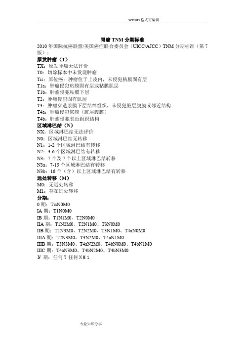
胃癌TNM分期标准2010年国际抗癌联盟/美国癌症联合委员会(UICC/AJCC)TNM分期标准(第7版):原发肿瘤(T)TX:原发肿瘤无法评价T0:切除标本中未发现肿瘤Tis:原位癌:肿瘤位于上皮内,未侵犯粘膜固有层T1a:肿瘤侵犯粘膜固有层或粘膜肌层T1b:肿瘤侵犯粘膜下层T2:肿瘤侵犯固有肌层T3:肿瘤穿透浆膜下层结缔组织,未侵犯脏层腹膜或邻近结构T4a:肿瘤侵犯浆膜(脏层腹膜)T4b:肿瘤侵犯邻近组织结构区域淋巴结(N)NX:区域淋巴结无法评价N0:区域淋巴结无转移N1:1-2个区域淋巴结有转移N2:3-6个区域淋巴结有转移N3:7个及7个以上区域淋巴结转移N3a:7-15个区域淋巴结有转移N3b:16个(含)以上区域淋巴结有转移远处转移(M)M0:无远处转移M1:存在远处转移分期:0期:TisN0M0IA期:T1N0M0IB期:T1N1M0、T2N0M0IIA期:T1N2M0、T2N1M0、T3N0M0IIB期:T1N3M0、T2N2M0、T3N1M0、T4aN0M0IIIA期:T2N3M0、T3N2M0、T4aN1M0IIIB期:T3N3M0、T4aN2M0、T4bN0M0、T4bN1M0IIIC期:T4aN3M0、T4bN2M0、T4bN3M0IV期:任何T任何NM1结直肠癌TNM分期美国癌症联合委员会(AJCC)/国际抗癌联盟(UICC)结直肠癌TNM分期系统(第七版)原发肿瘤(T)T x原发肿瘤无法评价T0无原发肿瘤证据Tis 原位癌:局限于上皮内或侵犯黏膜固有层T1肿瘤侵犯黏膜下层T2肿瘤侵犯固有肌层T3肿瘤穿透固有肌层到达浆膜下层,或侵犯无腹膜覆盖的结直肠旁组织T4a肿瘤穿透腹膜脏层T4b肿瘤直接侵犯或粘连于其他器官或结构区域淋巴结(N)N x区域淋巴结无法评价N0无区域淋巴结转移N1有1~3枚区域淋巴结转移N1a有1枚区域淋巴结转移N1b有2~3枚区域淋巴结转移N1c浆膜下、肠系膜、无腹膜覆盖结肠/直肠周围组织内有肿瘤种植(TD,tumor deposit),无区域淋巴结转移N2有4枚以上区域淋巴结转移N2a 4~6枚区域淋巴结转移N2b 7枚及更多区域淋巴结转移远处转移(M)M0无远处转移M1有远处转移M1a远处转移局限于单个器官或部位(如肝,肺,卵巢,非区域淋巴结)M1b远处转移分布于一个以上的器官/部位或腹膜转移解剖分期/预后组别:注:1 临床TNM分期(cTNM)是为手术治疗提供依据,所有资料都是原发瘤首诊时经体检、影像学检查和为明确诊断所施行的病理活检获得的。
软组织肉瘤诊断治疗

软组织肉瘤诊断治疗软组织肉瘤的表现开始大多为无痛性肿块出现,生长一般较快,从几个月到半年,也有生长较慢者,根据肿瘤的部位有些患者一开始便出现疼痛,一般到晚期均会出现疼痛。
恶性纤维组织细胞瘤发生部位除了肢体和躯干,内脏器官也可以发生。
滑膜肉瘤大多发生在肢体的大关节附近,但很少累及关节腔内。
脂肪肉瘤好发于臀部及大腿,在此部位发生肿瘤时,要考虑原发盆腔内肿瘤通过闭孔、坐骨大孔、耻骨弓甚至腹股沟韧带沿股管及肌肉间隙向下延伸。
横纹肌肉瘤多发生于肢体和躯干部,因此部位的横纹肌组织比较发达。
胚胎性横纹肌肉瘤多生长在头颈、眼眶周围,也可见于儿童的泌尿生殖器官。
腺泡状软组织肉瘤好发部位主要位于肢体,尤其是以下肢最多见,其中臀部和大腿占1/2以上,发生于躯干比较少见。
软组织肉瘤的体积与恶性程度没有必然的联系。
脂肪肉瘤的体积通常较大,其直径可以从数厘米到数十厘米,形状不规则。
软组织肉瘤的疼痛与发生的部位受肿瘤组织的来源及与神经的关系决定。
软组织肉瘤诊断软组织肉瘤的主要症状是局部肿块,单纯依靠X线,术前的诊断很困难,近年来影像学诊断的发展,如超声、CT、MRI、核素扫描、PET-CT 等检查对术前的诊断有了很大的帮助。
对手术计划的选择,术前治疗效果的评估有了不可低估的作用。
临床检查无痛性肿块是最主要的临床症状和体征。
软组织肉瘤多深在,边界不清,病史较短,或生长迅速,常伴一些压迫症状或骨侵犯。
恶性者大约3%~5%的病人出现区域淋巴结转移,相对较晚出现远隔转移,以肺转移为主。
1.症状对于软组织肉瘤,从发现肿瘤到来医院就诊,一般几个月以内的多见,但也有少数患者数年甚至是10余年的。
可以是无痛性的肿块,一般生长较快。
临床上可以看见生长多年的肿块突然增大,提示肿瘤恶性可能性大。
2.望诊一些表浅的肿瘤,皮肤的血管肉瘤可见多中心的血管窦样病灶。
神经纤维瘤病可见典型的咖啡斑。
上皮样肉瘤可见于手足部的腱鞘,晚期可形成难治的溃疡。
3.触诊一般检查肿瘤的大小、边界、软硬度、活动度、有无压痛、波动是否存在,有无搏动等。
软组织肉瘤的放射治疗
软组织肉瘤的放射治疗许宋锋;余子豪【摘要】软组织肉瘤(soft tissue sarcomas,STS)是起源于结缔组织的软组织恶性肿瘤,具有多种不同类型。
手术是STS主要治疗方法,放疗也是其重要的治疗方式并且是综合治疗早期选择之一。
对STS进行放疗已经超过50年历史,术前和术后放疗对于局部控制都有疗效,只是不良反应不同。
软组织肉瘤放疗技术包括远距离放疗(适形放疗、调强放疗、立体定向放疗等)、近距离放疗(组织间插植放疗、腔内后装放疗、术中放疗等)等。
放疗技术的进步,提高了放疗的精准性和确定性,降低了对病灶周围正常组织的损伤。
本文主要针对STS放疗技术以及适用原则进行综述。
%Soft tissue sarcomas (STS) consist of a heterogeneous group of rare malignancies with mesenchymal origin. Surgical resec-tion is the primary treatment for STS, but radiation therapy (RT) also plays an important role in the treatment. Radiotherapy for STS has advanced significantly over the past 50 years. Both preoperative and postoperative radiotherapies are equivalent in local control but are associated with different toxicity profiles. Boost techniques for STS include brachytherapy, intraoperative radiation therapy (IORT), and external beam. Long-term toxicities of RT to normal tissues have been reduced because of improvements in image guid-ance and intensity-modulated radiotherapy, which significantly increase the precision and delivery of RT. This review discusses RT tech-nologies and their acceptable treatment principles.【期刊名称】《中国肿瘤临床》【年(卷),期】2017(044)001【总页数】5页(P19-23)【关键词】软组织肉瘤;放疗;新辅助放疗;治疗原则【作者】许宋锋;余子豪【作者单位】国家癌症中心,中国医学科学院,北京协和医学院肿瘤医院骨科北京市100021;国家癌症中心,中国医学科学院,北京协和医学院肿瘤医院放疗科北京市100021【正文语种】中文余子豪,教授,主任医师,博士生导师,中国医学科学院肿瘤医院放疗科首席专家。
软组织肿瘤病理学-概述说明以及解释

软组织肿瘤病理学-概述说明以及解释1.引言1.1 概述软组织肿瘤是一种源自软组织结构的肿瘤,包括皮下组织、肌肉、筋膜、血管、神经等各种类型的肿瘤。
软组织肿瘤病理学是研究这些肿瘤在病理学上的特征、分类和诊断方法的学科。
软组织肿瘤病理学在临床诊断和治疗中起着关键作用,能够帮助医生准确地诊断肿瘤的性质和走向,制定合理的治疗方案。
本文将系统介绍软组织肿瘤的病理学特征,希望能够为临床医生提供一些帮助和指导。
1.2 文章结构文章结构:本文主要分为引言、正文和结论三部分。
引言部分将介绍软组织肿瘤的概述、文章结构和研究目的,为后续内容的阐述奠定基础。
正文部分将包括软组织肿瘤的概述、分类和病理学特征等内容,通过对软组织肿瘤的深入分析,揭示其病理学特点和分类规律。
结论部分将对前文进行总结,探讨软组织肿瘤病理学在临床上的意义,并展望未来软组织肿瘤病理学研究的发展方向。
1.3 目的"软组织肿瘤病理学"这篇文章的目的在于深入探讨软组织肿瘤的病理学特征,帮助读者更全面地了解这一领域的知识。
通过对软组织肿瘤的分类和病理学特征进行详细介绍,我们旨在帮助读者对软组织肿瘤的诊断、治疗和预后有更深入的认识。
同时,我们还希望通过本文的撰写,促进相关领域的学习和研究,为临床实践提供更多有益的帮助。
最终目标是促进软组织肿瘤领域的进步,提高对这一疾病的认识和管理水平。
2.正文2.1 软组织肿瘤概述:软组织肿瘤是指发生在身体软组织中的良性或恶性肿瘤。
相比于骨肿瘤,软组织肿瘤的发生率要低很多。
然而,由于软组织的种类繁多,软组织肿瘤的分类也变得复杂多样。
软组织肿瘤可以发生在任何部位的软组织中,包括肌肉、脂肪、血管、神经等组织,因此病理学特征和临床表现也各不相同。
软组织肿瘤的诊断常需要结合临床症状、影像学表现和病理学检查结果。
病理学在软组织肿瘤的诊断和鉴别诊断中起着关键作用,通过细胞学和组织学检查,可以明确肿瘤的性质、起源以及生长模式,为临床治疗提供重要依据。
软组织肿瘤

1) 多发生于大腿及腹膜后的软组织 深部。 2)瘤多见于40岁以上成人 3)肉眼观,大多数肿瘤呈结节状或 分叶状,表面常有一层假包膜,亦 可呈粘液性外观,或均匀一致呈鱼 肉样。
4)本瘤的瘤细胞形态多种多样,可 见分化差的星形、梭形、小圆形或异 型性明显的脂肪母细胞,胞浆内可见 多少和大小不等的脂滴空泡。 5)分型:高分化脂肉、粘液样型脂 肪肉瘤、圆形细胞型脂肪肉瘤、多形 性脂肪肉瘤
2、关于生物学潜能的命名
软组织肿瘤分为四类(2002年新分类): • 良性、 • 中间性(局部侵袭性)、 • 中间性(偶尔有转移) • 恶性
良性:大多数不复发,即使复发为非 破坏性,局部完整切除几乎都能治愈, 极罕见情况下(<1/5万),形态学良性 肿瘤可发生远处转移,但形态学检查完 全不能预测。
软组织肿瘤
南昌大学医学院 熊小亮
概况
• 大多数软组织肿瘤为良性,手术切除后 治愈率很高。恶性间叶性肿瘤占人类全 部恶性肿瘤的不到1%,但却危及生命 • 流行病学 良性软组织肿瘤的每年临床发病率(就 诊的新病例数)估计高达3000/百万人 口,而软组织肉瘤的每年发病率约30/ 百万人口,即占所有恶性肿瘤的不到 1%。
4、免疫组化在软组织肿瘤诊断中 的应用
常用抗体及意义(见下表)
• • • • • • • • • • • • • • • •
NCCN指南:软组织肉瘤(中文)

NCCN 肿瘤临床实践指南(NCCN 指南)®)软组织肉瘤版本2.2020—2020年5月28日继续工作基本信息:•在开始治疗前,强烈建议由具有肉瘤专业知识和经验的多学科综合治疗组对所有患者进行评价和管理a•H&P•原发性肿瘤的充分成像b适用于以下情况的所有病变特殊考虑胃肠道间质瘤(GIST)硬纤维瘤(侵袭性纤维瘤病)参见(GIST-1)见DESM-1合理的恶性几率仅适用于h参见NCCN•仔细计划的针芯[首选]或切除活检后充分成像(参见SARC-D)c►将活检标本沿未来的切除轴放置,分离最小,组织学尤文肉瘤指南骨癌小心止血►活检应确定分级和组织学亚型d►适当时,使用辅助诊断方法e•胸部影像b在特定情况下使用:f•根据指征进行额外成像;见成像原理(SARC-A)•下列疾病与肉瘤和其他癌症的发生率增加相关:►个人/家族史提示Li-Fraumeni综合征应考虑做进一步的遗传学评估。
见NCCN遗传/家族性高风险指南评估:乳腺癌、卵巢癌和胰腺癌►应考虑对遗传性非息肉病性结直肠癌(HNPCC或Lynch综合征)患者进行进一步评估参见NCCN遗传/家族高风险评估指南:四肢/体壁、头部/颈部其他软组织肉瘤i横纹肌肉瘤(RMS)I期可切除的II、III期功能结局可接受的疾病II、III期可切除疾病有不良功能结局或不可切除的原发疾病参见RMS-1参见主要部分(EXTSARC-2)参见主要部分(EXTSARC-3)参见主要部分(EXTSARC-4)结直肠►神经纤维瘤病患者g1型糖尿病的风险增加IV期同步疾病参见主要部分(EXTSARC-5)同时发生恶性外周神经鞘膜瘤(MPNSTs)和胃肠道间质瘤(GIST)。
复发性疾病参见主要部分(EXTSARC-6)主要目的治疗随访IA 期j /IB 期j (低级)广泛手术切除k,l肿瘤边缘未能获得肿瘤样本•康复评定(见严重程度亚组c-第2天,共2天)•2-3年,每3-6个月进行一次H&P ,然后每年一次•考虑胸部成像b•考虑获得术后基线MRI •主要部位成像b 基于以下估计风险如果复发,参见复发疾病(EXTSARC-6)►对于R2局部复发p,q前重新成像b1B 期肿瘤)b 见成像原理(SARC-A).j 见美国癌症联合委员会(AJCC)分期,第8版(ST-2和ST-3).k 见手术原理(SARC-D).l 对于非典型脂肪瘤肿瘤/高分化脂肪肉瘤(ALT/WDLS)患者,应量身定制切除手术,以尽量减少手术发病率。
软组织肉瘤
软组织肉瘤软组织肉瘤均起源于胚胎中胚层的机体间充质组织,具有局部侵袭性浸润生长以及易于发生血行转移的生物学行为行为和临床转归。
人体的软组织约占人体比重的75%,然而发生于间质组织的软组织肉瘤的构成比却相对很小,仅占成人全部恶性肿瘤的1%,儿童恶性肿瘤的15%。
但软组织肿瘤的种类最为繁多,有超过100种以上的分型。
软组织肉瘤可发生于任何年龄,在中国有两个发病高峰:第一高峰为从出生到5岁,第二高峰为从20--50岁。
软组织肉瘤50%--60%发生于肢体部位,这应该是易于被发现和早期获得诊断的条件,然而80%以上的病灶并没有表现出疼痛等能引起重视的症状,这使得确诊时50%的病例瘤体已大于5.0cm,而其中的一半已经超过10cm,约10%的病灶已侵犯大血管和神经组织,显然治疗会因此变得复杂而难以治愈。
软组织肉瘤的病因不是单一因素所致,临床发病与环境因素、细胞遗传学病因、分子病因学相关。
软组织肉瘤种类很多,按不同组织来源分为纤维组织、肌肉组织、脂肪组织、神经组织、血管组织、组织细胞、滑膜组织、骨与软骨组织、间皮组织及其他十类。
软组织肉瘤可发生于人体任何部位,不同类型发生部位不同,临床症状和体、体征也各具特色。
其中一般以上发生在四肢,其次为躯干,后腹膜,头颈等部位。
肿瘤的生长部位科提示它的起源。
纤维肉瘤大多来自躯干的皮肤和皮下组织;脂肪肉瘤多发生在脂肪组织较多的臀部,大腿和腹膜后;滑膜肉瘤多发生在四肢的大关节处;横纹肌肉瘤多发生在下肢深肌层内,其胚胎型常见于眼眶、耳道、鼻腔和泌尿生殖器官;间皮肉瘤多发生在胸膜、腹膜、心包腔、甚至鞘膜腔;平滑肌肉瘤易躯干和腹腔多见;腺泡状软组织肉瘤主要见于臀部和腹部肌肉。
症状和体征:1.疼痛疼痛是软组织肉瘤最常见症状,其程度根据发生部位、肿瘤来源及与神经组织的关系等因素决定。
血管平滑肌肉瘤及平滑肌肉瘤多有疼痛;纤维肉瘤则在肿瘤生长到一定程度才出现疼痛;滑膜肉瘤及横纹肌肉瘤等变化很大,有的患者疼痛与肿块同时出现,有的则先出现疼痛后出现肿块。
病理学的肉瘤名词解释
病理学的肉瘤名词解释为了更好地理解和探究医学领域中的肉瘤,我们需要对其进行深入的解释和了解。
从病理学的角度出发,肉瘤是一类肿瘤的名称,其主要特征是由异常增殖的恶性细胞组成。
这一类肿瘤通常生长迅速,并且存在着潜在的侵袭和转移的风险。
首先,我们需要明确肿瘤的定义。
肿瘤是人体组织异常增殖的结果,它可以分为良性和恶性两种类型。
良性肿瘤生长缓慢,通常局限于原发部位,并且很少侵犯周围组织或转移至其他部位。
恶性肿瘤则具有高度侵袭性和转移性,其细胞异常增殖能力较强,同时也具备从原发灶转移到身体其他部位的潜力。
接下来,我们将关注肉瘤这一特殊类别。
肉瘤是一类良性和恶性肿瘤的总称,它们的特点是细胞异型性高和侵袭力强。
这些特点使得肉瘤通常生长迅速,并可能迅速侵犯周围组织。
相比于其他肿瘤类型,肉瘤的发病率相对较低,但其致死率却相对较高。
具体而言,肉瘤可根据其起源组织的类型进行分类。
例如,骨肉瘤是一种最常见的恶性肉瘤,起源于骨组织。
骨肉瘤通常在年轻人中发生,并主要位于长骨的干骺端,如股骨、胫骨等。
它以快速生长和易转移著称,由于这种特点,骨肉瘤的治疗相对复杂。
除了骨肉瘤外,还有其他类型的恶性肉瘤。
例如软组织肉瘤,这种肉瘤起源于肌肉、血管、神经等组织,常见于软组织中,如肺褶、下肢、腹腔等部位。
软组织肉瘤生长迅速,可侵犯邻近组织,甚至造成全身转移。
良性肉瘤也是一种值得注意的类型。
虽然良性肉瘤生长缓慢,但其本质上仍然是一种异常增殖的肿瘤。
由于良性肉瘤通常不具备转移的能力,所以其对生命的威胁较小。
然而,某些良性肉瘤可能会对患者的健康产生负面影响,例如压迫周围结构和引起疼痛。
在病理学诊断中,对肉瘤的分析是至关重要的。
通过细胞形态学特征、组织学结构和免疫组化染色等方法,病理学家能够确定肉瘤的类型。
这对于制定个体化的治疗方案和预后评估具有重要意义。
此外,研究人员也在不断努力深入了解肉瘤的发病机制和治疗方法。
分子生物学的进展为肉瘤研究提供了新的途径。
软组织肉瘤会产生特殊蛋白,“策反”免疫细胞丨肿瘤新资讯汇总

软组织肉瘤会产生特殊蛋白,“策反”免疫细胞丨肿瘤新资讯汇总一、Cell Rep:揭示肿瘤细胞让免疫细胞“变坏”的机制来自西达赛奈医学中心等机构的科学家们通过研究发现,称之为软组织肉瘤的癌变肿瘤或许会产生一种特殊蛋白,它能让免疫细胞“变节”,从攻击肿瘤转变为帮助肿瘤发展,相关研究结果或有望帮助开发治疗人类软组织肉瘤的新型疗法。
这项研究中,研究人员重点对肿瘤微环境进行研究,肿瘤微环境是肿瘤招募血管和其它细胞的微生态系统,可以给肿瘤细胞提供所需营养并帮助其存活。
研究团队成员Jlenia Guarnerio说道,肿瘤同时也会招募免疫细胞,这些免疫细胞能够识别和攻击肿瘤细胞,但我们发现,肿瘤细胞可以分泌一种蛋白质,这种蛋白质会改变免疫细胞的生物学,使免疫细胞不会杀灭肿瘤细胞,而是做了相反的事情。
二、新型探针助力实现肿瘤早期诊断精准、“可视化”中国医学专家通过将肿瘤靶向的全人源纳米抗体与近红外荧光染料吲哚菁绿定点共价偶联,创新地研发了一种新型荧光免疫探针。
新型荧光免疫探针以全人源纳米抗体为靶向载体,与传统的荧光染料相比,该探针具有荧光信号强、检测灵敏度高、性能稳定等明显优势。
复旦大学基础医学院、上海市重大传染病和生物安全研究院应天雷教授、吴艳玲副研究员团队与复旦大学化学系李富友教授团队合作获得的相关研究成果以长文形式发表在最新的《生物材料》上。
三、科学家新发现破解晚期非小细胞肺癌患者耐药难题最新研究揭示,非小细胞肺癌患者寡转移后若接受高效、低毒、短平快的立体定向放疗联合相关抑制剂治疗,与单纯接受抑制剂治疗相比,患者可以延长中位生存期6个月左右。
在抑制剂耐药前查明脑部的寡转移病灶,针对性实施立体定向放疗,同样具有延长患者生存期、改善生活质量的潜力。
该系列研究从2016年开始启动,由复旦大学附属肿瘤医院专家朱正飞教授领衔,成果目前已经在美国放射肿瘤协会官方杂志《International Journal of Radiation Oncology Biology Physics》,肿瘤领域知名期刊《Clinical and Translational Medicine》《Lung Cancer》,知名杂志《British Journal of Radiology》等发表。
- 1、下载文档前请自行甄别文档内容的完整性,平台不提供额外的编辑、内容补充、找答案等附加服务。
- 2、"仅部分预览"的文档,不可在线预览部分如存在完整性等问题,可反馈申请退款(可完整预览的文档不适用该条件!)。
- 3、如文档侵犯您的权益,请联系客服反馈,我们会尽快为您处理(人工客服工作时间:9:00-18:30)。
Annals of Oncology21(Supplement5):v198–v203,2010doi:10.1093/annonc/mdq209 clinical practice guidelinesSoft tissue sarcomas:ESMO Clinical Practice Guidelines for diagnosis,treatment and follow-upP.G.Casali1&J.-Y.Blay2On behalf of the ESMO/CONTICANET/EUROBONET Consensus Panel of experts*1Department of Cancer Medicine,Istituto Nazionale dei Tumori,Milan,Italy;2INSERM U590,Claude Bernard University and Department of Oncology,Edouard Herriot Hospital,Lyon,FranceThe following recommendations apply to adult-type soft tissue sarcomas arising from limbs and superficial trunk. Recommendations on retroperitoneal sarcomas,desmoid-typefibromatosis,uterine sarcomas head and neck sarcomas and breast sarcomas are provided separately at the end of the chapter with regard to those main aspects by which they differ from more frequent soft tissue sarcomas.In general,the main principles of diagnosis and treatment may well apply to all soft tissue sarcomas,including the rarest presentations[e.g.visceral sarcomas other than gastrointestinal stromal tumours(GISTs)],which therefore are not specifically covered.Specific histological types,however, may deserve specific approaches,not necessarily covered hereafter,given the scope of these Recommendations. Extraskeletal Ewing sarcoma as well as embryonal and alveolar rhabdomyosarcoma are covered by other ESMO Clinical Practice Guidelines,inasmuch as they need completely different approaches.The same applies to GIST.Kaposi’s sarcoma is excluded from this chapter.incidenceAdult soft tissue sarcomas are rare tumours,with an estimated incidence averaging5/100000/year in Europe.diagnosisSoft tissue sarcomas are ubiquitous in their site of origin, and are often treated with multimodality treatment.A multidisciplinary approach is therefore mandatory in all cases (involving pathologists,radiologists,surgeons,radiation therapists,medical oncologists and paediatric oncologists if applicable).This should be carried out in reference centres for sarcomas and/or within reference networks sharing multidisciplinary expertise and treating a high number of patients annually.These centres are involved in ongoing clinical trials,in which sarcoma patients’enrolment is highly encouraged.This centralized referral should be pursued from the time of the clinical diagnosis of a suspect sarcoma.In practice,referral of all patients with a lesion likely to bea sarcoma would be recommended.This would mean referring all patients with an unexplained deep mass of soft tissues,or with a superficial lesion of soft tissues having a diameter of>5cm,or arising in paediatric age.In soft tissue tumours,MR is the main imaging modality, although radiographs should be thefirst step to rule out a bone tumour,to detect a bone erosion with a risk of fracture and to show calcifications.CT has a role in calcified lesions to rule*Correspondence to:ESMO Guidelines Working Group,ESMO Head Office,ViaL.Taddei4,CH-6962Viganello-Lugano,Switzerland;E-mail:clinicalrecommendations@Approved by the ESMO Guidelines Working Group:August2003,last update March 2010.This publication supercedes the previously published version—Ann Oncol2009; 20(Suppl4):iv132–iv136.Conflict of interest:Dr Casali has reported that he is currently conducting researchsponsored by Amgen Dompe´,Merck SD,Glaxo SK,Lilly,Novartis,Pfizer,PharmaMar,Sanofi-Aventis and Schering Plough.He had a consultancy role with and/or receivedhonoraria for lectures from Merck SD,Novartis,Pfizer,PharmaMar,Sanofi-Aventis.Hehas received travel coverage for medical meetings from Novartis and PharmaMar;Prof.Blay has reported that he is a consultant for Pfizer,Novartis,GSK,Roche andPharmamar.Consensus panel’s conflict of interest:Prof.Aglietta has reported that hehas received research grants from Bayer,Amgen,Roche and Novartis;Prof.Alvega˚rdhas reported no conflicts of interest;Dr Athanasou has reported no conflicts of interest;Dr Bihn has not reported any conflicts of interest;Dr Bonvalot has reported that she hasreceived honoraria from Novartis and grants from Pharmamar;Dr Boukovinas hasreported that he is a member of the speakers’bureau for Novartis;Prof.De Alava hasreported no conflicts of interest;Dr Dei Tos has reported no conflicts of interest;Dr Dileohas reported no conflicts of interest;Dr Eriksson has reported that he has receivedhonoraria from Novartis,MSD,Pfizer,Swedish Orphan and GSK;Dr A.Ferrari hasreported no conflicts of interest;Dr S.Ferrari has reported that he has participated inresearches sponsored by Pharmamar and Ariad;Dr Garcia Del Muro has reported noconflicts of interest;Dr Gronchi has reported no conflicts of interest;Dr Hall has reportedno conflicts of interest;Prof.Hassan has reported that at present he has no conflicts ofinterest,but he is planning trials with Takeda and Pharmamar;Prof.Hogendoorn hasreported no conflicts of interest;Dr Hohenberger has not reported any conflicts ofinterest;Dr Gelderblom has reported no conflicts of interest;Dr Grimer has reported noconflicts of interest;Prof.Issels has reported no conflicts of interest;Dr Joensuu hasreported no conflicts of interest;Dr Jost has reported no conflicts of interest;Prof.Judson has reported that he has received honoraria for participation in advisory boardsmeetings from Novartis,Pfizer,PharmaMar and Ariad;Dr Juergens has reported noconflicts of interest;Dr Le Cesne has reported that he has received honoraria fromNovartis,Pfizer and PharmaMar;Dr Leyvraz has not reported any conflicts of interest;DrMartin-Broto has reported no conflicts of interest;Dr Montemurro has reported noconflicts of interest;Prof.Nishida has reported that he is currently conducting researchpartly funded by Novartis Pharma;Dr Shreyaskumar has reported no conflicts ofinterest;Dr Reichardt has reported that he is a member of the speakers’bureau andAdvisory Board for PharmaMar;Dr Robinson has reported no conflicts of interest;DrRutkowski has reported that he has received honoraria from and that he is a member ofthe speakers’bureau of Novartis;Prof.Scho¨ffski has reported that he is conductingresearch sponsored by Novartis,Pfizer,PharmaMar,Eisai,GlaxoSmithKline,Infinity andGenentech and that he is a member of the speakers’bureau for Novartis,Pfizer,PharmaMar,Eisai,GlaxoSmithKline;Dr Schlemmer has reported that he is currentlyconducting research funded by Novartis;Dr Sleijfer has reported no conflicts of interest;Dr Van der Graaf has reported no conflicts of interest;Dr Vanel has reported no conflictsof interest;Prof.Verweij has reported no conflicts of interest;Dr Wardelmann has notreported any conflicts of interest;Dr Whelan has reported no conflicts of interest.ªThe Author2010.Published by Oxford University Press on behalf of the European Society for Medical Oncology. All rights reserved.For permissions,please email:journals.permissions@ at St Jude Childrens Research Hospital on September 4, Downloaded fromout a myositis ossificans,and in retroperitoneal tumours, where the performance is identical to MR.Following proper imaging assessment,the standard approach to diagnosis consists of multiple core needle biopsies(by using needles>16G).However,an excisional biopsy may be the most practical option for superficial lesions of<5cm.An open biopsy may be another option in selected cases.Immediate evaluation of tissue viability may be considered,to make sure the biopsy is adequate at the time it is done.However,a frozen-section technique for immediate diagnosis is not encouraged,because generally it does not allow a complete diagnosis,especially when a preoperative treatment is planned.Fine-needle aspiration is used only in some institutions,which have developed specific expertise on this procedure,and is not recommended outside these centres.A biopsy may underestimate the tumour malignancy grade, so that,when preoperative treatment is an option,radiological imaging may add to pathology in providing the clinician with information that helps to estimate the malignancy grade (e.g.necrosis).The biopsy should be performed by a surgeon or a radiologist,after interdisciplinary discussion,as needed. It should be planned in such a way that the biopsy pathway and the scar can be safely removed on definitive surgery. The biopsy entrance point is preferably tattooed.The tumour sample should befixed in formalin in due time(Bouinfixation should be banned,since it prevents molecular analysis).Histological diagnosis should be made according to the latest World Health Organization(WHO)classification.A pathological expert second opinion is recommended in all cases where the original diagnosis was made outside reference centres.The malignancy grade should be provided in all cases in which this is feasible based on available systems.The Federation Nationale des Centres de Lutte Contre le Cancer(FNCLCC) grading system is generally used,which distinguishes three malignancy grades based on differentiation,necrosis and mitotic rate.Whenever possible,the mitotic rate should be provided independently.An effort should be made to improve the reliability of mitotic count as actually recorded.Tumour site should be properly recorded.Tumour size and tumour depth(in relation to the muscular fascia)should be recorded,since they entail a prognostic value,along with malignancy grade.The pathology report after definitive surgery should mention whether the tumour was intact and should include an appropriate description of tumour margins(i.e.the status of inked margins and the distance between tumour edge and the closest inked margins).This allows assessment of marginal status(i.e.whether the minimum margin is intralesional,marginal,wide and distances from surrounding tissues).The pathological assessment of margins should be made in collaboration with the surgeon.If preoperative treatment was carried out,the pathology report should include a tumour response assessment.In contrast to osteosarcoma and Ewing sarcoma,however,no validated system is available at present in this regard,and no percentage of residual‘viable cells’is considered to havea specific prognostic significance.This depends on several factors,including the presence of non-treatment-related necrosis and haemorrhage and the heterogeneity of post-treatment changes.A multidisciplinary judgement is recommended,involving the pathologist and the radiologist. Pathological diagnosis relies on morphology and immunohistochemistry.It should be complemented by molecular pathology[fluorescent in situ hybridization(FISH), reverse transcription–polymerase chain reaction(RT–PCR)], especially when:(i)the clinical pathological presentation is unusual;(ii)the specific histological diagnosis is doubtful; (iii)it may have prognostic/predictive relevance.External quality assurance programmes are encouraged for laboratories performing molecular pathology assessments. Collection of fresh frozen tissue and tumour imprints(touch preps)is encouraged,because new molecular pathology assessments could be made at a later stage in the patient’s rmed consent for tumour banking should be sought enabling later analyses and research,as long as this is allowed by local and international guidelines.stage classification and risk assessmentThe American Joint Committee on Cancer(AJCC)/ International Union against Cancer(UICC)stage classification system stresses the importance of the malignancy grade in sarcoma.However,its use in routine practice is limited.In addition to grading,other prognostic factors are tumour size and tumour depth.Of course,tumour resectability is also important.staging proceduresThe surgical report,or patient chart,should provide details on: the preoperative and intraoperative diagnosis;the surgical conduct,including possible contamination(i.e.it should mention whether the tumour was opened,was‘seen’during the excision,etc.);surgical actual completeness vis-a`-vis planned quality of margins.A chest spiral CT scan is mandatory for staging purposes. Depending on the histological type and other clinical features,further staging assessments may be recommended (i.e.regional lymph node clinical assessment for synovial sarcoma,epithelioid sarcoma,alveolar soft part sarcoma,clear cell sarcoma;abdominal CT scan for myxoid liposarcoma,etc.). treatmentlimited diseaseSurgery is the standard treatment for all patients with adult-type,localized soft tissue sarcomas.It must be performed by a surgeon specifically trained in the treatment of this disease. The standard surgical procedure is a wide excision with negative margins(R0).This implies removing the tumour with a rim of normal tissue around.One centimetre has been selected as a cut-off in some studies,but it is important to realize that the margin can be minimal in the case of resistant anatomical barriers,such as muscular fasciae,periostium and perineurium.A marginal excision may be acceptable as anAnnals of Oncology clinical practice guidelinesVolume21|Supplement5|May2010doi:10.1093/annonc/mdq209|v199 at St Jude Childrens Research Hospital on September 4, Downloaded fromindividualized option in highly selected cases,in particular for extracompartmental atypical lipomatous tumours.Wide excision followed by radiation therapy is standard treatment in high-grade,deep lesions,>5cm.Radiation therapy is not given in the case of a truly compartmental resection of a tumour entirely contained within the compartment.With exceptions to be discussed in a multidisciplinary setting,and in the face of a lack of consensus across reference centres,also high-grade,deep,<5cm lesions are treated with surgery followed by radiation therapy.Radiation therapy is added in selected cases in the case of low-grade,superficial,>5cm, and low-grade,deep,<5cm soft tissue sarcoma.In the case of low-grade,deep,>5cm soft tissue sarcoma,radiation therapy should be discussed in a multidisciplinary fashion, considering the anatomical site and the related expected sequelae versus the histological aggressiveness.Overall, radiation therapy has been shown to improve local control, but not overall survival.Radiation therapy should be administered postoperatively,with the best technique available, at a dose of50–60Gy,with fractions of1.8–2Gy,possibly with boosts up to66Gy,depending on presentation and quality of surgery.Alternatively,radiotherapy may be carried out preoperatively,normally using a dose of50Gy.Intraoperative radiation therapy(IORT)and brachytherapy are options in selected cases.Re-operation in reference centres must be considered in the case of R1resections,if adequate margins can be achieved without major morbidity,taking into account tumour extent and tumour biology(e.g.it may be spared in extracompartmental atypical lipomatous tumours,etc.).In the case of R2surgery,re-operation is mandatory,possibly with preoperative treatments if adequate margins cannot be achieved,or surgery is mutilating.In the latter case,the use of multimodal therapy with less radical surgery requires shared decision-making with the patient under conditions of uncertainty.Plastic repairs and vascular grafting should be used as needed,and the patient should be properly referred if necessary.Radiation therapy will obviously follow marginal or R1–R2excisions,if these cannot be rescued through re-excision, even outside the usual indications(see above).In non-resectable tumours,or those amenable only to mutilating surgery(in this case,on an individualized basis after sharing the decision with the patient in conditions of uncertainty),chemotherapy and/or radiotherapy,or isolated hyperthermic limb perfusion with tumour necrosis factor-a(TNF a)+melphalan,if the tumour is confined to an extremity,or regional hyperthermia combined with chemotherapy,are options.Regional lymph node metastases should be distinguished from soft tissue metastases involving lymph nodes.They are rare,and constitute an adverse prognostic factor in adult-type soft tissue sarcomas.More aggressive treatment planning is therefore felt to be appropriate for these patients,although there is a lack of formal evidence to indicate that this improves clinical results.Surgery through wide excision(mutilating surgery is exceptionally done given the prognosis of these patients)may be coupled with adjuvant radiation therapy and adjuvant chemotherapy for sensitive histological types,as standard treatment for these presentations.Chemotherapy may be administered as preoperative treatment,at least in part.These treatment modalities adding to surgery should not be viewed as truly‘adjuvant’,the context being in fact that ofa likely systemic disease.In one large randomized phase III study(in patients with G2–3,deep,>5cm soft tissue sarcomas), regional hyperthermia in addition to systemic chemotherapy was associated with a local and disease-free survival advantage. Isolated limb perfusion may be an option in this patient population,along with chemotherapy and radiation therapy. Data have been provided that adjuvant chemotherapy might improve,or at least delay,distant and local recurrence in high-risk patients.A meta-analysis found a statisticallysignificant,limited benefit in terms of both survival and relapse-free survival.However,studies are conflicting,andafinal demonstration of efficacy is lacking.It is also unknown whether adjuvant chemotherapy may be especially beneficial in specific subgroups.Therefore,adjuvant chemotherapy is not standard treatment in adult-type soft tissue sarcomas,and can be proposed as an option to the high-risk individual patient (having a>G1,deep,>5cm tumour)for shared decision-making with the patient[II,C].Adjuvant chemotherapy is not used in histologies known to be insensitive to chemotherapy. If the decision is made to use chemotherapy as upfront treatment,it may well be used preoperatively,at least in part.A local benefit may be gained,facilitating surgery.In one large randomized phase III study(in patients with G2–3,deep,>5cm soft tissue sarcomas),regional hyperthermia in additionto systemic chemotherapy was associated with a local and disease-free survival advantage(no survival benefit demonstrated).If used,adjuvant chemotherapy should consist of the combination chemotherapy regimens proven to be most active in the advanced disease.The standard approach to local relapse parallels the approach to primary local disease,except for a wider resort to preoperative or postoperative radiation therapy,if not previously performed.extensive diseaseMetachronous resectable lung metastases without extrapulmonary disease are managed with complete excision of all lesions as standard treatment[IV,B].Chemotherapy may be added as an option,taking into account the prognostic factors(a short previous free interval and a high number of lesions are adverse factors,encouraging the addition of chemotherapy),although there is a lack of formal evidence that this improves results.Chemotherapy is preferably given before surgery,in order to assess tumour response and thus modulate the length of treatment.In the case of lung metastases being synchronous,in the absence of extrapulmonary disease, standard treatment is chemotherapy[IV,B].Especially when a patient benefit is achieved,surgery of completely resectable lung metastases may be offered as an option. Extrapulmonary disease is treated with chemotherapy as standard treatment[I,A].In highly selected cases,surgery of responding metastases may be offered as an option following a multidisciplinary evaluation,taking into consideration their site and the natural history of the disease in the individual patient.Standard chemotherapy is based on anthracyclines asfirst-line treatment[I,A].There is no formal demonstration thatclinical practice guidelines Annals of Oncologyv200|Casali&Blay Volume21|Supplement5|May2010 at St Jude Childrens Research Hospital on September 4, Downloaded frommultiagent chemotherapy is superior to single-agent chemotherapy with doxorubicin alone in terms of overall survival.However,a higher response rate may be expected,in particular in a number of sensitive histological types, according to several,although not all,randomized clinical trials.Therefore,multiagent chemotherapy with anthracyclines plus ifosfamide may be the treatment of choice,especially when a tumour response is felt to be able to give an advantage and patient performance status is good.In angiosarcoma,taxanes are an alternative option,given their antitumour activity in this specific histological type[III,B]. Imatinib is standard medical therapy for those rare patients with dermatofibrosarcoma protuberans who are not amenable to non-mutilating surgery or with metastases requiring medical therapy[III,B].After failure of anthracycline-based chemotherapy,or impossibility of using it,the following criteria may apply, although in the lack of high-level evidence.Patients who have already received chemotherapy may be treated with ifosfamide,if they did not receive it previously. High-dose ifosfamide( 14g/m2)may be an option also for patients who have already received standard-dose ifosfamide [IV,C].Trabectedin is a second-line option[II,B].It has proved effective in leiomyosarcoma and liposarcoma.In myxoid liposarcoma a peculiar pattern of tumour response has been reported,with an early phase of tissue changes preceding tumour shrinkage.Responses to trabectedin have also been obtained in other histological types,including synovial sarcoma.One trial showed that gemcitabine+docetaxel is more effective than gemcitabine alone as second-line chemotherapy, but data are conflicting and toxicity is different[II,C]. Gemcitabine was also shown to have antitumour activity in leiomyosarcoma as a single agent.Dacarbazine has some activity as second-line therapy(mostly in leiomyosarcoma).It could also be combined with gemcitabine.Best supportive care is an option for pretreated patients with advanced soft tissue sarcoma,all the more if further-line therapies have already been used in the patient.In general, advanced pretreated patients are candidates for clinical studies. With reference to selected histological types,there is anecdotal evidence of activity of some molecular targeted agents,possibly in the face of preclinical consistent data.These patients can be sent to reference centres,to be treated accordingly,preferably within clinical studies.follow-upThere are no published data to indicate the optimal routine follow-up policy of surgically treated patients with localized disease.The malignancy grade affects the likelihood and speed at which relapses may take place.The risk assessment based on tumour grade,tumour size and tumour site therefore helps in choosing a routine follow-up policy.High-risk patients generally relapse within2–3years,while low-risk patients may relapse later,although it is less likely.Relapses most often occur to the lungs.Early detection of local or metastatic recurrence to the lungs may have prognostic implications,and lung metastases are asymptomatic at a stage in which they are suitable for surgery.Therefore,routine follow-up may focus on these sites.Although the use of MRI to detect local relapse and CT to scan for lung metastases is likely to pick up recurrences earlier,it is yet to be demonstrated that this is beneficial,or cost effective,compared with clinical assessment of the primary site and regular chest X-rays.That said,while prospective studies are needed,a practical approach in place at several institutions is as follows.The surgically treated intermediate-/high-grade patient may be followed every3–4months in thefirst2–3years,then twice a year up to thefifth year and once a year thereafter.Low-grade sarcoma patients may be followed for local relapse every4–6months,with chest X-rays or CT scan at more relaxed intervals in thefirst3–5years,then yearly.special presentations and entities retroperitoneal sarcomasCore needle biopsies are the standard procedure for diagnosis in retroperitoneal sarcomas.They should not be performed through the peritoneum.An open biopsy may be an option in selected cases.In both cases,the pathway of the biopsy should be carefully planned to avoid contamination and complications.However,radiological imaging may be sufficient for the diagnosis of lipomatous tumours,if no preoperative treatment is planned.Standard treatment for localized lesions is surgery,which is best performed through a retroperitoneal quasi-compartmental resection,that is a complete excision of the mass,along with en-bloc visceral resections of adjacent organs and tissues covering the tumour[IV,B].The value of preoperative treatments in resectable tumours is not established.Thus,while not standard they are available options, including radiation therapy,chemotherapy,chemoradiation therapy,regional hyperthermia in addition to chemotherapy.If given,preoperative treatments are not meant to change the extent of surgery.Likewise,the value of adjuvant chemotherapy is not established.In general,postoperative radiation therapy to the whole tumour bed at doses recommended for sarcomas is not feasible at an acceptable toxicity.In selected cases,it may be an option for well-defined anatomical areas felt to be at high risk.uterine sarcomasThis group includes leiomyosarcomas,endometrial stromal sarcomas(formerly,low-grade endometrial stromal sarcomas), undifferentiated endometrial sarcomas and pure heterologous sarcomas.Carcinosarcomas(malignant Mullerian mixed tumours) are mixed epithelial and mesenchymal neoplasms,whose treatment should follow their mainly epithelial nature. Standard treatment for all these tumours,when localized,is total abdominal hysterectomy.The added value of bilateral salpingo-oophorectomy is not established.In endometrial stromal sarcoma bilateral salpingo-oophorectomy is generally performed,due to the hormonal sensitivity of these tumours, and lymphadenectomy may be an option,given the possibleAnnals of Oncology clinical practice guidelinesVolume21|Supplement5|May2010doi:10.1093/annonc/mdq209|v201 at St Jude Childrens Research Hospital on September 4, Downloaded fromhigher incidence of nodal involvement[IV,D].However,as far as leiomyosarcomas and high-grade undifferentiated sarcomas are concerned,bilateral salpingo-oophorectomy, particularly in premenopausal women,as well as lymphadenectomy,is not demonstrated to be useful in the absence of macroscopic involvement.Although retrospective studies have suggested a possible decrease in local relapses,radiation therapy did not improve survival and relapse-free survival in a randomized trial,and therefore is not recommended in leiomyosarcoma[II,C]. Therefore,its use as an adjuvant to surgery may only be an option in selected cases,after shared decision-making with the patient following multidisciplinary discussion,taking into account special risk factors for local relapse.The systemic treatment of metastatic endometrial stromal sarcomas exploits their sensitivity to hormonal therapies [V,D].Therefore,progestins are generally used,along with gonadotrphin-releasing hormone(GnRH)analogues and aromatase inhibitors.Tamoxifen is contraindicated,as well as hormonal replacement therapy containing estrogens.Surgery of lung metastases is an option,given the natural history of the disease.The medical treatment of leiomyosarcomas,undifferentiated endometrial sarcomas and pure heterologous sarcomas parallels that for adult-type soft tissue sarcomas.In any case,it should be kept distinct from malignant Mullerian mixed tumours. desmoid-typefibromatosisBeta catenin mutational analysis may be useful when the pathological differential diagnosis is difficult.Given the unpredictable natural history of the disease (with the possibility of long-lasting stable disease and even occasional spontaneous regressions,along with a lack of metastatic potential),and functional problems implied by some tumour anatomical locations,a watchful waiting policy may be the best option[IV,B],after shared decision-making with the patient,with the exclusion of potentially life-threatening extra-abdominal locations(e.g.head and neck region)and intra-abdominal desmoids(mesentericfibromatosis).Under such a policy,treatment is reserved for progressing cases. Preferred imaging is MRI,though considering that the tumour signal is not meaningful with regard to disease evolution.For progressing cases,optimal treatment needs to be individualized on a multidisciplinary basis and it may consist of surgery(without any adjuvant therapy),radiation therapy, observation,isolated limb perfusion(if the lesion is confined to an extremity)or systemic therapy(see below)[V,D]. Systemic therapies include:hormonal therapies(tamoxifen, toremifene,GnRH analogues),non-steroidal anti-inflammatory drugs;low-dose chemotherapy,such as methotrexate+vinblastine or methotrexate+vinorelbine; low-dose interferon;imatinib;full-dose chemotherapy(using regimens active in sarcomas).It is reasonable to employ the less toxic therapies before the more toxic ones in a stepwise fashion. head and neck sarcomasThese are sarcomas arising at a difficult anatomical location. Cases should be dealt with through a multidisciplinary approach,also involving head and neck surgeons.Radiation therapy is widely resorted to,given the surgical margins generally achievable.breast sarcomasBreast sarcomas encompass radiation-and non-radiation-induced sarcomas.Then sarcomas of the skin of the breast area should be conceptually distinguished from mammary gland sarcomas.Finally,angiosarcoma has a more aggressive behaviour than other histological types,while malignant phyllodes tumours[i.e.those having>10mitoses/10high-powerfields(HPF)and marked stromal overgrowth]havea20%–30%metastatic rate.The best treatment of breast sarcomas is far from beingdefined,given their rarity and heterogeneity.In general,breast-conserving surgery may be used,depending on the quality of margins versus the size of the tumour and the breast,along with the feasibility of radiation therapy.In addition, angiosarcomas of the mammary gland have such a tendency to recur that mastectomy(involving the muscular fascia)is generally preferred,even in combination with postoperative radiation therapy.Lymphadenectomy is not performed in the absence of clinical evidence of involvement.As far as adjuvant chemotherapy is concerned,the same principles as for soft tissue sarcoma apply.One may consider the high risk of local and systemic relapse of angiosarcoma in making a decision.noteThese Clinical Practice Guidelines have been developed following a consensus process based on a consensus event organized by ESMO in Lugano in November2009.This involved experts from the community of the European sarcoma research groups,sarcoma networks of excellence and ESMO Faculty.Their names are indicated hereafter.The text reflects an overall consensus among them,although each of them may not necessarilyfind it consistent with his/her own views.The EU-funded network of excellence CONTICANET(CONnective TIssue CAncers NETwork)and EUROBONET(EUROpean BOne NETwork)alsofinancially supported the consensus process. consensus panelMassimo Aglietta,Universita`degli Studi di Torino,Italy Thor Alvegaard,Lund University Hospital,Lund,Sweden Nick Athanasou,University of Oxford,Oxford,UKBui Binh,Institut Bergonie´,Bordeaux,FranceJean-Yves Blay,Centre Le´on Be´rard Lyon,FranceSlyvie Bonvalot,Institut Gustave Roussy,Villejuif,France Ioannis Boukovinas,Theagenion Cancer Hospital of Thessaloniki,GreecePaolo G.Casali,Istituto Nazionale Tumori,Milan,Italy Enrique De Alava,Centro de Investigacion del Cancer-IBMCC, Salamanca,SpainA.Paolo Dei Tos,Ospedale Civile,Treviso,ItalyPalma Dileo,Istituto Nazionale Tumori,Milan,ItalyMikael Eriksson,University Hospital,Lund,SwedenAndrea Ferrari,Istituto Nazionale Tumori,Milan,Italy Stefano Ferrari,Istituti Ortopedici Rizzoli,Bologna,Italy Solans Francisco Javier Garcia Del Muro,Institut Catala`d’Oncologia,Barcelona,Spainclinical practice guidelines Annals of Oncologyv202|Casali&Blay Volume21|Supplement5|May2010 at St Jude Childrens Research Hospital on September 4, Downloaded from。
