磁性转染试剂(MagnetofectionTM)
磁性纳米颗粒作为基因载体的研究发展概况

磁性纳米颗粒作为基因载体的研究发展概况*李 瑶,崔海信,刘 琪,崔金辉(中国农业科学院农业环境与可持续发展研究所,北京100081)摘 要: 磁性纳米颗粒作为非病毒基因载体介导的基因转染(即磁性转染)是基因治疗中极具运用前景的技术之一。
主要介绍了磁性纳米颗粒的种类和性质,介导基因转染的最新研究进展,在细胞内的定位和动态过程,面临的问题以及将来的发展前景。
关键词: 磁性纳米颗粒;磁性转染;基因治疗中图分类号: Q633;Q789文献标识码:A文章编号:100129731(2010)增刊Ñ200142061 引 言近年来非病毒基因载体的开发颇受关注,许多新的物理化学技术应运而生,作为运载工具,非病毒载体可避免病毒载体的安全性等问题。
磁性纳米颗粒作为非病毒载体介导的基因转染即磁性转染(magnetofection)是其中极具运用前景的技术之一。
磁性纳米基因载体技术是以磁性纳米颗粒作为基因载体,在细胞的摄粒作用下,磁性纳米颗粒携带DNA 、RNA 、PNA 、dsRNA 等基因治疗分子进入细胞并释放这些分子,在磁场作用下实现安全有效的靶向性基因治疗[1,2]。
该技术不仅加快转染速度,而且提高转染率。
磁性纳米基因载体具有目的基因高效稳定表达、安全无害、靶向性高、为非病毒型基因载体等优点,因此磁性纳米颗粒作为基因载体在肿瘤基因治疗中的应用得到了迅速发展。
本文将讨论磁性纳米基因载体的种类与性质、近年来在体内外应用的研究进展及其应用前景。
2 磁性纳米颗粒基因载体的种类与性质2.1 磁性纳米颗粒的种类磁性纳米颗粒由无机磁性材料与各种提供活性功能基团的材料复合制备而成。
目前无机磁性微粒的种类很多,较常用的有金属合金(Fe,Co,Ni)、氧化铁(C 2Fe 2O 3、Fe 3O 4)、铁氧体(CoFe 2O 4、BaFe 12O 19)、氧化铬(CrO 2)和氮化铁(Fe 4N )等,其中Fe 3O 4(magnetite)是应用最多的磁性颗粒,它很容易在水溶液中通过共沉淀或氧化共沉淀制备,其粒度、形状和组成可以根据调节反应条件得到控制[3],用于磁转化的磁性纳米颗粒粒径从几十到几百纳米不等。
广东转染试剂类型

广东转染试剂类型
广东转染试剂类型
广东省作为全国经济重心之一,其科技实力在行业内也是一直走在前列。
转染试剂作为生物科研中不可缺少的试剂之一,在广东制造商也
有相当的生产水平和供应能力。
转染试剂,是指将外来的目的基因(DNA、RNA等)引入到靶细胞内,并使其表达的试剂。
其种类繁多,在广东也有着相应的常规类型。
1. 磷脂体基转染试剂
主要由磷脂体和质粒DNA组成,优点是容易制备和操作,适合于大规模生产。
缺点是转染率相对不高,有一定的毒性。
2. 聚乙烯酰胺转染试剂
聚乙烯酰胺(PEI)是一种化学合成剂,使用时与DNA分子组合形成
复合物,可以将DNA有效转染到细胞内。
优点是转染率高且毒性低,目前在广东科研中得到广泛应用。
3. 蛋白质/肽转染试剂
蛋白质/肽转染试剂通常是根据蛋白质或肽与DNA分子的作用机制设计制备的,可以有效地将目的分子转染到靶细胞内。
优点是转染效率高且无毒性,但缺点是价格昂贵。
4. 常规转染剂
常规转染剂是一类较为通用的转染试剂,主要包括羧甲基纤维素钠(CMC-Na)、聚乙烯醇(PVA)、去离子水等,优点是价格低廉且易于操作,但其转染效率较低,通常适用于一些较为简单的试验。
总体来说,转染试剂在广东科研中扮演着不可或缺的角色,其种类繁多,需根据实验需要选择合适的试剂。
在使用转染试剂时,需注意试剂质量和实验条件,以确保实验结果的准确性和科学性。
fugene 4k 转染试剂 中文说明书

2022版 CTM694原英文技术手册TM694中 文 说 明 书适用产品目录号:E5911和E5912FuGENE ®4K TransfectionReagent普洛麦格(北京)生物技术有限公司Promega (Beijing) Biotech Co., Ltd 地址:北京市东城区北三环东路36号环球贸易中心B座907-909电话:************网址:技术支持电话:400 810 8133技术支持邮箱:*************************CTM 6942022制作1所有技术文献的英文原版均可在/ protocols 获得。
请访问该网址以确定您使用的说明书是否为最新版本。
如果您在使用该试剂盒时有任何问题,请与Promega 北京技术服务部联系。
电子邮箱:*************************1. 描述 (2)2. 产品组分和储存条件 (2)3. 一般注意事项 (2)3. A. 转染试剂与DNA的比例 (3)3. B. DNA (3)3. C. 时间 (3)3. D. 血清 (3)3. E. 细胞培养条件 (3)3. F. 稳定转染 (3)4. 推荐操作步骤 (4)4. A. 细胞铺板 (5)4. B. FuGENE® 4K Transfection Reagent准备 (5)4. C. 一般转染操作步骤 (6)4. D. 稳定转染操作步骤 (8)4. E. 转染优化 (9)4. F. 报告基因活性和细胞健康的多重检测方案 (11)5. 疑难解答 (12)FuGENE® 4K Transfection Reagent普洛麦格(北京)生物技术有限公司Promega (Beijing) Biotech Co., Ltd 地址:北京市东城区北三环东路36号环球贸易中心B座907-909电话:************网址:技术支持电话:400 810 8133技术支持邮箱:*************************CTM 6942022制作21. 描述FuGENE® 4K Transfection Reagent是一个多组分,非脂质体试剂,用于将DNA高效、低毒地转染至多种哺乳动物细胞系中,无需在加入试剂-DNA复合物后更换培养基。
转染试剂的转染过程及特点

转染是将核酸导入真核细胞中的过程。
相关实验方案和技术包括转染试剂(脂质体转染、阳离子聚合物转染等)、以及化学法(DEAE-葡聚糖法、磷酸钙法等)或物理方法(如电穿孔、基因枪粒子轰击法、显微注射等)。
其中,转染试剂已然成为实验室细胞转染主流方法。
转染试剂是新一代的阳离子聚合物基因转染试剂,已被广泛应用于体外细胞转染和动物体内转染、众多原代细胞和转化细胞株转染、瞬时转染和稳定转染、贴壁细胞和悬浮细胞转染等。
具有高效转染、细胞毒性很低,不被血清清除、操作简便易行,高重复性等特点。
特点
● 转染效率高
对原代培养细胞HUV-EC有较高的转染效率,而大多数阳离子脂质体对此细胞的转染效率很低。
● 细胞毒性低
在正确的转染方法及条件下,常用的细胞存活率高于90%。
● 试剂不被血清清除,转染操作简单
新型转染试剂不被血清清除,转染时可直接将转染试剂/DNA复合物直接加到细胞培养基中,无需更换培养基质和清洗细胞,整个操作过程可在半小时内完成。
图1.与传统转染实验程序比较
● 适应于体外细胞转染和动物体内转染、众多原代细胞和转化细胞株转染、瞬时转染和稳定转染、贴壁细胞和悬浮细胞转染等。
●新型转染试剂成功转染的部分细胞株:。
英格恩entranster转染试剂说明书

英格恩entranster转染试剂说明书一、产品概述英格恩 Entanster 转染试剂是一种用于转染外源 DNA/RNA 到细胞内的试剂。
它是经过优化的化学试剂组合,可以将外源 DNA/RNA 高效地传递到各种细胞中,并促进其定向表达。
本试剂适用于体外转染实验和基因工程研究,具有高转染效率、低细胞毒性、简单易用等优点。
二、试剂成分英格恩 Entanster 转染试剂主要成分包括转染缓冲液和转染增强剂。
转染缓冲液中包含有机溶剂、非离子表面活性剂等,用于稳定 DNA/RNA与转染剂的结合。
转染增强剂含有具有阴离子表面活性剂、脂质、蛋白质等物质,可以提高细胞膜通透性和转染效率。
三、使用说明1.储存和稀释:试剂应储存于-20℃的冰箱中,保持干燥和避光。
使用前需要将试剂溶解在适当体积的转染缓冲液中,最佳稀释比例为1:9、溶液需充分混匀,离心并去除任何沉淀物。
2.样品准备:在转染前,将目标DNA/RNA溶解在适当的缓冲液中,浓度通常在0.1-1μg/μL最佳。
确保样品充分溶解并无明显沉淀。
3.转染操作:对于已经培养至适当倍数的细胞,将细胞用预先暖和的转染缓冲液洗涤一次,并去除缓冲液。
将转染缓冲液和溶解好的目标DNA/RNA混合,稍微搅拌均匀。
与此同时,将适量的转染增强剂加入到混合物中,充分混合。
然后将混合物直接滴加到细胞上,并轻轻摇晃培养板以确保混合物均匀分布。
4.培养和检测:将含有转染混合物的培养板放回恒温培养箱中,保持恒定的温度和CO2浓度。
培养时间和温度根据需要进行调整,通常在24-48小时后可以进行下一步实验或观察。
四、注意事项1.试剂需保存在干燥、阴凉、避光的环境中,避免结冰或暴露在高温环境中。
2.操作中需佩戴手套并遵循生物安全实验操作规范。
3.使用前需充分摇匀试剂瓶,确保所有试剂成分充分混合均匀。
4.转染缓冲液中的有机溶剂可能对一些细胞有毒性,因此在选择试剂时应参考相关文献或尝试不同的浓度和时间,以确定最佳条件。
转染试剂的作用原理

转染试剂的作用原理一、转染试剂的基本概念转染试剂是一种用于将外源基因或其他生物分子转移到目标细胞中的化学物质。
转染试剂可以通过物理或化学方法改变细胞的通透性,使外源物质能够进入细胞内,并达到转染的目的。
转染试剂在基因治疗、基因表达研究和细胞工程等领域广泛应用。
二、转染试剂的分类转染试剂按照机理和性质的不同可分为多种类型,常见的转染试剂包括:1.脂质体(Liposome):脂质体是由磷脂双分子层构成的微小囊泡,可与细胞膜融合,将目标物质转移到细胞中。
2.内源蛋白介导的转染:通过利用特定的蛋白质,如病毒衣壳蛋白或细胞内运输蛋白,将目标物质转移到目标细胞中。
3.阳离子聚合物:阳离子聚合物具有正电荷,能够与负电荷的DNA或RNA结合形成复合物,进而转染入细胞。
4.高分子基质:高分子材料如凝胶、纤维或微球等可用于载体,将目标物质与细胞接触,实现转染。
5.电穿孔:利用电场或离子流导致细胞膜破裂,使目标物质通过细胞膜进入细胞质。
三、脂质体转染试剂的作用原理脂质体是一种常用的转染试剂,其作用原理主要包括以下几个步骤:1.与DNA结合:脂质体通过与目标DNA相互作用,形成脂质体-DNA复合物。
脂质体由于其亲油性能与DNA中的疏水部分相互作用,同时脂质体表面带有正电荷,可以与DNA的负电荷相吸引。
2.细胞摄取:脂质体-DNA复合物与细胞膜结合后,脂质体会在细胞膜上形成微囊泡。
随后,微囊泡与细胞膜融合,释放出脂质体-DNA复合物进入细胞质。
3.脱脂质体:脂质体在细胞质中逐渐失去其亲油性,释放出DNA。
由于脂质体-DNA复合物在细胞膜上的微囊泡中形成的电位差,使得DNA被吸引到细胞核附近。
4.核入:DNA由细胞质进入细胞核,最终与染色体结合或进入细胞核内的胞浆,实现转基因。
四、脂质体转染试剂的优缺点脂质体转染试剂具有一些优点和缺点,需要根据实际应用情况选择使用:优点:1.安全性:脂质体转染试剂大多采用合成非病毒载体,通常比病毒载体更安全,不会引起病毒感染及遗传毒性。
艾美捷 OZ Biosciences 纳米转染试剂解决方案

艾美捷OZ Biosciences 纳米转染试剂解决方案艾美捷OZ Biosciences专注于生物分子的递送技术,如DNA,RNA和蛋白质,用于体外和体内应用。
在分子递送系统领域建立了强大的地位,在磁转染、磁生物分解、磁辅助转导、聚转染、脂质转染和3D转染技术方面拥有多项专有技术。
OZ Biosciences全面发展:Magnetofection: 这是一种用于体外和体内应用的独特基因递送方法。
Lipofection:专有的可生物降解脂质转染试剂。
Transduction tools转导工具:用于生产、浓缩、储存和提高病毒效率。
3D transfection:允许核酸直接递送到3D matrix中。
Protein delivery systems蛋白质输送系统:包括抗体和基于可生物降解的脂质纳米颗粒。
Cell specific reagents细胞特异性试剂:用于特定细胞,如神经元和干细胞。
Vaccine Adjuvants疫苗佐剂:抗原和基因免疫的标准和新型佐剂。
OZ Biosciences特色研究:CP90000 CaPo Transfection Kit 15mL x 4NM50200 NeuroMag Transfection Reagent 200 uLRS30500 RmesFect Stem Transfection Reagent 500 uLPNC40200 PolyMag CRISPR kit 200 uLRMC70500 RmesFect CRISPR transfection Reagent 500 uLCAS9050 Optimized Cas9 Nuclease 50 ugVM40100 ViroMag Transduction Reagent 100 uLLVB1000 M ag4C Conservation Buffer 1 mLPI10100 Pro-DeliverIN Transfection Reagent 100 uLAM70100 AdenoMag Transfection Reagent 100 uLPL00010 pVectOZ Transfection Plasmids 25 ugIV-VM30250 In vivo ViroMag Transfection Reagent 250 uLIV-BF30100 BrainFectIN 100 uL。
EndoFectinTM-Max 转染试剂用户手册说明书

EndoFectin TM -Max 转染试剂高效转染核酸到哺乳动物细胞产品编号:EF003 / EF004 / EF003T 包装规格:1 mL / 3 mL / 100 μL储存条件:4℃~8℃密闭保存,可保持稳定至少12个月,常温运输。
产品概述EndoFectin TM -Max 转染试剂是以脂质体转染为原理的转染试剂,它能与核酸形成复合物,并使该复合物进入哺乳动物细胞。
EndoFectin TM -Max 转染试剂广泛适用于常见细胞系,如HEK-293、HEK293T 、CHO-K1、Hep G2、Hela 、MCF-7、NIH/3T3和A549等。
即使在有血清存在的情况下,该试剂仍能高效将核酸导入细胞。
GeneCopoeia 公司提供的EndoFectin TM -Max 转染试剂具有如下优点: y 转染效率更优良 y 细胞毒性低y 适用于多种细胞系的转染操作,操作简便y 与含血清的培养基相兼容,转染前不需去除细胞培养液或血清,转染后不需清洗细胞质量控制每批次EndoFectin TM -Max 转染试剂均经过转染测试。
将eGFP 表达质粒(GeneCopoeia Cat.No. EX-EGFP-M02)用EndoFectin TM -Max 转染试剂转入亚融合状态的HEK-293细胞,转染24小时后,超过95%的细胞表达eGFP 。
注意事项使用高质量的质粒:请务必使用高质量的转染级无内毒素质粒。
可通过260 nm 光吸收测定DNA 浓度,并以260 nm / 280 nm 比值确定DNA 纯度(比值应在1.8~2.0的范围内)。
如有可能,请通过琼脂糖凝胶电泳检测质粒的完整性。
保证细胞状态:请使用适当保存和经常传代的健康细胞,并确保培养基无细菌、真菌或支原体污染。
如果细胞是近期复苏的液氮冻存细胞,请在转染前至少传代两次。
实验材料y EndoFectin TM -Max 转染试剂、含目的基因的DNA 质粒y 无蛋白细胞培养液(如Opti-MEM I TM ,来自Life Technologies . 货号:31985-088) y 培养至70~80%汇合度的目的细胞条件摸索在进行正式转染前,推荐以EndoFectin TM -Max 转染试剂摸索目的细胞的最佳转染条件。
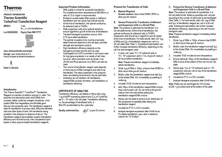
Thermo turbofect 转染试剂说明书

PRODUCT INFORMATIONThermo ScientificTurboFect Transfection Reagent#R0531 1 mLfor _in vitro transfectionsLot XXXXXXXX Expiry Date MM.YYYYFFFFR0531FFFFXXXXXXXXwww. /onebioStorage: Upon receipt store at 4°C.Product shipped at ambient temperature.hMade in LithuaniaIntroductionThe Thermo Scientific™ TurboFect™ Transfection Reagent is a solution of cationic polymer in water. The polymers and deoxyribonucleic acid (DNA) form compact, stable, positively charged complexes, which protect DNA from degradation and facilitate gene delivery into eukaryotic cells. The transfection reagent is ideal for transfection of primary cells, difficult-to-transfect cells and other types of cells with transfection being possible in the presence or absence of serum. The transfection reagent demonstrates superior transfection efficiency and minimal toxicity when compared to lipid-based or other polymer-based transfection reagents.Rev.11 c Important Product Information• DNA quality is critical for successful transfection.For excitation/emission absorbance at 260/280nm, a ratio of 1.8 or higher is recommended.• Endotoxin-contaminated DNA results in inefficient transfection and can cause high cellular toxicity. • At the time of transfection, the optimal confluency for adherent cells is 70-90%.• Plate suspension cells at an optimal density to ensure logarithmic growth at the time of transfection. • Transient transgene expression occurs within 2-72 hours after transfection.• The optimal incubation time must be empirically determined and depends on the cell type, promoterstrength and expression product.• High transfection efficiency depends on thetransgene promoter and the cell line used.• Cytomegalovirus (CMV) promoter is commonly used for high gene expression in a variety of cell lines;however, other promoters such as simian virus(SV40) and Rous sarcoma virus (RSV) can also beused.• The volume of transfection reagent used depends on the amount of DNA, transgene and cells to betransfected. The ratios presented in the protocolsbelow are starting amounts and may be optimized. • Antibiotics do not interfere with transfectionreagent/DNA complex formation, cell transfection ortransfection efficiency.CERTIFICATE OF ANALYSISTransfection efficiency was tested on HeLa cells using1 µg of eGFP expressing plasmid and2 µL of TurboFect per 5 × 10⁴ cells in 24-well plate. Transfection efficiency, i.e. the percentage of transfected cells, is90±10% as estimated by flow cytometry.Quality authorized by:Jurgita Zilinskiene Protocol for Transfection of CellsA. Material RequiredGrowth medium: Serum-free DMEM, RPMI orother growth mediumB. General Protocol for Transfection of Adherentand Suspension Cells in a 24-well PlateNote: The protocol is optimized for transfection in a24-well plate format. At the time of transfection, theoptimal confluency for adherent cells is 70-90%.Suspension cells should be in logarithmic growth phaseat the time of transfection. For best results, start with 1µgof DNA and 2µL of transfection reagent per well in a24-well plate (See Table 1). Subsequent optimization canfurther increase transfection efficiency, depending on thecell line and transgene used.1. In each well, seed ~5 × 10⁴ adherent cells or~5 × 10⁵ suspension cells in 1mL of growth medium24 hours before transfection.Note: Prepare transfection reagent immediatelybefore transfection.2. Dilute 1µg of DNA in 100µL of serum-free DMEM orother serum-free growth medium.3. Briefly vortex the transfection reagent and add 2µLto the diluted DNA. Mix immediately by pipetting orvortexing.4. Incubate 15-20 minutes at room temperature.5. Add 100µL of the transfection reagent/DNA mixturedrop-wise to each well. Do not remove the growthmedium from the cells before adding thetransfection reagent/DNA mixture.6. Gently rock the plate to achieve even distribution ofthe complexes immediately after adding thetransfection reagent.7. Incubate at 37°C in a CO2 incubator.8. Analyze transgene expression after 24-48 hours.For stable transfection, grow cells in selectivemedium for 10-15 days.C. Protocol for Reverse Transfection of Adherentand Suspension Cells in a 24-well PlateNote: The protocol is optimized for transfection in a24-well plate format. Scale-up quantities and volumesaccording to the number of cells/wells to be transfected(See Table 1). For best results, start with 1µg of DNAand 2µL of transfection reagent per well in a 24-wellplate. Subsequent optimization can further increasetransfection efficiency, depending on the cell line andtransgene used.Note: Prepare transfection reagent immediately beforetransfection.1. Dilute 1µg of DNA in 100µL of serum-free DMEMor other serum-free growth medium.2. Briefly vortex the transfection reagent and add 2µLto the diluted DNA. Mix immediately by pipetting orvortexing.3. Incubate 15-20 minutes at room temperature.4. Evenly distribute 100µL of the transfection reagent/DNA mixture at the bottom of the well of a 24-wellplate.5. Gently layer 1mL of ~105 adherent cells or ~106suspension cells per well on top of the transfectionreagent/DNA mixture.6. Incubate at 37°C in a CO2 incubator.7. Analyze transgene expression after 24-48 hours.Note: Plates can be centrifuged for 2-5 minutesat 200 × g to collect cells at the bottom of the plate.Table 1. Scale-up ratios for transfection of adherent and suspension cells using TurboFect Transfection Reagent.Tissue cultureplate Growth area(cm2/well)Volume ofmedium (mL)No. of adherent(suspension) cells toseed the day beforetransfection*Amount of DNAVolume of TurboFectTransfection Reagent (µL)(µg) (µL)** Recommended Range96-well plate 0.3 0.2 0.5-1.2 × 10⁴(2.0 × 10⁴)0.2 20 0.4 0.3-0.648-well plate 0.7 0.5 1.0-3.0 × 10⁴(5.0 × 10⁴)0.5 50 1.0 0.5-1.424-well plate 2.0 1.0 2.0-6.0 × 10⁴(1.0 × 10⁵)1.0 1002.0 1.0-2.812-well plate 4.0 2.0 0.4-1.2 × 10⁵(2.0 × 10⁵)2.0 200 4.0 2.6-6.06-well plate 9.5 4.0 0.8-2.4 × 10⁵(4.0 × 10⁵)4.0 400 6.0 4.0-8.060mm plate20 6.0 2.0-6.3 × 10⁵(1.0 × 10⁶)6.0 600 12.0 8.0-16.0*Values for suspension cells are in parentheses.**The volume of DNA should be 1/10 the volume of the culture medium used for dilution of the DNA.Note: These numbers were determined using HeLa and Jurkat cells. Actual values depend on the cell type. The amount of DNA and TurboFect Transfection Reagent used may require optimization.Additional InformationA. Cells successfully transfected with TurboFect Transfection Reagent.Permanently growing cell lines Primary cell culturesCos-7 African green monkey kidney cellsHeLa Human cervix adenocarcinoma cellsCHO Chinese hamster ovary cellsHEK293 Human embryonic kidney cellsB50 Rat nervous tissue neuronal cellsCalu1 Human lung epidermoid carcinoma cells RAW264 Mouse leukaemic monocyte-macrophage cells WEHI Mouse B cell lymphoma cellsMDCK Madin Darby Canine Kidney cellsRaji Human Burkitt’s lymphoma cellsCOLO Human colon adenocarcinoma cellsJurkat Human leukaemic T cellsSp2/Ag14 Mouse myeloma cellsHeLa S3 Human cervix carcinoma cellsHep2C Human larynx carcinoma cellsL929 Mouse connective tissue fibroblastsNIH3T3 Mouse embryo fibroblasts Rat fibroblastsMouse bone marrow-deriveddendritic cellsMouse bone marrow-derivedmacrophagesHuman lung fibroblasts (HLF)TroubleshootingProblem Possible Cause SolutionLow transfectionefficiencySuboptimal transfection reagent/DNAratioOptimize the amount of transfection reagent added tothe fixed amount of DNASuboptimal quantity of DNAOptimize the amount of DNA used for transfectionKeep the amount of transfection reagent constantPoor DNA qualityUse high-quality DNA with an absorbance ratio greaterthan 1.8 at 260/280nmSuboptimal cell confluencyOptimize cell plating conditionsEnsure adhered cells are 70-90% confluent at the timeof transfectionEnsure that suspension cells are in logarithmic growthphase at the time of transfectionMycoplasma contamination Regularly check cells for mycoplasma infectionHigh cellulartoxicityToxic transgene Verify if the expressed transgene is toxicSuboptimal incubation conditionsReduce incubation time of the polyplexes with the cellsReplace the transfection mixture 3-4 hours later withnew growth mediumExcess amount of DNA Reduce the quantity of DNA used for transfectionCell density was too lowIncrease the plating density of cells used fortransfectionEndotoxin or other toxic materials werepresent with transgeneEnsure transgene is free of toxic substancesRepeat insertion of gene into new toxin-free plasmidpreparationRelated Thermo Scientific Products16146-89Pierce® Luciferase Assay Kits and Reagents88273 High Capacity Endotoxin Removal Spin Columns, 0.1mL, 5/pkgNote:This product or the use of this product is covered by US patent application US20100041739A1 and corresponding counterparts. The purchase of thisproduct includes a non-transferable license to use this product for the purchaser's internal research. All other commercial uses of this product, includingwithout limitation product use for diagnostic purposes, resale of product in the original or any modified form or product use in providing commercial servicesrequire a separate license. For further information on obtaining licenses please contact @PRODUCT USE LIMITATIONThis product is developed, designed and sold exclusively for research purposes and in vitro use only. The product was not tested for use in diagnostics orfor drug development, nor is it suitable for administration to humans. Please refer to /onebio for Material Safety Data Sheet of theproduct.Current product instructions are available at /onebio.© 2012 Thermo Fisher Scientific, Inc. All rights reserved. Unless otherwise indicated, all trademarks are property of Thermo Fisher Scientific Inc. and itssubsidiaries. Printed in the Lithuania.。
lipofectamine rnaimax转染试剂的原理

lipofectamine rnaimax转染试剂的原理
Lipofectamine RNAiMAX是一种RNA干扰实验中常用的转染
试剂。
其原理主要涉及以下几个步骤:
1. 形成转染复合物:将RNA干扰试剂(如siRNA或miRNA)与Lipofectamine RNAiMAX转染试剂按照一定比例混合,形
成转染复合物。
2. 结合到细胞膜表面:转染复合物中的Lipofectamine能与细
胞膜表面的磷脂结合,形成Lipofectamine-RNA复合物。
3. 转染复合物进入细胞:通过细胞膜的内吞作用或细胞膜破损,转染复合物能够被细胞摄入。
4. 转染复合物释放RNA:一旦进入细胞,转染复合物会释放
出RNA干扰试剂。
5. RNA干扰效应:释放的RNA干扰试剂能够与细胞内的靶向mRNA相互结合,通过RNA酶PAS复合物介导的靶向降解或抑制翻译的机制,达到基因沉默或抑制的效果。
总之,Lipofectamine RNAiMAX转染试剂通过将RNA干扰试
剂转染入细胞内,调控目标基因的表达,从而实现基因沉默或抑制。
neofect转染试剂说明书中文

NeoFect是一种用于将DNA或RNA转染到真核细胞中的转染试剂。
以下是一个简化的、假设性的NeoFect转染试剂说明书的中文翻译,请注意,这不是官方翻译,仅供参考。
实际使用时,请参考随产品附带的正式说明书和安全数据表(SDS)。
---NeoFect转染试剂说明书【产品名称】通用名:转染试剂商品名:NeoFect【成分】主要成分为阳离子脂质体,用于促进核酸分子与细胞膜的融合。
【性状】本品为透明至微浑浊的液体,通常以小瓶或多孔板包装。
【适应症】用于科研实验中,将DNA或RNA高效转染到哺乳动物细胞中,以进行基因表达、基因沉默、基因编辑等研究。
【使用方法】1. 准备待转染的细胞和核酸溶液(DNA或RNA)。
2. 根据实验设计,将适量的NeoFect转染试剂加入无血清培养基中,轻轻混匀。
3. 将核酸溶液加入含有NeoFect的培养基中,轻轻混匀,室温孵育15-30分钟。
4. 将混合物加入到细胞培养皿中,轻轻摇晃使混合均匀。
5. 根据细胞类型和实验目的,孵育一定时间后更换为完全培养基继续培养。
【不良反应】本品仅供实验室使用,不适用于临床治疗。
【禁忌】对本品成分过敏者禁用。
【注意事项】1. 使用前请检查试剂是否清澈,如有沉淀或颜色变化请勿使用。
2. 避免反复冻融,分装后请立即使用。
3. 使用过程中请遵守实验室安全操作规程,佩戴适当的个人防护装备。
4. 请在无RNA酶和DNA酶的环境中操作RNA转染实验。
5. 转染效率受多种因素影响,如细胞状态、核酸浓度、孵育时间等,建议优化实验条件。
【贮藏】存放于4°C冰箱中,避免冷冻。
【有效期】请参考包装标签上的说明。
【生产批号】见包装标签。
【生产企业】(请填写生产企业名称和联系方式)---以上信息仅供参考,实际使用时请遵循产品附带的正式说明书和安全数据表(SDS)的内容。
在操作前,务必了解所有相关的安全和健康信息。
上海常用转染试剂配方

上海常用转染试剂配方
转染试剂是在细胞培养研究中必不可少的试剂。
它将核酸或蛋白质带入目标细胞中,以达到研究目的。
本文将介绍上海常用的几种转染试剂配方。
1. Lipofectamine 2000转染试剂
Lipofectamine 2000是一种常用的脂质体转染试剂,其用途广泛且转染效率高。
其配方如下:
无血清培养基:500μL
要转染的DNA:2μg(根据实验需要调整)
将Lipofectamine 2000试剂与无血清培养基混合,放置5分钟,再加入要转染的DNA 液体,混合后放置15分钟,滴加在细胞上。
2. PEI转染试剂
聚乙烯亚胺(PEI)是一种阳离子高分子,由于其能够形成与DNA负电荷的缔合物,使其成为一种有效的转染试剂。
PEI转染试剂可用于转染细胞以获得较高的转染效率。
其配方如下:
PEI试剂:4μg/μL,将其与等体积的无血清培养基混合。
3. PolyJet转染试剂
PolyJet是一种高效、无毒、具有低细胞损伤的DNA转染试剂。
其配方如下:
4. FuGENE HD转染试剂
细胞的转染操作应该注意以下几点:
1. 转染时间应该控制在半小时至1小时内,过长或过短都会对转染效率造成影响。
2. 转染的DNA浓度应该适当,过高的DNA浓度会对细胞产生毒性。
3. 转染试剂与DNA的混合比例也应该适当,过多或过少都会导致转染效率降低。
总之,合理选择转染试剂和配方,并严格按照操作步骤进行转染可以提高实验的可靠性和成功率。
磁性抗体转染结肠癌干细胞的最佳浓度探索

【 关键词】 磁性抗体; 肿瘤干细胞; 结肠癌; 最佳浓度 【 中图分类号】 R 3 7 8 . 1 【 文献标志码】 A 【 收稿日期】 2 0 1 5 — 1 1 - 0 7
Op t i ma I c o n c e n t r a t i o n o f ma g n e t i c a n t i b o d i e s t r a n s f e c t e d
i n t o c o l o n c a n c e r s t e m c e l l s/ n v i t r o
Wa n g Z h e n g x i o n f, L i u Mi n , C h e n H u a y i n f, Me i X u e q i a n 3 , L i X u e h o n f, L i Q i a n f, T a n S h u d e I L i u Y u a n b i n f, Z h a o X i a o ( 1 . D e p a r t m e n t o f R a d i o l o g y ; 2 . D e p a r t m e n t o f O b s t e t r i c s nd a G y n e c o l o y; g 3 . D e p a r t m e n t fI o n f o r m a t i o n ,
验) 进行 组 间均数多 重 比较 , 结 果显示 , 5 0 m g / L组与 7 5 m g / L 、 1 0 0 m g / L各组信号 强度 之间差 异无 统计学 意义 ( P = 0 . 9 4 5 , P =
纳米Fe3O4的制备及在油墨中的应用研究

科研应用纳米Fe 3O 4的制备及在油墨中的应用研究杨勃(湖南工业大学湖南·株洲412007)中图分类号:O614.81文献标识码:ADOI :10.16871/ki.kjwhb.2016.05.080作者简介:杨勃(1994—),就读于湖南工业大学印刷工程专业。
摘要磁性防伪油墨是一种新型防伪技术,其防伪技术的核心是磁性材料。
Fe 3O 4是目前最常使用的磁性材料。
本文采用共沉淀法,利用硫酸亚铁为单一铁源,以醋酸钠作静电保护剂,在一定条件下成功制备了形貌基本均一、平均粒径为100nm 的棒状Fe 3O 4。
然后利用实验室自制的Fe 3O 4纳米材料作为磁性防伪材料和颜料,配以一定比例的连接料和助剂,制备出了磁性能良好防伪油墨,其细度均匀、墨色浓厚,流动性能良好,适于印刷。
关键词纳米Fe 3O 4磁性油墨共沉淀法磁性能印刷适性Research on the Preparation of Nano Fe 3O 4and Its Appli 原cation to Painting Ink //Yang BoAbstract Magnetic anti-counterfeiting ink is a new anti-coun-terfeiting technique,whose core is magnetic materials.Fe 3O 4is the most commonly used magnetic material at present.Through co-precipitation method,with ferrous sulfate as the single source of iron and sodium acetate as the static protection agent,rod-like Fe 3O 4,with generally average appearance and an average diame-ter of 100nm,is prepared under certain conditions.Then with laboratory made Fe 3O 4nano material as the magnetic anti-coun-terfeiting material and pigment,supplemented by a certain pro-portion of binder and auxiliary,anti-counterfeiting painting ink with good magnetic property is prepared,and it is well suitable for printing with its average size,ink density and good flowability.Key words nano Fe 3O 4;magnetic painting ink;co-precipitation method;magnetic property;printability1引言当今社会假冒伪劣产品充斥市场,各种防伪技术层出不穷。
转染试剂使用说明书

2. 3).
配制转染工作液: ( 6 孔板或 35 mm 平皿, 2 ml 培养液) 取 5~8μ g DNA (起始用量 5μ g) ,加入稀释液中至总体积为 100μ l,轻轻混匀,室 温放置。
4).
先将 GenFectin TM 涡旋振荡混匀。取 GenFectinTM 1~4μl(起始用量 2μl,) ,加入稀 释液中至总体积为 100μl,轻轻混匀,室温放置 5 分钟。
5).
将稀释的 GenFectinTM 逐滴加入稀释的 DNA 溶液中,轻轻混匀,所得的转染工作 液在室温放置 15 分钟。
6).
将转染工作液轻轻混匀,逐滴加入 2 ml 培养液中,轻轻混匀培养液,置 37℃,5% CO 2 培养。
3. 细胞后续处理: 7). 24~48 小时后,观察或收取细胞。
-4-
8).
稳定转染时,于转染后 24~48 小时消化细胞分至 3~5 个培养皿中,加适当浓度的 相应抗生素(如 G418)筛选。
建议的起始转染条件 : 培养容器
96 孔板 24 孔板 6 孔板 35mm 培养皿 60mm 培养皿
转染前一天 接种细胞数
1-1.510 个 0.5-1 10 个 2-4 10 个 2-4 10 个 4-6 105 个
5 5 5 4
转染时 培养液体积
0.1 ml 0.5 ml 2 ml 2 ml 4 ml
DNA 用量 与稀释后体积
0.25 μg 1.25 μg 5 μg 5 μg 10 μg 5 μl 25 μl 100 μl 100 μl 200 μl
GenFectin 量 与稀释后体积
0.1 μl 0.5 μl 2 μl 2 μl 4 μl 5 μl 25 μl 100 μl 100 μl 200 μl
- 1、下载文档前请自行甄别文档内容的完整性,平台不提供额外的编辑、内容补充、找答案等附加服务。
- 2、"仅部分预览"的文档,不可在线预览部分如存在完整性等问题,可反馈申请退款(可完整预览的文档不适用该条件!)。
- 3、如文档侵犯您的权益,请联系客服反馈,我们会尽快为您处理(人工客服工作时间:9:00-18:30)。
磁性转染试剂(Magnetofection TM-)-Qwbio
产品名称:Magnetofection TM
产品描述:
磁性转染(Magnetofection TM)(启维益成有售)是一种新颖、简单且高效的转染培养细胞的方法。
它利用磁力将与磁性颗粒结合的基因载体吸向,甚至可能吸入靶细胞。
这样所有加入的载体在几分钟之内即在细胞表面富集,使得100%的细胞与显著剂量的载体接触。
这具有以下重要效果:
. 与标准转染方法相比,极大的提高了基于被转染细胞百分比的转染率
. 与标准转染方法相比,短时间孵育即可获得提高达几千倍的转基因表达
. 使用极低剂量的载体即可获得高转染率和高转基因表达水平,节省昂贵的转染试剂
. 极短的处理时间。
细胞与基因载体孵育几分钟即足以得到高转染率,而标准方法需几小时。
PolyMAG是一种用于高效递送核酸的通用磁性颗粒制剂。
它与欲转染的核酸在一个步骤中混合,成功转染了质粒DNA、反义寡核苷酸和siRNAs。
CombiMAG是为了与市售的如多聚阳离子脂质等任何转染试剂结合使用而设计的磁性颗粒
制剂。
它可与质粒DNA、反义寡核苷酸、siRNAs或病毒结合。
保存:
Magnetofection TM试剂盒的所有组分在打开前应在室温(20-25o C)下保存。
第一次使用后在4o C保存。
•不要冻存磁性纳米颗粒
•不要在磁性纳米颗粒储存液中加入任何物质
•运输条件:室温
参考文献:
9.001 Plank C., Scherer F., Schillinger U., Anton M. Magnetofection: Enhancement and localization of gene delivery with magnetic particles under influence of a magnetic fields. J. Gene Med.2000; 2((5) Suppl.): S24.
9.002 Scherer F., Anton M., Schillinger U., Henke J., Bergemann C., Krüger A., Gänsbacher B. and Plank C. Magnetofection: enhancing and targeting gene delivery by magnetic force in vitro and in vivo. Gene Therapy 2002; 9: 102-109.
9.003 Plank C., Schillinger U., Scherer F., Bergemann C., Remy J.-S., Krötz F., Anton M., Lausier J. and Rosenecker J. The Magnetofection Method: Using Magnetic Force to Enhance Gene Delivery. Biol. Chem. May 2003; 384: 737-747.
9.004 Plank C., Anton M., Rudolph C., Rosenecker J. and Krötz F. Enhancing and targeting nucleic acid delivery by magnetic force. Expert Opinion on Biological Therapy 2003; 3: 745-758.
9.005 Plank C., Scherer F., Schillinger U., Bergemann C., Anton M. Magnetofection: enhancing and targeting gene delivery with superparamagnetic nanoparticles and magnetic fields. J.Liposome Res. 2003; 13(1): 29-32.
9.06 Plank C. Magnetofection: enhancing and targeting gene delivery with lipid-DNA vectors by magnetic force. J. Liposome Res. 2003; 13(1): 105-106.
9.007 Krötz F., Son HY., Gloe T., Planck C. Magnetofection Potentiates Gene Delivery to Cultured Endothelial. Journal of Vascular Research 2003; 40: 425-434.
9.008 Krötz F., Wit C., Sohn HY., Zahler S., Gloe T., Pohl U. and Plank C. Magnetofection-A highly efficient tool for antisense oligonucleotide delivery in vitro and in vivo. Mol. Ther. May 2003;7(5): 700-710.。
