LT SV 40 Large T Antigen
酵母双杂实验操作手册和注意事项
酵母双杂实验操作⼿册和注意事项酵母双杂(Yeast two-hybrid)实验操作⼿册和注意事项⼀. 酵母双杂的原理1989年,Song和Field建⽴了第⼀个基于酵母的细胞内检测蛋⽩间相互作⽤的遗传系统。
很多真核⽣物的位点特异转录激活因⼦通常具有两个可分割开的结构域,即DNA特异结合域(DNA-binding domain,BD)与转录激活域(Transcriptional activation domain ,AD)。
这两个结构域各具功能,互不影响。
但⼀个完整的激活特定基因表达的激活因⼦必须同时含有这两个结构域,否则⽆法完成激活功能。
不同来源激活因⼦的BD区与AD结合后则特异地激活被BD结合的基因表达。
基于这个原理,可将两个待测蛋⽩分别与这两个结构域建成融合蛋⽩,并共表达于同⼀个酵母细胞内。
如果两个待测蛋⽩间能发⽣相互作⽤,就会通过待测蛋⽩的桥梁作⽤使AD与BD形成⼀个完整的转录激活因⼦并激活相应的报告基因表达。
通过对报告基因表型的测定可以很容易地知道待测蛋⽩分⼦间是否发⽣了相互作⽤。
酵母双杂交系统由三个部分组成:(1)与BD融合的蛋⽩表达载体,被表达的蛋⽩称诱饵蛋⽩(bait)。
(2)与AD融合的蛋⽩表达载体,被其表达的蛋⽩称靶蛋⽩(prey)。
(3)带有⼀个或多个报告基因的宿主菌株。
常⽤的报告基因有HIS3,URA3,LacZ和ADE2等。
⽽菌株则具有相应的缺陷型。
双杂交质粒上分别带有不同的抗性基因和营养标记基因。
这些有利于实验后期杂交质粒的鉴定与分离。
根据⽬前通⽤的系统中BD来源的不同主要分为GAL4系统和LexA系统。
后者因其BD来源于原核⽣物,在真核⽣物内缺少同源性,因此可以减少假阳性的出现。
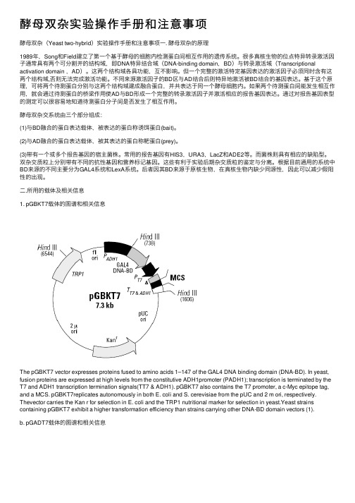
⼆.所⽤的载体及相关信息1. pGBKT7载体的图谱和相关信息The pGBKT7 vector expresses proteins fused to amino acids 1–147 of the GAL4 DNA binding domain (DNA-BD). In yeast, fusion proteins are expressed at high levels from the constitutive ADH1promoter (PADH1); transcription is terminated by the T7 and ADH1 transcription termination signals(TT7 & ADH1). pGBKT7 also contains the T7 promoter, a c-Myc epitope tag, and a MCS. pGBKT7replicates autonomously in both E. coli and S. cerevisiae from the pUC and 2 m ori, respectively. Thevector carries the Kan r for selection in E. coli and the TRP1 nutritional marker for selection in yeast.Yeast strains containing pGBKT7 exhibit a higher transformation efficiency than strains carrying other DNA-BD domain vectors (1).b. pGADT7载体的图谱和相关信息pGADT7-T encodes a fusion of the SV40 large T-antigen (a.a. 86–708) and the GAL4 AD (a.a. 768–881). The SV40 large T DNA (GenBank LocusSV4CG) was derived from a plasmid referenced in Li & Fields (1993) and was cloned into pGADT7 using the EcoR I and Xho I sites. pGADT7-T has not been sequenced.三.实验主要流程A.需要准备的药品和设备1.两种酵母菌种(AH109,Y187)2.酵母培养所需的药品: Yeast nitrogen base without amino acidsAgar (for plates only)sterile 10×Dropout Solution单缺-T,-L(clontech公司)⼆缺-T/-L (clontech公司)四缺-T/-L/-Ade/-His(clontech公司)3.酵母转化所需的药品: 10×TE buffer10×LiAc40%PEGcarrier DNA4.酵母显⾊所需要的药品: x- -GAL5.其他仪器设备: 30℃恒温培养箱30℃摇床.⽔浴锅分光光度计B.DNA-BD和DN-AD fusion protein 载体的分别构建。
pAD-SV40T使用说明

pAD-SV40T编号载体名称北京华越洋VECT76070pAD-SV40TpAD-SV40T载体图谱:pAD-SV40T载体简介:The pAD-SV40T control plasmid expresses a hybrid protein which contains the NF-κB transcription activation domain fused to amino acids84–708of the SV40large T-antigen.5pAD-SV40T载体相关的哺乳动物表达载体:SuperCos I pDsRed2-Bid pNFκB-MetLuc2-ReporterpYr-adshuttle-3pAcGFP1-N1pEF1α-IRES-DsRed-Express2pVitro2-neo-mcs pSecTag2A pCMV-DsRed-Express2pUB6/V5-His/LacZ pGL4.27pcDNA3.1/NT-GFP-TOPOpUB6/V5-His C pGL4.26pEF1α-IRES-ZsGreen1pUB6/V5-His B pACT pCMV-Tag2ApUB6/V5-His A pBIND-Id Control pCMV-Tag5BpTracer-CMV2pTRE2pAcGFP1-C In-Fusion ReadypSV-β-Galactosidase pRevTRE p3XFLAG-CMV-14pSI pTK-hyg p3XFLAG-CMV-8pSG5pTRE3G-Luc pFLAG-CMV-2pSFV1pSwitch pcDNA3.3-TOPOpSecTag2/Hygro A pcDNA4/His C pcDNA6.2/cLumio-DESTpSecTag B c-Flag pcDNA3pCMV-tdTomatopRluc-N2pcDNA4/TO/Myc-His A pAcGFP1-MitopPICZalpha D pcDNA6/myc-His B pAcGFP1-N In-Fusion Ready pORF-lacZ pcDNA6/V5-His B pDsRed-Monomer-N In-Fusion Ready pORF-HSV1tk pcDNA6.2/nTC-Tag-DEST pcDNA4/TO/Myc-His BpOG44pOptiVEC-TOPO pIRES2-EGFPpNTAP-B pcDNA5/FRT pcDNA3.1/His CpMEP4pGL4.30pcDNA3.1/CT-GFP-TOPOpLVX-ZsGreen-miRNA-Puro pGL4.19pEF1α-IRES-AcGFP1pLVX-IRES-Puro-3xFlag pACT-MyoD pcDNA3.2/V5/GW/D-TOPO pLPCX pCMV-BD pcDNA4/TO/Myc-His/LacZ pLEGFP-N1pCMV-Tet3G pcDNA4/HisMax-TOPOpKH3pTet on advanced p3XFLAG-CMV-13pIRES-puro2pTRE-Tight p3xFLAG-CMV-10phRL-TK pIND pFLAG-CMV-3pG5lac pGene/V5-His B pcDNA4/TO/Myc-His CpFR-luc pOPRSVI pcDNA6.2/C-YFP-DESTpEF6/myc-His C pcDNA4/HisMax A pcDNA6.2/cTC-Tag-DESTpEF4/V5-His A pIRESpuro3pcDNA6.2/nGeneBLAzer-DESTpEF1/myc-His lacZ pIRESneo3pCRE-MetLuc2-ReporterpEF1/myc-His C pIRESneo2pEF1α-DsRed-Express2pEF1/myc-His B pcDNA4/myc-His A pDsRed-Express-C1pEF1/myc-His A pCMV-PKA pEF1α-DsRed-Monomer-N1 pECFP-Mito pAcGFP1-N3pDD-AmCyan1ReporterpECFP-ER pcDNA5/FRT/TO pCRE-DD-AmCyan1pDP8rs pBApo-CMV-Pur pIRES2-DsRed-Express2pDP5rs pBApo-EF1α-pur pDsRed-Express-N1pDP4rs ptdTomato-N1pcDNA6.2/nLumio-DESTpDP3rs pAcGFP1-Golgi pcDNA6/myc-His ApDP2rs pAcGFP1-p53pcDNA6/V5-His ApDP1rs pAcGFP1-Actin pcDNA5/FRT/TO-TOPOpCMV-Myc-C pBI-CMV2pcDNA6.2/V5/GW/D-TOPOpCMV-HA-N pEF1α-tdTomato pcDNA6.2/cGeneBLAzer-DEST pCMV-HA-C pEF1α-tdTomato pcDNA6.2/nGeneBLAzer-GW/D-TOPO pCMV6-XL4pEBVHis A pcDNA5/TOpCMV6-AC-GFP pGL4.10pBApo-CMV-neopCMV5pGL4.29pDsRed-MonomerpCI pGL4.13pIRESpCGN pG5luciferase pIRES-hrGFP-1apcDNA6.2/EmGFP-Bsd/V5-DEST pCMV-AD pDsRed-Express2-N1pcDNA4/V5-His A pRevTet-Off pCMV-Tag3BpcDNA3-mRFP pTet-Off pCRE-hrGFPpcDNA3-CFP pTRE2-hygro pDsRED2-MitopcDNA3.1(+)/myc-His C pVgRxR pAcGFP1-FpcDNA3.1(+)/myc-His A pOPI3CAT pAcGFP1-C1pcDNA3.1(+)/CAT pBK-RSV pAsRed2-C1pcDNA3.1(-)/myc-His C pIRES2-DsRed2pAsRed2-N1pcDNA3.1/Zeo(+)pCMV-Myc pAcGFP1-LampcDNA3.1/Zeo(-)pCMV-Tag2C pAcGFP1-CpcDNA3.1/V5-His C pCMV-Tag5A pSEAP2-BasicpcDNA3.1/Hygro(+)pCMV-Tag3C pBI-CMV3pcDNA3.1/Hygro(-)p3XFLAG-CMV-7pNFkB-DD-tdTomatopcDNA3.1/GS p3XFLAG-CMV-9pcDNA3.1/His ApBS185CMV-Cre pFLAG-CMV-4pEBVHis BpBIND-GFP pBI-CMV4pGL4.75pBiFC-VN155(I152L)pcDNA4/His A pGL4.20pBiFC-CC155pcDNA4/myc-His B pCMV-SPORT6pBiFC-CrN173pcDNA4/HisMax C pCMV-SPORT6pBiFC-bJunVN155(I152L)pCMV-Tag4A pBINDpBD-p53pcDNA6/myc-His C pBD-NF-κBpBD-NF-kB pCMV-Tag2B pRevTet-OnpBC1pGRN145pTet-OnpAD-TRAF pCMV-MEK1pTRE3GpAD-SV40T pCMV-Tag3A pcDNA4/TOpAAV-ZsGreen-miRNA pCMVLacI pcDNA4/His B pAAV-tTS-shRNA pBI-CMV1pcDNA4/HisMax B pIREShyg3pEF1α-AcGFP1-N1pcDNA4/myc-His C pcDNA6/TR pCMV-LacZ pcDNA3.1/His B pDsRed-Express2-C1pCMV-Tag4B pcDNA6/V5-His C pBI-CMV5pCMV-Tag5C pCHO1.0 pAmCyan1-C1plRES2-ZsGreen1pGL3-Promoter pAcGFP1-Mem p3XFLAG-CMV-7.1pCMV-MEKK1 pAcGFP1-C3pFLAG-CMV-5a pFLAG-CMV2 pTT5pBudCE4.1pAcGFP1-C2 pSEAP2-Control pREP4ptdTomato-C1 pIRES2-AcGFP1pBApo-CMV pCRE-DD-tdTomato pAcGFP1-Hyg-C1。
large T-antigen

Thanks !
• 此时大T抗原: 95%将会返回细胞核。一旦在细胞核内,大T 抗原就结合3个病毒DNA位点,I,II和III。自动 调节早期的RNA合成。
• SV40病毒基因组早期基因编码两种肿瘤抗原 (tumor antigen,T抗原),其分子量分别为 94000(大T抗原)和17000(小T抗原). • T抗原有以下的作用: ①大T抗原为细胞转化的启动所必需; ②转化细胞表型的维持必需要有大T抗原的连续 表达; ③小T抗原对于细胞的转化不是必需的,但可起 加强作用.
SV40
• 由于SV40结构简单,被首• 增强子序列。其长度为72bp,以串联重复的形式 位于基因组DNA复制起点的附近,它的主要功能 是促进病毒DNA发生有效的早期转录,而且具有 一般增强子所共有的基本特性。
SV40
• 病毒在细胞核内,由细胞的 RNA 聚合酶II的 行为促进早期基因表达。其结果是一条拼接 成两段的mRNA。大小T抗原就是它的结果。
SV40 Large T-antigen
SV40
• 这种病毒是在1960年,于用来 生产脊髓灰质炎疫苗的恒河猴肾 脏细胞中发现。 • 是猴空泡病毒40,猿猴病毒40 或猴病毒40的缩写,多瘤病毒科, 这是在人类和猴子都发现的致瘤 病毒。 • SV40病毒的基因组是一种环形 双链的DNA,基因组5.2kb,病 毒的直45nm,病毒成熟部位细 胞核,无被膜,62个核壳粒亚单 位,这种大小很适于基因操作。 同时它也是第一个完成基因组 DNA全序列分析的动物病毒。
SV40 Large T-antigen
• 大T抗原能转化各种不同的基因表型。TAg的活性 阻碍了肿瘤抑制基因pRB和p53的表达。 • 大T抗原还绑定了几个其他的细胞因子,包括转 录激活剂P300和CBP。
c-Myc标签Co-IP试剂盒中文说明
23620描述ProFound™ c-Myc Tag IP/Co-IP Kit 包含了足够25个与c-Myc –标签蛋白IP或Co-IP的成分,包括10个c-Myc –标签阳性对照的实验试剂盒成分:Immobilized Anti-c-Myc, 62.5 g固定的珠状琼脂加上125g 抗体,以含25%的溶液和0.1%叠氮钠的PBS 的形式提供(总体积为250ul)。
BupH™ Tris Buffered Saline Pack,一个包装,用500 ml超纯水稀释后,溶液成分为25 mM Tris, 0.15 M NaCl, pH 7.2Elution Buffer, 50 ml, pH 2.8Lane Marker Non-Reducing Sample Buffer (5X), 5 ml, 包含0.3 M Tris•HCl, pH 6.8, 5% SDS, 50% 甘油, 电泳指示剂。
Handee™ Spin Columns Accessory Pack, 含提前放置好的玻璃原料,顶盖和底盖的27Collection Tubes and Caps Accessory Pack, 100 有刻度的2 ml 管子和塞盖c-Myc-tagged Positive Control, 500ul,1 mg/ml E. coli 提取的包含c-Myc-标签GST(古胱氨酸S-转移酶)储藏: 收到后,将c-Myc-标签阳性对照放在-80度冰箱,(在-20度可放6个月),将其他所用到的成分放在4度,c-Myc-标签阳性对照用干冰运输。
注意:ProFound™ c-Myc Tag IP/Co-IP Kit是ProFound™ c-Myc Tag IP/Co-IP Application Set (23622) 和c-Myc-tagged Positive Control (23633)的联合体。
介绍ProFound™ c-Myc Tag IP/Co-IP Kit与用蛋白A/G琼脂糖的IP方法相比,传统的提供了一个简单快速的方法来研究c-Myc-标签蛋白。
plat-e病毒包装细胞文献
Gene Therapy (2000)7,1063–1066©2000Macmillan Publishers Ltd All rights reserved 0969-7128/00$/gtVIRAL TRANSFER TECHNOLOGY BRIEF COMMUNICATIONPlat-E:an efficient and stable system for transient packaging of retrovirusesS Morita,T Kojima and T KitamuraDepartment of Hematopoietic Factors,Institute of Medical Science,University of Tokyo,4-6-1Shirokanedai Minato-ku,Tokyo 108-8639,JapanA potent retrovirus packaging cell line named Platinum-E (Plat-E)was generated based on the 293T cell line.Plat-E is superior to existing packaging cell lines regarding efficiency,stability and safety.The novel packaging constructs utilized in establishment of Plat-E ensure high and stable expression of viral structural proteins.Conventional packaging con-structs made use of the promoter of MuLV-LTR for expression of viral structural genes gag-pol and env,while our packaging constructs utilized the EF1␣promoter,which is 100-fold more potent than the MuLV-LTR in 293T cells in combination with the Kozak’s consensus sequence upstream of the initiation codon resulting in high expression Keywords:packaging cell;retroviruses;ecotropic;EF1␣promoterRetroviral vectors and packaging cells are important tools for gene transfer applications.Introduction of retroviral vectors containing the gene of interest into suitable pack-aging cells enables production of infectious retroviruses,and these particles can infect target cells and stably trans-mit the gene of interest into chromosomes.In conven-tional strategies,stable high producers of a retrovirus vector harboring a gene of interest were established by transducing the retrovirus construct into NIH3T3-based packaging cells such as PA317,1and 2–3months were usually needed to acquire high producers.Pear et al 2developed a unique packaging system by which high titer retroviruses can be obtained in 3days by transient transfection.The expression of viral structural genes was driven by the MuLV LTR in Bosc23cells.For transient transfection,the combination of Bosc23cells and the pMX-neo vector 3produced 1–3×10⁶/ml viruses,assessed based on the number of neomycin resistant col-onies of the infected NIH3T3cells (data not shown).Since Bosc23cells carry the large T antigen,we attempted to increase titers of the retroviruses by introducing the SV40origin to the pMX vector for amplification of the vector.However,this proved unfeasible (data not shown),sug-gesting that the limiting factor was the expression level of the viral structural proteins in the packaging cells.Bosc23was obtained by cotransfection of the plasmid encoding gag-pol together with the plasmid encoding the hygromycin-resistant gene and the plasmid encoding envCorrespondence:T KitamuraReceived 3November 1999;accepted 3March 2000of virus structural proteins in Plat-E cells.To maintain the high titers of retroviruses under drug selection pressure,we inserted the IRES (internal ribosome entry site)sequence between the gene encoding gag-pol or env,and the gene encoding a selectable marker in the packaging constructs.Plat-E cells can stably produce retroviruses with an average titer of 1×10⁷/ml for at least 4months.In addition,as we used only the coding sequences of viral structural genes to avoid inclusion of unnecessary retrovirus sequences in the packaging constructs,the probability of generating the repli-cation competent retroviruses (RCR)by recombination can virtually be ruled out.Gene Therapy (2000)7,1063–1066.together with the plasmid encoding another selection gene GPT (guanine phosphoribosyl transferase),one after the other.Therefore,expression of selectable markers did not guarantee the expression of gag-pol or env genes,which may account for the instability of the cells in producing high-titer retroviruses.A similar packaging cell line Phoenix-E 4has also been developed.In Phoenix-E cells,the plasmids encoding the gag-pol and env genes were cotransfected with selection markers,which did not warrant the stable expression of the gag-pol and env genes in the selection drug,hygromy-cin and diphtheria toxin,respectively.There were several improvements in the Phoenix-E cells when compared with Bosc23cells.First,the RSV and CMV promoters,which are much stronger than MuLV-LTR in 293T cells,were used to express the gag-pol and env genes,respect-ively.Second,the internal ribosome entry site (IRES 5)sequence was used to express gag-pol and a cell surface marker CD8simultaneously which enables sorting of high expressers of the gag-pol gene.However,one needs to sort cells from time to time to maintain the expression levels of the gag-pol gene.To design a packaging cell line which can stably pro-duce retroviruses with high titer,we searched for a strong promoter to drive expression of viral structural proteins in 293T cells using the FACS-GAL assay.6Among seven promoters tested,the EF1␣and CMV pro-moters induced high expression of lacZ (Figure 1).The activities driven by these promoters were 100-fold higher than those by LTR utilized in Bosc23cells,and even exceeded those by SV40and SR ␣promoters,which enable amplification of vectors in 293T cells expressingPlat-E:a system for transient packaging of retrovirusesS Morita et al1064 GeneTherapyFigure1Activities of various promoters in293T cells.The activities ofthe seven promoters,SV40,SR␣,EF1␣,RSV,TK,MuLV LTR and CMVwere evaluated by expression of lacZ under the control of each promoter in293T7cells by the FACS-GAL assay as described.6Briefly,cells(1×10⁶)transfected with each promoter construct were suspended in50l of phos-phate buffered saline(PBS),then incubated for5min at37°C.FDG(fluorescein di--d-galactopyranoside;Molecular Probes,Eugene,OR,USA)was dissolved in distilled water,warmed at37°C and50l of2m m FDG solution was added to50l of cell suspension.After1min ofincubation at37°C,1ml of PBS was added followed by incubation on icefor2h.To stop the reaction,20l of50m m PETG(phenylethyl--d-thiogalactoside;Sigma,St Louis,MO,USA)was added,and the prep-aration was placed on ice until being subjected to FACS analysis.the SV40large T antigen.Because we thought that thepromoters of housekeeping genes were more suitablethan the viral promoters for driving stable geneexpression in mammalian cells,we used the EF1␣pro-moter to express the viral structural proteins in293T cells(Figure2).In addition,IRES was inserted between thegag-pol or env gene and the selection marker in the pack-aging constructs described here.Therefore,expression ofthe selection marker is a direct reflection of gag-pol or envexpression in the same cells.Packaging constructs pEnv-IRES-puro r and pGag-pol-IRES-bs r,which were constructed as described above,were sequentially transfected into293T cells and50sub-clones resistant to both puromycin and blasticidin wereisolated.Among50subclones,clone1named Platinum-E(Plat-E)produced the retrovirus which had the highestinfection efficiency and was used for further analysis.Thetiter of the retroviruses was about1×107/ml when testedon NIH3T3cells,using serially diluted virus super-natants of Plat-E cells transfected with pMX-lacZ(datanot shown).We next compared early passages of Plat-Ecells with those of Bosc23cells and Phoenix-E cells withregards to long-term stability to produce high-titer retro-viruses by transient transfection(Figure3).Culture con-ditions of the three packaging cell lines were as follows:Bosc23cells were grown in DMEM with10%fetal bovineserum containing the GPT selection reagents as indicatedby the manufacturer(Specialty Media,Lavallette,NJ,USA).Phoenix-E cells were sorted by FACS forexpression of CD8and maintained in DMEM with10%Figure2Schematic diagrams of packaging constructs.The packagingconstructs used for development of Plat-E are shown.The fragment carry-ing the selectable marker,the blasticidin resistant gene(bs r)or the puro-mycin resistant gene(puro r),was obtained by PCR using a pair of oligo-nucleotides(for bs r:5Ј-AAAACATTTAACATTTCTCAACAAG-3Ј,5Ј-ACGCGTCGACTTAATTTCGGGTATATTTGAGTG-3Ј,for puro r:5Ј-ACCGAGTACAAGCCCACG-3Ј,5Ј-ACGCAGATCTTCAGGCACCGGGCTTG-3Ј),and were inserted in the NcoI and SalI site(for bs r),or inthe NcoI and BglII site(for puro r)of pMX-IRES-EGFP.8The fragmentscontaining the IRES sequence and either of bs r and puro r were excisedfrom the vector by NotI and SalI for bs r,or NotI and BglII for puro r,respectively.The viral structural genes,gag-pol and env were amplifiedby PCR,using the MoMuLV genome as a template,and the oligonucleo-tide primers were used as follows.Each primer contains either the EcoRIsite or the NotI site(underlined)and the5Јprimers also contain a Kozak’sconsensus sequence GCCGCCACC located upstream of the initiationcodon.gag-pol:5Ј-CGAATTCGCCGCCACCATGGGCCAGACTGTTACCACTCCCTTAA-3Ј;5Ј-TACGCGGCCGCTCTGAGCATCAGAAGAA-3Ј;env:5Ј-cGAATTCGCCGCCACCATGGCGCGTTCAACGCTCTCAAAA-3Ј;5Ј-TACGCGGCCGCTATGGCTCGTACTCTAT-3Ј.Theresulting PCR fragments were digested with the EcoRI and the NotI frag-ment.Finally,the fragment containing the viral structural genes,and thefragment containing the IRES sequence and the selection marker wereinserted downstream of the EF1␣promoter in the pCHO vector,a deriva-tive of pEF-BOS.9For construction of the pGag-pol-IRES-bs r,pCHO wasdigested with BamHI,and converted to a blunt end by Klenow reaction,and then ligated with SalI linker(Stratagene,La Jolla,CA,USA).TheEcoRI–NotI fragment of gag-pol,and the NotI–SalI fragment of IRES-bs rwere inserted into the EcoRI and the SalI site of pCHO by triple ligation.To construct pEnv-IRES-puro r,pCHO was digested with EcoRI andBamHI,and the EcoRI–NotI fragment of env,and the NotI–SalI fragmentof IRES-puro r were inserted into the EcoRI and the BamHI sites of pCHO.Packaging constructs pEnv-IRES-puro r and pGag-pol-IRES-bs r weresequentially transfected into293T cells using Fugene(BoehringerMannheim,Germany)according to the manufactuer’s recommendations.One day after transfection with pEnv-IRES-puro r,293T cells were selectedin DMEM containing1g/ml puromycin.The selected cells were thentransfected with the pGag-pol-IRES-bs r vector,and subcloned in the pres-ence of puromycin and blasticidin(10g/ml).The selected clones weretested for their potential to produce retroviruses.EF1␣,EF1␣promoter;IRES,internal ribosome entry site;bs r,blasticidin resistant gene;puro r,puromycin resistant gene.fetal bovine serum containing hygromycin(300g/ml)and diphtheria toxin(1g/ml)for1week,then cellswere transferred to DMEM with10%fetal bovine serumwithout hygromycin and diphtheria toxin.Plat-E cellswere always maintained in DMEM with10%fetal bovineserum containing blasticidin(10g/ml)and puromycin(1g/ml).On one hand,infection efficiency of retro-viruses produced from Bosc23was decreased within3months,and that of retroviruses produced from the Pho-enix-E cells also decreased in time(Figure3).On theother hand,Plat-E produced retroviruses with an infec-tion efficiency greater than75%with a titer of about1×107/ml for at least4months under drug selectionpressure.To compare the expression level of gag-pol and envmRNA in Plat-E,Bosc23and Phoenix-E packaging cellPlat-E:a system for transient packaging of retroviruses S Morita et al1065Figure3Long-term stability of Plat-E in producing high titer retro-viruses.The infection efficiencies of Ba/F3cells using retroviruses derivedfrom pMX-GFP produced by Plat-E,Bosc23and Phoenix E were exam-ined at the indicated times.pMX-GFP was constructed as follows.TheGFP fragment was excised from the pEGFP-N1vector(Clontech,PaloAlto,CA,USA)by EcoRI and NotI,and was inserted into the EcoRI–NotI site of the pMX vector.3Transfection and infection were performedas described10except that we used Fugene(Boehringer Mannheim)insteadof LipofectAmine(Gibco-BRL,Rockville,MD,USA).lines,Northern blot analysis was done using the cells cul-tured for3weeks.The expression levels of gag-pol andenv mRNA was four-fold and10-fold higher,respect-ively,in Plat-E cells than in the other packaging cell lines(Figure4a).The RT activity in the cell lysate was alsoanalyzed.Plat-E produced at least twice more RT activitywhen compared with Bosc23and Phoenix-E cells(Figure4b).In addition,the expression level of env protein wasmuch greater than that of Bosc23and Phoenix-E(Figure4c)when evaluated by antibody staining raised againstthe env gene product.As the retroviral structural genes were encoded on thetwo different plasmids,three recombination events arenecessary to generate the replication competent retro-viruses(RCR).In addition,the probability of recombi-nation was minimized by using only the coding sequenceof gag-pol and env genes isolated by PCR from MuLV gen-ome in the packaging constructs.In fact,production ofRCR was tested by the XC plaque assay,13and no RCRwas detected from Plat-E cells after transfection of pMX-GFP.As for a positive control,a supernatant ofMoMuLV-infected C3H2K cells(a gift from Dr Hoshino)was used after serial dilutions,and the viral titer of thewild-type MoMuLV produced from C3H2K cells wasestimated as1×104/ml.In conclusion,we report here a stable retrovirus pack-aging cell line Plat-E which has several advantages overthe existing packaging cell lines.First,the EF1␣promoterin the packaging constructs,in combination with theKozak’s consensus sequence,allows production of retro-viruses with a titer of1×107/ml.Second,a bicistronicvector carrying the IRES sequence was used in the pack-aging constructs to ensure stable expression of the viralstructural genes under the drug selection pressure,whichGeneTherapyFigure4Comparison of gag-pol and env expression in Bosc23,Phoenix-E and Plat-E.(a)Northern hybridization of gag-pol and env.Expressionof gag-pol and env in Plat-E(3)was compared with that in Bosc23(1)and Phoenix-E(2)by Northern hybridization.The probes used were theEcoRI–NotI fragment of pGag-pol-IRES-bs r and pEnv-IRES-puro r.(b)RTassay of cell lysate of Bosc23,Phoenix-E and Plat-E.Cell lysate of293Tcells was used as a negative control(1),1ng of HIV RT was mixed withthe cell lysate of293T cells(2),and was used as a positive control.RTactivities derived from Bosc23(3),Phoenix-E(4),Plat-E(5)were meas-ured as described.11(c)Expression of env in Bosc23,Phoenix E and Plat-E cells.The expression of env was determined by cell surfacefluorescenceof Bosc23,Phoenix-E and Plat-E using the rat monoclonal antibody raisedagainst the env proteins termed83A25.12Staining procedure was perfor-med as described12and then subjected to FACS analysis.As a control,these cells were stained only with the second antibody(FITC-conjugatedgoat anti-rat IgG as second antibody).Plat-E:a system for transient packaging of retrovirusesS Morita et al1066Gene Therapymakes it possible to maintain the titer of retroviruses derived from the Plat-E cells by simply culturing the cells in the presence of selection drugs.Finally,to lessen the possibility of generation of RCR,the minimum virus sequences were used in the packaging constructs.Thus,Plat-E cells can stably produce helper-free retroviruses at high titers for a long time.Using retroviruses produced by Plat-E cells,we can efficiently transfer genes to many different cells including cells in primary culture such as T cells and mast cells (data not shown).Recently,it has been reported that by introducing the coding region of the polyomavirus early gene into the packaging cell lines,the titers of recombi-nant retrovirus produced by these cell lines were 10–100times higher than those produced by the parent cell line.14Introduction of the polyomavirus early region into Plat-E may lead to more efficient production of retro-viruses with high titer.Plat-E is an ecotropic packaging cell line and generation of its amphotropic counterpart,the Plat-A cell line should prove useful in human gene therapy.AcknowledgementsWe would like to thank Dr Hiroo Hoshino (Department of Hygiene and Virology,Gunma University School of Medicine)for his kind gift of MoMuLV-infected C3H2K cells,and Dr Leonard H Evans (Laboratory of Persistent Viral Disease,National Institute of Allergy and Infectious Disease)for anti-Env antibody,and Dr Kunitada Shimo-tohno (Department of Viral Oncology,Institute for Virus Research,Kyoto University)for his valuable discussions.We also thank Ms Mariko Ohara for her providing langu-age assistance.This work was supported in part by grant-in-aid from the Ministry of Education,Science,Sports,and Culture and the Ministry of Health and Welfare ofJapan.The Department of Hematopoietic Factors is supported by Chugai Pharmaceutical Company Ltd.References1Miller AD,Buttimore C.Redesign of retrovirus packaging cell lines to avoid recombination leading to helper virus production.Mol Cell Biol 1986;6:2983–2902.2Pear WS,Nolan GP,Scott ML,Baltimore D.Production of high-titer helper-free retroviruses by transient transfection.Proc Natl Acad Sci USA 1993;90:8392–8396.3Onishi M et al .Application of retrovirus-mediated expression cloning.Exp Hematol 1996;24:324–329.4/group/nolan/.5Gattas IR,Sanesm JR,Major JE.The encephalomyocarditis virus internal ribosome entry site allows efficient coexpression of two genes from a recombinant provirus in cultured cells and in embryo.Mol Cell Biol 1991;11:5848–5859.6Fiering SN et al .Improved FACS-Gal:flow cytometric analysis and sorting of viable eukaryotic cells expressing reporter gene constructs.Cytometry 1991;12:291–301.7Dubridge RB,Tang P,Hsia HC.Analysis of mutation in human cells by using an Epstein–Barr virus shuttle system.Mol Cell Biol 1987;7:379–387.8Nosaka T et al .STAT5as a molecular regulator of proliferation,differentiation,and apoptosis in hematopoietic cells.EMBO J 1999;18:4754–4765.9Mizushima S,Nagata S.pEF-BOS,a powerful mammalian expression vector.Nucleic Acids Res 1990;18:5332.10Kitamura T et al .Efficient screening of retroviral cDNAexpression libraries.Proc Natl Acad Sci USA 1995;92:9146–9150.11Mathias S et al .Reverse transcriptase encoded by a human trans-posable element.Science 1991;254:1808–1810.12Evans LH et al .A neutralizable epitope common to the envelopeglycoproteins of ecotropic,polytropic,xenotropic,and ampho-tropic murine leukeia viruses.J Virol 1990;64:6176–6183.13Rowe WP,Pugh WE,Hartly JW.Plaque assay techniques formurine leukemia viruses.Virology 1970;42:1136–1139.14Yoshimatsu T et al .Improvement of retroviral packaging celllines by introducing the polyomavirus early region.Hum Gene Ther 1998;9:161–172.。
SV40包装病毒系统
实验证明,一些具有DNA基因组或其生活史中出现有DNA阶段的真核生物的病毒,经过改建之后,都可以发展成为用于转移动物基因的分子载体。
这主要是由于动物病毒具有如下的特点:①动物病毒含有能够被真核细胞识别的有效的启动子。
②有许多种动物病毒,在其感染周期中都能够持续地复制,使其基因组拷贝数达到相当高的水平。
③有些动物病毒具有控制自己复制的顺式元件和反式作用因子。
④有些动物病毒,在它们的复制过程中能高效稳定地整合到寄主核基因组上。
⑤病毒的外壳蛋白质能够识别细胞接受器(acceptor)。
用病毒外壳蛋白质包装重组质粒DNA形成的假病毒颗粒(pseudovirions),即构成了一种高效的转化体系。
一 .SV40病毒的基本生物学特性猿猴空泡病毒40(Simian vacuolating virus 40, SV40)是迄今为止研究得最为详尽的乳多空病毒(Papova viruses)之一。
SV40病毒的基因组是一种环形双链的DNA,其大小仅有5243bp,很适于基因操作。
(l)SV40病毒的生命周期根据SV40病毒感染作用的不同效应,可将其寄主细胞分成三种不同的类型。
SV40病毒在感染了CV-1和AGMK猿猴细胞之后,便产生感染性的病毒颗粒,并使寄主细胞裂解。
我们称这种感染效应为裂解感染(lytic infection),而猿猴细胞则叫做受纳细胞(permissive cell)。
但如果感染的是啮齿动物(通常是仓鼠和小鼠)的细胞,就不会产生感染性颗粒,此时病毒基因组整合到寄生细胞的染色体上,于是细胞便被转化,也就是说发生了癌变。
我们称这种啮齿动物细胞为SV40病毒的非受纳细胞(non-permissive cell)。
人体细胞是SV40的半受纳细胞(semi-permissive cell),因为同SV40病毒接触的人体细胞中,只有1%~2%会产生出感染性的病毒。
SV40病毒对猿猴细胞的裂解感染可分成三个不同的时相。
SV40-LT基因的原核表达及免疫原性检测
SV40-LT基因的原核表达及免疫原性检测付玉志;李传峰;陈宗艳;王超;陈铁桥;刘光清【摘要】为了获得纯化的SV40-大T抗原(SV40-LT)及其特异性抗体,本研究利用原核表达系统高效表达了SV40大T抗原的高抗原指数区域,然后将纯化的大T抗原免疫试验兔,制备了相应的特异性抗体.经蛋白免疫电泳和ELISA方法检测,结果表明本研究获得的原核表达产物,不仅具有较好的免疫原性,而且能够诱导试验兔产生较高的抗体水平(抗体效价为1:6400).%In order to obtain the purified SV40-large T antigen (SV40-LT) and its specific antibodies, the high antigen index region of SV40-LT antigen was over-expressed in DE3 cells firstly, and then the purified LT antigen was vaccinated into rabbit, and induced specific antibody against SV40-LT. The results of western blot and ELJSA showed that the expression products not only had well immunogenicity, but also could induce rabbit to produce higher levels of antibodies (antibody titre 1:6400).【期刊名称】《浙江农业学报》【年(卷),期】2012(024)002【总页数】5页(P208-212)【关键词】SV40-LT基因;原核表达;特异性抗体;ELISA【作者】付玉志;李传峰;陈宗艳;王超;陈铁桥;刘光清【作者单位】湖南农业大学动物医学院,湖南长沙410128;中国农业科学院上海兽医研究所,上海200241;中国农业科学院上海兽医研究所,上海200241;中国农业科学院上海兽医研究所,上海200241;中国农业科学院上海兽医研究所,上海200241;湖南农业大学动物医学院,湖南长沙410128;中国农业科学院上海兽医研究所,上海200241【正文语种】中文【中图分类】S852.65SV40的全名是猿猴空泡病毒40(simian virus 40),该病毒于1960年,在恒河猴肾脏细胞中发现,属于乳多空病毒科多型瘤病毒属中的一种小DNA肿瘤病毒,具有转化动物细胞和诱发肿瘤的特性。
永生化胚胎肝细胞系的构建及其生物学特性
永生化胚胎肝细胞系的构建及其生物学特性作者:李羽,白雪帆,王军,徐哲,张岩【关键词】永生化;肝细胞;细胞系;生物学特性;SV40;脂质体Establishment and biological characterization of an immortalized human fetal hepatocyte line【Abstract】 AIM: To establish an immortalized human fetal hepatocyte line and to study its biological characteristics and functions. METHODS: Human fetal hepatocytes were transfected with pcDNA3.1 recombined vector containing the Simian Virus 40 large T antigen. Characteristics of the immortalized fetal hepatocyte line were evaluated by morphologic and functional detection. RESULTS: One of the hepatocyte clones displaying highly differentiated in liver function and immortalized characteristics had been screened after 40 day selection of 700-300 mg/L G418. The immortalized hepatocyte line maintained the most morphologic characteristics of primary hepatocyte. The cell cloneforming rat was 31.2%. The immortalized human fetal hepatocytes had a normal karyotype andwere not able to grow in soft agar culture. It had the ability of composing albumin and the immunohistochemical test showed that the SV40T gene had been integrated into the transformed cell genome. CONCLUSION: The immortalized human fetal hepatocyte line has the similar most morphologic characteristics and biological function to primary hepatocytes, and it would become ideal cell material on the study of bioartificial liver and hepatocyte transplantation.【Keywords】 immortalized; hepatocytes; cell line; characterization; SV40; liposomes【摘要】目的: 构建永生化肝细胞系并对其生物学特性及某些功能进行研究. 方法:利用SV40T基因和真核表达载体pcDNA3.1经脂质体转染至体外分离培养的人胚胎肝细胞,使其永生化,进一步鉴定其形态学特征和生物学功能. 结果:经G418 700~300 mg/L筛选,40 d后获得一株阳性克隆. 形态学观察发现,该细胞具有原代培养肝细胞的大多数典型特征. 永生化胚胎肝细胞克隆形成率为31.2%,染色体核型分析表明细胞核型无明显异常,软琼脂集落形成试验表明细胞在软琼脂中不能生长,免疫组化证明SV40T基因已整合入细胞,而且该细胞具有合成白蛋白的功能. 结论:新建胚胎肝细胞系具有与原代肝细胞类似的形态特征及生物学功能,可以成为生物人工肝及肝细胞移植研究中的理想细胞材料.【关键词】永生化;肝细胞;细胞系;生物学特性;SV40;脂质体【中图号】 R512.60引言原位肝移植可以有效治疗各种原因引起的肝功能衰竭,因而成为治疗肝功能衰竭的唯一有效手段. 但由于受到技术复杂、价格昂贵尤其是供肝短缺的困扰,使它的临床应用受到很大限制,难以及时有效地挽救患者的生命. 研究显示生物人工肝及肝细胞移植可以作为一种有效的替代治疗手段,帮助患者渡过危险期等待肝移植甚至直接取得令人满意的疗效,因而为肝衰竭的治疗提供了一种新的选择[1-5]. 而该项技术的应用需要足够数量肝细胞材料,动物肝细胞或者各种肝细胞系由于存在病毒感染及潜在的致瘤危险,无法成为理想的选择. 从理论上讲人源性肝细胞应该是最佳材料,但原代细胞体外培养增殖及传代困难,而且来源同样受到很大限制,为此我们采用脂质体转染的方法,构建了一株永生化胚胎肝细胞系,并对其生物学特性进行研究,希望可以为生物人工肝及肝细胞移植的临床开展提供理想而充足的细胞材料.。
sv40蛋白分子结构

sv40蛋白分子结构## SV40 Large T Antigen: Structure and Function.Simian virus 40 (SV40) large T antigen (TAg) is a multifunctional protein that plays a critical role in viral replication, regulation of host gene expression, and immortalization of primary cells. The structure of TAg has been extensively studied, and it has been shown to have a complex architecture consisting of multiple domains with distinct functions.### Overall Structure.The TAg protein consists of 708 amino acids and has a molecular weight of approximately 94 kDa. It is composed of three major domains:The N-terminal domain (NTD) is responsible for binding to the viral origin of replication and is essential for initiating viral DNA replication.The central domain (CD) contains the ATPase and helicase activities that are required for unwinding the viral DNA duplex during replication.The C-terminal domain (CTD) is involved in binding to cellular proteins and regulating host gene expression.### N-terminal Domain (NTD)。
核定位信号的名词解释

核定位信号的名词解释核定位信号的名词解释核定位信号(Nuclear Localization Signal,NLS)•定义:核定位信号(Nuclear Localization Signal,NLS)是一段蛋白质序列,能够指导蛋白质在细胞中定位到细胞核。
•例子:经典的核定位信号包括monopartite NLS和bipartite NLS。
例如,一些转录因子中含有monopartite NLS,如辅转录因子p53,它的核定位信号是一个简单的short peptide序列。
另外,一些病毒蛋白质也含有核定位信号,可以帮助它们定位到细胞核内进行复制。
核排除信号(Nuclear Export Signal,NES)•定义:核排除信号(Nuclear Export Signal,NES)是一段蛋白质序列,可引导蛋白质从细胞核转运到胞质中。
•例子:核排除信号通常与核运输蛋白结合,并促使蛋白质从细胞核转移至胞质。
一个常见的核排除信号是leucine-rich NES,它包含多个亮氨酸(leucine)残基。
一个例子是Rev蛋白,它是HIV-1病毒的转录调节蛋白,并含有一个NES序列,可以将Rev蛋白从细胞核导出到胞质中。
细胞定位•定义:细胞定位指的是蛋白质在细胞内的位置分布,包括细胞核内、细胞质中或其他亚细胞结构中。
•例子:许多蛋白质的定位对于其功能至关重要。
例如,转录因子通常需要定位到细胞核才能调控基因表达。
细胞定位可以通过核定位信号和核排除信号等序列来实现。
与此同时,一些蛋白质可能在细胞质中发挥功能,如细胞质骨架蛋白。
因此,蛋白质的细胞定位对于理解其功能和相互作用至关重要。
核转运蛋白•定义:核转运蛋白(Nuclear Transport Protein)是一类帮助蛋白质在细胞核与胞质之间进行转运的蛋白质。
•例子:核转运蛋白参与调节细胞核与胞质之间的物质传输。
其中,核孔复合体(Nuclear Pore Complex,NPC)是一个复杂的蛋白质结构,包含多种核转运蛋白,它们负责调控细胞核-胞质转运。
GeneCopoeia CMV-SV40T (Puro) Lentifect
CMV-SV40T (Puro) Lentifect™ Purified Lentiviral ParticlesCat No. LPP-SV40T-Lv105-025-C,LPP-SV40T-Lv105-100-CReady-to-use lentiviral particles for the transduction of a variety of mammalian cells including difficult-to-transfect, primary, stem and non-dividing cells.DescriptionGeneCopoeia Lentifect™ Lentiviral Particles are produced from a standardized protocol using purified plasmid DNA and the proprietary reagents, EndoFectin™ Lenti (for transfection) and TiterBoost™ solution. The protocol uses a third generation self-inactivating packaging system meeting BioSafety Level 2 requirements.The Lentifect particles include a CMV promoter for efficient expression of non-tagged, SV40 small T and large T antigen protein in target cells and use a puromycin resistance marker for selection of stably transduced cells.Contents and storageProvided as 1 vial of 25 µl or 4 vials of 25 µl purified SV40T lentiviral particles with titers of ~1 x 10⁸ TU/ml.Lentifect particles are shipped on dry ice and must be stored at -80°C immediately upon receipt. Avoid repeated freeze-thaw cycles as this will reduce titers.Quality controlThe lentiviral expression construct was validated by full-length sequencing, restriction enzyme digestion and PCR-size validation using gene-specific and vector-specific primers. Product is confirmed free of bacteria, fungi and common Mycoplasma contamination.Viral titerThe transduction unit (TU or IFU) of the lentiviral particles was estimated using the formula- 1TU=100 copies of viral genomic RNA. The physical copy numbers of the viral genomic RNA was determined using qRT-PCR. The customer should test the transduction at MOI=0.3, 1, 3, 5, 10 for their specific cell lines in order to get the best transduction efficiency.Overview of productionThe SV40 Small T and Large T Antigen OmicsLink™ ORF lentiviral expression plasmid (GeneCopoeia Cat. No. EX-SV40T-Lv105) was constructed using GeneCopoeia proprietary RecJoin™ technology. This plasmid was co-transfected into 293Ta cells (GeneCopoeia Cat. No. LT008) with the Lenti-Pac HIV Packaging Mix (GeneCopoeia Cat. No. LT001-01). Lentivirus-containing supernatants were harvested 48 hours after transfection and stored at -80°C.User manualPlease contact GeneCopoeia for a copy or download at:/product/lentiviral/pdf/packaging_kit_manual.pdfGeneCopoeia, Inc.9620 Medical Center Drive, Suite 101Rockville, Maryland 20850Tel: 301-762-0888 Fax: 301-762-8333Email: ***********************Web: GeneCopoeia Products are for Research Use Only Copyright © 2019GeneCopoeia Inc. Trademarks: Lentifect™, Lenti-Pac™, OmicsLink™, EndoFectin™, TiterBoost™ (GeneCopoeia Inc.) LPPSV40T081519 。
卵母细胞人工激活的研究进展

·102 ·中国性科学 2021年4月 第30卷 第4期 ChineseJournalofHumanSexuality, April2021, Vol.30,No.4βhydroxybutyrateconcentrationaffectsglucosemetabolismindairycowsbeforeandafterparturition[J].JDairySci,2017,100(3):2323.[8] 郑乐朋,徐莉敏,刘春艳,等.极低出生体质量早产儿和足月儿外周血T淋巴细胞亚群及血清IgG水平比较[J].现代免疫学,2017,37(4):328 330.[9] MazzoniE,DiSM,FioreJR,etal.SerumIgGantibodiesfrompregnantwomenreactingtomimotopesofsimianvirus40largeTantigen,theviraloncoprotein[J].FrontImmunol,2017,8(1):411.[10] 李丽红,肖泽兰,莫培晖,等.糖尿病患者妊娠期感染及免疫状态变化观察[J].中华医院感染学杂志,2016,26(17):4047 4049.[11] BehairyBE,EhsanN,AnwerM,etal.ExpressionofintrahepaticCD3,CD4,andCD8Tcellsinbiliaryatresia[J].ClinExpHepatol,2018,4(1):7 12.[12] Nguyen Thi DieuT,Le Thi ThuH,Duong QuyS.Theprofileofleucocytes,CD3+,CD4+,andCD8+Tcells,andcytokineconcentra tionsinperipheralbloodofchildrenwithacuteasthmaexacerbation[J].JIntMedRes,2017,45(6):1658 1669.[13] 陈春莹,程丽琴,付艳霞.妊娠期糖尿病孕妇IGF 1表达与新生儿免疫球蛋白和T细胞亚群的相关性分析[J].中南医学科学杂志,2018,46(3):68 74.[14] 张宝,孙磊,郑阳,等.性激素结合球蛋白、胰岛素信号转导蛋白和葡萄糖转运蛋白在妊娠期糖尿病胎盘组织中的表达及相关性分析[J].中国医科大学学报,2017,46(2):97 102.[15] AbbasalizadehF,SalehP,DoustiR,etal.Effectsofatorvastatinonproteinuriaoftype2diabeticnephropathyinpatientswithhistoryofgestationaldiabetesmellitus:aclinicalstudy[J].NigerMedJ,2017,58(2):63 67.[16] 李晶晶,陆婧,毕艳,等.胎儿葡萄糖窃取在妊娠合并糖尿病中的病理生理作用及临床意义[J].医学综述,2018,24(5):833 837.[17] SantamariaA,CorradoF,BavieraG,etal.Secondtrimesteramnioticfluidmyo inositolconcentrationsinwomenlaterdevelopinggestationaldiabetesmellitusorpregnancy inducedhypertension[J].JMaternFetalMed,2016,29(14):2245 2247.[18] Ortega SenovillaH,Schaefer GrafU,HerreraE.Pregnantwomenwithgestationaldiabetesandwithwellcontrolledglucoselevelshavede creasedconcentrationsofindividualfattyacidsinmaternalandcordserum[J].Diabetologia,2020,63(9):1 11.(收稿日期:2020 03 20)△【通讯作者】程东凯,E mail:759430426@qq.comDOI:10.3969/j.issn.1672 1993.2021.04.032·妇科与生殖医学·卵母细胞人工激活的研究进展赵竃朋 程东凯△ 于洪君 李宝山沈阳菁华医院生殖中心,沈阳110005【摘要】 受精时,精子诱导卵母细胞胞浆内Ca2+发生一系列升高,成熟的卵母细胞激活进而继续发育,这个过程称为钙振荡。
SV40 large T antigen NLS GlpBio
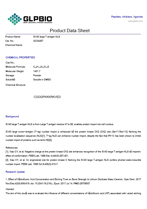
Peptides, Inhibitors, AgonistsProduct Data SheetProduct Name: SV40 large T antigen NLSCat. No.:GC34257Chemical Name:CHEMICAL PROPERTIESCas No.:Molecular Formula: C58H104N20O18SMolecular Weight: 1401.7Storage: PowderSolubility: Soluble in DMSOChemical Structure:BackgroundSV40 large T antigen NLS is from Large T antigen residue 47 to 55, enables protein import into cell nucleus.SV40 large tumor−antigen (T−ag) nuclear import is enhanced by the protein kinase CK2 (CK2) site (Ser111Ser112) flanking the nuclear localization sequence (NLS)[1]. T−ag NLS can enhance nuclear import, despite the fact that PK−C has been shown to inhibit nuclear import of proteins such as lamin B2[2].References:[1]. Xiao CY, et al. Negative charge at the protein kinase CK2 site enhances recognition of the SV40 large T-antigen NLS by importin: effect of conformation. FEBS Lett. 1998 Dec 4;440(3):297-301.[2]. Xiao CY, et al. An engineered site for protein kinase C flanking the SV40 large T-antigen NLS confers phorbol ester-inducible nuclear import. FEBS Lett. 1998 Oct 9;436(3):313-7.Research Update1. Effect of Hydrofluoric Acid Concentration and Etching Time on Bond Strength to Lithium Disilicate Glass Ceramic. Oper Dent. 2017 Nov/Dec;42(6):606-615. doi: 10.2341/16-215-L. Epub 2017 Jul 14. PMID:28708007AbstractThe aim of this study was to evaluate the influence of different concentrations of hydrofluoric acid (HF) associated with varied etchingtimes on the microshear bond strength (μSBS) of a resin cement to a lithium disilicate glass ceramic. Two hundred seventy-five ceramic blocks (IPS e.max Press [EMX], Ivoclar Vivadent), measuring 8 mm × 3 mm thickness, were randomly distributed into fiv e groups according to the HF concentrations (n=50): 1%, 2.5%, 5%, 7.5%, and 10%.2. Does acid etching morphologically and chemically affect lithium disilicate glass ceramic surfaces? J Appl Biomater Funct Mater. 2017 Jan 26;15(1):e93-e100. doi: 10.5301/jabfm.5000303. PMID:27647389AbstractBACKGROUND: This study evaluated the surface morphology, chemical composition and adhesiveness of lithium disilicate glass ceramic after acid etching with hydrofluoric acid or phosphoric acid.METHODS: Lithium disilicate glass ceramic specimens polished by 600-grit silicon carbide paper were subjected to one or a combination of these surface treatments: airborne particle abrasion with 50-μm alumina (AA), etching with 5% hydrofluoric acid (HF) or 36% phosphoric acid (Phos), and application of silane coupling age nt (Si).3. Fatigue failure load of feldspathic ceramic crowns after hydrofluoric acid etching at different concentrations. J Prosthet Dent. 2018 Feb;119(2):278-285. doi: 10.1016/j.prosdent.2017.03.021. Epub 2017 May 26. PMID:28552291AbstractSTATEMENT OF PROBLEM: Hydrofluoric acid etching modifies the cementation surface of ceramic restorations, which is the same surface where failure is initiated. Information regarding the influence of hydrofluoric acid etching on the cyclic loads to failure of ceramic crowns is lacking.PURPOSE: The purpose of this in vitro study was to evaluate the influence of different hydrofluoric acid concentrations on the fatigue failure loads of feldspathic ceramic crowns.。
large T-antigen

SV40 Large T-antigen
• 大T抗原能转化各种不同的基因表型。TAg的活性 阻碍了肿瘤抑制基因pRB和p53的表达。 • 大T抗原还绑定了几个其他的细胞因子,包括转 录激活剂P300和CBP。
SV40 Large T-antigen
• 在大鼠,这种致瘤的SV40大T抗原被用来建立关 于原始神经外胚层瘤和成神经管细胞瘤的脑肿瘤 模型。 • SV40大T抗原以作为蛋白质模型来研究核定为信 号(nuclear localisation signals ,NLSs)。第一 个NLS 就是在SV40中发现的PKKKRKV序列。
SV40
• 由于SV40结构简单,被Байду номын сангаас先应用于真核生物复制 的研究。增强子首先发现于SV40基因组内。 • 增强子序列。其长度为72bp,以串联重复的形式 位于基因组DNA复制起点的附近,它的主要功能 是促进病毒DNA发生有效的早期转录,而且具有 一般增强子所共有的基本特性。
SV40
• 病毒在细胞核内,由细胞的 RNA 聚合酶II的 行为促进早期基因表达。其结果是一条拼接 成两段的mRNA。大小T抗原就是它的结果。
Thanks !
• 此时大T抗原: 95%将会返回细胞核。一旦在细胞核内,大T 抗原就结合3个病毒DNA位点,I,II和III。自动 调节早期的RNA合成。
• SV40病毒基因组早期基因编码两种肿瘤抗原 (tumor antigen,T抗原),其分子量分别为 94000(大T抗原)和17000(小T抗原). • T抗原有以下的作用: ①大T抗原为细胞转化的启动所必需; ②转化细胞表型的维持必需要有大T抗原的连续 表达; ③小T抗原对于细胞的转化不是必需的,但可起 加强作用.
SV40 Large T-antigen
用sv40 t抗原能永生化原理
用sv40 t抗原能永生化原理下载提示:该文档是本店铺精心编制而成的,希望大家下载后,能够帮助大家解决实际问题。
文档下载后可定制修改,请根据实际需要进行调整和使用,谢谢!本店铺为大家提供各种类型的实用资料,如教育随笔、日记赏析、句子摘抄、古诗大全、经典美文、话题作文、工作总结、词语解析、文案摘录、其他资料等等,想了解不同资料格式和写法,敬请关注!Download tips: This document is carefully compiled by this editor. I hope that after you download it, it can help you solve practical problems. The document can be customized and modified after downloading, please adjust and use it according to actual needs, thank you! In addition, this shop provides you with various types of practical materials, such as educational essays, diary appreciation, sentence excerpts, ancient poems, classic articles, topic composition, work summary, word parsing, copy excerpts, other materials and so on, want to know different data formats and writing methods, please pay attention!用SV40 T抗原实现细胞永生化的原理细胞的永生化是现代细胞生物学中的重要课题,其中SV40 T抗原被广泛用于实现细胞的永生化。
_干扰素信号通路及功能相关基因_季旻珺

有 GTP 结合序列 。研究发现 , IGTP、IRG247 和 LRG2 47 等抗病原体感染的作用时效不同 ,且针对不同的 疾病谱 。LRG247 和 IGTP[7] 均能有效限制弓形虫的 早期感染 ,而 IRG247 在弓形虫感染的晚期才发挥部 分抗性 。IGTP 是重要的抗原虫感染因子 ,但不能有 效清除李斯特菌 。LRG247 基因缺陷小鼠完全失去 了对李斯特菌感染的抗力 ,而 IRG247 基因缺陷小鼠 则没有表现出任何的功能缺失[8] 。季 等[9]在研
IFN2γ功能的有效发挥有赖于细胞膜上受体的 完整性 。研究表明 IFN2γ受体 ( IFN2γR) 由α和β两 种亚基组成 。IFN2γR 的α、β亚基分别定位于人第 6 和 21 对染色体上 ,以及小鼠第 10 和 16 对染色体 上 。α亚基即配体结合亚基 ,是二价的高亲和力受 体 ,与同型二聚体的 IFN2γ 结合形成稳定的 1∶2 中 间复合体 ,其胞外区与 IFN2γ的 N 末端结合 ,而胞液 区与 C 末端结合 。随后 ,这一中间复合体作为结合 模板 ,结合 2 分子β亚基 ,生成有活性的 1∶2∶2 信号 复合体 ,从而介导信号向细胞内传递 。 2. IFN2γ的 JAK2STAT 信号转导通路
激酶 异 二 聚 化 相 互 激 活 使 IFN2γRα 亚 基 的 Tyr440磷酸化 , 它可选择性地与 STAT1α( P91) 结合 。 STAT1 的第 569~700 位氨基酸序列类似于 Src 同源 结构 (SH2) ,其中 Arg602突变将会丧失其与 IFN2γ 受 体的结合 。STAT1 与磷酸化的 IFN2γR 结合后 ,导致 STAT1 中 Try701和 Ser727磷酸化 ,形成同源二聚体 (又 称 IFNγ活化因子 , GAF) , 进而激活 STAT1 潜在的 DNA 结合活性 。STAT1 的活化需要 2 类胞内信号事
- 1、下载文档前请自行甄别文档内容的完整性,平台不提供额外的编辑、内容补充、找答案等附加服务。
- 2、"仅部分预览"的文档,不可在线预览部分如存在完整性等问题,可反馈申请退款(可完整预览的文档不适用该条件!)。
- 3、如文档侵犯您的权益,请联系客服反馈,我们会尽快为您处理(人工客服工作时间:9:00-18:30)。
1696
LI E T A L .
largely unknown (Urban and Schreiber, 1992). Their charac- was confirmed by sequence analysis. Reti-ovirases were proterization requires tumor-specific cytotoxic T cells whose cul- duced as described (Cayeux et al, 1995). The amphotropic tivation and specificity depends on the availabUity of the tamor packaging cell lines PA317 and GP-envA12 were infected witii line. A notable exception is malignant melanoma that is easier ecotropic viras of psi2 or G P ^ E-86 that wereti-ansfectedwith to culture and for which a number oftamor-associatedor spe- plasmid D N A of Lox P-HyTk-large T and Cre-puro by use of cific antigens have been cloned (Boon et al, 1994); (c) In gen- a mammalian transfected kU (Stratagene). PA317 cells that proeral, for many tumors, the availability of early passage lines duced virastitersof 1 X 10= (LoxP-HyTk-large T) and 5 X IO'' would allow to phenotypically characterize them in a more care- cfu/ml (Cre-puro) were used. GP+envA12/LT, which produced ^ E-86 ful way. The large T antigen of SV40 has been shown to pow- viras titer of 7 X 10*, were obtained by cloning G P erfuUy immortalize primary cells of both mouse and human ori- viras-infected GP-l-envA12 and identifying by immunofluoresgin (Fanning and Knippers, 1992; Manfredi and Prives, 1994). cent staining of large T. In some experiments, vUus was conHowever, the continuous presence of large T may change the centrated to 5 X 10^ cfu/ml by centrifugation. For mock-infecantigenicity of the cells, alter gene expression, or cause muta- tion tgLS(4-)HyTk or Babe-Puro viras was used. tions not specific for the tumor. Transient expression may be achieved by use of temperature-sensitive large T mutants (Jat Cell culture and virus infection and Sharp, 1989). However, this poses theriskof 'leakiness' Tumor specimens of breast or colon carcinomas were obor a not so total absence of the protein. The bacteriophage re- tained from surgery with patients' informed consent. Tumor tiscombinase Cre catalyses recombination in a sequence (LoxP) sues were washed with R P M I 1640 medium supplemented with specific way and, if two LoxP sites are oriented as direct re- antibiotic cocktail. Stroma and necrotic areas were removed, peats, the intervening sequence can be deleted and is lost by the tumor samples minced to about 1 m m ^ pieces, and digested the ceUs (Gu et al, 1993; Bergemann et al, 1995). Here, w e for 1-3 h (colon carcinoma) or 3-16 h for breast carcinoma at show that by efficient gene transfer through retrovimses and 37°C in 10-20 ml of medium plus 1 mg/ml collagenase type use of the Cre/LoxP system large T can be expressed in a time- IV (Sigma), 0.01% hyaluronidase type V (Sigma), and 0.001% dependent fashion. Furthermore, retroviral large T expression DNAse I (Boehringer, Mannheim). After organoids had formed, seems to delay the crisis primarytamorsusually undergo in cul- they were placed in 25-cm^ flasks coated for 2-4 h with a mixture. This system facilitates the ability to rapidly obtain larger ture of 30 /xg/ml of Collagen I and 10 /ig/ml BSA. The cells amounts of primary cells and thus minimize theriskof cell culwere cultured in low-calcium and low-semm D M E M / F 1 2 ture artifacts. medium supplemented with 0.1 m M calcium, 5 ng/ml EGF, 1 /xg/ml hydrocortisone, 1 /tg/ml insulin, 1.7 /ig/ml transferrin, 5 ng/ml sodium selenite, 35 /ig/ml pituitary extract, 0.01 m M deoxycholic acid, 2 % fetal brovine seram (FBS), 100 units/ml penicillin, and 100 /ig/ml streptomycin for colon carcinoma, and with 12.5 /ig/ml EGF, 1 /tg/ml hydrocortisone, 35 /ig/ml pitaitary extract, 10 n M triiodothyronine, 2 n M Estradiol, 1 ng/ml cholera toxin, 1 /ig/ml insulin, 10 /ig/ml transferrin, 15 n M sodium selenite, 2 m M L-glutamine, 5 % F B S and antibiotics as just described for breast cancer specimens. Cancer cells as well as primary human fibroblasts, mouse primary kidney cells, and NIH3T3 cells were infected by exposure to the viras containing supematants in the presence of polybrene (8 /ig/ml) for 24 h as described (Cayeux et al, 1995). Selection was begun between 24 h (NIH3T3) and 72 h for primary cells after infection. Selection occurred in medium containing hygromycin B (500 /ig/ml), puromycin (2 //.g/ml), and gancyclovir (GCV) (1 /ig/ml) for 10-14 days.
'Max-Delbriick-Center for Molecular Medicine, Robert-Rossle-Strasse 10, 13122 Berlin, FRG. 2Robert-Rossle-Hospital, Humboldt University, Lindenberger Weg 80, Berlin 13125. 1695
