代谢综合征合并慢性肾病中醛固酮的激活作用
醛固酮排酸保碱机制
醛固酮排酸保碱机制
醛固酮是一种重要的激素,在人体中发挥着调节钠和水平衡的功能。
它通过影响肾脏
的排酸和保碱作用来维持酸碱平衡。
下面是醛固酮排酸保碱的机制:
1. 醛固酮的合成和释放:醛固酮主要由肾上腺皮质合成,受到肾素-血管紧张素-醛
固酮系统的调节。
当血容量减少、血压降低或者血中的钠浓度增加时,肾素的分泌会增加,从而刺激醛固酮的合成和释放。
2. 醛固酮的作用机制:醛固酮主要通过两种方式对肾脏产生作用。
- 排酸作用:醛固酮会刺激肾小管上皮细胞中氢离子的分泌。
这些氢离子会与尿中的
氨基酸结合形成氨,进而被排出体外。
通过排出过多的氢离子,醛固酮可以起到排酸的作用,维持体内酸碱平衡。
总结:
醛固酮通过促进酸的排泄和碱的保留,起到调节体内酸碱平衡的作用。
这一过程主要
通过醛固酮的合成和释放受到肾素-血管紧张素-醛固酮系统的调节。
醛固酮排酸保碱机制
还受到多种因素的调控,如醛固酮受体的活性调节、醛固酮合成酶的表达和活性等。
对于
正常的醛固酮排酸保碱机制的研究,有助于深入理解人体内酸碱平衡的调节机制,并为相
关疾病的诊断和治疗提供新的线索。
醛固酮名词解释

醛固酮名词解释
醛固酮是一种内源性的类固醇激素,它属于酮固酮类激素家族。
它主要由肾上腺皮质产生,对水、电解质平衡和血压调节起着重要作用。
醛固酮的主要功能是通过肾脏调节体内钠、钾、氯等电解质的平衡。
它通过抑制肾小管对钠的重吸收,增加钠的排出量,同时促进钾的重吸收,减少钾的排出量。
这种电解质的调节作用对于维持正常的血压和体液平衡至关重要。
除了对电解质平衡的调节作用外,醛固酮还参与了许多生理过程。
它在心血管系统中起着重要作用,可以增加血管的紧张度,提高血压。
此外,醛固酮还对心脏和血管的纤维化、炎症反应等过程产生影响,参与了心血管疾病的发展和进展。
在肾脏中,醛固酮也参与了尿液浓缩的调节过程。
它通过影响肾小管的重吸收和尿液的分泌,调节尿液的浓缩和稀释,维持体内水分的平衡。
醛固酮还与其他激素相互作用,如抗利尿激素和抗利尿素激素等,共同调节体内水平衡和血压。
它还参与了炎症反应、免疫调节等生理过程。
总体而言,醛固酮是一种重要的内源性类固醇激素,对于体内电解质平衡、血压调节和一系列生理过程起着至关重要的作用。
了解醛固酮的功能和调节机制对于理解和治疗与其相关的疾病具有重要意义。
醛固酮的生理功能

醛固酮的生理功能醛固酮是一种由肾上腺皮质分泌的类固醇激素,它在人体内起着重要的生理作用。
本文将从以下几个方面详细介绍醛固酮的生理功能。
一、醛固酮的合成和分泌1. 醛固酮的合成醛固酮是由肾上腺皮质合成的一种类固醇激素,其合成过程包括胆固醇转化为孕酮、孕酮转化为17-羟孕酮和17-羟孕酮转化为醛固酮三个步骤。
这些步骤均需要多种激素和调节因子的参与,如促肾上腺皮质激素(ACTH)、去甲肾上腺素(NE)和血管紧张素 II(Ang II)等。
2. 醛固酮的分泌在正常情况下,肾上腺皮质分泌的大部分类固醇激素都是由ACTH调节的。
ACTH通过刺激肾上腺皮质细胞中胆固醇转化为孕酮,从而促进醛固酮的合成和分泌。
此外,血中的钠离子浓度也能影响醛固酮的分泌。
当血中钠离子浓度降低时,肾素-血管紧张素-醛固酮系统被激活,从而促进醛固酮的分泌。
二、醛固酮在水盐代谢中的作用1. 醛固酮与钠离子的关系在肾小管上皮细胞中,醛固酮能够促进钠离子的重吸收,并增加钠离子通道的表达量。
这样可以提高体内钠离子的浓度,并保持体内水平稳定。
2. 醛固酮与钾离子的关系除了对钠离子有调节作用外,醛固酮还能影响体内钾离子的代谢。
它能够抑制肾小管上皮细胞对钾离子的重吸收,并促进肾小球滤过率增加,从而导致体内钾离子排出增加。
三、醛固酮在心血管系统中的作用1. 醛固酮与血压的关系醛固酮能够通过促进肾小管上皮细胞对钠离子的重吸收,从而提高体内钠离子浓度,同时也会导致水分潴留。
这样会增加血容量和心脏负荷,从而提高血压水平。
2. 醛固酮与心肌保护作用除了调节血压外,醛固酮还能够发挥心肌保护作用。
研究表明,醛固酮能够促进心肌细胞内钙离子的代谢,并减少心肌细胞死亡。
四、醛固酮在代谢调节中的作用1. 醛固酮与葡萄糖代谢的关系研究表明,长期使用高剂量的类固醇激素(如强效糖皮质激素)会导致机体对葡萄糖的敏感性下降,并增加胰岛素抵抗风险。
而一些研究也发现,低剂量使用醛固酮能够改善胰岛素敏感性,从而降低糖尿病的风险。
代谢综合征与慢性肾脏病
内脏脂肪( 腹腔 内脂肪 ) 蓄积
腰围 男 性 ≥8 e 5m 女 性 >9 c  ̄ 0m
在日 本肾脏疾病的流行病学数据主要为井
阴等 的 研究 组 针对 冲绳 研究 的数 据 , 阴等就 井
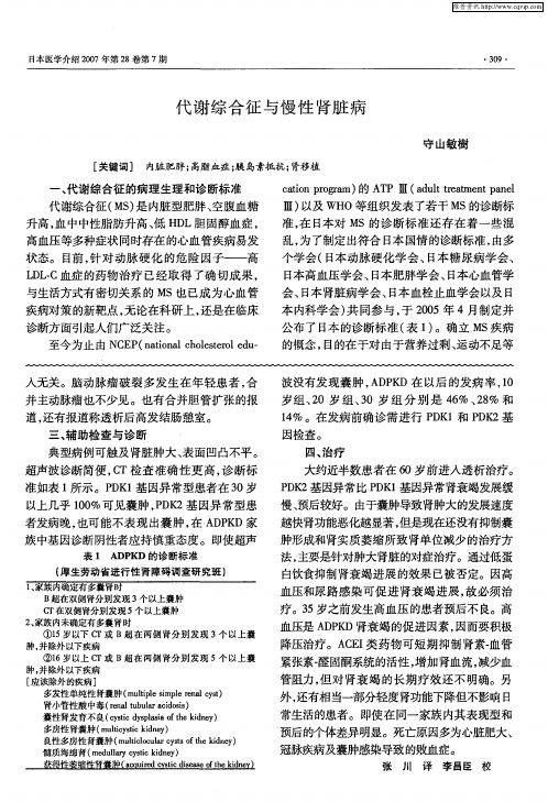
代谢综合征( S 是 内脏型肥胖 、 M) 空腹血糖
升高 , 中中性 脂肪 升高 、 HD 血 低 L胆 固醇血 症 ,
Ⅲ) 以及 WH O等组织发表了若干 M S的诊断标
准 , 日本 对 MS的诊 断标 准 还存 在 着 一些 混 在
高血压等多种症状 同时存在 的心血管疾病易发 状态。目前 , 针对动脉硬化 的危险因子——高 L LC血症 的药物治疗 已经取得 了确切成果 , D. 与生活方式有密切关系 的 M 也 已成为心血管 S 疾病对策 的新靶点 , 无论在科研上 , 还是在临床 诊 断方 面引起人 们广 泛关 注 。 至今 为止 由 N E ( a o a c o s rl d 一 C P nt nl h l t o eu i ee 人无关 。脑动脉瘤破裂多发生在年轻患者 , 合 并主动脉瘤也不少见。也有合并胆管扩张的报 道, 还有报道称透析后高发结肠憩室 。 三 、 助检查 与诊 断 辅 典型病例可触及肾脏肿大、 表面凹凸不平。 超声波诊断简便 , T检查准确性更高 , C 诊断标 准如表 1 所示 。P K 基因异常型患者在 3 岁 D1 0 以上几乎 10 0 %可见囊肿 ,D 2基 因异常型患 PK 者发病晚, 也可能不表 现出囊肿 , A P D家 在 DK
( 生劳动省进行性 肾障碍调查研究班 ) 厚
B 超在双侧肾分别发现 3 个以上囊肿 C T在双侧肾分别发现 5个 以上囊肿 2 家族内未确定有多囊肾时 、 ①1 岁 以下 C 5 T或 B超 在 两侧 肾分别 发现 3个 以上囊 肿, 并除外以下疾病 ②1 岁 以上 C 6 T或 B超 在 两侧 肾分别 发现 5个 以上囊 肿, 并除外以下疾病 [ 应该除外的疾病 ] 多发性单纯性 肾囊肿 ( u i e ipe ea cs m lp m l r l yt tl s n ) 肾小管性酸 中毒 ( n b l i s ) r a t u r c os el u a a d i 囊性肾发育不 良(y idsl iot i e) csc y a a fh k ny t p s e d 多房性 肾囊肿 ( u i s i e) m l y i k ny t t d e e 良性多房性肾囊肿( leoua yt o ek ny mut l lr ss fh i e ) i e c t d 髓质海绵 肾( eu a sc i e) m d l r c t d y ly y ik n
代谢综合征患者胰岛素抵抗与血浆醛固酮相关性研究
昆明医科大学学报2012,(12):102~106CN53-1221/R Journal of Kunming Medical University代谢综合征患者胰岛素抵抗与血浆醛固酮相关性研究丘红梅,沈国清,刘开平,李薇,周晓芳,姚纪瑜(昆明医科大学第六附属医院内分泌科,云南玉溪653100)[摘要]目的探讨代谢综合征(metabolicsyndrome,MS)患者胰岛素抵抗及血浆醛固酮(aldosterone,ALD)水平的关系.方法79例MS患者,根据MS不同组合成份分为MS合并2型糖尿病及高血压组(MS1组,31例)、MS合并2型糖尿病组(MS2组,28例)、MS合并高血压组(MS3组,20例).所有受试对象均行3h口服葡萄糖耐量试验及卧立位醛固酮试验,以稳态模型公式计算胰岛素抵抗指数(HOMAIR).结果MS各亚组血ALD、胰岛素曲线下面积(INSAUC)、HOMA-IR及HOMA-IR≥2.18者所占比例高于对照组(P<0.05).MS1组血ALD、胰岛素曲线下面积(INSAUC)、HOMA-IR及HOMA-IR≥2.18者所占比例高于MS2组及MS3组,差异有显著性(P<0.05).MS2组血ALD、胰岛素曲线下面积(INSAUC)、HOMA-IR及HOMA-IR≥2.18者所占比例略高于MS3组,但差异无统计学意义(P>0.05).Pearson相关分析显示,血浆醛固酮与BMI、WC、TG、FPG、FINS、HOMA-IR、DBP呈正相关,与HDL-C呈负相关.多元回归分析表明,HOMA-IR是MS患者血浆醛固酮水平的独立影响因素(β=0.12,P<0.05).结论MS患者血浆醛固酮水平增高,增高的醛固酮与HOMA-IR和MS大部分组分相关.[关键词]代谢综合征;胰岛素抵抗;醛固酮[中图分类号]R589[文献标识码]A[文章编号]1003-4706(2012)12-0102-05TheRelationshipofPlasmaAldosteroneandInsulinResistanceinPatientswithMetabolicSyndromeQIUHong-mei,SHENGuo-qing,LIUKai-ping,LIWei,ZHOUXiao-fang,YAOJi-yu(Dept.of Endocrinology,The Sixth Affiliated Hospital of Kunming Medical University,Yuxi Yunnan653100,China)[Abstract]ObjectiveToanalysetherelationshipofplasmaaldosteroneandinsulinresistanceanditscomponentsinpatientswiththemetabolicsyndrome(MS).Methods79metabolicsyndromepatientsweredividedintothreegroupsaccordingtothedifferentcomponentsofMS:31patientswithtype2diabetesmellitusandhypertension(MS1group),28patientswithtype2diabetesmellitus(MS2group)and20patientswithhypertension(MS3group).20normalsubjectsservedascontrols.Allpatientsweregiven3horalglucosetolerancetestandorizontalandverticalpositionofaldosteronetest,HOMA-insulinresistanceindex(HOMA-IR)wascalculatedbyHomeostasisModel.ResultsPlasmaaldosterone,theinsulinareaundercurve(INSAUC),HOMA-IRandprevalenceofinsulinresistanceweresignificantlyhigherintheclusteringofMScomponentsthancontrolgroup(P<0.05).TheseindexesweresignificantlyhigherinMS1thanMS2andMS3groupintheclusteringofMScomponents(P<0.05).TheseindexeswerehigherinMS2groupthaninMS3group,buttherewasnosignificantdifference(P>0.05).Pearsoncorrelationanalysisshowedthatplasmaaldosteronewaspositivelycorrelatedwithbodymassindex(BMI),waistcircumference(WC),triglyceride(TG),fastingplasmaglucose(FPG),fastinginsulin(FINS),HOMA-IRanddiastolicbloodpressure(DBP)(r=0.562,P<0.01;r=0.502,P<0.01;r=0.394,P<0.01;r=0.225,P<0.01;r=0.586,P<0.01;r=0.385,P<0.01;r=0.299,P<0.01),negativelycorrelatedwithhighdensitylipoproteincholesterol(HDL-C)(r=-0.298,P<[作者简介]丘红梅(1966~),女,广东梅县人,医学学士,主任医师,主要从事内分泌代谢性疾病临床工作.0.01).ThemultipleregressionanalysisshowedthatHOMA-IRwasanindependentfactorofplasmaaldosteronelevelinpatientswiththemetabolicsyndrome(β=0.12,P<0.05).ConclusionTheincreasingofplasmaaldosteroneinpatientswiththemetabolicsyndromemayberelatedwithIHOMA-IRandsomecomponentsofMS.[Ke y words]Metabolicsyndrome;Insulinresistance;Aldosterone代谢综合征(metabolicsyndrome,MS)特征是向心性肥胖、糖代谢紊乱、脂代谢紊乱和高血压等多重代谢危险因素的聚集,其共同病理基础是胰岛素抵抗,研究发现MS可明显增加糖尿病和心脑血管疾病的发病危险和死亡[1].肾素-血管紧张素-醛固酮系统在血压调节中起重要作用,但其对MS影响的研究相对较少.国外Srividya等[2]研究发现,高醛固酮血症与MS的多个组分有相关性。
醛固酮知识点总结

醛固酮知识点总结醛固酮的合成与分泌醛固酮是由肾上腺的肾上腺皮质细胞合成,并且是由胆固醇前体合成而来。
在合成过程中,胆固醇首先通过一系列酶的催化作用进入肾上腺皮质的内质网,然后经过酶的催化作用转变为醛固酮。
醛固酮主要是由醛固酮合成酶(CYP11B2)介导的醛固酮合成过程中合成的。
醛固酮合成酶是在肾上腺皮质的邻近区域细胞中合成的,主要功能是将17-羟基孕酮(17-OH-Progesterone)转化为醛固酮。
此外,醛固酮的合成还受到一些生物活性物质如血管紧张素Ⅱ(Ang Ⅱ)和钾离子的调节。
在肾上腺皮质分泌的激素中,醛固酮本身并不参与调节自身的分泌,而是通过调节肌醇交换的代谢和Na+和k+离子的转运来实现对醛固酮的调节。
醛固酮的生理作用醛固酮主要通过调节肾小管上皮细胞对钠和水的重吸收来维持血容量和血压的稳定。
具体来说,醛固酮的主要生理作用包括:1. 促进肾小管上皮细胞对钠的重吸收。
在肾小管上皮细胞中,醛固酮通过结合到钠通道和钠钾泵上来促进对钠的重吸收,并且将钠重新吸收到肾小管上皮细胞内,然后再由Na+-K+-ATPase泵将钠离子排出细胞外,最终进入血液循环。
2. 促进肾小管上皮细胞对水的重吸收。
醛固酮促进了肾小管上皮细胞对钠的重吸收后,还会使得水分子通过渗透压的作用重新吸收到肾小管上皮细胞内,然后再由细胞内的水通道蛋白促进水通过渗透压的作用进入血液循环。
3. 调节钾离子的排泄。
醛固酮不仅促进对钠的重吸收,还会促进对钾的排泄,使得体内的钾离子浓度维持在一个相对恒定的水平。
总的来说,醛固酮主要通过调节肾脏对钠和水的重吸收来维持血容量和血压的稳定,对于维持体内内环境的稳定起着至关重要的作用。
醛固酮的调节醛固酮的分泌受到多种生物活性物质的调节,包括血浆容量变化、醛固酮合成酶活性和表情和血清钠离子和钠离子之间的比例。
主要调节醛固酮分泌的激素包括:1. 血浆容量变化。
血浆容量的变化是调节醛固酮分泌的主要因素之一。
醛固酮的作用
醛固酮的作用
醛固酮是一种甾体类固醇激素,常被称为“盐类保留激素”。
它主要由肾上腺皮质产生,并对肾脏和其他器官起到重要作用。
下面将详细介绍醛固酮的作用。
首先,醛固酮参与维持体液和电解质平衡。
它通过作用于肾小管上皮细胞,增加对钠离子的重吸收,从而促进水分的重吸收。
这样,尿液中的钠离子含量减少,其水分便被保留在体内,维持了体液的正常容量和压力。
这也是醛固酮被称为“盐类保留
激素”的原因。
其次,醛固酮可促进肾小管对钾离子的排泄。
它作用于肾小管上皮细胞中的细胞膜钠钾泵,使其增加对钾的分泌。
这样可以调节体内钾离子的平衡,保持其在正常范围内。
此外,醛固酮还参与调节酸碱平衡。
它通过作用于肾小管上皮细胞中的质子泵,增加对氢离子的分泌,从而促进酸性物质的排泄,调节体内酸碱平衡。
醛固酮还对心血管系统具有一定影响。
它可增加血管平滑肌对肾上腺素和血管加压素的敏感性,使血管收缩,提高血压。
此外,醛固酮还参与调节心血管系统中的血容量和心肌肥大。
此外,醛固酮对中枢神经系统也有影响。
它可以增强体液调节中枢对低钠刺激的敏感性,从而调节摄盐和排盐的平衡,保持体内的电解质平衡。
最后,醛固酮还参与调节免疫和炎症反应。
它可以影响白细胞的增殖和分化,调节免疫细胞的功能。
总之,醛固酮在体内起着重要的调节作用,主要包括维持体液和电解质平衡、促进肾小管对钾的排泄、调节酸碱平衡、调节心血管系统和中枢神经系统的功能,以及调节免疫和炎症反应等。
对于维持人体正常的生理功能和稳态平衡具有重要作用。
原发性醛固酮增多症与糖代谢紊乱的研究进展

·综述·基金项目:江苏省卫生健康委面上项目(LKM2022042);无锡市科学技术协会软科学研究课题(KX 22 B43)作者简介:李晓玉,硕士研究生,Email:xiaoyu_li2020@163.com通信作者:洪侃,主任医师,Email:2898456291@qq.com原发性醛固酮增多症与糖代谢紊乱的研究进展李晓玉,陆守荣,芦嘉琪,李昕逸,杨颖,洪侃南京医科大学附属无锡人民医院内分泌科,无锡214023[摘要] 原发性醛固酮增多症作为内分泌性高血压中最常见的类型伴有相当比例的糖代谢异常,是内分泌性糖尿病的一种病因,可能机制包括自主醛固酮过量分泌并通过盐皮质激素受体及非盐皮质激素受体等导致外周组织和肝脏等胰岛素敏感性下降、胰岛功能受损。
近期研究表明,原发性醛固酮增多症合并皮质醇共分泌发生率较高,皮质醇作为一种糖皮质激素,可以通过糖皮质激素受体及盐皮质激素受体发挥作用,促进糖异生、抑制糖原合成、降低胰岛素敏感性及胰岛素分泌等途径影响糖代谢。
盐皮质激素受体拮抗剂对糖代谢的影响目前仍存在争议,针对合并皮质醇共分泌且不宜手术的原发性醛固酮增多症患者,盐皮质激素受体拮抗剂联合糖皮质激素受体拮抗剂或许对糖代谢发挥作用。
[关键词] 醛固酮增多症;葡萄糖代谢障碍;氢化可的松;盐皮质激素受体拮抗剂;综述DOI:10.3969/J.issn.1672 6790.2023.03.029ResearchprogressofprimaryaldosteronismandglucosemetabolismdisorderLiXiaoyu,LuShourong,LuJiaqi,LiXinyi,YangYing,HongKanDepartmentofEndocrinology,WuxiPeople′sHospitalofNanjingMedicalUniversity,Wuxi214023,ChinaCorrespondingauthor:HongKan,Email:2898456291@qq.com[Abstract] Asthemostcommontypeofendocrinehypertensionwithaconsiderableproportionofabnormalglucosemetabolism,primaryaldosteronismisacauseofendocrinediabetes.Thepossiblemechanismsincludeexces sivesecretionofautonomicaldosterone,whichleadstodecreasedinsulinsensitivityinperipheraltissuesandliver,andimpairedpancreaticisletfunctionthroughmineralocorticoidreceptorandnonmineralocorticoidreceptor.Recentstud ieshaveshownthatprimaryaldosteronismwithco secretionofcortisolhasahighincidenceandhasreceivedmoreandmoreattention.Asaglucocorticoid,cortisolcanplayarolethroughglucocorticoidreceptorandmineralocorticoidre ceptor,promotinggluconeogenesis,inhibitingglycogensynthesis,reducinginsulinsensitivityandinsulinsecretiontoaffectglucosemetabolism.Theeffectofmineralocorticoidreceptorantagonistsonglucosemetabolismisstillcontrover sial.Forpatientswithprimaryaldosteronismcombinedwithcortisolco secretionandunsuitableforsurgery,mineralo corticoidreceptorantagonistscombinedwithglucocorticoidreceptorantagonistsmayplayadominantroleinglucosemetabolism.[Keywords] Hyperaldosteronism;Glucosemetabolismdisorders;Hydrocortisone;Mineralocorticoidreceptorantagonists;Review 原发性醛固酮增多症(PA)是指肾上腺皮质自主分泌过多醛固酮,肾素-血管紧张素系统活性受抑制,导致水钠潴留,容量负荷增多,尿钾排泄增多,临床上主要表现为高血压和低血钾[1]。
醛固酮水平与心血管疾病研究进展

①甘肃省人民医院 甘肃 兰州 730000通信作者:王楠醛固酮水平与心血管疾病研究进展马慧元① 王楠① 杨立霞① 【摘要】 原发性醛固酮增多症是继发性高血压的常见病因,在原发性高血压、心力衰竭、心律失常、冠心病等心血管疾病患者中醛固酮水平同样升高。
本文通过综述醛固酮在高血压、心力衰竭、心律失常、冠心病、阻塞性睡眠呼吸暂停、糖尿病、卒中、肾功能不全等疾病中的作用机制,探讨醛固酮水平对心血管系统的影响及临床价值,推断醛固酮受体拮抗剂通过降低醛固酮水平有望对上述疾病的治疗提供帮助。
【关键词】 醛固酮 肾素-血管紧张素 心血管疾病 Research Progress of Aldosterone Level and Cardiovascular Disease/MA Huiyuan, WANG Nan, YANG Lixia. //Medical Innovation of China, 2023, 20(22): 176-180 [Abstract] Primary aldosteronism is a common cause of secondary hypertension. Aldosterone levels are also increased in patients with primary hypertension, heart failure, arrhythmia, coronary heart disease and other cardiovascular diseases. This article reviews the mechanism of aldosterone in hypertension, heart failure, arrhythmia, coronary heart disease, obstructive sleep apnea, diabetes, stroke, renal insufficiency and other diseases, discusses the effect of aldosterone level on cardiovascular system and its clinical value, and infers that aldosterone receptor antagonists may help the treatment of these diseases by reducing aldosterone level. [Key words] Aldosterone Renin-angiotensin Cardiovascular disease First-author's address: Gansu Provincial People's Hospital, Lanzhou 730000, China doi:10.3969/j.issn.1674-4985.2023.22.042 心血管系统疾病是全球引发致死和致残率最高的疾病,在我国心血管疾病的死亡率居首位,其中高血压是首要、可干预的心血管疾病[1]。
天然AMPK激活剂防治代谢综合征的研究进展

天然AMPK激活剂防治代谢综合征的研究进展摘要:能量代谢紊乱是代谢综合征产生的根本原因。
AMPK (AMP-activated protein kinase)作为细胞乃至整个机体的能量调节嚣在能量较低的状态下被激活,促进分解代谢,抑制合成代谢,从而恢复能量平衡状态。
因此,AMPK有型成为防治代谢综合征的新型靶点。
单纯的药物治疗代谢综合征难以达到理想效果,许多天然营养分子也具有激活AMPK的功效,提示营养干预可能成为缓解代谢综合征的另一种新型有效手段。
关键词:腺苷酸活化蛋白激酶(AMPK);代谢综合征:营养干预代谢综合征的慨念在1988年首次由Reaven提出。
它是临床上具有肥胖、高血压、血脂紊乱、受损的糖耐量或糖尿病以及胰岛素抵抗等一系列代谢失常症状的并称,是多种代谢成分异常聚集的的病理状念。
引起上述代谢失常症状的因素有很多,包括胰岛素抵抗、炎症反应、脂肪组织分泌的各种细胞因子的参与、生活方式及遗传因素等,但其根本原因还是能量代谢的失衡。
随着社会经济的发展和人们生活方式的改变,以肥胖、糖尿病、脂肪肝等代谢异常为特征的代谢综合征的发病几率在全球范围内日益增加,已成为危害人类健康的重大公共卫生问题。
因此,代谢综合征治疗手段的探索和相关药物的开发已成为国内外科学工作者关注的热点。
腺苷酸活化蛋白激酶(AMP-activated protein kinase,AMPK)是细胞层面乃至整个机体层面的能量调节器,能够维持能量代谢供求平衡。
AMPK在机体能量较低的状态下被激活(即胞内AMP:ATP比值上调时),通过直接磷酸化下游代谢相关靶蛋白调控其活性,或者调控代谢相关基因的表达,从而促进分解代谢,增加ATP的产生过程(如脂肪酸氧化、糖酵解),同时抑制合成代谢,减少ATP的消耗过程(如脂肪酸合成、糖异生),使机体恢复能量稳态:基于AMPK 对能量代谢的调控作用,AMPK的激活剂可能成为防治肥胖、糖尿病等代谢失常症状的有效手段。
醛固酮的代谢过程
醛固酮的代谢过程醛固酮啊,这可是我们身体里一个超级神奇的东西呢!想知道它的代谢过程吗?那就跟着我一起好好探索一番吧。
咱们先得知道醛固酮是从哪儿来的。
醛固酮主要是由肾上腺皮质球状带细胞合成和分泌的。
这就好比是身体里有一个小小的工厂,肾上腺皮质球状带细胞就是那些勤劳的工人,每天都在制造醛固酮这个特殊的产品。
当身体里的肾素 - 血管紧张素系统被激活的时候,就像是吹响了生产醛固酮的号角。
肾素这个小家伙会作用于血管紧张素原,把它变成血管紧张素Ⅰ。
嘿,这血管紧张素Ⅰ可不会闲着呢,它又会在血管紧张素转换酶的作用下变成血管紧张素Ⅱ。
血管紧张素Ⅱ就像是一个严厉的监工,它会跑到肾上腺皮质球状带细胞那儿,大喊着:“嘿,工人们,加快生产醛固酮!”于是,醛固酮的合成和分泌就增多啦。
那醛固酮生产出来之后,它要到哪儿去发挥作用呢?醛固酮会随着血液循环到达它的目的地——肾脏。
这就像是一个信使带着重要的信件,长途跋涉地来到了收件人那里。
在肾脏里,醛固酮会和肾小管上皮细胞里面的受体相结合。
这一结合啊,就像是一把钥匙开了一把锁,然后神奇的事情就发生了。
醛固酮会促使肾小管上皮细胞对钠离子进行重吸收。
这钠离子啊,就像是一群调皮的小娃娃,本来在肾小管里晃悠着,准备被排出体外呢。
但是醛固酮一来,就像是一个严厉的老师,把这些小娃娃都赶回了身体里。
与此同时呢,醛固酮还会促进钾离子的排泄。
这钾离子就像是那些不太听话的学生,醛固酮就把它们赶到了外面。
有人可能就会问了:“为啥要这么做呢?”其实啊,这样做是为了维持身体里的电解质平衡。
如果钠离子都跑出去了,钾离子都留在体内,那身体可就乱套了,就像一个班级里没有了纪律一样。
醛固酮在肾脏里的这些作用还不是它的全部本事呢。
它还会影响到肾脏对水的重吸收。
你想啊,钠离子被重吸收回来了,就像在身体里建了一个个小小的“吸水站”,水就会跟着钠离子一起被重吸收回来。
这就像是一条小河里的水,跟着一群小鸭子(钠离子)一起回到了身体这个大池塘里。
醛固酮
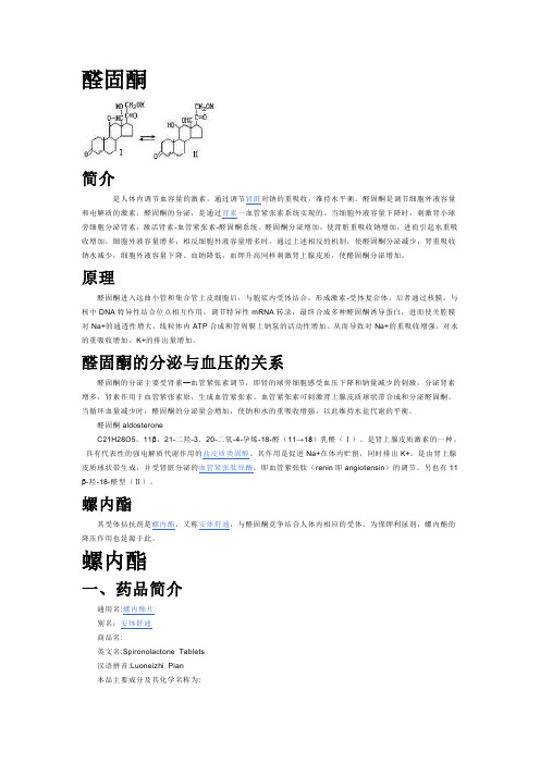
醛固酮简介是人体内调节血容量的激素,通过调节肾脏对钠的重吸收,维持水平衡。
醛固酮是调节细胞外液容量和电解质的激素,醛固酮的分泌,是通过肾素一血管紧张素系统实现的。
当细胞外液容量下降时,刺激肾小球旁细胞分泌肾素,激活肾素-血管紧张素-醛固酮系统、醛固酮分泌增加,使肾脏重吸收钠增加,进而引起水重吸收增加,细胞外液容量增多;相反细胞外液容量增多时,通过上述相反的机制,使醛固酮分泌减少,肾重吸收钠水减少,细胞外液容量下降。
血钠降低,血钾升高同样刺激肾上腺皮质,使醛固酮分泌增加。
原理醛固酮进入远曲小管和集合管上皮细胞后,与胞浆内受体结合,形成激素-受体复合体,后者通过核膜,与核中DNA特异性结合位点相互作用,调节特异性mRNA转录,最终合成多种醛固酮诱导蛋白,进而使关腔膜对Na+的通透性增大,线粒体内ATP合成和管周膜上钠泵的活动性增加。
从而导致对Na+的重吸收增强,对水的重吸收增加,K+的排出量增加。
醛固酮的分泌与血压的关系醛固酮的分泌主要受肾素—血管紧张素调节,即肾的球旁细胞感受血压下降和钠量减少的刺激,分泌肾素增多,肾素作用于血管紧张素原,生成血管紧张素。
血管紧张素可刺激肾上腺皮质球状带合成和分泌醛固酮。
当循环血量减少时,醛固酮的分泌量会增加,使钠和水的重吸收增强,以此维持水盐代谢的平衡。
醛固酮aldosteroneC21H28O5。
11β,21-二羟-3.20-二氧-4-孕烯-18-醛(11→18)乳醛(Ⅰ)。
是肾上腺皮质激素的一种。
具有代表性的强电解质代谢作用的盐皮质类固醇。
其作用是促进Na+在体内贮留,同时排出K+。
是由肾上腺皮质球状带生成,并受肾脏分泌的血管紧张肽原酶,即血管紧张肽(renin即angiotensin)的调节。
另也有11β-羟-18-醛型(Ⅱ)。
螺内酯其受体拮抗剂是螺内酯,又称安体舒通,与醛固酮竞争结合人体内相应的受体,为保钾利尿剂,螺内酯的降压作用也是源于此。
螺内酯一、药品简介通用名:螺内酯片别名:安体舒通商品名:英文名:Spironolactone Tablets汉语拼音:Luoneizhi Pian本品主要成分及其化学名称为:主要成分:螺内酯化学名称:17β-羟基-3-氧-7α-(乙酰硫基)-17α-孕甾-4-烯-21-羧酸γ-内酯。
醛固酮吸钠排钾的原理
醛固酮吸钠排钾的原理
醛固酮是一种重要的激素,它在我们体内发挥着重要的调节作用。
它主要通过抑制肾脏的钠吸收和钾排泄来维持体内的电解质平衡。
醛固酮的作用原理可以简单概括为“吸钠排钾”。
具体来说,醛固酮通过结合到肾小管上皮
细胞的醛固酮受体,促使这些细胞合成和激活一系列的蛋白质,最终导致钠的吸收和钾的排泄。
醛固酮作用于远曲小管,促使细胞内的钠-钾泵活性增加。
这种泵可以将细胞内的钠排出,同时将细胞内的钾吸收进来。
这样一来,体内的钠浓度就会增加,而钾浓度则会降低。
醛固酮还可以增加远曲小管上皮细胞中的钠通道的数量和活性。
这些钠通道可以让钠离子更容易地从尿液中吸收进来,进而增加体内的钠浓度。
醛固酮还可以抑制远曲小管中的钾通道的活性。
这些钾通道可以让钾离子从细胞内排出,但在醛固酮的作用下,这些通道的活性减弱,从而减少了钾的排泄。
通过这一系列的作用,醛固酮可以调节体内钠和钾的平衡。
当体内钠浓度过低时,醛固酮会增加钠的吸收,从而提高钠浓度;而当体内钾浓度过高时,醛固酮会增加钾的排泄,从而降低钾浓度。
醛固酮通过吸钠排钾的原理,维持了体内电解质的平衡。
这一机制的正常调节对于我们的健康至关重要。
醛固酮的生理功能
醛固酮的生理功能概述醛固酮是一种主要由肾上腺产生的激素,它在机体中起着重要的生理调节作用。
醛固酮主要通过调节血压和电解质等平衡来维持体内的内环境稳定。
本文将重点介绍醛固酮的生理功能,并深入探讨其在血压调节、水盐平衡、抗炎反应等方面的作用。
血压调节醛固酮对血压调节起着重要的作用。
它通过下丘脑-垂体-肾上腺(HPA)轴的调节,促使肾上腺皮质释放醛固酮。
醛固酮在肾小管中起到抑制钠的排泄和钾的吸收的作用,从而增加体内钠和水分的重吸收,增加血容量,提高血压。
此外,醛固酮还通过抑制一氧化氮合酶的活性,减少一氧化氮的生成,从而收缩血管,增加外周阻力,进一步增加血压。
水盐平衡醛固酮在水盐平衡调节中也发挥着重要作用。
它通过对肾小管的作用,增加钠和水分的重吸收,减少钠的排泄,从而增加体内的钠储备,维持机体的水盐平衡。
此外,醛固酮还通过抑制醛固酮释放激素的释放,减少尿液的生成,进一步降低水分量,维持体内的血浆渗透压稳定。
抗炎反应除了在血压调节和水盐平衡方面的作用,醛固酮还具有抗炎反应的功能。
它通过抑制炎症介质的释放,调节免疫系统的功能,抑制炎症反应的发生。
研究发现,醛固酮可以通过调节炎症细胞的活性,减少炎症细胞的浸润,减轻炎症反应,起到抗炎作用。
这一发现为醛固酮在治疗炎症性疾病方面的应用提供了新的思路。
醛固酮受体与疾病醛固酮的生理功能主要通过与醛固酮受体(MR)的结合而实现。
醛固酮受体广泛分布于肾脏、心血管系统、免疫系统等多个组织中。
与醛固酮受体的结合可以调节多种基因的表达和信号通路的激活,从而影响相关生理过程。
高醛固酮血症与高血压高醛固酮血症是指体内醛固酮水平异常升高,常见于原发性醛固酮增多症和肾上腺皮质功能亢进症等疾病。
高醛固酮血症与高血压密切相关。
当醛固酮水平升高时,它会抑制肾小管中钠的排泄和钾的吸收,导致体内钠和水分的潴留,增加血容量,从而引起血压升高。
此外,在高醛固酮血症的状态下,醛固酮还通过直接或间接影响血管收缩剂和舒张剂的生成和释放,增加外周阻力,进一步升高血压。
醛固酮拮抗剂对代谢综合征合并慢性心力衰竭患者胰岛素抵抗指数和C-反应蛋白的影响

醛固酮拮抗剂对代谢综合征合并慢性心力衰竭患者胰岛素抵抗指数和C-反应蛋白的影响胡怡;宋今丹;刘立旻;张斌;高阳;熊盈;魏敏;毛宇强;陈松;陈思娇【期刊名称】《中国全科医学》【年(卷),期】2008(11)15【摘要】目的观察醛固酮拮抗剂对代谢综合征合并慢性心力衰竭(心衰)患者胰岛素抵抗指数(HOMA-IR)、C-反应蛋白(CRP)的影响.方法将86例代谢综合征合并慢性心衰患者随机分成两组,常规治疗组(40例)使用洋地黄、利尿剂、血管紧张素转化酶抑制剂或血管紧张素受体拮抗剂、β-受体阻滞剂、硝酸酯类、降糖等治疗;醛固酮拮抗剂组(46例)在上述常规治疗基础上,加用醛固酮拮抗剂螺内酯20 mg,2次/d,逐渐减至1次/d.治疗6个月.评估治疗前后两组患者临床疗效、HOMA-IR、CRP、血脂〔高密度脂蛋白胆固醇(HDL-C)、低密度脂蛋白胆固醇(LDL-C)、三酰甘油(TG)〕水平和血压、心率.结果治疗后两组患者临床疗效间差别有统计学意义(P<0.01);HOMA-IR及CRP、HDL-C、LDL-C、TG水平、血压、心率间差别有统计学意义(P<0.05).结论代谢综合征合并心衰患者在常规治疗的基础上加用醛固酮拮抗剂可能对提高胰岛素敏感性、降低CRP有一定作用.【总页数】3页(P1332-1334)【作者】胡怡;宋今丹;刘立旻;张斌;高阳;熊盈;魏敏;毛宇强;陈松;陈思娇【作者单位】110001,辽宁省沈阳市,中国医科大学附属第一医院老年病教研室;中国医科大学医学分子生物学研究所卫生部细胞生物学重点实验室;中国医科大学七年制医学专业;中国医科大学七年制医学专业;110001,辽宁省沈阳市,中国医科大学附属第一医院老年病教研室;110001,辽宁省沈阳市,中国医科大学附属第一医院老年病教研室;110001,辽宁省沈阳市,中国医科大学附属第一医院老年病教研室;中国医科大学七年制医学专业;中国医科大学七年制医学专业;110001,辽宁省沈阳市,中国医科大学附属第一医院老年病教研室【正文语种】中文【中图分类】R541.6【相关文献】1.醛固酮受体拮抗剂对慢性心衰患者胰岛素抵抗及细胞因子的影响 [J], 王煜;张涛;邓洁;李轶炜;王瑞萍;杨莉;齐云萍2.大血管病变合并代谢综合征患者临床特征与C-反应蛋白的关系 [J], 曾宪辉;叶燕华3.初诊2型糖尿病合并代谢综合征患者血清高分子量脂联素水平与胰岛素抵抗、C 反应蛋白的相关性 [J], 俞淑琴;王苏;潘瑞蓉;胡浩;孙文君;杨奇超;刘媛馨;蒋丹;王东4.初诊2型糖尿病合并代谢综合征患者颈围与胰岛素抵抗、超敏C反应蛋白的相关性 [J], 钱唯韵;俞淑琴;朱天一;汤冰倩;孙文君;胡浩;叶菁菁;王东;王济芳5.缬沙坦对高血压糖尿病合并胰岛素抵抗和勃起功能障碍患者C-反应蛋白及性功能的作用 [J], 陈思娇;祁虹;魏敏;高阳;胡怡;熊盈;李廷富因版权原因,仅展示原文概要,查看原文内容请购买。
醛固酮 渗透压 生理学
醛固酮渗透压生理学
醛固酮是一种重要的激素,对于人体的渗透压调节起着重要的作用。
渗透压是指溶液中溶质的浓度决定的压力差异,影响着细胞内外液体的平衡。
醛固酮通过调节肾脏的排尿量和浓度,维持人体的渗透压稳定。
人体内部的渗透压是一个复杂的平衡过程,涉及到多个器官和激素的相互作用。
醛固酮是由肾上腺分泌的一种激素,它的主要作用是促进肾小管对钠离子的重吸收,同时促进钾离子的排泄。
这种调节机制可以帮助维持体液中钠离子和钾离子的平衡,从而维持人体的渗透压稳定。
当人体处于高渗透压环境下时,醛固酮的分泌会增加。
这是因为高渗透压环境会导致体内水分的流失,为了保持体液的稳定,肾上腺会释放更多的醛固酮,进而促使肾小管对钠离子的重吸收,减少尿液中的钠排泄量。
这样一来,体内的钠离子浓度就会增加,从而提高体液的渗透压,以补充水分的流失。
醛固酮的分泌还受到其他因素的调节,例如血容量和血压的变化。
当血容量减少或者血压下降时,肾上腺会释放更多的醛固酮,以增加肾小管对钠离子的重吸收,从而增加体液的渗透压,维持血压的稳定。
醛固酮还与抗利尿激素ADH(抗利尿激素)密切相关。
ADH主要作
用于肾小管,促进水分的重吸收,从而减少尿液的产生。
醛固酮与ADH的相互作用,可以调节尿液的渗透压,进一步维持体液的平衡。
醛固酮对于人体的渗透压调节起着重要的作用。
通过调节肾脏的排尿量和浓度,它可以维持体液中钠离子和钾离子的平衡,保持体液的渗透压稳定。
这种调节机制对于人体的正常功能和健康至关重要,因此对醛固酮的研究也成为了生理学的一个重要领域。
醛固酮排钾机制
醛固酮排钾机制醛固酮是一种主要由肾上腺分泌的激素,其在体内发挥着重要的生理功能。
其中最为重要的一个作用就是通过排钾调节体内的电解质平衡。
本文将主要围绕醛固酮排钾机制展开讨论,从分子生物学层面、肾脏生理学角度以及临床应用等方面进行探讨。
1. 醛固酮的生物合成及分泌醛固酮的生物学合成主要发生在肾上腺的醛固酮合成区。
这个过程主要依赖于胆固醇合成途径所提供的原料,通过一系列酶的协调作用最终合成出醛固酮。
醛固酮的合成与分泌主要受到Renin-Angiotensin-Aldosterone系统 (RAAS)的调控。
当血压下降、血容量减少、钠离子浓度下降等情况出现时,肾脏会释放出Renin 活性,使其切割Angiotensinogen为Angiotensin I;然后Angiotensin I被转化为Angiotensin II,这是一种强烈的血管收缩剂,同时也是一种激活RAAS所必需的先导物质。
Angiotensin II通过作用于肾上腺中的醛固酮合成区,刺激醛固酮的分泌,这样就将皮质醇转化为醛固酮。
相应的,由于醛固酮能够很快地去钾,急性的钠离子的重视就会对应地造成钾离子的流失。
因此,醛固酮排钾机制与RAAS系统尤其密切相关。
2. 醛固酮的功能特点醛固酮在体内的作用是多方面的,但最引人注目的还是它通过腺上皮细胞及肾上皮细胞上的醛固酮受体(mineralocorticoid receptor MR)参与到排钾调节中。
醛固酮能够通过增加利钠肽和减少Angiotensin II等方式提高Na+的重视,进而导致K+离子分泌。
在健康人体内,夜间及轻微的运动都能出现轻度的醛固酮水平上升,这是一种正常的生理现象。
醛固酮在肾脏以及肝脏中也有多种其他的生理功能。
3. 醛固酮排钾机制在肾脏中的作用钾离子是肾脏排分泌的重要离子之一。
肾脏搜集尿液后,将其分解为草酸、磷酸盐及有机酸等,将其中的K+离子从尿液中析出,然后通过肾小管向集合管发展,这是一种主要的钾离子排泄途径。
醛固酮的功能

醛固酮的功能醛固酮是一种肾上腺激素,主要作用是调节体内水盐平衡。
下面将详细介绍醛固酮的功能。
1. 保持体内水盐平衡醛固酮的主要作用是维持体内的水盐平衡,保持正常生理状态。
醛固酮通过调节肾脏排出的水和电解质的比例,确保体内水和钠的含量维持在一定范围之内。
当体内水分和电解质过多时,醛固酮能够促进肾脏排出更多的水和钠,从而达到维持体内水盐平衡的效果。
2. 维持血压稳定醛固酮还能够促进钠离子的重吸收,增加血容量,从而使血液循环更加畅通,维持血压的稳定。
当血容量增加时,血液中的钠离子浓度也会增加,从而使血压得到一定程度的调节。
因此,醛固酮是维持体内血压稳定的一个非常重要的调节因素。
3. 维持酸碱平衡醛固酮能够帮助维持体内的酸碱平衡。
它能够促进钠离子和氢离子的交换,使体内的氢离子在肾脏排出之前与钠离子结合,从而减少氢离子的洛伦兹效应,维持体内pH值的稳定。
4. 提高心肌收缩力研究表明,醛固酮能够提高心肌的收缩力,从而增加心脏的泵血能力。
醛固酮还能够抑制水钠负荷引起的心肌细胞增生,防止心脏病变。
5. 促进肾上腺素的合成醛固酮能够促进肾上腺素的合成和分泌,增加代谢率,提高身体的抵抗力和应激能力。
6. 抵抗压力醛固酮能够抵抗体内的压力,增强人体的应激反应。
它能够影响大脑皮层的神经元活动,并通过影响下丘脑向肾上腺皮质释放ACTH激素,从而调节肾上腺皮质激素的分泌,进一步抵抗压力。
总之,醛固酮在维持体内水盐平衡、调节血压稳定、维持酸碱平衡、提高心肌收缩力、增强应激能力等方面都扮演着重要的角色。
如果身体出现水肿、低血压、酸碱失衡等问题,可能与醛固酮功能失调有关。
因此,维持醛固酮的正常功能是维持人体健康的重要因素。
病理生理学简答
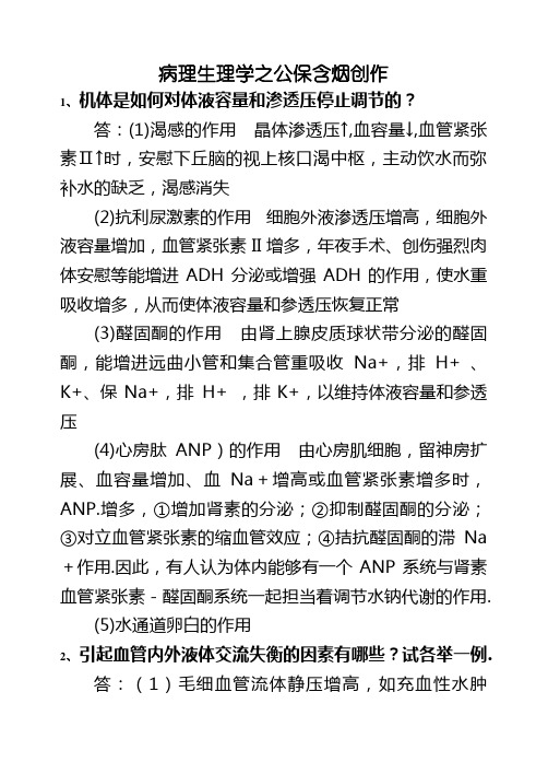
病理生理学之公保含烟创作1、机体是如何对体液容量和渗透压停止调节的?答:(1)渴感的作用晶体渗透压↑,血容量↓,血管紧张素Ⅱ↑时,安慰下丘脑的视上核口渴中枢,主动饮水而弥补水的缺乏,渴感消失(2)抗利尿激素的作用细胞外液渗透压增高,细胞外液容量增加,血管紧张素II增多,年夜手术、创伤强烈肉体安慰等能增进ADH分泌或增强ADH的作用,使水重吸收增多,从而使体液容量和参透压恢复正常(3)醛固酮的作用由肾上腺皮质球状带分泌的醛固酮,能增进远曲小管和集合管重吸收Na+,排H+ 、K+、保Na+,排H+ ,排K+,以维持体液容量和参透压(4)心房肽ANP)的作用由心房肌细胞,留神房扩展、血容量增加、血Na+增高或血管紧张素增多时,ANP.增多,①增加肾素的分泌;②抑制醛固酮的分泌;③对立血管紧张素的缩血管效应;④拮抗醛固酮的滞Na +作用.因此,有人认为体内能够有一个ANP系统与肾素血管紧张素-醛固酮系统一起担当着调节水钠代谢的作用.(5)水通道卵白的作用2、引起血管内外液体交流失衡的因素有哪些?试各举一例.答:(1)毛细血管流体静压增高,如充血性水肿时,全身毛细血管流体静压增高.(2)血浆胶体渗透压下降,如肝硬化时,卵白质分解增加.(3)微血管通透性升高,如炎性水肿时,炎症介质是微血管的通透性升高,(4)淋巴回流受阻,如丝虫病,可引起阻塞淋巴性水肿.3、哪型脱水易造成休克?为什么?答:低渗性脱水容易.原因(1)细胞外液丧失,血容量增加;(2)细胞外液向细胞内转移;(3)渴感不明显,病人不主动饮水;(4)早期ADH分泌增加所以容易发作休克.相反高渗性脱水时,1、口渴明显:自动找水喝;2、ADH分泌增多;3、细胞内液向胞外转移;4、醛固酮:重者可增多,增进钠水重吸收——不容易发作休克4、试述水肿的发病机制.答:水肿发病的根本机制是血管内液体交流失衡.前者包括毛细血管流体静压增高、血浆胶体渗透压降低、微血管通透性增加以及淋巴回流受阻,这些因素均招致血管内胶体滤出年夜于回收而使组织液生成过多;另一方面是体内外液体交流失衡,包括GFR下降和近曲小管、髓袢以及远曲小管于集合管重吸收增多,招致体内钠水潴留.5、试述酸中毒对机体的主要影响.答:⑴心血管系统:①血管对儿茶酚胺的反响性降低;②心肌收缩力削弱;③心肌细胞能量代谢障碍;④高钾血症引起心律失常.故严重代谢酸中毒的病人易并发休克、DIC、心力衰竭.⑵中枢神经系统:主要表现是抑制,患者可有疲乏、觉得愚钝、嗜睡甚至神清不清、苏醒.⑶呼吸系统:呈现年夜而深的呼吸.糖尿病酸中毒时,呼出气中带有烂苹果味(丙酮味).⑷水和电解质代谢:血钾升高、血氯降低和血钙升高.⑸骨骼发育:影响骨骼的生长发育,重者发作骨质蔬松和佝偻病,成人则可招致骨软化病.6、影响氧合血红卵白解离曲线的因素有哪些?答:答:影响氧合血红卵白解离曲线的因素有红细胞内2,3—DPG含量、血【H+】、CO2浓度和血温.这四因素上升时,均可使Hb与O2亲和力降低,以至在相同的氧分压下血氧饱和度降低,氧解离曲线右移;相反,当这四因素数值下降时,氧解离曲线左移.7、比拟发热三期的临床表现和热代谢特点答:、I体温上升期:体温调定点,体温上升,产热↑散热↓,产热>散热.临床表现:畏寒和皮肤苍白(皮肤血管收缩,血流增加),寒颤(骨骼肌周期收缩),竖毛肌收缩(鸡皮)(交感兴奋)II、高热继续期:当体温上升到与新的调定点水平相适应的高度后,就坚定于该高度左近,产热↑散热↑,产热=散热.临床表现:炎热(血温升高→使皮肤温度升高→安慰温觉感受器),皮肤发红、枯燥III、退热期:调定点恢复正常,体温调定点,体温下降,产热↓散热↑,散热>产热.临床表现:血温仍偏高,出汗(皮肤血管扩张,汗腺分泌增加)8、应激性溃疡发作机制答:a.胃黏膜缺血(根本条件):胃腔内H+向黏膜内的逆向弥散(需要条件);缺血→胃黏膜屏障破坏;缺血→弥散至黏膜内的H+不能被中和或带走b.糖皮质激素的作用,抑制胃黏膜的分解和分泌;胃肠黏膜细胞的卵白质分解增加,细胞更新减慢,再生能力降低c.其它:胆汁返流、酸中毒、PGE2分解↓9、缺血-再灌注时自由基生成增多的机制(1)黄嘌呤氧化酶形成增多:缺血:ATP↓,Ca离子泵功用↓,Ca离子进入细胞激活Ca离子依赖卵白酶,促使少量的黄嘌呤脱氢酶转酿成黄嘌呤转化酶;再灌注:ATP 分解代谢增强,组织中次黄嘌呤少量堆积.少量氧分子进入缺血组织.黄嘌呤氧化酶催化次黄嘌呤转酿成黄嘌呤进而催化黄嘌呤转酿成尿酸,释放出少量电子,以氧分子作为电子受体发作少量超氧阴离子和H2O2.(2)中性粒细胞聚集和激活:缺血:补体系统激活或经细胞膜分解发作多种趋化因子,吸引、激活中性粒细胞.再灌注:氧供给迅速、少量↑,发作少量自由基——呼吸迸发或氧迸发.(3)、线粒体功用损伤:缺血:ATP↓,Ca离子进入线粒体↑,线粒体功用受损,氧分子经单电子复原形成超氧阴离子↑;Ca离子进入线粒体↑,SOD、过氧化氢酶增加和活性↓.再灌注:氧供给迅速、少量↑,超氧阴离子↑(4)儿茶酚氨的自身氧化10、失血性休克发作什么类型的缺氧?血氧指标有何变卦?答:失血性休克时既有少量失血又有休克,少量失血造成血液型缺氧,血氧变卦有血氧含量和血氧容量降低,动态脉血氧含量差增加;休克造成微循环性缺氧,动态脉血氧含量差增年夜.总的变卦血氧含量和血氧容量均降低.11、DIC发作普遍出血的机制.答:(1)凝血物质被消耗而增加:DIC时,少量微血栓形成进程中,消耗了少量血小板和凝血因子,血液中纤维卵白原、凝血酶原、FⅤ、FⅧ、FⅩ等凝血因子及血小板明显增加,使凝血进程障碍,招致出血.(2)纤溶系统激活:DIC时纤溶系统亦被激活,激活的原因主要为:①在FⅫ激活的同时,激肽系统也被激活,发作激肽释放酶,激肽释放酶可使纤溶酶原酿成纤溶酶,从而激活了纤溶系统;②有些富含纤溶酶原激活物的器官,如子宫、前列腺、肺等,由于少量微血栓形成,招致缺血、缺氧、变性坏死时,可释缩小量纤溶酶原激活物,激活纤溶系统;③应激时,肾上腺素等作用血管内皮细胞分解、释放纤溶酶原激活物增多;④缺氧等原因使血管内皮细胞损伤时,内皮细胞释放纤溶酶原激活物也增多,从而激活纤溶系统,纤溶系统的激活可发作少量纤溶酶.纤溶酶是活性较强的卵白酶,除可使纤维卵白降解外,尚可水解凝血因子如:FⅤ、FⅧ、凝血酶、FⅫ等,从而招致出血.(3)FDP的形成:纤溶酶发作后,可水解纤维卵白原(Fbg)及纤维卵白(Fbn).发作纤维卵白(原)降解产物(FgDP或FDP)这些片段中,X,Y,D片段均可阻碍纤维卵白单体聚合.Y,•E片段有抗凝血酶作用.此外,少数碎片可与血小板膜结合,降低血小板的粘附、聚集、释放等功用.这些均使患者出血倾向进一步减轻.12、试述DIC的发病机制.答:DIC的发病机制包括:(1)组织严重破坏,使少量组织因子入血,启动外源性凝血系统,招致DIC的发作开展.(2)血管内皮细胞普遍损伤,激活XII因子,启动内源性凝血系统;同时激活激肽释放酶,激活纤溶和补体系统,招致DIC.(3)血细胞少量破坏,血小板被激活,招致DIC.(4)胰卵白酶、蛇毒等促凝物质进入血液,也可招致DIC.13、试述休克与DIC的关系.答:休克和DIC互为因果,相互影响,恶性循环.休克晚期由于微循环障碍,血液浓缩,血细胞聚集,血液粘滞度增高,血液处于高凝状态;血流变慢,减轻酸中毒,易于形成血栓;败血症休克时病原微生物与蛇毒均可损伤内皮,激活内源性凝血途径;严重的创伤性休克,组织因子入血,可启动外源性凝血系统;异型输血引起溶血,容易诱发DIC.急性DIC时由于微血管内少量微血栓形成,使回心血量明显增加;普遍出血使血容量增加;心肌损伤,使心输出量增加;补体及激肽系统的激活和PDF少量生成,造成微血管平滑肌舒张,通透性增高,使外周阻力降低.这些因素均可增进休克的发作和开展.14、什么是休克?休克发作的始动环节是什么?答:有效循环血量增加,引起重要生命器官血液灌流缺乏,从而招致细胞功用紊乱,称为休克.引起有效循环血量增加的始动环节是:血容量增加,血管库容量增加,心泵功用障碍.15、试述休克缺血性缺氧期病人的典型临床表现及其微循环变卦特点.答:临床变现:神色苍白、四肢冰凉、出冷汗、脉搏细速、尿少、焦躁不安、血压下降也可正常.微循环特点:微循环痉挛、少灌少流,灌少于流、A—V短路开放.16、试述休克淤血性缺氧期病人的典型临床表现及其微循环变卦特点.答:临床表现:血压停止性下降,心搏无力,心音低钝,神智冷淡.可进入苏醒;少尿,脉细速,静脉塌陷,皮肤可呈现发钳、花斑.微循环特点:微循环瘀滞,灌而少流,灌年夜于流.17、为什么休克缺血性缺氧期又称为代偿期?答:此期的代偿表现有:(1)微静脉及储血库收缩“自身输血”;(2)组织液反流入血管“自身输液”;(3)血液重新散布担保心脑供给.其他有心收缩力增强,外周阻力增加,动脉血压维持正常.18、为什么休克淤血性缺氧期又称为失代偿期?答:此期的失代偿表现有:微循环血管床少量开放瘀滞,回心血量锐减,心输出量血压停止性下降,引起交感—肾上腺髓质更增强烈兴奋,组织灌流量更低,形成恶性循环,毛细血管后阻力年夜于前阻力,血浆外渗,血液浓缩;MAP<7Kpa(1Kpa=7.5mmHg),心脑血管失去自我调节,心脑功用障碍.19、影响DIC发作开展的因素答:单核吞噬细胞系统功用受损肝功用严重障碍血液高凝状态微循环障碍20、什么叫心力衰竭?其根本病因是什么?答:在各种致病因素的作用下心脏的收缩和舒张功用发作障碍,使心输出量相对或相对下降,以至不能满足机体代谢需要的病理进程称为心力衰竭.根本病因是原发性心肌舒缩功用障碍和心脏负荷过重.21、心功用不全时机体的代偿答:神经-体液的代偿心内代偿(一)功用代偿(心率放慢,收缩力增强)(二)构造代偿(心室重塑)心外代偿(一)血容量增加(二)血流重散布(三)红细胞增多(四)组织细胞应用氧的能力增强22、心力衰竭的发病机制答:一、心肌收缩功用降低(一)收缩相关卵白质改动(二)心肌能量代谢紊乱(三)心肌兴奋收缩藕联障碍二、心肌舒张功用障碍(一) Ca2+复位延迟(ATP增加)(二)肌球-肌动卵白复合体解离障碍(三)心室舒张势能增加(四)心室顺应性降低三、心脏各部舒缩活动不协调(一)、心律失常破坏心脏各部舒缩活动的协调性(二)、同一心室心肌梗死区域性散布,招致心室活动不协调23、试述肺通气障碍的类型和原因.答:通气障碍有限制性通气缺乏和阻塞性通气缺乏两种类型.前者的原因有呼吸肌活动障碍、胸廓和肺的顺应性降低,胸腔积液和气胸;后者的原因有气道狭窄或阻塞,多因气道痉挛、炎症、异物或肿瘤所致.24、阻塞性通气缺乏中阻塞部位分歧呈现的呼吸困难形式有何分歧?为什么?答:阻塞性通气缺乏可分为中央性气道阻塞和外周性气道阻塞.中央性气道阻塞为气管分叉处以上的气道阻塞,阻塞要位于胸外部位,吸气时气体流经病灶狭窄处引起压力降低,使气道内压明显低于年夜气压,招致气道狭窄减轻,发作吸气性呼吸困难;阻塞部位若位于胸内部位,呼气时由于胸内压升高而压迫气道,使气道狭窄减轻,表现为呼气性呼吸困难.外周气道阻塞使位于内径<2mm无软骨的细支气管阻塞,细支气管与周围肺泡构造严密相连,呼气时小气管变窄,小气道阻力年夜年夜增加,表现为呼气性呼吸困难.25、发作肺内气体弥散障碍的原因有哪些?血气变卦如何?答:原因是:(1)肺泡膜面积增加,见于肺不张,肺实变;(2)肺泡膜厚度增加,见于肺间质性水肿,肺泡透明膜形成和肺纤维化等.弥散障碍时,因CO2的弥散能系数比O2年夜20倍,如无伴发通气障碍,只有缺氧,即PaO2降低,而无CO2潴留,既无PaCO2升高.26、在肺泡通气与血流比例失调中,造成Va/Q变卦的原因有哪些?对机体有什么影响?答:造成VA/Q降低的原因为肺水肿、肺纤维化所致的限制性通气障碍和支气管哮喘、慢性支气管炎、阻塞性肺气肿所致的阻塞性通气障碍,因肺泡通气量(NA)增加而使VA/Q下降,使流经通气缺乏的肺泡的血液未很好的氧合而入动脉血内,造胜利用性分流.造成VA/升高的原因未肺动脉栓塞、肺血管收缩和微血栓形成等,因肺血流量增加而使VA/Q升高,因患部肺泡血流增加而通气正常,肺泡通气不能被直接应用,造成死腔样通气.功用性分流可有正常时的3%--50%.死腔样通气可有正常的30%上升至60%--70%.均严重影响换气功用,招致机体缺氧,发作缺氧性病感性变卦.27、呼吸衰竭的机制与呼衰的血气指标.答:(1)肺通气功用障碍:包括限制性通气缺乏和阻塞性通气缺乏此为Ⅱ型呼衰,P a CO2增加,P a O2降低(2)肺换气功用障碍:弥散障碍,此为Ⅰ型呼衰,其一般不呈现血气异常或仅P a O2降低通气血流比例失调,包括局部肺泡通气缺乏和局部肺泡血流缺乏,P a O2降低,P a CO2可正常或降低,极严重时可升高解剖分流增加,此为Ⅰ型呼衰,P a O2降低28、肝性脑病患者为什么会有高氨血症?答:血氨升高的原因(1)氨的生成过多,因胆汁分泌增加,肠菌丛生,分解产物产氨(2)高卵白饮食和上消化道年夜出血时卵白质在肠菌作用下少量产氨.(3)氨清除缺乏,肝脏、严重损伤时,肝内酶系统遭破坏及底物缺失,使将氨分解尿素的鸟氨酸循环难以正常停止而有血氨增加.29、氨对脑组织有哪些毒性作用?答:(1)搅扰脑组织的能量代谢.氨于脑内a—同戊二酸结合,同时又消耗了少量NADH,阻碍呼吸链中递氢进程,以至ATP发作缺乏,不能维持中枢神经系统兴奋活动.(2)使脑神经递质发作改动,脑内氨增多可使脑内兴奋性神经递质增加和抑制性神经递质增多,致使神经递质间作用失去平衡,招致脑功用紊乱.(3)氨对神经细胞质膜的作用30、肾功用不全的根本发病环节答:一、肾小球滤过功用障碍:1. 肾血流量↓;2. 肾小球有效滤过压↓;3. 肾小球有效滤面积↓;4. 肾小球效滤膜通透性改动二、肾小管功用障碍:近端小管、髓袢、远端小管、集合管功用障碍三、肾内分泌功用障碍31、急性肾功用不全发病机制答:↓(二)肾小管因素1. 肾小管阻塞;2. 原尿反流32、何谓多尿?慢性肾衰为什么会发作多尿?答:24小时尿量超越2000ml,称为多尿.慢性肾衰呈现多尿的机制是:(1)残留肾单元滤过的原尿多,流速快,未能及时重吸收.(2)原尿中的溶质多,发作渗透性利尿.(3)髓质间质高渗区破坏,肾小管浓缩功用障碍. 33、试述慢性肾功用衰竭的四个学说:答:健存肾单元学说;矫枉失衡学说;肾小球过度滤过学说;肾小管细胞和间质细胞损伤假说34、试述肾性贫血的机制答:由各种因素造成EPO发作缺乏或尿毒症血浆中一些毒性物质搅扰红细胞生成与代谢而招致的贫血.机制有:1. EPO↓;2. 骨髓造血功用抑制;3. 出血;4. RBC 寿命缩短;5. 铁和叶酸缺乏:肾毒性物质致肠道吸收增加或应用障碍35、试述急性肾小管坏死时少尿的发作机制.答:(1)肾缺血.如肾灌流压下降、肾血管收缩和血液流变学的变卦;(2)肾小管阻塞.如异型输血、挤压综合症、磺胺结晶的引起急性肾小管坏死,脱落的上皮细胞碎片、肌红卵白、血红卵白等阻塞肾小管管腔;(3)肾小管原尿返流.因肾小管上皮细胞普遍坏死,基膜断裂,原尿流经断裂的基膜分散到肾间质,引起间质水肿,进一步压迫肾小管和毛细血管;(4)肾小球超滤系数降低.因系膜细胞收缩招致肾小球滤过面积增加和滤过系数降低,致使GFP下降.注:非教师提供,仅供参考。
- 1、下载文档前请自行甄别文档内容的完整性,平台不提供额外的编辑、内容补充、找答案等附加服务。
- 2、"仅部分预览"的文档,不可在线预览部分如存在完整性等问题,可反馈申请退款(可完整预览的文档不适用该条件!)。
- 3、如文档侵犯您的权益,请联系客服反馈,我们会尽快为您处理(人工客服工作时间:9:00-18:30)。
REVIEW ARTICLEActivation of the aldosterone/mineralocorticoid receptor system in chronic kidney disease and metabolic syndromeMiki NagaseReceived:3January 2010/Accepted:13May 2010/Published online:9June 2010ÓJapanese Society of Nephrology 2010Abstract Recent clinical and experimental studies have shown that aldosterone is a potent inducer of proteinuria and that mineralocorticoid receptor (MR)antagonists confer efficient antiproteinuric effects.We identified glo-merular epithelial cells (podocytes)as novel targets of aldosterone;activation of MR injures podocytes possibly via oxidative stress,resulting in disruption of glomerular filtration barrier,proteinuria,and progression of chronic kidney disease.We also demonstrated that SHR/cp,a rat model of metabolic syndrome,was susceptible to podocyte injury and proteinuria.Aldosterone excess caused by adipocyte-derived aldosterone-releasing factors was sug-gested to underlie the nephropathy.High salt intake aug-mented MR activation in the kidney and exacerbated the nephropathy.Furthermore,we identified an alternative pathway of MR activation by small GTPase Rac1.Rho-GDI a knockout mice,a model with Rac1activation in the kidney,showed albuminuria,podocyte injury,and glo-merulosclerosis.Renal injury in the knockout mice was accompanied by enhanced MR signaling in the kidney despite normoaldosteronemia,and was ameliorated by an MR antagonist,eplerenone.Moreover,Rac-specific inhib-itor significantly reduced the nephropathy,concomitantly with repression of MR activation.In vitro transfection studies provided direct evidence of Rac1-mediated MR activation.In conclusion,our findings suggest that MRactivation plays a pivotal role in the pathogenesis of chronic kidney disease in metabolic syndrome,and that MR may be activated both aldosterone dependently (via aldosterone-releasing factors)and independently (via Rac1).MR antagonists are promising antiproteinuric drugs in metabolic syndrome,although long-term effects on renal outcomes,mortality,and safety need to be established.Keywords Proteinuria ÁPodocyte injury ÁSalt ÁAldosterone-releasing factors ÁRac1IntroductionSince its isolation in 1953,aldosterone (formerly called ‘‘electrocortin’’)has been recognized as a hormone that regulates electrolyte,fluid volume,and blood pressure (BP)homeostasis.Aldosterone acts on its receptor,mineralo-corticoid receptor (MR)[1],in the epithelial cells of the aldosterone-sensitive distal nephron in the kidney (late distal convoluted tubules,the connecting tubules,and the principal cells of the collecting ducts),thereby controlling sodium reabsorption and potassium excretion.This classic mode of action has been substantiated by many studies of genetic disorders and knockout mice.Recently,however,aldosterone has emerged as a dele-terious hormone in the cardiovascular and renal system [2–4].Accumulating clinical evidence suggests the role of aldosterone in the pathogenesis of hypertension,meta-bolic syndrome,cardiac outcomes,and renal munity-based prospective studies revealed that higher serum aldosterone levels within the physiologic range confer increased risk of development of hypertension and metabolic syndrome [5,6].Higher circulating con-centrations of aldosterone were associated with increasedThis article was presented as the Oshima Award memorial lecture at the 52nd annual meeting of the Japanese Society of Nephrology,held at Yokohama,Japan,in 2009.M.Nagase (&)Division of Chronic Kidney Disease,Department of Nephrology and Endocrinology,University of Tokyo Graduate Schoolof Medicine,7-3-1Hongo,Bunkyo-ku,Tokyo 113-8655,Japan e-mail:mnagase-tky@umin.ac.jpClin Exp Nephrol (2010)14:303–314DOI 10.1007/s10157-010-0298-8mortality among patients with chronic heart failure[7]and with ST-elevation myocardial infarction[8].As for renal dysfunction,plasma aldosterone concentrations were in most instances elevated in chronic renal disease(creatinine clearance below50%of normal)with normal serum potassium and renin levels[9].Quinkler et al.[10]reported that serum aldosterone concentrations were negatively correlated with creatinine clearance and positively associ-ated with renal scarring in chronic kidney disease(CKD) patients.We recently found that the glomerular epithelial cell (podocyte),finalfiltration barrier in the glomerulus,is a novel target of aldosterone[11],and activation of MR in podocyte causes proteinuria and glomerulosclerosis,pos-sibly via oxidative stress,in metabolic syndrome model [12].We also identified a novel mechanism of MR acti-vation by small GTPase Rac1and its contribution to CKD [13].Herein,we summarize recent advances in aldosterone research and introduce our data on the roles and regulation of the aldosterone/MR system in CKD and metabolic syndrome.Aldosterone and target organ injuryHistorically,it was already known in the1940s(even before isolation of aldosterone and MR)that MR activation causes inflammation andfibrosis in target organs;for example,Hans Selye[14],an advocate of‘‘stress theory(non-specific adaptation response to stressor by glucocorticoids),’’reported that administration of desoxy(11-deoxy)corticosterone acetate(DOCA),thefirst synthetic steroid which stimulates MR,to rats induces inflammatory andfibrotic changes in the heart and kidney.Uninephrectomy and salt loading were performed as conditioning factors.He considered that these phenotypes were due to mineralocorticoid actions of DOCA.Furthermore,he proposed that glucocorticoids have anti-inflammatory actions in adaptation to stress, whereas mineralocorticoids may have pro-inflammatory effects.Half a century later,aldosterone,the physiological mineralocorticoid in our body,was shown to cause myo-cardial and vascularfibrosis[15,16].MR was found to be expressed not only in the epithelia of the kidney and colon but also in various nonepithelial cells.Based on these experimentalfindings,together with aldosterone break-through phenomenon(see below),Pitt et al.[17,18]con-ducted two milestone randomized clinical trials,RALES and EPHESUS,in which addition of low-dose MR antag-onist,spironolactone or eplerenone,dramatically improved outcomes of heart failure patients already taking standard medications including angiotensin-converting enzyme inhibitors(ACEIs),angiotensin II(Ang II)type1receptor blockers(ARBs),diuretics,and b-blockers.The organ-protective efficacy of MR antagonists was further con-firmed by subsequent clinical studies[19,20].The aldosterone/MR system plays a pathogenic role also in the progression of CKD[2,3].MR is present in various nonepithelial cells,where aldosterone causes inflammation andfibrosis through genomic and nongenomic actions. Multiple factors are proposed to be involved:plasminogen activator inhibitor(PAI)-1,transforming growth factor (TGF)-b1,connective tissue growth factor,proinflammatory cytokines such as osteopontin and monocyte chemoattrac-tant protein(MCP)-1,and reactive oxygen species(ROS).MR binds both mineralocorticoids and glucocorticoids with high affinity[1].Because plasma glucocorticoid con-centrations are100–1000times higher than those of aldo-sterone,aldosterone target cells have to equip machinery for preventing occupation of MR by glucocorticoids.In the Na?-transporting epithelia,such as the renal distal nephron, the enzyme11b-hydroxysteroid dehydrogenase type2 (11b HSD2)confers specificity on aldosterone to activate MR,by converting cortisol and corticosterone into inactive 11-keto derivatives,which have low affinity for MR[21].In nonepithelial tissues without11b HSD2,glucocorticoids activate MR or,in some cases,glucocorticoids just occupy and do not activate MR.In the kidney,11b HSD2is abun-dantly expressed in the distal tubules and collecting ducts, but is also found in the glomeruli,proximal tubules,cultured podocytes,and mesangial cells[22–24].The enzyme is also expressed in vascular endothelial and smooth muscle cells, while its expression is minimal or absent in the myocardium, hippocampus,adipocytes,and macrophages. Aldosterone and proteinuriaCumulative evidence suggests that aldosterone is a potent inducer of rge-scale,randomized clinical trials have proved the efficacy of renin–angiotensin–aldo-sterone system(RAAS)blockade by ACEIs or ARBs in reducing proteinuria and retarding progression of kidney disease in diabetic and nondiabetic patients[25–30]. Conventionally,Ang II has been regarded as the primary factor responsible for injurious actions of the RAAS in the kidney.Recent studies,however,indicated aldosterone as an additional pathogenetic factor.According to the report by Greene et al.[31],Ang II blockade by losartan and enalapril markedly attenuated proteinuria in rat remnant kidney,a model with enhanced RAAS activity.However, these drugs not only inhibited the action of Ang II but also blocked aldosterone synthesis,and reversal of plasma aldosterone levels by exogenous aldosterone infusion recapitulated the nephropathy.Conversely,adrenalecto-mized rats with glucocorticoid replacement were protectedfrom renal-ablation-induced proteinuria[32].Similarfind-ings were observed in stroke-prone spontaneously hyper-tensive rats(SHR)[33,34],in which renal MR contents as well as plasma aldosterone levels are elevated[35].Indeed, Ang II excess models,such as Ang II/salt rats and double transgenic rats carrying both human renin and human angi-otensinogen genes,develop severe albuminuria and kidney injury,which were abrogated by aldosterone synthase inhibitor FAD286,adrenalectomy or MR antagonists [36,37].The pivotal role of aldosterone was directly proved by creating mineralocorticoid excess models,such as uni-nephrectomized,high-salt-fed rats loaded with aldosterone or DOCA,which were quite prone to proteinuria and glo-merulosclerosis,despite suppressed plasma renin activity and Ang II concentrations[38,39].Serum-and glucocorti-coid-inducible kinase1(Sgk1),an aldosterone effector kinase,might mediate the proteinuric effect of aldosterone, because mice lacking Sgk1were protected against DOCA/ salt-induced albuminuria[40].As for clinical evidence supporting the association between aldosterone and proteinuria,Conn et al.[41] described that85%of patients with primary aldosteronism (PA),a high-aldosterone state,were positive for protein-uria.More recent studies documented that urinary protein excretion was higher in patients with PA than in those with essential hypertension[42,43].Conversely,MR antago-nists were shown to effectively ameliorate proteinuria in patients with hypertension,diabetes mellitus,and CKD. For example,Williams et al.[20]compared the efficacy of eplerenone and enalapril in patients with stage1or2 hypertension.After12months,the extent of BP reduction was similar between the two groups,whereas eplerenone was superior to enalapril in reducing albuminuria(-62% versus-26%;P=0.01).A recent double-blind,placebo-controlled trial by Mehdi et al.[44]showed that addition of spironolactone,but not losartan or placebo,afforded greater antiproteinuric effect in diabetic hypertensive patients already under maximal ACE inhibition.They carefully controlled factors known to affect protein excretion in diabetes including BP,sodium and protein intake,and glycemia.After48weeks,albuminuria decreased by34.0%in the spironolactone group and by 16.8%in the losartan group compared with the placebo group.Regarding safety,however,incidence of asymp-tomatic hyperkalemia(C6.0mEq/L on at least one occasion)was highest in the spironolactone group(14out of27patients).A recent systematic review and meta-analysis by Navaneethan et al.[45]revealed that MR antagonists could significantly reduce proteinuria in CKD patients receiving ACEI and/or ARB,but increase the risk of hyperkalemia.It should be noted,however,that long-term effects on renal outcomes,mortality,and safety need to be established.Aldosterone breakthroughThe role of aldosterone as a mediator of proteinuria was also suggested by the‘‘aldosterone breakthrough’’phenomenon [46].Staessen et al.[47]first described in1981that treat-ment of hypertensive patients with high dose of ACEI captopril lowered plasma aldosterone from74to21pg/mL after1month,but plasma aldosterone began to rise there-after,and after1year reached a level of165pg/mL.Plasma Ang II levels remained suppressed throughout the long-term therapy.This phenomenon was originally known as ‘‘aldosterone escape’’but is now termed‘‘aldosterone breakthrough,’’because‘‘aldosterone escape’’also means loss of salt-retaining ability of excess aldosterone.Although the renin–angiotensin system(RAS)is the principal regu-lator of aldosterone synthesis and secretion in the adrenal glands,aldosterone synthesis is also regulated by various non-RAS stimuli including plasma potassium levels and adrenocorticotropic hormone.These non-RAS secreta-gogues may stimulate aldosterone release under long-term RAS blockage.Sato et al.[48]reported that aldosterone breakthrough was observed in40%of patients with type2diabetes and early nephropathy during40weeks of trandolapril therapy (plasma aldosterone concentration:112.0±18.7and 53.2±15.1pg/mL in patients with and without aldoste-rone breakthrough,respectively).Trandolapril reduced urinary albumin excretion in patients without aldosterone breakthrough(119±95mg/g Cre),whereas the antialbu-minuric effect of trandolapril was lost in patients with aldosterone breakthrough(368±142mg/g Cre).The contribution of unsuppressed aldosterone was further sup-ported by the observation that addition of low-dose spiro-nolactone on top of trandolapril for24weeks significantly reduced albuminuria without BP change in patients with aldosterone breakthrough.Bianchi et al.[49]reported that spironolactone(25mg/day)is useful in reducing protein-uria and retarding the decrease in estimated glomerular filtration rate(GFR)after1year of therapy in nondiabetic CKD patients already treated with ACEIs and/or ARBs. They found that baseline aldosterone levels under Ang II blockade were positively correlated with protein-uria,and predicted the degree of reduction in proteinuria with spironolactone.These studies indicate that aldoste-rone breakthrough indeed contributes to albuminuria and decline in GFR.Aldosterone evokes podocyte injury:role of oxidative stressThe glomerularfiltration barrier comprises three layers:the fenestrated glomerular endothelium,the glomerularbasement membrane,and the podocytes.Podocytes line the outer aspect of the glomerular basement membrane,and serve as thefinal barrier against urinary protein loss[50, 51].Podocytes are injured in many types of proteinuric renal diseases,including nephrotic syndrome,lupus nephritis,diabetic and hypertensive nephropathy,and obesity-related glomerulopathy[39,52–55].The intimate relationship between aldosterone and pro-teinuria prompted us to investigate the effects of aldoste-rone on podocytes.Wefirst analyzed proteinuria and podocyte injury in uninephrectomized,high-salt-fed rats continuously infused with aldosterone(0.75l g/h via osmotic minipump)[11].This is an established chronic mineralocorticoid excess model in which plasma aldoste-rone levels rise to those seen in human congestive heart failure,whereas plasma renin activity and circulating Ang II are quickly suppressed.After4weeks,aldosterone-infused rats developed hypertension and massive protein-uria.Glomerular expressions of slit diaphragm-associated molecules nephrin and podocin were markedly decreased, whereas expression of a damaged podocyte marker desmin was upregulated in aldosterone-infused rats.Electron microscopic analysis revealed podocyte foot process effacement.Podocytes were already impaired at2weeks of aldosterone infusion,when proteinuria was modestly increased.Proteinuria and podocyte damage were com-pletely reversed by selective MR antagonist eplerenone. Podocyte injury was also reported in another mineralo-corticoid excess model,uninephrectomized DOCA/salt rats [39].Thesefindings suggest that podocyte injury underlies the pathogenesis of proteinuria caused by aldosterone administration.So,how did aldosterone evoke podocyte injury?First, aldosterone-induced hypertension should be an important contributor to the podocyte damage.Our uninephrectom-ized aldosterone/salt rats developed hypertension.Systemic BP load,especially in the presence of impaired autoregu-latory vasoconstriction of the preglomerular arterioles, enhances transmission of systemic pressure to the glome-ruli,and the resultant glomerular hypertension/hyperfiltra-tion predisposes to hypertensive glomerular damage[56]. Indeed,a micropuncture study proved these hemodynamic factors as the basis for glomerular injury in uninephrec-tomized DOCA/salt rats[57].Mechanical stress is postu-lated as a key mediator of podocyte damage[51]. Podocytes,whose foot processes overlie glomerular capil-lary tufts,are sensitive to mechanical stress.Cultured podocytes exposed to mechanical strain were reported to enhance expression of proinflammatory cytokines,TGF-b, and Ang II type1receptor,resulting in podocyte damage [58,59].Alternatively,aldosterone may modulate podocyte function directly by acting on MR within podocytes.MR transcripts and proteins were actually detected in in vitro cultured podocytes as well as in vivo glomerular podo-cytes.Exposure of cultured podocytes to aldosterone resulted in nuclear translocation of MR,activation of reduced nicotinamide adenine dinucleotide phosphate (NADPH)oxidase,and ROS increment.In addition,aldo-sterone upregulated expression of an aldosterone effector kinase,Sgk1,in cultured podocytes as well as in the kidney of aldosterone-infused rats,which was prevented by MR antagonist.Taken together,our results suggest that at least some of the proteinuric effects of aldosterone are attribut-able to direct actions on podocytes.We next examined the role of oxidative stress,because ROS are proposed as important mediators of aldosterone-induced target organ injury.Aldosterone-infused rats exhibited enhanced oxidative stress markers such as increased urinary excretion of thiobarbituric acid reactive substances,elevated renal NADPH oxidase activity deter-mined using lucigenin chemiluminescence assay,and stimulation of membrane translocation of NADPH oxidase cytosolic components p67phox and Rac1.Indeed,an anti-oxidant,tempol,significantly reduced oxidative stress markers and corrected podocyte damage and proteinuria. Oxidative stress markers were also suppressed by eplere-none.These results suggest that aldosterone causes podo-cyte injury and proteinuria,possibly through induction of oxidant stress.Metabolic syndrome,CKD,and aldosterone Metabolic syndrome,a constellation of comorbidities that include visceral obesity,hypertension,glucose intolerance, and dyslipidemia,has become a major public health con-cern.Metabolic syndrome is known as a highly predis-posing condition for cardiovascular disease(CVD).Recent epidemiological studies revealed that metabolic syndrome is an important modifiable risk factor for proteinuria and CKD as well.For instance,a cross-sectional study based on the data of NHANES III showed that metabolic syndrome subjects have higher incidence of microalbuminuria (12.3%versus4.7%)and CKD(6.0%versus1.2%)[60]. According to a prospective longitudinal study,based on the data of community-living nondiabetic adults with normal kidney function,having metabolic syndrome increased the risk of developing CKD by approximately50%over 9years,and the risk increased with more traits of the syndrome[61].However,the mechanisms linking meta-bolic syndrome to CKD have not been clearly elucidated.It cannot be solely attributed to the additive effects of indi-vidual component such as hypertension and diabetes mel-litus[61],and some unifying underlying mechanism has been suggested.It is widely accepted that the RAAS plays a crucial role in the pathogenesis of metabolic syndrome.Recent clinical evidence suggests a role for aldosterone per se.Two cross-sectional clinical studies of patients of African descents demonstrated that plasma aldosterone concentration is independently associated with metabolic syndrome,while plasma renin activity was rather suppressed [62,63].In another study,the C allele of -344C/T variant in the promoter of the aldosterone synthase (CYP11B2)gene,which is associated with hyperaldosteronemia,was shown to increase susceptibility to metabolic syndrome in European men [64].Fallo et al.[65]observed that patients with PA had higher incidence of metabolic syndrome than those with essential hypertension (41.1%versus 29.6%;P \0.05).Podocyte injury and proteinuria in metabolic syndrome rats:contribution of aldosterone/MR activation Considering the interrelationships among metabolic syn-drome,CKD,and aldosterone,we postulated a working hypothesis that aldosterone/MR activation plays a critical role in progression of CKD in metabolic syndrome.We used SHR/NDmcr-cp (SHR/cp,obese SHR)as a rat model of metabolic syndrome (Fig.1)[12].This rat is a derivative of SHR with introduction of leptin receptor gene cpmutation,and manifests a clustering of obesity,hyperten-sion,hyperinsulinemia,and hypertriglyceridemia.Obese SHR/cp developed marked proteinuria in an age-dependent manner,while urinary protein excretion remained low in nonobese SHR despite similar BP level.Proteinuria in SHR/cp was accompanied by podocyte injury,as indicated by attenuation of nephrin,induction of desmin,and foot process effacement under electron microscopic analysis.Notably,serum aldosterone levels were higher in obese SHR/cp than in nonobese SHR,and there was positive correlation between circulating aldosterone concentration and proteinuria.Expression of Sgk1was upregulated in the glomerular fraction as well as in the whole kidney of SHR/cp,supporting the causative role of aldosterone/MR acti-vation.Indeed,selective MR antagonist eplerenone effec-tively reduced proteinuria and podocyte damage in SHR/cp.These data suggest that aldosterone-provoked podocyte injury plays a pivotal role in pathogenesis of proteinuria in SHR/cp.We further explored the role of oxidative stress.Similar to the case of aldosterone-infused rats,oxidative stress markers were upregulated in SHR/cp.Importantly,eplerenone reversed the increased oxidative stress,suggesting partici-pation of endogenous aldosterone.Treatment with antioxi-dant tempol also significantly alleviatedproteinuriaFig.1Hyperaldosteronemia in SHR/cp (metabolic syndrome rats),and antiproteinuric effects of MR antagonist eplerenone in SHR/cp and SHR/cp fed a high salt (8%NaCl)diet.a Serum aldosterone concentrations in SHR at 17weeks old and SHR/cp at 17and 24weeks old.b Proteinuria in SHR,SHR/cp,and SHR/cp treated with Epl (SHR/cp ?Epl).Epl wasadministered from 13weeks old for 4weeks.c Proteinuria and nephrin staining in SHR/cp fed a normal diet (SHR/cp),SHR/cp fed a high salt diet (SHR/cp ?HS),and SHR/cp ?HS treated with Epl (SHR/cp ?HS ?Epl).*P \0.05,**P \0.01versus SHR;##P \0.01versus SHR/cp;$$P \0.01versus SHR/cp ?HS.Epl,eplerenoneand podocyte injury in SHR/cp.These data suggest that oxidative stress is an important mediator of aldosterone-induced podocyte injury in this model.Mechanisms of aldosterone excess in SHR/cpWe further investigated the mechanisms of high-aldoste-rone state in SHR/cp.Expression of CYP11B2was enhanced in the adrenal glands of SHR/cp compared with nonobese SHR,but was below the detection level in the kidney,suggesting that aldosterone production in the adrenals is responsible for high circulating aldosterone. However,aldosterone excess in this model was not attrib-utable to conventional aldosterone secretagogues,such as Ang II or hyperkalemia.Recent evidence suggests the presence of nonclassical pathways of adrenal aldosterone production in obesity and metabolic syndrome.Goodfriend et al.[66,67]demon-strated correlation between plasma aldosterone concentra-tion and the amount of visceral fat,and proposed the role of oxidized products of linoleic acid.Ehrhart-Bornstein et al.[68]reported that adipocytes from obese subjects secrete potent aldosterone-releasing factors(ARFs).Although ARFs themselves are as yet unidentified,we compared the aldosterone-releasing activity of adipocytes between SHR/ cp and SHR,according to their method[68].Interestingly, aldosterone production in H295R adrenocortical cells was markedly increased by adipocyte conditioned medium from SHR/cp but not that from nonobese SHR.In parallel, adipocyte conditioned medium from SHR/cp but not from SHR stimulated expression of CYP11B2and steroidogenic acute regulatory protein,another key factor in aldosterone synthesis that mediates transfer of cholesterol to mito-chondria in H295R cells.The activity was not recapitulated by known adipocytokines,including angiotensinogen. Thesefindings suggest involvement of adipocyte-derived substances other than Ang II in hyperaldosteronism in SHR/cp.Aldosterone excess has been reported in other animal models of metabolic syndrome.For example,plasma aldosterone as well as renal Sgk1expression were increased in obese db/db mice,which have a nonfunctional leptin receptor[69,70].Complement-C1q TNF-related protein1(CTRP1)was suggested to participate in hyper-aldosteronemia in db/db mice.This32-kDa adipokine is expressed at high level in adipose tissues of obese db/db mice and Zucker diabetic fatty(fa/fa)rats and has ability to stimulate aldosterone secretion in adrenal cortical cell line H295R[71,72].It should be noted that ARF-mediated hyperaldosteron-emia is not inhibitable by ACEIs or ARBs.Thus,eplere-none should have benefit over Ang II blockade in situations where such factors are overproduced.We expect that ARFs can be a novel target of therapy in metabolic syndrome.Taken together,our data indicate that adipocytes pro-duce ARFs in SHR/cp,which might contribute to elevated circulating aldosterone,podocyte injury,and proteinuria in this model.Salt accelerates renal and cardiac damagein SHR/cp via MR activationRecent clinical studies showed increased salt sensitivity of target organ injury in obese subjects.For example,Verhave et al.[73]demonstrated that higher sodium intake increa-ses urinary albumin excretion in overweight subjects but not in nonoverweight people.However,the mechanisms have not been clearly elucidated.The deleterious effect of aldosterone is known to be augmented under high salt intake.So we examined the effects of salt loading in SHR/cp.High-salt feeding for4weeks markedly enhanced pro-teinuria in SHR/cp.Notably,MR antagonist eplerenone perfectly inhibited the salt-induced exacerbation,suggest-ing involvement of aldosterone/MR cascade.Although salt loading suppressed circulating renin and aldosterone,it paradoxically activated renal MR signaling,as revealed by increased MR in the nuclear fraction,induction of aldo-sterone effector kinase Sgk1,and upregulation of putative mediators of aldosterone-evoked organ damage,such as PAI-1,MCP-1,and TGF-b1in kidney of salt-loaded SHR/ cp.Eplerenone completely inhibited these MR-dependent cascades.The paradoxical MR activation might be attributable in part to adipocyte-derived ARFs.While RAS-regulated aldosterone generation is counterbalanced by salt,our preliminary data suggest that aldosterone production by ARFs lacks negative feedback regulation in response to high salt intake.As a result,suppression of circulating aldosterone level might be less than expected,causing inappropriately high aldosterone for the amount of salt intake.Cross-talk between the kidney and the heart has recently become a major topic.We examined whether the same mechanism can be extrapolated to the pathogenesis of cardiac injury in our rats[74].Left ventricular diastolic function,as revealed by cardiac catheterization study and Doppler echocardiographic analysis,was impaired in salt-loaded SHR/cp,which was accompanied by increased ROS and perivascularfibrosis in the heart.Again,cardiac abnormalities were fully recovered by eplerenone.Thesefindings corroborate our hypothesis that obesity and salt,two cardinal features of modern society,cause MR activation,leading ultimately to CKD and CVD.。