c Jun NH2 Terminal Kinase Interacting Protein 3
c-Jun_N-terminal_kinase抑制剂_激动剂_MCE

JNKc-Jun N-terminal kinaseprotein kinase family, and are responsive to stress stimuli, such ascytokines,ultraviolet irradiation, heat shock, and osmotic shock. JNKsplay a role in T cell differentiation and the cellular apoptosis pathway.Activation occurs through a dual phosphorylation of threonine (Thr)and tyrosine (Tyr) residues within a Thr-Pro-Tyr motif located inkinase subdomain VIII. Activation is carried out by two MAP kinases,MKK4 and MKK7 and JNK can be inactivated by Ser/Thr and Tyrprotein phosphatases. Downstream molecules that are activated byJNK include c-Jun, ATF2, ELK1, SMAD4, p53 and HSF1. JNKs canassociate with scaffold proteins JNK interacting proteins as well as their upstream kinases JNKK1 and JNKK2 following their activation. JNK activity regulates several important cellularfunctions including cell growth, differentiation, survival and apoptosis.JNK Inhibitors & ModulatorsAS601245 is an inhibitor of the c-Jun NH2-terminal kinase (JNK)(hJNK1: IC50=150nM, hJNK2: IC50=220nM and hJNK3: IC50=70 nM),CC-401 is a second generation ATP-competitive anthrapyrazolone c-Jun N terminal kinase (JNK) inhibitor with potential antineoplastic anthrapyrazolone c-Jun N terminal kinase (JNK) inhibitor with JNK1/JNK2/JNK3(IC50=61/7/6 nM) inhibitor and is currently under clinical development for fibrotic and infammatory indications.DB07268 is a potent and selective JNK1 inhibitor with an IC50 value JNK-IN-7 is a relatively selective JNKs inhibitor(IC50= 1.54/1.99/0.75for JNK1/2/3); also bound to IRAK1, PIK3C3, PIP5K3 and PIP4K2C.Email: sales@Cat. No.: HY-11010Cat. No.: HY-13022A(CC 401 hydrochloride; CC401 hydrochlorCat. No.: HY-13022Cat. No.: HY-15495Cat. No.: HY-P0069Cat. No.: HY-15737Cat. No.: HY-100233Cat. No.: HY-15617SR-3306 is a brain penetrant small molecule JNK inhibitor from the TCS JNK 5a(JNK Inhibitor IX) is a selective inhibitor of JNK2 and JNK3(pIC50 values are 6.7, 6.5, <5.0 and <4.8 for JNK3, JNK2, JNK1and p38(alpha) respectively); displays no significant activity at a range of other protein kinases including EGFR, ErbB2, cdk2,Cat. No.: HY-12829Cat. No.: HY-15881。
外胚层发育不良受体EDA2R的研究进展

肿瘤坏死因子受体超级家族(tumor necrosis fac⁃tor receptor superfamily,TNFRSF)的死亡受体(death receptor)以及它们的配体在胚胎正常发育及机体免疫和炎症反应过程中扮演了重要角色。
外胚层发育不良受体(ectodysplasin A2receptor,EDA2R)是一个在20年前被鉴定出来的TNFRSF成员(TNFRSF27)[1],在肿瘤发生、雄激素性脱发等过程中起到重要的作用,但对于该受体作系统性介绍的综述文章尚未见报道。
本文就该受体的研究进展作一系统性的综述,旨在为相关研究提供新的思路。
1EDA2R的蛋白结构和配体1.1EDA2R的蛋白结构EDA2R基因位于人类染色体Xq12,全长约43kb,有6个外显子(GenBank登录号:NG_013271),外胚层发育不良受体EDA2R的研究进展蓝希钳1,2,肖海婷1,2,罗怀容1,2,陈建宁1,2(西南医科大学药学院:1衰老与再生医学实验室,2药理学教研室,四川泸州646000)【摘要】外胚层发育不良受体EDA2R(ectodysplasin A2receptor)是肿瘤坏死因子受体超级家族(tumor necrosis factor recep⁃tor superfamily,TNFRSF)中的一个较新的成员,在发育中的胚胎里有很高的表达,在成年人和动物的多个器官组织中也有表达。
与其它TNFRSF成员不同,尽管EDA2R蛋白在胞内没有死亡结构域(death domain,DD),但它仍可激活NF-κB和JNK通路,并介导细胞的凋亡。
本文广泛回顾了近年来与EDA2R有关的文献,就该蛋白分子的相关研究进展进行综述,以期为与该蛋白相关的分子功能或其介导的相关疾病的研究提供新的思路。
【关键词】EDA2R受体肿瘤坏死因子受体超级家族死亡结构域凋亡【中图分类号】R34文献标志码A doi:10.3969/j.issn.2096-3351.2021.03.018Research progress of ectodysplasin A2receptorLAN Xi-qian1,2,XIAO Hai-ting1,2,LUO Huai-rong1,2,CHEN Jian-ning1,2 1Key Laboratory for Aging and Regenerative Medicine;2Department of Pharmacology,School of Pharmac,South⁃west Medical University,Luzhou646000,Sichuan,China【Abstract】Ectodysplasin A2receptor(EDA2R)is a relatively new member of the tumor necrosis factor re⁃ceptor superfamily(TNFRSF),and it is highly expressed in developing embryos and is also expressed in multiple organs and tissues of adult human and animals.Different from other TNFRSF members,EDA2R protein does not contain the death domain in the intracellular region,but it can still activate the NF-κB and JNK pathways and medi⁃ate cell apoptosis.This article reviews related articles on EDA2R in recent years and related research advances in this protein,in order to provide new ideas for research on molecular functions associated with EDA2R or related dis⁃eases mediated by EDA2R.【Key words】Ectodysplasin A2receptor Tumor necrosis factor receptor superfamily Death domain Apoptosis基金项目:泸州市科技局-西南医科大学联合项目(2018LZXNYD-ZK12);西南医科大学-泸州市中医医院基地项目(2019-LH005)第一作者简介:蓝希钳,博士。
MAPK信号通路
MAPK信号通路2008-06-04 21:50MAPK,丝裂原活化蛋白激酶(mitogen-activated protein kinases,MAPKs)是细胞内的一类丝氨酸/苏氨酸蛋白激酶。
研究证实,MAPKs信号转导通路存在于大多数细胞内,在将细胞外刺激信号转导至细胞及其核内,并引起细胞生物学反应(如细胞增殖、分化、转化及凋亡等)的过程中具有至关重要的作用。
研究表明,MAPKs信号转导通路在细胞内具有生物进化的高度保守性,在低等原核细胞和高等哺乳类细胞内,目前均已发现存在着多条并行的MAPKs信号通路,不同的细胞外刺激可使用不同的MAPKs信号通路,通过其相互调控而介导不同的细胞生物学反应。
1并行MAPKs信号通路的组成及其活化特点在哺乳类细胞目前已发现存在着下述三条并行的MAPKs信号通路[1]。
1.1ERK(extracellular signal-regulated kinase)信号通路1986年由Sturgill等人首先报告的MAPK。
最初其名称十分混乱,曾根据底物蛋白称之为MAP2K、ERK、MBPK、RSKK、ERTK等。
此后,由于发现其具有共同的结构和生化特征,而被命名为MAPK。
近年来,随着不同MAPK家族成员的发现,又重新改称为ERK。
在哺乳类动物细胞中,与ERK相关的细胞内信号转导途径被认为是经典MAPK信号转导途径,目前对其激活过程及生物学意义已有了较深入的认识。
研究证实,受体酪氨酸激酶、G蛋白偶联的受体和部分细胞因子受体均可激活ERK信号转导途径。
如:生长因子与细胞膜上的特异受体结合,可使受体形成二聚体,二聚化的受体使其自身酪氨酸激酶被激活;受体上磷酸化的酪氨酸又与位于胞膜上的生长因子受体结合蛋白2(Grb2)的SH2结构域相结合,而Grb2的SH3结构域则同时与鸟苷酸交换因子SOS(Son of Sevenless)结合,后者使小分子鸟苷酸结合蛋白Ras的GDP解离而结合GTP,从而激活Ras;激活的Ras进一步与丝/苏氨酸蛋白激酶Raf-1的氨基端结合,通过未知机制激活Raf-1;Raf-1可磷酸化MEK1/MEK2(MAP kinase/ERK kinase)上的二个调节性丝氨酸,从而激活MEKs;MEKs为双特异性激酶,可以使丝/苏氨酸和酪氨酸发生磷酸化,最终高度选择性地激活ERK1和ERK2(即p44MAPK和p42MAPK)。
RLR信号通路在病毒感染中作用机制研究进展
RLR信号通路在病毒感染中作用机制研究进展李园园;史伟峰【摘要】固有免疫反应构成了机体免疫系统的第一道防线,在抵抗病毒感染的过程中发挥着重要作用.在此过程中宿主细胞通过模式识别受体(pattern recognition receptor,PRR)识别侵入的病原微生物的病原体相关分子模型(pathogen associated molecular pattern,PAMP),激活下游信号级联反应,诱导宿主细胞释放促炎症细胞因子及Ⅰ型干扰素,抑制病毒的复制及感染.其中,维甲酸诱导基因Ⅰ受体(RIG-Ⅰ like receptors,RLR)定位于胞浆,是识别胞浆中病毒RNA的主要受体,在抗病毒固有免疫反应起着非常重要的作用.本文就RLR信号通路在病毒感染中作用机制的研究进展作一综述.【期刊名称】《临床检验杂志》【年(卷),期】2014(032)011【总页数】4页(P852-855)【关键词】维甲酸诱导基因Ⅰ受体信号通路;病毒感染【作者】李园园;史伟峰【作者单位】常州市第一人民医院检验科,江苏常州213000;常州市第一人民医院检验科,江苏常州213000【正文语种】中文【中图分类】R392.1RLRs属于含有DExD/H-box 结构域的RNA 解旋酶家族(RNA helicase family),在大多数组织细胞中都能表达。
目前发现的RLRs家族成员主要有维甲酸诱导基因Ⅰ(retinoic acid-induced gene Ⅰ,RIG-Ⅰ )、黑色素瘤分化相关基因5(melanoma differentiation associated gene 5,MDA5)和LGP2(laboratory of genetics and physiology 2,LGP2)。
RIG-Ⅰ、MDA5和LGP2分别由925、1 025和678氨基酸残基组成,其中RIG-Ⅰ和MDA5均包含N端2个半胱氨酸天冬氨酸蛋白酶激活和募集结构域(caspase activation and recruitment domain,CARD)、1个具有ATP酶活性的DExD/H-box解旋酶结构域和C端的1个阻遏子结构域(repressor domain,RD),而LGP2则没有CARD结构域[1]。
轴突假说
一、项目立项依据:【研究意义】阿尔茨海默病(Alzheimer’s disease, AD) 是老年痴呆中最常见的一种类型, 发病率随年龄而增加,表现为隐袭起病、不可逆进行性发展的记忆减退、认知、语言障碍及人格的改变等。
阿尔茨海默病是最常见和最重要的脑变性疾病,全球患有痴呆的患者人数在3年期间增长了22%,达到4400万人。
我国是世界上老年人口基数最大的国家,也是老年痴呆患者基数最多的国家,据估计,我国已有600-700万的老年痴呆患者,且在65岁及其以上年龄段的老龄人口中每年以5-7%的速度增长[1]。
且受传统文化的影响,我国痴呆患者照料者96%未经过正规培训,且医生和群众对痴呆知晓度较低,导致痴呆患者的低就诊率(23.3%)和低治疗率(21.3%)。
随着中国不断进入老龄化阶段,有关AD发病机制、诊疗相关研究将一直保持热点。
AD的特征性神经病理学改变是不同脑区(如海马、皮层和皮层下结构等)神经元纤维缠结(neurofibrillary tangle , NFT)和细胞外老年斑(senile plaque , SP)的形成。
老年斑是由嵌入细胞外的营养不良性神经突起(dystrophic neuritis)和嗜刚果红原纤维样淀粉蛋白组成的网状结构。
这些神经突起和淀粉样沉积物通常伴有明显的神经胶质反应。
与此同时,细胞内也发现成对螺旋丝聚集形成的神经纤维缠结。
此外,AD的大脑普遍表现许多神经递质严重紊乱和在不同解剖部位(如边缘系统和基底前脑)的广泛突触和神经元的缺失。
与此伴随的是严重的轴突和树突骨架的破裂,这表明在某种程度上说,轴突转运的异常和轴突病变是疾病的进展。
尽管对AD的发病机制和病因学研究在过去20多年的研究中有了较大的发展,但是到目前为止其确切的神经病理机制仍然是不清楚的,因此对于AD的早期诊断、预防和治疗仍然没有获得理想的结果。
早期对SP和NFT化学成分的分析发现SP的主要成分为β淀粉样肽(β-amyloid peptide , Aβ),Aβ来源于淀粉样前体蛋白(amyloid precursor protein , APP) 的水解, 它含有38~43个氨基酸残基。
高良姜素抗肿瘤作用机制的研究进展
高良姜素抗肿瘤作用机制的研究进展陈郑;哈文波【摘要】高良姜素是一种黄酮类化合物,具有多种药理学作用,其中显著的抗肿瘤作用成为目前的研究热点.高良姜素能够干预周期蛋白及其周期蛋白依赖性激酶等表达而抑制肿瘤细胞增殖,还可通过线粒体及内质网凋亡通路诱导肿瘤细胞凋亡,并能抑制黏着斑激酶的表达和上皮间质转化进而抑制肿瘤细胞侵袭和转移.另外,高良姜素还能下调抗凋亡蛋白表达从而增加耐药肿瘤的化疗敏感性.%Galangin is a kind of flavonoids with many kinds of pharmacological action,among which the significant anti-tumor effect has become the hot research topic.Galangin can inhibit tumor cell proliferation not only by intervening cyclin and cyclin dependent kinase expression and inducing apoptosis through the mitochondria and endoplasmic reticulum apoptotic pathways,but also with the function of inhibiting tumor cell invasion and metastasis by inducing focal adhesion kinase and epithelial-mesenchymal transition.In addition,galangin can downregulate the expression of anti-apoptotic protein to increase the chemosensitivity of drug-resistant tumors.【期刊名称】《医学综述》【年(卷),期】2017(023)009【总页数】5页(P1752-1756)【关键词】高良姜素;抗肿瘤;细胞凋亡【作者】陈郑;哈文波【作者单位】哈尔滨医科大学附属第五医院神经外科,黑龙江大庆 163316;哈尔滨医科大学附属第五医院神经外科,黑龙江大庆 163316【正文语种】中文【中图分类】R285高良姜素是一种黄酮类化合物,又称为高良姜黄素,3,5,7-三羟基黄酮,分子式C15H10O5,分子量为270.24,主要从姜科植物高良姜(alpinia officinarum hance)的根中提取,其在蜂胶中含量也很丰富,在亚洲地区作为一种香料和中草药用于治疗多种疾病已有几百年的历史[1]。
基于线粒体功能障碍和内质网应激探讨非酒精性脂肪性肝病发病机制
· 综 述·基于线粒体功能障碍和内质网应激探讨非酒精性脂肪性肝病发病机制*薛春燕 饶晨怡 吴 玲 黄晓铨 陈世耀 李 锋#复旦大学附属中山医院消化科(200032)摘要 非酒精性脂肪性肝病(NAFLD )是一种肝脏细胞脂肪异常堆积引起的慢性肝病,其患病率在全世界范围内呈上升趋势,已成为慢性肝病的最常见原因。
NAFLD 发病机制复杂多样,胰岛素抵抗、遗传和表观遗传因素、慢性全身性炎症、线粒体功能障碍、内质网应激、饮食和肠道菌群等均是NAFLD 发生、发展的重要因素。
本文主要讨论了线粒体功能障碍和内质网应激等参与NAFLD 形成的机制,旨在为NAFLD 防治提供新的认识和治疗思路。
关键词 非酒精性脂肪性肝病; 线粒体功能障碍; 内质网应激; 氧化性应激; 非折叠蛋白质应答Exploring Pathogenic Mechanisms of Non⁃alcoholic Fatty Liver Disease Based on Mitochondrial Dysfunction and Endoplasmic Reticulum Stress XUE Chunyan, RAO Chenyi, WU Ling, HUANG Xiaoquan, CHEN Shiyao, LI Feng. Department of Gastroenterology, Zhongshan Hospital, Fudan University, Shanghai (200032)Correspondence to: LI Feng, Email: li.feng2@zs⁃Abstract Non⁃alcoholic fatty liver disease (NAFLD) is a chronic liver disease caused by abnormal accumulation offat in the hepatocytes. Its prevalence is rising globally and has become the most common cause of chronic liver disease worldwide. The pathogenesis of NAFLD is multifaceted, involving insulin resistance, genetic and epigenetic factors, chronic systemic inflammation, mitochondrial dysfunction, endoplasmic reticulum stress, diet, gut microbiota, and other significantcontributors. This article primarily delves into the mechanisms of mitochondrial dysfunction and endoplasmic reticulum stress in the development of NAFLD, aiming to provide new insights and therapeutic strategies for NAFLD.Key words Non⁃Alcoholic Fatty Liver Disease; Mitochondrial Dysfunction; Endoplasmic Reticulum Stress; Oxidative Stress; Unfolded Protein ResponseDOI : 10.3969/j.issn.1008⁃7125.2022.12.007*基金项目:复旦大学附属中山医院科研基金⁃308(2019ZSFZ09)#本文通信作者, Email: li.feng2@zs⁃非酒精性脂肪性肝病(non⁃alcoholic fatty liver disease, NAFLD )指在无酒精作用下,以肝内细胞脂肪过度沉积为特征的慢性渐进性肝病。
c-Jun N-terminal kinases
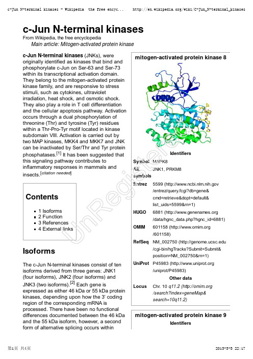
mitogen-activated protein kinase 8IdentifiersSymbol MAPK8Alt.symbols JNK1, PRKM8Entrez5599 ( /entrez/query.fcgi?db=gene&cmd=retrieve&dopt=default&list_uids=5599&rn=1)HUGO 6881 ( /data/hgnc_data.php?hgnc_id=6881)OMIM 601158 ( /601158)RefSeq NM_002750 (/cgi-bin/hgTracks?Submit=Submit&position=NM_002750&rn=1)UniProt P45983 (/uniprot/P45983)Other dataLocusChr. 10 q11.2 ( /search?index=geneMap&search=10q11.2)mitogen-activated protein kinase 9Identifiersc-Jun N-terminal kinasesFrom Wikipedia, the free encyclopediaMain article: Mitogen-activated protein kinase c-Jun N-terminal kinases (JNKs), were originally identified as kinases that bind and phosphorylate c-Jun on Ser-63 and Ser-73within its transcriptional activation domain.They belong to the mitogen-activated protein kinase family, and are responsive to stress stimuli, such as cytokines, ultravioletirradiation, heat shock, and osmotic shock.They also play a role in T cell differentiation and the cellular apoptosis pathway. Activation occurs through a dual phosphorylation of threonine (Thr) and tyrosine (Tyr) residues within a Thr-Pro-Tyr motif located in kinase subdomain VIII. Activation is carried out by two MAP kinases, MKK4 and MKK7 and JNK can be inactivated by Ser/Thr and Tyr protein phosphatases.[1] It has been suggested that this signaling pathway contributes toinflammatory responses in mammals and insects.[citation needed ]Contents1 Isoforms2 Function3 References4 External linksIsoformsThe c-Jun N-terminal kinases consist of ten isoforms derived from three genes: JNK1(four isoforms), JNK2 (four isoforms) and JNK3 (two isoforms).[2] Each gene isexpressed as either 46 kDa or 55 kDa protein kinases, depending upon how the 3' coding region of the corresponding mRNA isprocessed. There have been no functional differences documented between the 46 kDa and the 55 kDa isoform, however, a second form of alternative splicing occurs withinUn Re gi st er edSymbol MAPK9Alt.symbols JNK2, PRKM9Entrez5601 ( /entrez/query.fcgi?db=gene&cmd=retrieve&dopt=default&list_uids=5601&rn=1)HUGO 6886 ( /data/hgnc_data.php?hgnc_id=6886)OMIM602896 ( /602896)RefSeq NM_002752 (/cgi-bin/hgTracks?Submit=Submit&position=NM_002752&rn=1)UniProt P45984 (/uniprot/P45984)Other dataLocusChr. 5 q35 ( /search?index=geneMap&search=5q35)mitogen-activated protein kinase 10IdentifiersSymbol MAPK10Alt.symbols JNK3, PRKM10Entrez 5602 ( /entrez/query.fcgi?db=gene&cmd=retrieve&dopt=default&list_uids=5602&rn=1)HUGO 6872 ( /data/hgnc_data.php?hgnc_id=6872)OMIM602897 ( /602897)RefSeq NM_002753 (/cgi-bin/hgTracks?Submit=Submit&position=NM_002753&rn=1)UniProt P53779 (/uniprot/P53779)Other datatranscripts of JNK1 and JNK2, yielding JNK1-α, JNK2-α and JNK1-β and JNK2-β.Differences in interactions with protein substrates arise because of the mutually exclusive utilization of two exons within the kinase domain.[1]c-Jun N-terminal kinase isoforms have the following tissue distribution:JNK1 and JNK2 are found in all cells and tissues.[3]JNK3 is found mainly in the brain, but is also found in the heart and the testes.[3]FunctionInflammatory signals, changes in levels of reactive oxygen species, ultraviolet radiation,protein synthesis inhibitors, and a variety of stress stimuli can activate JNK. One way this activation may occur is through disruption of the conformation of sensitive protein phosphatase enzymes; specificphosphatases normally inhibit the activity of JNK itself and the activity of proteins linked to JNK activation.[4]JNKs can associate with scaffold proteins JNK interacting proteins as well as their upstream kinases JNKK1 and JNKK2following their activation.JNK, by phosphorylation, modifies the activity of numerous proteins that reside at the mitochondria or act in the nucleus.Downstream molecules that are activated by JNK include c-Jun, ATF2, ELK1, SMAD4, p53and HSF1. The downstream molecules that are inhibited by JNK activation include NFAT4, NFATC1 and STAT3. By activating and inhibiting other small molecules in this way, JNK activity regulates several important cellular functions including cell growth,differentiation, survival and apoptosis.JNK1 is involved in apoptosis,neurodegeneration, cell differentiation andproliferation, inflammatory conditions and cytokine production mediated by AP-1 (activationUn Re gi st er edprotein 1) such as RANTES, IL-8 and GM-CSF.[5]Recently, JNK1 has been found to regulate Jun protein turnover by phosphorylation and activation of the ubiquitin ligase Itch.References^ a b Ip YT, Davis RJ (April 1998). "Signal transduction by the c-Jun N-terminal kinase (JNK)--from inflammation to development". Curr. Opin. Cell Biol. 10 (2): 205–19.doi:10.1016/S0955-0674(98)80143-9 (/10.1016%2FS0955-0674%2898%2980143-9) . PMID 9561845 (// /pubmed/9561845) .1.^ Waetzig V, Herdegen T (2005). "Context-specific inhibition of JNKs: overcoming the dilemma of protection and damage". Br. J. Pharmacol 26 (9): 455–61. doi:10.1016/j.tips.2005.07.006(/10.1016%2Fj.tips.2005.07.006) . PMID 16054242 (// /pubmed/16054242) .2.^ a b Bode AM, Dong Z (August 2007). "The Functional Contrariety of JNK"(///pmc/articles/PMC2832829/) . Mol. Carcinog. 46 (8): 591–8.doi:10.1002/mc.20348 (/10.1002%2Fmc.20348) . PMC 2832829(///pmc/articles/PMC2832829) . PMID 17538955 (// /pubmed/17538955) . ///pmc/articles/PMC2832829/. "The protein products of jnk1 and jnk2 are believed to be expressed in every cell and tissue type, whereas the JNK3protein is found primarily in brain and to a lesser extent in heart and testis"3.^ Vlahopoulos S, Zoumpourlis VC (August 2004). "JNK: a key modulator of intracellular signaling". Biochemistry Mosc. 69 (8): 844–54. doi:10.1023/B:BIRY .0000040215.02460.45(/10.1023%2FB%3ABIRY .0000040215.02460.45) . PMID 15377263(///pubmed/15377263) .4.^ Oltmanns U, Issa R, Sukkar MB, John M, Chung KF (July 2003). "Role of c-jun N-terminal kinase in the induced release of GM-CSF , RANTES and IL-8 from human airway smoothmuscle cells" (///pmc/articles/PMC1573939/) . Br. J. Pharmacol. 139 (6):1228–34. doi:10.1038/sj.bjp.0705345 (/10.1038%2Fsj.bjp.0705345) .PMC 1573939 (///pmc/articles/PMC1573939) . PMID 12871843(///pubmed/12871843) . ///pmc/articles /PMC1573939/.5.External linksJNK+Mitogen-Activated+Protein+Kinases (/cgi/mesh/2011/MB_cgi?mode=&term=JNK+Mitogen-Activated+Protein+Kinases) at the US National Library of Medicine Medical Subject Headings (MeSH)Getting a Handle on Cellular JNK (/?p=1026) (from Beaker Blog)MAP Kinase Resource (http://www.mapkinases.eu)Retrieved from "/w/index.php?title=C-Jun_N-terminal_kinases&oldid=541721429"Categories: Genes on chromosome 10Genes on chromosome 5Cell signaling Signal transduction EC 2.7.11This page was last modified on 2 March 2013 at 13:46.T ext is available under the Creative Commons Attribution-ShareAlike License;Un Re gi st er edadditional terms may apply. See T erms of Use for details.Wikipedia® is a registered trademark of the Wikimedia Foundation, Inc., a non-profit organization.deretsigeRnU。
CaN信号转导机制及其与疾病相关性研究现状-病理学论文-基础医学论文-医学论文
CaN信号转导机制及其与疾病相关性研究现状-病理学论文-基础医学论文-医学论文——文章均为WORD文档,下载后可直接编辑使用亦可打印——1、概述1.1CaN发现钙调神经磷酸酶(CalcinurinCaN)也称依赖钙调蛋白的磷酸酯酶、神经贮钙蛋白、蛋白磷酸酶2B(PP2B),首次由张槐耀教授、王学荆教授在猪脑中提纯成功,后由Klee将其命名为Calcinurin,汉译为钙调神经磷酸酶,是目前所发现的唯一受Ca2+和钙调素(CaM)调节的丝/苏氨酸蛋白磷酸酶。
1.2CaN分布、编码基因及组成CaN在真核生物中具有高度保守性,广泛存在于各组织脏器中,动物脑内含量最高,提示CaN可能是参与多种细胞功能调节的多功能信号酶,与疾病的发生有关。
CaN是由一个催化亚单位(CnA)和一个调节亚单位(CnB)组成的异源二聚体。
其中CNA和CNA分别位于4号、10号(10q21y7q22)染色体上,CaNB位于2号(p16yp15)染色体上,其复杂的同工酶形式是通过选择性剪接产生的。
A亚基(CNA)是全酶的活性中心,分子量61kD,包括5个不同结构域,其中最为重要的是:催化区、CnB结合区、CaM结合区及自动抑制区,后者与钙调素结合域缺失后会导致CaN持续激活,而当CaM结合域向后弯曲,形成螺旋状封闭酶的底物结合区域起到了抑制磷酸酶活性的作用。
1.3CaN调节机制及酶学特性Ca2+是CaN的上游信号分子,具体过程如下:结合2个Ca2+的CaM可以与CaN形成复合物,但无活力,只有再结合上1-2个Ca2+后才有活性,这样Ca2+浓度持续升高才是激活CaN过程的限速步骤,而不是CaM与CaN的结合,持续激活的CaN又可负反馈调节细胞内游离Ca2+浓度。
CaN内还含有一个双核的金属中心,分别是Zn2+和Fe3+,Wang等发现,Zn2+和Fe3+位点的氧化损伤引起酶的失活,除此之外,CaN还可以被Mn2+、Ni2+等活化。
病理生理学专业词汇中英文对照
A2,3-diphosphoglyceric acid, 2,3-DPG 2,3-二磷酸甘油酸5-hydroxytryptamine receptor 5-羟色胺受体5-hydroxytryptamine, 5-HT 五羟色胺A cyclosporin A 环胞霉素A1 annexin A1 膜联蛋白a11ergy 变态反应abdominal distention 腹胀abdominal pain 腹痛acetylcholine Ach 乙酰胆碱acetylcholine receptor,AchR Ach受体Acid phosphatase, ACP 酸性磷酸酶acid sensitive ionic channels ASICs 酸敏感离子通道acid-base balance 酸碱平衡acid-base disturbance 酸碱平衡紊乱acid-base imbalance 酸碱失衡actin 肌动蛋白activated partial thromboplastin time APTT 活化部分凝血活酶时间activation function 1,AF-1 激活功能1activation function 2,AF-2 激活功能2actual bicarbonate, AB 实际碳酸氢盐acute phase protein 急性期蛋白acute phase response 急性期反应acute promyelocytic leukemia 急性早幼粒细胞白血病acute psychogenic reaction 急性心因性反应acute pulmonary edema 急性肺水肿acute pulmonary injury, ALI 急性肺损伤acute renal failure,ARF 急性肾功能衰竭acute respiratory distress syndrome, ARDS 急性呼吸窘迫综合征acute tubular necrosis,A TN 急性肾小管坏死adapter protein 适配体蛋白adaptor 接头蛋白adaptor protein 适配体蛋白adenylate cyclase AC 腺苷酸环化酶adhesion molecule 黏附分子adipokines 脂肪因子adiponectin 脂联素adipose tissue,A T 脂肪组织adjustment disorders 适应障碍adrenalin 肾上腺素adrenergic receptor 肾上腺素能受体adrenocorticotropic hormone ACTH 促肾上腺皮质激素adrenocorticotropic hormone ACTH 促肾上腺皮质激素释放激素adriamycin 阿霉素advanced glycosylation end products, AGE 38 39 高级糖基化终产物aflatoxin 黄曲霉毒素after depolarization 后除极aging 老化agonosia 失认aheolar ventilation 肺泡通气量alarm stage 警觉期aldosterone 醛固酮a-L-fucosidase,AFU a-L岩藻糖苷酶alignment of the chromosomes 染色质联会alkylating agents 烷化剂allelic heterogeneity 等位异质性allelic loss 等位基因丢失alpha-fetoprotein,AFP 甲胎蛋白alternative pathway 替代途径alternative splicing 选择性剪接alternative splicing,AS 选择性剪接alveolar PCO2, PACO2 肺泡气二氧化碳分压alveolar PO2,PAO2 肺泡气氧分压Alzheimer disease, AD 阿尔茨海默病a-Melanocyte stimulating hormone, a-MSH 黑素细胞刺激素American College of Cardiology,ACC 美国心脏病学会American Heart Association,AHA 美国心脏协会ammonia 氨中毒amplification of signal 信号放大amygdala 杏仁核amyloid precursor protein,ARP 淀粉样蛋白前体anaphase 后期anaphylactic shock 过敏性休克anaphylactoid purpura 过敏性紫癜Anaplasma phagocytophilum 嗜吞噬无形体anasarca 全身性水肿anatomic shunt 解剖分流angiogenin 基因扩增angiotensin Ⅰ,Ang Ⅰ血管紧张素Ⅰangiotensin Ⅱ,AngⅡ血管紧张素Ⅱangiotensin-converting enzyme, ACE 血管紧张素转化酶angiotension converting enzyme inhibitor,ACEI 血管紧张素转换酶抑制剂anion gap, AG 阴离子间隙ankyrin 锚蛋白annexin 2 膜联蛋白2annexin-1 膜联蛋白-1anorexia 食欲不振anterior cingule cortex ACC 大脑前扣带皮质antidiuretic hormone,ADH 抗利尿激素antimetabolite 血管生长素antiostain 抗代谢药物antisense 反义antithrombin III AT III 抗凝血酶IIIanuia 无尿aphasia 失语aplastic anemia 再生障碍性贫血apolipoprotein A1 血管抑制素apoptosis 凋亡apoptosis 细胞凋亡apoptosis inducing factor,AIF 凋亡诱导因子apoptosome 凋亡复合体apoptotic bodies 凋亡小体apoptotic effector 凋亡效应因子apoptotic lesion 凋亡性损伤apoptotic protease activating factor,Apaf 凋亡蛋白酶激活因子apraxia 失用aquaporin,AQP 水通道arachidonic acid AA 花生四烯酸aralkylating agents 载脂蛋白A1ARAS 上行网状激动系统arcuate nucleus, ARC 弓状核Arg-Gly-Asp,GRD 芳烷化剂arginine vasopressin, A VP 精氨酸加压素arginine,R 精氨酸aromatic amino acids,AAA 芳香族氨基酸arrhythmia 心律失常arylhydroxylamines 精-甘-天冬氨酸asbestos 芳基异羟肟胺类ascending reticular activating system ARAS 脑干上行网状激动系统ascending reticular inhibitory system ARIS 上行网状抑制系统asymbolia for pain 疼痛说示不能综合征asymmetric cell division 细胞的不对称分裂ata 石棉ataxia and rad3 related,ATR 共济失调毛细血管扩张症Rad3相关蛋白Ataxia telangiectasia,AT 共济失调性毛细血管扩张症ataxia-telangiectasia,AT 毛细血管扩张性共济失调症突变基因ataxia-telangiectasia-mutated,A TM 毛细血管扩张性共济失调突变蛋白atherosclerosis,AS 动脉粥样硬化athogenesis 发病学ATLL T 细胞白血病淋巴瘤ATP binding pocket ATP结合口袋ATP receptor ATP受体atrial natriuretic peptide ANP 心房利尿钠肽autocrine motility factor,AMF 自分泌运动因子autoimmune disease 自身免疫性疾病automatic external defibrillator,AED 自动体外除颤器automatic impulse 自主性激动autophagic cell death 自噬性细胞死亡autophagin 自噬素autophagolysosome 自噬溶酶体autophagosome,AP 自噬体autophagy 细胞自噬autophagy 自噬autophagy 自噬性程序性细胞死亡autophagy-related proteins,Atg 细胞自噬相关蛋白autophosphorylation 自主磷酸化azotemia 氮质血症α-lactalbumin α-乳白蛋白BB1 cyclin B1 细胞周期蛋白bacterial translocation 细菌移位BaP 苯并芘base excess, BE 碱剩余base excision repair,BER 碱基切除修复baterial endotoxin 细菌内毒素Bax-interacting factor 1,BIF-1 Bax作用因子1B-cell lymphoma 2,Bcl-2 B细胞淋巴瘤2Bcl-2 homology,BH Bcl-2同源结构域beme oxygenase 血红素加氧酶benign tumor 良性肿瘤benzediazepine,BZ 苯二氮卓Beta-amyloid,Aββ-淀粉样蛋白biallelic 二等位基因bi-allelie marker 双等位标记big MAP kinase,BMK1 大丝裂原活化蛋白激酶bilinogen enterohepatic circulation 胆素原的肠肝循环bilirubin 胆红素bilirubin encephalopathy 胆红素性脑病biliverdin reductase 胆绿素还原酶biofeedback therapy 生物反馈治疗bioinformatics 生物信息学blebbing 水泡化blood urea nitrogen,BUN 血浆尿素氮bone marrow hematopoietic stem cells 骨髓造血干细胞bradykinin BK 缓激肽brain derived neurotrophic factor,BDNF 脑源神经营养因子brain stem 脑干brain-derived neurotrophic factor BDNF 脑源性神经营养因子branched-chain amino acids,BCAA 支链氨基酸breakage-fusion-brige, BFB 断裂-融合-桥breast cancer susceptibility gene C terminus,BRCT 乳腺癌易感基因羧基末端结构域brown adipo 棕色脂肪组织Brugada Syndrome Brugada综合征buffer base, BB 缓冲碱Burkholderia pseudomallei 类鼻疽伯克霍尔德菌burn shoch 烧伤性休克butyrolactone 丁内酯bystander cell 旁观者细胞β2-Microglobulin,β2Mβ2-微球蛋白β-amyloid precursor protein β-淀粉样前体蛋白β-amyloid protein β-淀粉样蛋白β-endorphin β-内啡肽β-lactams β-内酰胺类CC cytochrome C,Cyto-C 细胞色素C1-esterase inhibitor C1酯酶抑制物Ca2+ ionophore Ca2+离子载体cadherin 钙离子依赖的细胞黏附素cadherins 钙粘蛋白calcitonin ,CT 降钙素calcitonin gene related protein CGRP 降钙素基因相关蛋白calcium dyshomeostasis 钙稳态失衡calcium leak Ca2+泄漏calcium overload 钙超载calcium paradox 钙反常calcium transient Ca2+瞬变calmodulin,CaM 钙调蛋白calmodulin-dependent kinase II,CaMK II 钙调蛋白依赖性激酶II calmodulin-dependent kinase,CaMK 钙调蛋白依赖性激酶cancer 癌症cancer stem cells,CSC 肿瘤干细胞capping protein 加帽蛋白carbon dioxide narcosis 二氧化碳麻醉carbon monoxide, CO 一氧化碳carbonic anhydrase, CA 碳酸酐酶carboxyhemoglobin, HbCO 碳氧血红蛋白carcinoembryonic antigen,CEA 癌胚抗原carcinogen 致癌物cardiac asthma 心源性哮喘cardiac edema 心性水肿cardiac index,CI 心脏指数cardiac insufficiency 心功能不全cardiac output,CO 心输出量cardiac resynchronization therapy,CRT 心脏再同步治疗cardiogenic shock 心源性休克carnitine palmitoyl transferase-1,CPT-1 肉碱脂酰转移酶-1 caspase 半胱天冬酶caspase-like apoptosis regulatory protein,CLARP 半胱天冬酶样凋亡调节蛋白Caspase-recruitment domain,CARD 凋亡蛋白酶募集结构域catabolic process 分解代谢过程catecholamine 儿茶酚胺catecholamine,CA 儿茶酚胺Catypical protein kinase C,aPKC 非典型性蛋白激酶CDK inhibitor,CKI CDK抑制因子CDK-activating kinase,CAK CDK活化激酶CEA 癌胚抗原cell adhesion molecule,CAM 细胞粘附分子cell cycle 细胞周期Cell Damage 细胞损伤cell differentiation 细胞分化cell division 细胞分裂cell division cycle 2,CDC2 细胞分裂周期2cell pathology 细胞病理学cell proliferation 细胞增殖cell signaling 细胞信号传递cell stress 细胞应激cellular hydration 细胞水合cellular oncogene 细胞癌基因central nervous system CNS 中枢神经系统central pain 中枢性疼痛central venous pressure,CVP 中心静脉压centrosome 中心体cerebral cortex 大脑皮层chaperokine 伴侣因子chaperon mediated autophagy,CMA 分子伴侣介导的自噬checkpoint 检测点chemokine 趋化因子chemokine receptors 趋化因子受体chemoprevention 化学预防cholecystokinin CCK 胆囊收缩素choline acetyl transferase ChAT 胆碱乙酰基转移酶chondroitin sulfate 硫酸软骨素chromatin body 染色质小体chromosomal aberration 染色体畸变chromosomal instability,CIN 染色体不稳定chromosome instability 染色体不稳定性chronic bronchitis 慢性支气管炎chronic inflammationsyndrome 慢性炎症综合征chronic obstructive pulmonary disease, COPD 慢性阻塞性肺疾病chronic renal failure,CRF 慢性肾功能衰竭ciliary neurotrophic factor CNTF 睫状神经营养因子ciliary neurotrophic factor, CNTF 睫状神经营养因子circulatory hypoxia 循环性缺氧cirrhosis of liver 肝硬化cis-acting element 顺式作用元件cis-acting element 顺式作用元件cis-regulatory module 顺式调节模块c-Jun N terminal kinase,JNK c-Jun N端蛋白激酶class I PI3K protein I类PI3K蛋白clot 血凝块cluster 集簇coactivator 共激活因子coagulation 凝血Coding-region SNPs,cSNPs 基因编码区SNPs cognitive appraisal 认知评价cognitive disorder,cognitive handicap 认知障碍cold inducible RNA-binding protein 寒冷诱导的RNA结合蛋白cold sensitive neuron 冷敏神经元cold shock 冷休克cold shock protein 冷休克蛋白cold shock response 冷休克反应cold stress 冷应激collagen 胶原collagenases 胶原酶coma 昏迷代偿性抗炎反应综合征compensatory anti-inflammatory responsesyndrome, CARScompensatory stage of shock 休克代偿期complement,C 补体complete recovery 完全康复complex multisite variants 复合性多位点变异concentric hypertrophy 向心性肥大confusion 意识模糊congestive hypoxia 淤血性缺氧conjugated bilirubin 结合胆红conscious disturbance 意识障碍consciousness 意识constipation 便秘constitutive expression 基础表达content of consciousness 意识内容contraction alkalosis 浓缩性碱中毒copy number variant, CNV 。
肽基脯氨酰顺反异构酶1_在肝癌中的研究进展
㊀㊀[摘要]㊀肽基脯氨酰顺反异构酶1(PIN1)在肝癌中异常表达,作为肿瘤催化分子在肝癌的增殖㊁侵袭㊁凋亡和血管生成等过程中发挥着重要作用㊂该文从PIN1的分子结构与功能㊁PIN1在肝癌中的表达及其在肝癌发生㊁发展中的作用等几方面进行综述㊂㊀㊀[关键词]㊀肽基脯氨酰顺反异构酶1;㊀肝癌;㊀进展㊀㊀[中图分类号]㊀R735 7㊀[文献标识码]㊀A㊀[文章编号]㊀1674-3806(2023)10-1100-04㊀㊀doi:10.3969/j.issn.1674-3806.2023.10.24Researchprogressofpeptidyl⁃prolylcis/transisomeraseNIMA⁃interacting1inhepatocellularcarcinoma㊀MOZhu⁃ning,HUANGLi.DepartmentofBloodTransfusion,thePeopleᶄsHospitalofGuangxiZhuangAutonomousRegion(GuangxiAcademyofMedicalSciences),Nanning530021,China㊀㊀[Abstract]㊀Peptidyl⁃prolylcis/transisomeraseneverinmitosisA(NIMA)⁃interacting1(PIN1)isabnormallyexpressedinhepatocellularcarcinoma,andplaysanimportantroleintheproliferation,invasion,apoptosisandangio⁃genesisofhepatocellularcarcinomaasatumorcatalyticmolecule.Thispaperreviewsthemolecularstructureandfunc⁃tionofPIN1,theexpressionofPIN1inhepatocellularcarcinomaanditsroleintheoccurrenceanddevelopmentofhepatocellularcarcinoma.㊀㊀[Keywords]㊀Peptidyl⁃prolylcis/transisomeraseneverinmitosisA(NIMA)⁃interacting1(PIN1);㊀Hepato⁃cellularcarcinoma;㊀Progress㊀㊀肝癌是全球第七大常见实体肿瘤,也是第二大致死恶性肿瘤[1]㊂国内肝癌的防治也依然严峻[2]㊂尽管肝癌的诊断水平在不断提高,但早期肝癌的有效诊断仍然十分困难㊂在治疗方面,虽然肝癌患者能够通过手术切除或消融,甚至肝移植获得有效治疗,但仍有部分患者存在肿瘤复发,导致肝癌的5年生存率较低[3⁃5]㊂因此,迫切需要探究肝癌发生㊁发展过程中涉及的潜在关键分子,为肝癌的诊断㊁治疗和预后提供有效的理论基础㊂肽基脯氨酰顺反异构酶1[peptidyl⁃prolylcis/transisomeraseneverinmitosisA(NIMA)⁃interacting1,PIN1]属于肽脯氨酰基顺反异构酶家族,普遍存在于生命体中,能够特异地识别并与蛋白质中磷酸化的丝/苏⁃脯氨酸基序结合,催化其中酰胺键的顺反异构,继而调节蛋白的生物活性㊁稳定性㊁磷酸化水平及亚细胞定位[6]㊂PIN1在细胞生命进程中通过对蛋白的调控,诱导众多原癌及抑癌基因的蛋白功能发生变化,从而参与多种细胞信号转导及通路调节,促进肿瘤的发生和发展[7⁃8]㊂研究证实PIN1在肝癌中高表达,并通过抑制肿瘤细胞凋亡,促进肿瘤细胞生长㊁侵袭㊁转移和肿瘤血管生成的方式发挥作用㊂因此,PIN1与肝癌的发生㊁发展密切相关㊂本文对PIN1的分子结构与功能㊁PIN1在肝癌中的表达及其在肝癌发生㊁发展中的作用等综述如下㊂1㊀PIN1的分子结构与功能PIN1定位于19号染色体短臂13带,由163个氨基酸组成,相对分子质量为18ˑ103,广泛分布于各种原核㊁真核生物体及各种组织,多数定位于胞质,但部分也可存在于酵母㊁果蝇和哺乳动物的内质网㊁红色面包霉的线粒体基质及大肠杆菌的外周质等[9⁃10]㊂PIN1发挥生物学功能主要通过以下2个功能结构区域实现:一个是由1 39位氨基酸构成的氨基末端色氨酸⁃色氨酸中心结构区域,主要参与底物的识别,使PIN1特异性地与底物中磷酸化的丝/苏⁃脯氨酸肽段的蛋白结合;另一个是由45 163位氨基酸构成的羧基末端肽基脯氨酰异构酶活性结构域,可诱导蛋白的构象和功能变化,特异性地异构化磷酸化的丝/苏⁃脯氨酸酰胺键[11]㊂这两个结构域紧密合作催化底物蛋白的构象变化,随后通过多种途径激活并放大各类致癌转导信号,从而引起中心体的异常扩增和基因组序列的不稳定,导致细胞恶性转化,最终导致肿瘤的发生与发展㊂2㊀PIN1在肝癌中的表达PIN1表达紊乱通常导致细胞增殖失控和肿瘤形成㊂PIN1表达水平与癌症间存在相关性首先在乳腺癌中得到证实,其在细胞中的定位与肿瘤的病理类型相关[12]㊂研究发现,PIN1在多种肿瘤中均呈现高表达,包括肝癌㊁脑癌㊁宫颈癌㊁结肠癌㊁肺癌和前列腺癌等[13]㊂多项研究证实,与邻近的非肿瘤肝组织相比,PIN1的核糖核酸(ribosenucleicacid,RNA)和蛋白在肝癌中均呈高水平表达[14⁃16]㊂Shinoda等[17]采用免疫组化和蛋白免疫印迹技术对肝癌患者中PIN1表达进行研究,发现PIN1表达越高的患者,其肿瘤体积越大㊁门静脉侵犯频率也越高,患者的预后越差㊁总生存率越低,而3年内复发率也越高㊂Pang等[18]还发现肝癌组织中PIN1的表达与乙型肝炎病毒(hepatitisBvirus,HBV)X蛋白(HBx)的表达呈显著正相关㊂另一项研究结果也表明PIN1在约70%的HBV阳性肝癌患者中呈高水平表达[19]㊂PIN1基因启动子的单核苷酸多态性也可能参与PIN1表达的调控,其基因型⁃842CC与外周血单个核细胞内PIN1蛋白表达水平降低有关,表明PIN1与肝癌的遗传易感性之间具有相关性[20⁃22]㊂由此可见,PIN1的表达与肝癌的发生㊁发展密切相关㊂3㊀PIN1在肝癌发生、发展中的作用3 1㊀PIN1与肝癌增殖㊀HBV是肝癌最常见的病因,其编码的HBx具有致癌性㊂研究表明,PIN1和HBx可以在特定的丝氨酸⁃脯氨酸基序上相互作用,从而增强HBV感染状态下肝细胞的增殖作用[23]㊂另一项研究发现,肝内胆管癌中PIN1与c⁃Jun氨基端激酶(c⁃JunN⁃terminalproteinkinase,JNK)活性呈正相关,其主要机制是JNK可直接结合PIN1中丝氨酸115位点并使其磷酸化,这一磷酸化作用阻止了PIN1在赖氨酸117位点的泛素化及其蛋白酶降解㊂JNK通路的激活主要是通过影响PIN1蛋白稳定性来促进肝内胆管细胞增殖,表明PIN1是肿瘤发生的关键驱动因子[24]㊂Bae等[25]通过体外细胞实验对p53基因突变与PIN1表达间的相关性研究发现,PIN1介导的脯氨酸异构化可通过调控靶蛋白p53诱导细胞周期阻滞和生长抑制㊂随着研究的深入,发现p53突变对肝癌生长的作用机制,主要是通过周期蛋白依赖激酶4(cyclin⁃dependentkinase4,CDK4)促进p53突变的丝氨酸249磷酸基团与PIN1相结合并依赖PIN1定位于核内,而且结合物随着PIN1的增加而增多,从而促进p53突变的核定位,导致p53突变与c⁃Myc相互作用,增强依赖c⁃Myc的核糖体合成,证实了CDK4⁃PIN1⁃p53⁃RS⁃c⁃Myc通路在肝癌进程中的作用[26]㊂Farra等[27]发现,PIN1可通过上调多种细胞周期蛋白和E2F转录因子促进肝癌细胞的增殖,表明PIN1⁃E2F1是控制肝癌细胞生长的另一个具有吸引力的靶点㊂β⁃连环蛋白是Wnt信号通路中的重要分子,它能在Ras相关C3肉毒素底物1(Ras⁃relatedC3botulinumtoxinsubstrate1,Rac1)的辅助下,迁移至胞核,与转录因子LEF/TCF及多种蛋白形成转录复合体,从而启动靶基因的转录,影响细胞的增殖与分化㊂张钰[28]发现PIN1与β⁃连环蛋白的表达呈正相关,其机制可能是PIN1激活了Wnt/β⁃连环蛋白通路,从而促进肝细胞癌的发生和发展㊂微小RNA(microRNA,miRNA)结合与功能实验表明,过表达miR⁃140⁃5p不仅可以降低PIN1的表达,还可以抑制多种PIN1依赖的癌症途径,并抑制小鼠的肿瘤生长[29]㊂由此可见,PIN1可用不同的方式参与肝癌细胞的增殖㊂3 2㊀PIN1与肝癌转移㊀早期发现或预测肝癌转移的分子标志物,对于肝癌患者的治疗管理和确定新的治疗靶点以抑制肝癌的进展和转移至关重要㊂Ng等[15]通过随机对照试验对139例肝癌患者中PIN1的表达水平与肿瘤转移的相关性进行研究,发现肝癌标本中PIN1转录水平显著高于配对的癌旁组织,且PIN1高表达与肝癌患者的肿瘤转移和无复发生存率密切相关,这也是预测肝癌根治切除术后转移复发的独立危险因素㊂另一项研究发现,瑞格拉非尼耐药的肝癌细胞株,不仅可通过敲除或过表达PIN1调控上皮间质转化相关分子(上皮钙黏附素和Snail)来实现相应抑制或促进肝癌细胞的迁移和侵袭,而且可通过与上皮间质转化调节因子Gli1的相互作用,影响肝癌细胞的上皮间质转化㊁迁移㊁侵袭和转移等生物学行为[30]㊂Sun等[31]研究发现,PIN1可通过改变细胞外调节蛋白激酶磷酸化后输出蛋白⁃5(exportin⁃5)的构象,导致miR⁃122负载减少,以增加微管动力学相关蛋白的表达,从而参与肝癌的迁移㊂因此,PIN1与肝癌的转移活性密切相关㊂3 3㊀PIN1与肝癌血管生成㊀肿瘤血管的形成能促进肿瘤的发生㊁发展,而肝癌作为一种高度侵袭性的多血管性肿瘤,其疾病进展有赖于活跃的血管生成㊂肿瘤血管生成主要受血管内皮生长因子(vascularendo⁃thelialgrowthfactor,VEGF)的表达调控,其通过刺激血管通透性诱导肿瘤血管生成,促进肿瘤发展进程[32⁃33]㊂研究发现,PIN1高表达可增加人肝星形细胞激活蛋白⁃1(activatorprotein⁃1,AP⁃1)的转录活性和VEGF蛋白水平㊂AP⁃1的转录活性通过与c⁃Fos和c⁃Jun形成异二聚体进行调控,而PIN1可通过结合c⁃Fos和c⁃Jun来增加其转录活性,进而增强AP⁃1的活化活性,导致VEGF基因转录的增加㊂这表明在肝细胞中PIN1可通过调控AP⁃1活性促进VEGF介导的血管生成[34]㊂此外,另一项细胞体外实验显示,抑制PIN1表达可降低肝癌细胞中VEGF的蛋白表达量,从而抑制核因子κB(nuclearfactorkappa⁃B,NF⁃κB)通路的磷酸化,降低NF⁃κB的活化,阻碍肿瘤血管生成[17]㊂因此,PIN1能通过底物蛋白活化调控VEGF介导的肝癌血管生成㊂3 4㊀PIN1与肝癌细胞凋亡㊀PIN1除了参与上述功能,还影响肝癌细胞的程序性死亡,即细胞凋亡㊂Zheng等[19]发现,索拉非尼能通过Rb/E2F途径抑制PIN1的转录,从而下调PIN1RNA和蛋白的表达㊂敲除PIN1可导致Mcl⁃1的表达降低,从而增强索拉非尼在肝癌中诱导细胞凋亡的能力㊂此外,抑制并最终诱导癌细胞中活性PIN1的降解,也可通过依赖含半胱氨酸的天冬氨酸蛋白水解酶(cysteinylaspartatespecificpro⁃teinase,caspase)的方式增加索拉非尼诱导的肝癌细胞凋亡敏感性㊂这些结果表明,PIN1在索拉非尼抗肿瘤细胞凋亡过程中发挥重要作用㊂生存素(survivin)的抗凋亡功能在包含肝癌在内的多种癌症中起着关键作用,其在肝癌中与PIN1的表达呈正相关㊂通过细胞系和异种移植模型研究发现,过表达PIN1可抑制caspase⁃3和caspase⁃9活性导致凋亡减弱㊂此外,在过表达PIN1的细胞中下调survivin可减弱PIN1诱导的抗凋亡作用,提示抑制凋亡可通过PIN1⁃survivin相互作用介导㊂免疫共沉淀分析也表明PIN1可通过磷酸化的苏氨酸34⁃脯氨酸35基序与survivin相互作用,并增强苏氨酸34位点磷酸化的survivin㊁HBx蛋白和caspase⁃9前体之间的结合㊂证实PIN1可通过survivin蛋白参与肝癌细胞的凋亡功能[35]㊂此外,Leong等[36]研究证实,miR⁃874⁃3p在肝癌细胞系可通过靶向PIN1抑制细胞生长和集落形成,从而促进细胞凋亡㊂综上所述,PIN1在肝癌细胞的凋亡过程中可通过调控多种底物蛋白发挥重要作用㊂4㊀结语肝癌是一种具有很强侵袭性且预后不良的癌症,其发生㊁发展过程中重要分子靶点的识别可能促进新的诊疗方法的发展㊂PIN1表达失调与肝癌肿瘤大小㊁分期和复发等临床特征密切相关㊂PIN1通过调控底物蛋白磷酸化异构在多种通路中参与肝癌进程㊂因此,PIN1高表达所产生的多种致癌效应使得PIN1很有可能成为肝癌诊断和治疗的靶点,但仍需要进一步的研究和临床试验来检验PIN1在肝癌诊治中的安全性和有效性,这也将是今后研究的重点㊂相信随着研究的深入,将逐步揭示PIN1在肝癌发生㊁发展中的机制,为肝癌的诊断和治疗提供更多的研究基础和理论依据㊂参考文献[1]SungH,FerlagJ,SiegelRL,etal.Globalcancerstatistics2020:GLOBOCANestimatesofincidenceandmortalityworldwidefor36cancersin185countries[J].CACancerJClin,2021,71(3):209-249.[2]贺君剑,魏长慧,李㊀众,等.中国居民2004 2016年肝癌死亡趋势分析[J].郑州大学学报(医学版),2020,55(1):119-122.[3]ChapmanBC,PanicciaA,HosokawaPW,etal.Impactoffacilitytypeandsurgicalvolumeon10⁃yearsurvivalinpatientsundergoinghepaticresectionforhepatocellularcarcinoma[J].JAmCollSurg,2017,224(3):362-372.[4]朱晓峰.肝癌肝移植的研究进展与挑战[J].中国临床新医学,2020,13(12):1190-1193.[5]吕天石,邹英华.肝癌微创介入治疗进展[J].中国临床新医学,2020,13(3):211-215.[6]MinSH,ZhouXZ,LuKP.TheroleofPin1inthedevelopmentandtreatmentofcancer[J].ArchPharmRes,2016,39(12):1609-1620.[7]PuW,ZhengY,PengY.ProlylisomerasePin1inhumancancer:function,mechanism,andsignificance[J].FrontCellDevBiol,2020,8:168.[8]ChengCW,TseE.TargetingPIN1asatherapeuticapproachforhepa⁃tocellularcarcinoma[J].FrontCellDevBiol,2020,7:369.[9]LuKP,HanesSD,HunterT.Ahumanpeptidyl⁃prolylisomeraseessen⁃tialforregulationofmitosis[J].Nature,1996,380(6574):544-547.[10]RanganathanR,LuKP,HunterT,etal.StructuralandfunctionalanalysisofthemitoticrotamasePin1suggestssubstraterecognitionisphosphorylationdependent[J].Cell,1997,89(6):875-886.[11]ZhangM,FrederickTE,VanPeltJ,etal.Coupledintra⁃andinter⁃domaindynamicssupportdomaincross⁃talkinPin1[J].JBiolChem,2020,295(49):16585-16603.[12]WulfGM,RyoA,WulfGG,etal.Pin1isoverexpressedinbreastcancerandcooperateswithRassignalinginincreasingthetranscrip⁃tionalactivityofc⁃JuntowardscyclinD1[J].EMBOJ,2001,20(13):3459-3572.[13]ChengCW,TseE.PIN1incellcyclecontrolandcancer[J].FrontPharmacol,2018,9:1367.[14]AoR,ZhangDR,DuYQ,etal.ExpressionandsignificanceofPin1,β⁃cateninandcyclinD1inhepatocellularcarcinoma[J].MolMedRep,2014,10(4):1893-1898.[15]NgL,KwanV,ChowA,etal.OverexpressionofPin1andRhosig⁃nalingpartnerscorrelateswithmetastaticbehaviorandpoorrecurrence⁃freesurvivalofhepatocellularcarcinomapatients[J].BMCCancer,2019,19(1):713.[16]许晓梅,胡怀东,廖㊀勇,等.应用组织芯片检测Pin1在肝炎㊁肝硬化㊁肝细胞性肝癌组织中的表达[J].重庆医科大学学报,2010,35(9):1377-1380.[17]ShinodaK,KubokiS,ShimizuH,etal.Pin1facilitatesNF⁃κBacti⁃vationandpromotestumourprogressioninhumanhepatocellularcar⁃cinoma[J].BrJCancer,2015,113(9):1323-1331.[18]PangR,LeeTK,PoonRT,etal.Pin1interactswithaspecificserine⁃prolinemotifofhepatitisBvirusX⁃proteintoenhancehepatocarci⁃nogenesis[J].Gastroenterology,2007,132(3):1088-1103.[19]ZhengM,XuH,LiaoXH,etal.InhibitionoftheprolylisomerasePin1enhancestheabilityofsorafenibtoinducecelldeathandinhibittumorgrowthinhepatocellularcarcinoma[J].Oncotarget,2017,8(18):29771-29784.[20]SegatL,PontilloA,AnnoniG,etal.PIN1promoterpolymorphismsareassociatedwithAlzheimerᶄsdisease[J].NeurobiolAging,2007,28(1):69-74.[21]HuangL,MoZ,LaoX,etal.PIN1geneticpolymorphismsandthesusceptibilityofHBV⁃relatedhepatocellularcarcinomainaGuangxipopulation[J].TumourBiol,2016,37(5):6599-6606.[22]SegatL,MilaneseM,CrovellaS.Pin1promoterpolymorphismsinhepatocellularcarcinomapatients[J].Gastroenterology,2007,132(7):2618-2620.[23]ZhouQ,YanL,XuB,etal.ScreeningoftheHBxtransactivationdomaininteractingproteinsandthefunctionofinteractorPin1inHBVreplication[J].SciRep,2021,11(1):14176.[24]LeporeA,ChoyPM,LeeNCW,etal.Phosphorylationandstabili⁃zationofPIN1byJNKpromoteintrahepaticcholangiocarcinomagrowth[J].Hepatology,2021,74(5):2561-2579.[25]BaeJS,NohSJ,KimKM,etal.PIN1inhepatocellularcarcinomaisassociatedwithTP53genestatus[J].OncolRep,2016,36(4):2405-2411.[26]LiaoP,ZengSX,ZhouX,etal.Mutantp53gainsitsfunctionviac⁃MycactivationuponCDK4phosphorylationatserine249andcon⁃sequentPIN1binding[J].MolCell,2017,68(6):1134-1146.e6.[27]FarraR,DapasB,BaizD,etal.ImpairmentofthePin1/E2F1axisintheanti⁃proliferativeeffectofbortezomibinhepatocellularcarci⁃nomacells[J].Biochimie,2015,112:85-95.[28]张㊀钰.肽基脯氨酰顺反异构酶在肝细胞癌中的表达及其临床病理意义[D].福州:福建医科大学,2014.[29]YanX,ZhuZ,XuS,etal.MicroRNA⁃140⁃5pinhibitshepatocel⁃lularcarcinomabydirectlytargetingtheuniqueisomerasePin1toblockmultiplecancer⁃drivingpathways[J].SciRep,2017,7:45915.[30]WangJ,ZhangN,HanQ,etal.Pin1inhibitionreversestheacquiredresistanceofhumanhepatocellularcarcinomacellstoRegorafenibviatheGli1/Snail/E⁃cadherinpathway[J].CancerLett,2019,444:82-93.[31]SunHL,CuiR,ZhouJ,etal.ERKactivationgloballydownregu⁃latesmiRNAsthroughphosphorylatingexportin⁃5[J].CancerCell,2016,30(5):723-736.[32]NaitoH,IbaT,TakakuraN.Mechanismsofnewblood⁃vesselforma⁃tionandproliferativeheterogeneityofendothelialcells[J].IntImmunol,2020,32(5):295-305.[33]MelincoviciCS,BoşcaAB,ŞuşmanS,etal.Vascularendothelialgrowthfactor(VEGF) keyfactorinnormalandpathologicalangio⁃genesis[J].RomJMorpholEmbryol,2018,59(2):455-467.[34]YangJW,HienTT,LimSC,etal.Pin1inductioninthefibroticliveranditsrolesinTGF⁃β1expressionandSmad2/3phosphoryla⁃tion[J].JHepatol,2014,60(6):1235-1241.[35]ChengCW,ChowAK,PangR,etal.PIN1inhibitsapoptosisinhep⁃atocellularcarcinomathroughmodulationoftheantiapoptoticfunc⁃tionofsurvivin[J].AmJPathol,2013,182(3):765-775.[36]LeongKW,ChengCW,WongCM,etal.MiR⁃874⁃3pisdown⁃regulatedinhepatocellularcarcinomaandnegativelyregulatesPIN1expression[J].Oncotarget,2017,8(7):11343-11355.[收稿日期㊀2021-11-08][本文编辑㊀韦㊀颖]本文引用格式莫柱宁,黄㊀莉.肽基脯氨酰顺反异构酶1在肝癌中的研究进展[J].中国临床新医学,2023,16(10):1100-1103.。
病理生理学-专业词汇-中英文对照
A2,3-diphosphoglyceric acid, 2,3-DPG 2,3-二磷酸甘油酸5-羟色胺受体5-hydroxytryptamine receptor五羟色胺5-hydroxytryptamine, 5-HT环胞霉素A cyclosporin A膜联蛋白A1 annexin A1变态反应a11ergy腹胀abdominal distention腹痛abdominal pain乙酰胆碱acetylcholine AchAchacetylcholine receptor,AchR 受体酸性磷酸酶Acid phosphatase, ACP酸敏感离子通道acid sensitive ionic channels ASICs酸碱平衡acid-base balance酸碱平衡紊乱acid-base disturbance酸碱失衡acid-base imbalance肌动蛋白actin活化部分凝血活酶时间activated partial thromboplastin time APTT 激活功能,activation function 1AF-1 1激活功能,AF-22activation function 2实际碳酸氢盐actual bicarbonate, AB急性期蛋白acute phase protein急性期反应acute phase response急性早幼粒细胞白血病acute promyelocytic leukemia急性心因性反应acute psychogenic reaction急性肺水肿acute pulmonary edema急性肺损伤acute pulmonary injury, ALI急性肾功能衰竭acute renal failure,ARF急性呼吸窘迫综合征acute respiratory distress syndrome, ARDS急性肾小管坏死,ATN acute tubular necrosis适配体蛋白adapter protein接头蛋白adaptor适配体蛋白adaptor protein腺苷酸环化酶adenylate cyclase AC黏附分子adhesion molecule脂肪因子adipokines脂联素adiponectin脂肪组织T adipose tissue,A适应障碍adjustment disorders肾上腺素adrenalin肾上腺素能受体adrenergic receptor促肾上腺皮质激素adrenocorticotropic hormone ACTH促肾上腺皮质激素释放adrenocorticotropic hormone ACTH激素adriamycin阿霉素高级糖基化终产物advanced glycosylation end products, AGE 38 39黄曲霉毒素aflatoxin后除极after depolarization老化aging失认agonosia肺泡通气量aheolar ventilation警觉期alarm stage醛固酮aldosteronea-L岩藻糖苷酶a-L-fucosidase,AFU染色质联会alignment of the chromosomes烷化剂alkylating agents等位异质性allelic heterogeneity等位基因丢失allelic loss甲胎蛋白alpha-fetoprotein,AFP替代途径alternative pathway选择性剪接alternative splicing选择性剪接,alternative splicing AS肺泡气二氧化碳分压alveolar PCO2, PACO2肺泡气氧分压,PAO2 alveolar PO2阿尔茨海默病Alzheimer disease, AD黑素细胞刺激素a-Melanocyte stimulating hormone, a-MSH美国心脏病学会ACC American College of Cardiology,美国心脏协会,American Heart AssociationAHA氨中毒ammonia信号放大amplification of signal杏仁核amygdala淀粉样蛋白前体,amyloid precursor proteinARP后期anaphase过敏性休克 anaphylactic shock过敏性紫癜 anaphylactoid purpura嗜吞噬无形体Anaplasma phagocytophilum全身性水肿 anasarca解剖分流anatomic shunt基因扩增 angiogenin血管紧张素ⅠⅠ,Ⅰ angiotensin Ang血管紧张素ⅡⅡ,Ⅱ angiotensin Ang血管紧张素转化酶angiotensin-converting enzyme, ACE血管紧张素转换酶抑制剂angiotension converting enzyme inhibitorACEI ,阴离子间隙 anion gap, AGankyrin 锚蛋白2 膜联蛋白annexin 2-1 annexin-1 膜联蛋白食欲不振anorexiaanterior cingule cortex ACC 大脑前扣带皮质抗利尿激素, ADH antidiuretic hormone血管生长素antimetabolite抗代谢药物antiostain反义antisense抗凝血酶antithrombin III AT III III无尿 anuia失语aphasia再生障碍性贫血aplastic anemia血管抑制素apolipoprotein A1凋亡 apoptosis细胞凋亡 apoptosis凋亡诱导因子, apoptosis inducing factorAIF凋亡复合体 apoptosome凋亡小体 apoptotic bodies凋亡效应因子apoptotic effector凋亡性损伤 apoptotic lesion凋亡蛋白酶激活因子,apoptotic protease activating factorApaf失用apraxia水通道 aquaporin,AQP花生四烯酸 arachidonic acid AA载脂蛋白A1aralkylating agents上行网状激动系统ARAS弓状核arcuate nucleus, ARC芳烷化剂GRDArg-Gly-Asp,精氨酸加压素VP arginine vasopressin, A精氨酸,arginine R芳香族氨基酸 aromatic amino acids,AAA心律失常 arrhythmia精-甘-天冬氨酸arylhydroxylamines芳基异羟肟胺类asbestos脑干上行网状激动系统ascending reticular activating system ARAS上行网状抑制系统ascending reticular inhibitory system ARIS疼痛说示不能综合征asymbolia for pain细胞的不对称分裂asymmetric cell division石棉ata共济失调毛细血管扩张症,ataxia and rad3 relatedATRRad3相关蛋白共济失调性毛细血管扩张Ataxia telangiectasia,AT症毛细血管扩张性共济失调,ataxia-telangiectasiaAT症突变基因毛细血管扩张性共济失调,ataxia-telangiectasia-mutatedTMA 突变蛋白,动脉粥样硬化 AS atherosclerosis发病学athogenesis细胞白血病淋巴瘤ATLL TATP结合口袋ATP binding pocket受体 ATP receptorATP心房利尿钠肽 atrial natriuretic peptide ANP 自分泌运动因子,AMF autocrine motility factor自身免疫性疾病autoimmune disease自动体外除颤器AED automatic external defibrillator,自主性激动automatic impulse自噬性细胞死亡 autophagic cell death自噬素 autophagin自噬溶酶体 autophagolysosome自噬体,autophagosome AP细胞自噬 autophagy自噬 autophagy自噬性程序性细胞死亡 autophagy细胞自噬相关蛋白,autophagy-related proteinsAtg自主磷酸化 autophosphorylation氮质血症azotemiaα--lactalbumin 乳白蛋白αB细胞周期蛋白B1 cyclin B1细菌移位bacterial translocation苯并芘BaP碱剩余base excess, BE碱基切除修复base excision repair ,BER细菌内毒素baterial endotoxinBax,Bax-interacting factor 1BIF-1 作用因子1BBcl-2 B-cell lymphoma 2,细胞淋巴瘤2Bcl-2Bcl-2 homology同源结构域,BH血红素加氧酶beme oxygenase良性肿瘤 benign tumor苯二氮卓benzediazepine,BZ淀粉样蛋白, AβBeta-amyloid- β二等位基因biallelic双等位标记bi-allelie marker大丝裂原活化蛋白激酶big MAP kinase,BMK1胆素原的肠肝循环bilinogen enterohepatic circulation胆红素 bilirubin胆红素性脑病bilirubin encephalopathy胆绿素还原酶biliverdin reductasebiofeedback therapy 生物反馈治疗生物信息学bioinformatics水泡化 blebbing血浆尿素氮BUNblood urea nitrogen,骨髓造血干细胞 bone marrow hematopoietic stem cells缓激肽bradykinin BK脑源神经营养因子brain derived neurotrophic factor,BDNF脑干 brain stem脑源性神经营养因子brain-derived neurotrophic factor BDNF支链氨基酸branched-chain amino acids,BCAA断裂-融合-桥breakage-fusion-brige, BFB乳腺癌易感基因羧基末端结构域,breast cancer susceptibility gene C terminusBRCT棕色脂肪组织brown adipoBrugadaBrugada Syndrome 综合征缓冲碱 buffer base, BB类鼻疽伯克霍尔德菌Burkholderia pseudomallei烧伤性休克burn shoch丁内酯butyrolactone旁观者细胞bystander cellβ2-,β2-Microglobulinβ2M微球蛋白β-淀粉样前体蛋白-amyloid precursor protein ββ-淀粉样蛋白β-amyloid proteinβ-内啡肽-endorphin ββ-内酰胺类-lactamsβC细胞色素, Cyto-C C cytochrome C C1酯酶抑制物C1-esterase inhibitor2+离子载体Ca2+ ionophore Ca钙离子依赖的细胞黏附素cadherin钙粘蛋白cadherins降钙素, calcitonin CT降钙素基因相关蛋白calcitonin gene related protein CGRP钙稳态失衡calcium dyshomeostasis2+泄漏calcium leak Ca钙超载 calcium overload钙反常 calcium paradox2+瞬变 calcium transient Ca钙调蛋白CaM,calmodulin钙调蛋白依赖性激酶CaMK II ,calmodulin-dependent kinase IIII钙调蛋白依赖性激酶CaMK calmodulin-dependent kinase,癌症cancer肿瘤干细胞, cancer stem cellsCSC加帽蛋白 capping protein二氧化碳麻醉 carbon dioxide narcosis一氧化碳 carbon monoxide, CO碳酸酐酶 carbonic anhydrase, CA碳氧血红蛋白 carboxyhemoglobin, HbCO癌胚抗原, carcinoembryonic antigenCEA致癌物 carcinogen心源性哮喘 cardiac asthma心性水肿 cardiac edema心脏指数, cardiac indexCI心功能不全 cardiac insufficiency心输出量, COcardiac output心脏再同步治疗, CRT cardiac resynchronization therapy心源性休克 cardiogenic shock肉碱脂酰转移酶,-1 carnitine palmitoyl transferase-1CPT-1半胱天冬酶 caspase半胱天冬酶样凋亡调节蛋白CLARP caspase-like apoptosis regulatory protein,CARD 凋亡蛋白酶募集结构域Caspase-recruitment domain,分解代谢过程 catabolic process儿茶酚胺 catecholamine儿茶酚胺 catecholamine,CA非典型性蛋白激酶, Catypical protein kinase CaPKC抑制因子, CKICDKCDK inhibitor活化激酶, CAK CDKCDK-activating kinase癌胚抗原CEA细胞粘附分子, cell adhesion moleculeCAM细胞周期 cell cycle细胞损伤 Cell Damage细胞分化 cell differentiation细胞分裂 cell division细胞分裂周期,2 cell division cycle 2CDC2细胞病理学 cell pathology细胞增殖 cell proliferation细胞信号传递 cell signaling细胞应激 cell stress细胞水合 cellular hydration细胞癌基因 cellular oncogene中枢神经系统 central nervous system CNS中枢性疼痛central pain中心静脉压, central venous pressureCVP中心体 centrosome大脑皮层 cerebral cortex伴侣因子chaperokine分子伴侣介导的自噬CMA,chaperon mediated autophagycheckpoint 检测点趋化因子chemokine趋化因子受体 chemokine receptors化学预防 chemoprevention胆囊收缩素cholecystokinin CCK胆碱乙酰基转移酶 choline acetyl transferase ChAT硫酸软骨素 chondroitin sulfate染色质小体 chromatin body染色体畸变chromosomal aberration染色体不稳定CIN chromosomal instability,染色体不稳定性chromosome instability慢性支气管炎chronic bronchitis慢性炎症综合征 chronic inflammationsyndrome慢性阻塞性肺疾病chronic obstructive pulmonary disease, COPD慢性肾功能衰竭,CRFchronic renal failure睫状神经营养因子ciliary neurotrophic factor CNTF睫状神经营养因子 ciliary neurotrophic factor, CNTF循环性缺氧circulatory hypoxia肝硬化 cirrhosis of liver顺式作用元件cis-acting element顺式作用元件cis-acting element顺式调节模块cis-regulatory modulec-Jun N端蛋白激酶,c-Jun N terminal kinaseJNKI类PI3K蛋白class I PI3K protein血凝块clot集簇cluster共激活因子 coactivator凝血coagulation基因编码区SNPs ,Coding-region SNPscSNPs认知评价cognitive appraisal认知障碍cognitive handicap ,cognitive disorder寒冷诱导的RNA结合蛋白cold inducible RNA-binding proteincold sensitive neuron 冷敏神经元冷休克 cold shock冷休克蛋白cold shock protein冷休克反应cold shock response冷应激cold stress胶原 collagen胶原酶collagenases昏迷 coma代偿性抗炎反应综合征 anti-inflammatory compensatory responsesyndrome, CARS休克代偿期 compensatory stage of shock补体complement,C完全康复 complete recovery复合性多位点变异complex multisite variants向心性肥大 concentric hypertrophy意识模糊 confusion淤血性缺氧congestive hypoxia结合胆红conjugated bilirubin意识障碍 conscious disturbance意识 consciousness便秘 constipation基础表达constitutive expression意识内容content of consciousness浓缩性碱中毒contraction alkalosis拷贝数目变异copy number variant, CNV 。
顺铂诱导的肾损伤中的细胞死亡

生物技术进展 2023 年 第 13 卷 第 5 期 718 ~ 724Current Biotechnology ISSN 2095‑2341进展评述Reviews顺铂诱导的肾损伤中的细胞死亡张明娇 , 朱杰夫 , 吴雄飞*武汉大学人民医院肾内科,武汉 430060摘 要:顺铂作为干预细胞周期的非特异性药物,广泛应用于临床抗肿瘤治疗中,但顺铂可诱导肾细胞死亡,损害肾功能。
调节性细胞死亡(regulated all death, RCD)是指具有明确病理机制的受控细胞程序性死亡的过程。
近年来,对于各种类型急性肾损伤(acute kidney injury, AKI )发病机制相关的细胞死亡方式研究较多,但细胞死亡在顺铂诱导的肾损伤中的作用及机制研究仍存在空缺。
因此,全面了解顺铂诱导的肾毒性中细胞死亡的机制可能会为顺铂诱导的肾病提供新的治疗靶点。
综述了顺铂诱导的肾损伤中的细胞死亡方式,包括细胞凋亡、坏死性凋亡、焦亡和铁死亡,以期为在肿瘤治疗中制定降低肾损伤的顺铂用药方案提供参考。
关键词:顺铂;肾损伤;细胞凋亡;坏死性凋亡;焦亡;铁死亡DOI :10.19586/j.2095‑2341.2023.0036 中图分类号:Q291, R692 文献标志码:ACell Death in Cisplatin -induced Kidney InjuryZHANG Mingjiao , ZHU Jiefu , WU Xiongfei *Department of Nephrology , Renmin Hospital of Wuhan University , Wuhan 430060, ChinaAbstract : As a non -specific drug that interferes with cell cycle , cisplatin is widely used in clinical anti -tumor therapy. However , cisplatin can induce renal cell death and impair renal function. Regulated cell death (RCD ) refers to a process of well -controlled programmed cell death with well -defined pathological mechanisms. In recent years , there have been many studies on the cell death modes related to the pathogenesis of various types of acute kidney injury (AKI ), but the role and mechanism of cell death in cisplatin -induced kidney injury remains vacant. Therefore , a comprehensive understanding of the mechanism of cell death in cisplatin -induced nephrotoxicity may provide new therapeutic targets for improving cisplatin -associated nephropathy. This papersummarized the cell death mode in cisplatin -induced kidney injury , including apoptosis , necrotic apoptosis , pyroptosis and ferroptosis , which was expected to provide a possible scheme for the use of cisplatin in tumors therapy with less renal injury.Key words :cisplatin ; kidney injury ; apoptosis ; necrotic apoptosis ; pyroptosis ; ferroptosis顺铂(cisplatin , CDDP )是用于治疗肿瘤的有效药物之一,可单独或与其他抗肿瘤药物联合使用,也被用作手术或放疗后的辅助治疗[1]。
真核生物内质网应激信号传导调控研究

真核生物内质网应激信号传导调控研究引言真核生物细胞中的内质网(Endoplasmic Reticulum, ER)是一个重要的细胞器,承担着蛋白质合成、修饰和折叠等多种生物功能。
然而,细胞内外环境的变化常常会导致内质网功能的紊乱,从而引发内质网应激。
内质网应激信号传导调控是一种细胞内响应机制,通过一系列的信号传导通路实现对内质网应激的调节和适应,对真核生物的细胞生存和适应环境具有重要意义。
内质网应激信号传导通路内质网应激信号传导调控的主要通路是IRE1(Inositol-Requiring Enzyme 1)、PERK(Protein Kinase RNA-like ER kinase)和ATF6(Activating Transcription Factor 6)这三个受内质网应激激活的传感器。
这三个传感器在细胞内受到应激刺激后,激活启动子的酶活性,并进一步激活一系列下游效应因子,调控内质网应激相应的基因表达。
IRE1信号通路主要通过其对于非编码RNA(XBP1)的剪切,进而调控细胞内内质网应激下基因表达的变化。
PERK通过其激酶活性,在内质网应激下抑制泛素连接酶导致的细胞内蛋白酶降解过程,同时通过下游ATF4(Activating Transcription Factor 4)的激活提高内质网和线粒体之间的融合,并增强线粒体的内膜电位。
ATF6主要是通过其转运至高尔基体和区分细胞核的方式运行,调控应激下的内质网功能。
内质网应激调控的生物学功能内质网应激信号传导调控在真核生物中具有重要的生物学功能。
首先,内质网应激调控对蛋白质合成和折叠具有直接的影响。
细胞在内质网应激下会增加内质网蛋白折叠和蛋白质合成的容量,以增强蛋白质的抗应激能力。
其次,内质网应激调控还可以通过调节细胞的凋亡和存活信号通路来维持细胞的生存。
在淋巴细胞等细胞中,内质网应激通路的激活可以诱导细胞的凋亡,以维持机体的稳态。
此外,内质网应激还可以调节细胞的代谢过程,影响脂质的合成和胰岛素的信号传导等重要生物功能。
c-Jun氨基末端激酶活性测定
c-Jun氨基末端激酶活性测定蔡琪;李晓玫;王海燕【期刊名称】《北京大学学报(医学版)》【年(卷),期】1999(031)003【摘要】@@ 丝裂素活化蛋白激酶(mitogen-activated protein kinase,MAPKs)是细胞内重要的信号转导酶类,它们被细胞外刺激因子激活后,可通过使不同的转录因子磷酸化,调节特定基因的表达,转导细胞增殖、肥大或细胞分化的信号.研究表明,MAPKs家族至少有3个亚类,分别为细胞外信息调节激酶(extracellular signal-regulated kinases,ERK1/ERK2)、c-Jun氨基末端激酶(c-Jun NH2-terminal kinase,JNK)和p38激酶,其中JNK又称应激活化蛋白激酶(stress-activated protein kinase,SAPK),已知它可被炎症介质(TNF-α、IL-1)、应激刺激(热休克、高渗)、紫外线、缺血/再灌注等激活.【总页数】2页(P283-284)【作者】蔡琪;李晓玫;王海燕【作者单位】北京医科大学第一医院肾内科,北京,100034;北京医科大学第一医院肾内科,北京,100034;北京医科大学第一医院肾内科,北京,100034【正文语种】中文【中图分类】R3【相关文献】1.加味脑泰方对卵巢摘除脑缺血大鼠海马细胞外信号调节蛋白激酶1/2和c-Jun氨基末端蛋白激酶蛋白活化的影响 [J], 秦莉花;李晟;成邵武;刘林;刘洋;黄娟;龚胜强;程诚;葛金文2.丹参酮Ⅱ A对肥厚心肌细胞内C-jun氨基末端激酶活性的影响 [J], 胡志华;梁黔生;郑智3.木犀草素对慢性肾衰竭大鼠c-Jun氨基末端激酶/p38丝裂原活化蛋白激酶信号通路及肾小球基膜增生的影响 [J], 周燕;郭珊岚;兰秀君;杨君华4.黄芪当归合剂通过抑制c-Jun氨基末端激酶活性影响肾小球系膜细胞表型变化[J], 王荣;李晓玫5.人参皂苷Rg3通过活性氧/c-Jun氨基末端激酶对骨肉瘤细胞凋亡与自噬影响[J], 李曼玲;白曼莫;吴海波因版权原因,仅展示原文概要,查看原文内容请购买。
c-Jun氨基末端激酶在慢性移植物抗宿主病狼疮样小鼠肾组织的表达及氟伐他汀的干预作用

c-Jun氨基末端激酶在慢性移植物抗宿主病狼疮样小鼠肾组织的表达及氟伐他汀的干预作用王美美;兰灵【期刊名称】《中国药科大学学报》【年(卷),期】2010()2【摘要】建立慢性移植物抗宿主病(cGVHD)狼疮样小鼠模型,观察c-Jun氨基末端激酶(JNK)在狼疮样小鼠肾组织中的表达及氟伐他汀的干预作用。
(C57BL/6J×DBA/2)F1代小鼠随机分为正常对照组、氟伐他汀高剂量组(10mg/kg)、低剂量组(5mg/kg)和模型组,16周后处死,采用双缩脲比色法观察各组小鼠24h尿蛋白量,免疫组织化学法定位检测JNK、p-JNK在肾组织的表达,RT-PCR法检测各组JNK mRNA表达及Western blotting半定量检测JNK、p-JNK 蛋白表达。
结果显示,模型组小鼠24h尿蛋白量、JNK mRNA及JNK、p-JNK蛋白表达较正常对照组均显著增加(P<0.01);氟伐他汀干预组较模型组上述指标均明显降低(P<0.01);高剂量组较低剂量组降低更明显(P<0.05)。
JNK可能参与了狼疮肾炎病情进展,氟伐他汀可能通过影响JNK的表达从而延缓狼疮肾炎肾纤维化进程。
【总页数】6页(P180-185)【关键词】慢性移植物抗宿主病;狼疮肾炎;Jun氨基末端激酶;肾纤维化;氟伐他汀【作者】王美美;兰灵【作者单位】东南大学附属中大医院风湿免疫科【正文语种】中文【中图分类】R593.24;R33【相关文献】1.慢性移植物抗宿主病狼疮样小鼠肾组织CTGF表达及氟伐他汀的干预作用 [J], 王美美;张薇2.慢性移植物抗宿主病狼疮样小鼠肾组织集落刺激因子-1表达及泼尼松的调控作用 [J], 许韩师;杨岫岩;梁柳琴;叶玉津;詹钟平;阳晓3.慢性移植物抗宿主病狼疮样小鼠肾组织RANTES表达及泼尼松的调控作用 [J], 许韩师;杨岫岩;梁柳琴;阳晓;梁鸣;叶玉津;尹培达4.氟伐他汀对慢性移植物抗宿主病狼疮性肾炎模型小鼠肾组织p38 MAPK表达的影响 [J], 张芹;王美美5.MKR转基因2型糖尿病小鼠脂肪组织p38丝裂原活化蛋白激酶、c-jun氨基末端激酶蛋白的表达及左归复方的保护作用 [J], 吴勇军;喻嵘;成细华;夏金华;吴慧因版权原因,仅展示原文概要,查看原文内容请购买。
自噬与脂质代谢的研究新进展

自噬与脂质代谢的研究新进展宁洁;刘娇;廖端芳【摘要】自噬是细胞内分解、清除聚合大分子及老损细胞器的一系列重要的代谢过程.肝脏和脂肪组织是脂质代谢的主要部位."噬脂"是除了胞浆脂肪酶途径外,脂肪分解的另一重要途径.脂质急需期、短期内脂质增加,肝脏自噬水平增强,长期高脂饮食和肥胖者,肝脏自噬水平减弱.同时,自噬干预脂肪细胞分化方向.调控自噬可作为治疗非酒精性脂肪性肝病、肥胖等慢性代谢性疾病的一个新靶点.【期刊名称】《中南医学科学杂志》【年(卷),期】2017(045)005【总页数】4页(P531-534)【关键词】自噬;脂质代谢;药物【作者】宁洁;刘娇;廖端芳【作者单位】深圳市龙华区中心医院,广东深圳518110;湖南省中医药大学干细胞中药调控与应用研究室;湖南省第二人民医院;深圳市龙华区中心医院,广东深圳518110;湖南省中医药大学干细胞中药调控与应用研究室【正文语种】中文【中图分类】R589.2自噬是细胞在自噬相关基因(autophagy related gene,Atg)的调控下降解胞内大分子物质及老化或损坏的细胞器,如高尔基体、线粒体、内质网、过氧化物酶体等的一系列重要的代谢过程。
自噬是唯一能够转运大分子到溶酶体的途径,该途径参与细胞自主清除胞内物质,降解产物氨基酸、脂肪酸被运送到细胞浆去,供细胞重新利用,生成ATP提供能量,残渣被排出细胞外,最终实现细胞的物质代谢、能量代谢、细胞更新。
但如果该功能受损,细胞只能通过其他途径导致细胞死亡,然后被巨噬细胞吞噬。
细胞内脂质成分以甘油三酯(triglyceride,TG)和胆固醇酯(cholesteryl ester,CE)为主,以脂滴(lipid drop,LD)的形式存在,LD并非脂肪细胞特有,还可存在于肝细胞和肌肉细胞等组织细胞中。
LD在脂类代谢、存储、膜转运、蛋白降解中起着重要的作用。
除胞浆脂肪酶途径外,LD发生自噬是调节细胞内脂质平衡的另一个重要途径。
载脂蛋白E受体2的研究进展

载脂蛋白E受体2的研究进展卢东赫;付滨丽;高飞;张文静;刘彦虹【摘要】载脂蛋白 E 受体2(ApoER2)是一种细胞表面糖蛋白,广泛存在于体内多种组织细胞中,且能够与多种结构和功能各异的配体相互作用,在信号转导中起重要作用。
文章主要对 ApoER2的结构与生物学功能以及ApoER2的表达与疾病相关性进展作一综述。
%Apolipoprotein E receptor 2 (ApoER2),a kind of cell surface glycoprotein, widely existing in many tissues of body cells, can interact with a variety of different structures and functions of ligand, and plays an important role in signal transduction.This paper mainly reviews the research progress about the structure of ApoER2 and its biological function, ApoER2 expression and relative diseases.【期刊名称】《检验医学》【年(卷),期】2015(000)005【总页数】4页(P537-540)【关键词】载脂蛋白 E 受体 2;结构;配体;功能【作者】卢东赫;付滨丽;高飞;张文静;刘彦虹【作者单位】哈尔滨医科大学附属第二医院检验科,黑龙江哈尔滨 150086;哈尔滨医科大学附属第二医院检验科,黑龙江哈尔滨 150086;哈尔滨医科大学附属第二医院检验科,黑龙江哈尔滨 150086;哈尔滨医科大学附属第二医院检验科,黑龙江哈尔滨 150086;哈尔滨医科大学附属第二医院检验科,黑龙江哈尔滨150086【正文语种】中文【中图分类】R446.1在含有网格蛋白的特殊被膜窦区的细胞中有一系列细胞表面膜受体,通过受体介导的细胞内吞作用将大分子物质运输到细胞内。
- 1、下载文档前请自行甄别文档内容的完整性,平台不提供额外的编辑、内容补充、找答案等附加服务。
- 2、"仅部分预览"的文档,不可在线预览部分如存在完整性等问题,可反馈申请退款(可完整预览的文档不适用该条件!)。
- 3、如文档侵犯您的权益,请联系客服反馈,我们会尽快为您处理(人工客服工作时间:9:00-18:30)。
Cellular/Molecularc-Jun NH2-Terminal Kinase-Interacting Protein-3Facilitates Phosphorylation and Controls Localization of Amyloid-Precursor ProteinZoia Muresan and Virgil MuresanDepartment of Physiology and Biophysics,Case Western Reserve University School of Medicine,Cleveland,Ohio44106-4970Abnormal phosphorylation of amyloid-precursor protein(APP)is a pathologic feature of Alzheimer’s disease.To begin to understand the mechanism of APP phosphorylation,we studied this process in differentiating neurons under normal physiological conditions.We found that c-Jun NH2-terminal kinase(JNK),not cyclin-dependent kinase5,is required for APP phosphorylation,leading to localized accumulation of phosphorylated APP(pAPP)in neurites.We show that JNK-interacting protein-3(JIP-3),a JNK scaffolding protein that does not bind APP,selectively increases APP phosphorylation,accumulation of pAPP into processes,and stimulates process extension in both neurons and COS-1cells.Downregulation of JIP-3by small interfering RNA impairs neurite extension and reduces the amount of localized pAPP.Finally,whereas stress-activated JNK generates pAPP only in the cell body,concomitant expression of JIP-3restores pAPP accumulation into neurites.Thus,APP phosphorylation,transport of the generated pAPP into neurites,and neurite extension are interdependent processes regulated by JIP-3/JNK,in a pathway distinct from stress-activated JNK signaling.Key words:amyloid-precursor protein;c-Jun NH2-terminal kinase;JNK-interacting protein;Alzheimer’s disease;kinesin;axonal transportIntroductionAlzheimer’s disease(AD),the prevalent neurodegenerative dis-order in humans later in life(Hedera and Turner,2002),is a multifactorial syndrome linked to abnormal metabolism of the transmembrane protein,amyloid-precursor protein(APP) (Selkoe,2001).The extracellular deposits known as neuritic plaques,a typical neuropathological feature in AD brains,con-tain large amounts of amyloid-peptide(A),which is gener-ated from APP by two endoproteolytic cleavages(Sisodia and St George-Hyslop,2002).In neurons,newly synthesized APP follows the secretory path-way and is targeted into axons by fast axonal transport(Selkoe, 1998).A fraction of full-length APP reaches the plasma mem-brane and re-enters the cell via endocytosis.APP is thought to interact via its cytoplasmic domain(ACD)with several proteins that may regulate its metabolism,including Ageneration,and may participate in APP function(De Strooper and Annaert,2000; King and Turner,2004).Many of these proteins(e.g.,Mint-1/X11␣,mDab1,Fe65,Shc,and Numb)bind to a conserved motif (YENPTY)in ACD via their phosphotyrosine-binding(PTB)do-main(De Strooper and Annaert,2000).The microtubule motor that transports APP into axons,kinesin-1(Muresan,2000;Law-rence et al.,2004),may also bind to ACD via its light chain sub-unit(Kamal et al.,2000).APP is phosphorylated in its cytoplasmic domain at several residues,including Thr668(numbering for the major neuronal isoform APP695)(Suzuki et al.,1994).Interestingly,Thr668-phosphorylated APP(pAPP)is preferentially localized to neurite endings(Ando et al.,1999;Iijima et al.,2000),suggesting that phosphorylation might regulate APP transport into neurites. Phosphorylation of APP has also been implicated in neuronal differentiation(Ando et al.,1999),APP processing by secretases (Lee et al.,2003),and localization of an APP fragment to the nucleus(Muresan and Muresan,2004).Importantly,increased levels of pAPP have been found in AD brains that might be di-rectly linked to the overproduction of A(Lee et al.,2003).The neuronal kinase that phosphorylates APP in vivo is not known.Potential candidates are cyclin-dependent kinase5 (Cdk5)(Iijima et al.,2000),c-Jun NH2-terminal kinase(JNK) (Standen et al.,2001;Taru et al.,2002;Scheinfeld et al.,2003), and glycogen synthase kinase3(Aplin et al.,1996).These ki-nases phosphorylate Thr668in vitro and,under certain experi-mental conditions,in vivo.Identifying the pathways that lead to APP phosphorylation in vivo,in normal physiological state,could help understand important aspects of APP transport and metabolism.Using a CNS-derived neuronal cell line,we investigated APPReceived Jan.12,2005;revised March2,2005;accepted March4,2005.ThisworkwassupportedbyNationalInstitutesofHealthGrant1RO1GM068596-01A1,aMountSinaiHealthCareFoundation scholarship,and Pilot Grant AG08012from the Alzheimer’s Disease Research Center at the UniversityHospitalsofClevelandandCaseWesternReserveUniversity(V.M.).WethankDrs.DonaChikaraishiandJamesWangfor kindly providing the CAD cell line,Drs.Li-Huei Tsai and Ming-Sum Lee for kindly providing cDNA constructs andan antibody to phosphorylated APP that was used in preliminary experiments,and Drs.Luciano D’Adamio,RogerDavis,and Ben Margolis for kindly providing cDNA constructs.Correspondence should be addressed to Virgil Muresan,Department of Physiology and Biophysics,Case WesternReserve University School of Medicine,E553,10900Euclid Avenue,Cleveland,OH44106-4970.E-mail:virgil.muresan@.DOI:10.1523/JNEUROSCI.0152-05.2005Copyright©2005Society for Neuroscience0270-6474/05/253741-11$15.00/0The Journal of Neuroscience,April13,2005•25(15):3741–3751•3741phosphorylation in the absence of external treatments.We found that JNK,not Cdk5,is responsible for APP phosphorylation at Thr668in differentiated neurons.We also found that JNK-interacting protein-3(JIP-3),a JNK scaffolding protein that does not directly interact with APP,facilitates this phosphorylation and induces neurite elongation.Finally,we found that,whereas stress activation of JNK can produce pAPP in the cell body,the generation of pAPP in unchallenged cells and the localization of the produced pAPP to process endings require JIP-3.These find-ings directly implicate JNK and JIP-3in APP phosphorylation and transport in neurons.Materials and MethodsAntibodies.Primary antibodies used in this study are as follows:goat anti-JIP-1(E-19)and anti-JIP-3(E-18),rabbit anti-Cdk5(C-8)and p35 (C-19)(Santa Cruz Biotechnology,Santa Cruz,CA);rabbit antibodies to phospho-c-Jun(Ser73),stress-activated protein kinase(SAPK)/JNK, phospho-SAPK/JNK(Thr183/Tyr185),and Bcr(Cell Signaling Technol-ogy,Beverly,MA);mouse anti-human amyloid-protein(clone6E10; Signet,Dedham,MA);rabbit anti-APP(#2452;raised against a polypep-tide from the APP cytoplasmic domain;Cell Signaling Technology).The rabbit anti-APP antibody recognizes total APP(phosphorylated and nonphosphorylated)(Muresan and Muresan,2004)and was used for immunocytochemistry and immunoblotting.The following two anti-bodies were used to detect pAPP in immunoblots:#2451(Cell Signaling Technology)and#44-336Z(BioSource International,Camarillo,CA). They recognize specifically the Thr668-phosphorylated,but not non-phosphorylated,forms of APP(Muresan and Muresan,2004)(data not shown)and were both used in immunoblots.Antibody#44-336Z was also used in immunocytochemistry.As in Western blot analysis,the an-tibody showed strict specificity for pAPP in immunocytochemistry(sup-plemental Fig.1,available at as supplemental mate-rial).Unlike#2451,the antibody#44-336Z detected strongly cytoplasmic APP but detected poorly the previously described intranuclear pool of phosphorylated APP C-terminal fragment(Muresan and Muresan, 2004).A mouse antibody(MAB1614;clone H2)to kinesin heavy chain was obtained from Chemicon(Temecula,CA),mouse anti-␣-tubulin (clone B-5-1-1)was obtained from Sigma(St.Louis,MO),and rabbit anti-actin was obtained from ICN Biomedicals(Aurora,OH).The fol-lowing mouse antibodies to tags were used:anti-c-myc(OP10;Oncogene Research Products,San Diego,CA)and anti-FLAG epitope(M2;Strat-agene,La Jolla,CA).Cell cultures.The mouse CNS catecholaminergic cell line,CAD(Qi et al.,1997),was grown in1:1F-12:DMEM containing8%FBS and peni-cillin/streptomycin.Differentiation was induced by culturing cells in serum-free medium.COS-1cells,originally derived from African green monkey kidney,were grown in high-glucose DMEM containing10% FBS and penicillin/streptomycin.Human embryonic kidney293 (HEK293)cells were grown in Roswell Park Memorial Institute-1640 medium containing8%FBS.Transfection.CAD,HEK293,and COS-1cells were transfected either with FuGene6(Roche Diagnostics,Indianapolis,IN)or by electropora-tion using Nucleofector technology(Amaxa,Gaithersburg,MD). Nucleofection gave consistently higher transfection efficiencies.Con-structs of human APP695and green fluorescent protein(GFP)-tagged, dominant-negative Cdk5(dnk5-GFP)(Niethammer et al.,2000)were obtained from Dr.Li-Huei Tsai(Harvard Medical School,Howard Hughes Medical Institute,Boston,MA).JIP constructs(pcMV-JIP-1-FLAG,pcDNA3-JIP-2-FLAG,and pcDNA3-JIP-3-FLAG)were obtained from Dr.Roger Davis(University of Massachusetts Medical School, Howard Hughes Medical Institute,Worcester,MA)(Whitmarsh et al., 1998;Yasuda et al.,1999;Kelkar et al.,2000).Constructs of FLAG-tagged mouse JIP-1b,mouse JIP-1b lacking the JNK binding domain,and hu-man JIP-1e were obtained from Dr.Luciano D’Adamio(Albert Einstein College of Medicine,Bronx,NY)(Scheinfeld et al.,2002).Myc-tagged Fe65(cloned into RK5vector)was obtained from Dr.Ben Margolis (University of Michigan,Howard Hughes Medical Institute,Ann Arbor, MI)(Borg et al.,1996).After transfections,CAD cells were plated in complete medium(toallow attachment)and then transferred to differentiation medium with-out serum.At24–48h after transfection,cells were fixed for immuno-cytochemistry or extracted in lysis buffer(10m M Tris-HCl,pH7.5,50m M NaCl,4m M EDTA,1%NP-40)containing leupeptin,pepstatin,aprotinin(10g/ml each),5m M benzamidin,1m M PMSF,and phos-phatase inhibitors(1m M Na vanadate,5m M NaF,25m M -glycerophosphate)and analyzed by immunoblotting(Muresan and Arvan,1997).When used,kinase inhibitors were added directly to the culture me-dium of differentiated CAD cells(transfected or not)as follows:JNKinhibitor SP600125(Biomol Research Laboratories,Plymouth Meeting,PA),25M,1–8h(Bennett et al.,2001;Han et al.,2001);Cdk5inhibitor roscovitine(Calbiochem,La Jolla,CA),20M,6h or overnight (Niethammer et al.,2000).Control cultures received equal amounts of DMSO.For activation of the JNK stress pathway,cells were treated with sorbitol(0.4M;30min at37°C)(Inomata et al.,2003)or anisomycin(1g/ml;30min at37°C)(Kyriakis et al.,1994).RNA interference experiments.For immunocytochemistry,CAD cellswere cotransfected with pmaxGFP(Amaxa)and either JIP-1or JIP-3-specific small interfering RNA(siRNA)(Santa Cruz Biotechnology).Cells were allowed to attach to the coverslip in the presence of serum andthen cultured for24–48h in the absence of serum.For biochemistryanalysis,cells were cotransfected with FLAG-tagged JIP-1plus JIP-3inthe presence of either JIP-1or JIP-3-specific siRNA.After differentiation,cells were extracted in sample buffer,and the expression of JIP-1or JIP-3was analyzed by immunoblotting.Pull-down and coimmunoprecipitation experiments.COS-1cells,trans-fected with cDNAs encoding FLAG-tagged JIPs or myc-tagged Fe65,were rinsed with PBS and extracted30min at4°C in lysis buffer.Extractswere centrifuged for10min at16,000ϫg and incubated12–16h at4°Cwith biotinylated polypeptides corresponding to the entire ACD.Proteincomplexes containing the polypeptides were collected on streptavidin-Sepharose and analyzed by SDS-PAGE and immunoblots with anti-tagantibodies.To test JIP-1binding to APP in the presence of Fe65,HEK293cells were cotransfected with JIP-1-FLAG and Fe65-myc.NP-40cell ex-tracts were subjected to pull-down on biotinylated polypeptides corre-sponding to ACD,as described above.Immunocytochemistry.Transfected or nontransfected CAD or COS-1cells were fixed for20min in PBS containing4%formaldehyde and4%sucrose,permeabilized with0.3%Triton X-100(20min,20°C),andprocessed for antigen labeling as described previously(Muresan et al.,2000).Secondary antibodies coupled to Alexa dyes(488and594)werefrom Molecular Probes(Eugene,OR).The specificity of the anti-pAPPantibodies was tested in the presence of phosphorylated or nonphospho-rylated polypeptides(100:1molar excess over IgG)that encompassedeither a12amino acid region centered on(p)Thr668or the entire ACD(supplemental Fig.1,available at as supplementalmaterial).Transfected,nontagged,human APP was selectively detectedwith the antibody6E10that does not detect endogenous mouse APP.Digital images were obtained with a Nikon Optiphot microscope(100ϫoil,20ϫ,and40ϫobjectives)equipped with cooled CCD camera andcollected using Optronics Magnafire image analysis software.In somecases,samples were analyzed by confocal microscopy.Analysis of neurite outgrowth.The ability of exogenous JIPs to promoteneurite outgrowth was analyzed in CAD cells that were allowed to attachfor8h posttransfection in medium containing8%serum,followed bydifferentiation for24h in medium containing0.4%serum.Under theseconditions,less than one-third of the nontransfected control cells ex-tended processes,which were consistently short.To visualize the pro-cesses,cells were stained with an antibody to␣-tubulin.Cells were scored as having processes if these were longer than one cell body diameter. ResultsCAD cells as a model system for studying phosphorylationof APPAPP is phosphorylated at Thr668under physiological and patho-logical conditions,but the mechanism and significance of this phosphorylation are poorly understood.To gain insight into this3742•J.Neurosci.,April13,2005•25(15):3741–3751Muresan and Muresan•JIP-3-Mediated APP Phosphorylationphenomenon,we used the CNS catecholaminergic cell line CAD. When cultured in serum-free medium,CAD cells stop dividing, grow processes,and acquire a neuronal-like phenotype,a condi-tion that can be maintained for longer than6weeks in culture(Qi et al.,1997).Differentiated CAD cells express,transport into neu-rites,and proteolytically process APP(Muresan and Muresan, 2004).As in primary neurons,the majority of APP is detected in the cell body,whereas a fraction is localized within neurites(Fig. 1a,b).In addition,in CAD cells,APP is phosphorylated at Thr668(Muresan and Muresan,2004),and pAPP localizes primarily to neurite endings(Fig.1c,d),similar to PC12cells(Ando et al., 1999)and cultured primary neurons(Iijima et al.,2000;Lee et al., 2003;Muresan and Muresan,2004).Although phosphorylation targets both the mature and immature forms of APP(Liu et al., 2003),the mature form is preferentially phosphorylated(Fig.1g), as reported previously for PC12cells and primary neurons(Ando et al.,1999;Iijima et al.,2000).Although only a small fraction of the total APP is phosphorylated(Fig.1),we found that the ma-chinery responsible for APP phosphorylation is capable to pro-duce a substantial amount of pAPP in cells that overexpress APP695(Fig.1h,i).This suggests a tight control of this process. Like endogenous APP,a large amount of the exogenous APP that becomes phosphorylated accumulates in the neurites(Fig.1h).What is the identity of the kinase that phosphorylates APP in CAD cells?Cdk5and JNK have been proposed to perform this task in cultured neurons(Iijima et al.,2000;Taru et al.,2002).As in primary neurons(Nikolic et al.,1996;Gdalyahu et al.,2004),in CAD cells,these kinases are localized in the cell body and at neurite endings(Fig.1j,l).Also,the activator of Cdk5,p35,and JIP-1,a scaffolding protein necessary for JNK activity toward some of its substrates(Whitmarsh et al.,1998),localize primarily to the terminals of CAD cell processes(Fig.1e,f,k).In addition, we found that active,phosphorylated(pThr183/pTyr185)JNK (pJNK)preferentially localizes to distal neurites(Fig.1m),as does pAPP.Thus,both active Cdk5and JNK display the spatial distri-bution that would allow them to phosphorylate APP.JNK phosphorylates an APP fraction that accumulatesin neuritesTo determine the implication of Cdk5and JNK in the phosphor-ylation of APP in CAD cells,we used drugs and dominant-negative inhibitors of these two kinases(Fig.2).To block Cdk5 activity,we expressed a well-characterized,dominant-negative construct of Cdk5(dnk5-GFP)(Niethammer et al.,2000)that has no kinase activity and competes with endogenous Cdk5for substrates and activators.This construct caused neither signifi-cant reduction in pAPP levels nor change in pAPP distribution when expressed in CAD cells(data not shown).Similarly,treat-ment of cells with the potent Cdk5inhibitor roscovitine did not block APP phosphorylation and localization to distal neurites,as detected immunocytochemically(Fig.2a–d,j)or in biochemical experiments(Fig.2m).These results suggest that Cdk5plays only a minor role in APP phosphorylation in CAD cells.To test whether JNK phosphorylates APP in CAD cells,we used a dominant-negative approach that used overexpression of JIP-1,a scaffold for components of the JNK signaling pathway (Whitmarsh et al.,1998).Overexpressed JIP-1markedly inhibits JNK by sequestering the kinase away from its substrates(Hall and Davis,2002).Our results show that overexpression of JIP-1-FLAG did not interfere with cell differentiation,because trans-fected cells extended normal processes(Fig.2g–i).However,in sharp contrast with nontransfected cells,where most process ter-minals contained pAPP,pAPP was undetectable inϾ85%of the processes of CAD cells expressing JIP-1-FLAG(Fig.2g–i).This result was confirmed by biochemical analysis of extracts from CAD cells transfected with human APP695alone or in combina-tion with JIP-1-FLAG.Consistent with a role of JNK in APP phosphorylation,overexpression of JIP-1caused a dramatic re-duction in pAPP,although APP695was expressed at similar lev-els(Fig.2k).This result was further confirmed in experiments in which we used the JNK inhibitor SP600125.Immunocytochem-istry showed that treatment with SP600125significantly reducedFigure1.Localization of total APP(tAPP),pAPP,and kinases(Cdk5,JNK)potentially in-volved in APP phosphorylation in cultured CAD cells.a–f,Immunocytochemical detection oftAPP,pAPP,and JIP-1,a JNK scaffolding protein that interacts with APP.Note that pAPP ispreferentiallylocalizedtothedistalprocesses(c).Arrowspointtothetipsofprocesses.Alsonotethat tAPP is most abundant in the cell body(a),and only a small fraction is detected at theprocess terminal,where pAPP is concentrated(compare a with c).This implies that only arelatively small fraction of tAPP,which is localized to the neurite tip,might be phosphorylated.g,Immunoblot of extracts of CAD cells transfected with APP695probed with antibodies to tAPPand pAPP.The distribution pattern of the APP-reactive bands is similar to that reported previ-ously(Buxbaum et al.,1998;Iijima et al.,2000)and consists of immature(bottom band)andmature(partially and fully glycosylated;top two bands)APP forms.Note that the anti-pAPPantibody detects all forms but preferentially the fully matured form of APP(arrow).As reportedpreviously(Ando et al.,1999;Iijima et al.,2000),in nontransfected cells,only the matureendogenous pAPP forms are detected(data not shown).h,i,Increased levels of pAPP aredetected throughout the process of a CAD cell transfected with human APP695.ExogenouslyexpressedAPPisdetectedwithanantibodytohumanAPP(6E10)thatdoesnotcross-reactwithmouse APP(i).The arrow in h points to terminals containing endogenous pAPP.j–m,Immu-nocytochemical detection of Cdk5,p35,total JNK,and pJNK.Note that the Cdk5activator,p35,and active pJNK are preferentially localized within neurites(compare k with j and mwith l).b,d,f,Phase-contrast micrographs.Scale bars:(in d)a–f,20m;(in i)h,i,20m;(in m)j–m,50m.Muresan and Muresan•JIP-3-Mediated APP Phosphorylation J.Neurosci.,April13,2005•25(15):3741–3751•3743the level of pAPP within neurites and in the cell body(Fig.2e,f,j).Similarly,immu-noblots of extracts from treated cells showed a gradual decrease in pAPP,but not total APP,level during incubation with SP600125(Fig.2l).Together,these results suggest that generation of most pAPP detected in CAD cells requires the activity of JNK.JIP-1and JIP-2,but not JIP-3,bindto APPTo understand the mechanism of APP phosphorylation by JNK,we tested whether proteins that might recruit JNK to APP,such as the JIPs,are involved in this process.Recent reports(Matsuda et al., 2001;Scheinfeld et al.,2002;Taru et al., 2002)have shown that the PTB domain of JIP-1binds to the YENPTY-containing site in APP.This makes JIP-1a likely can-didate for recruiting JNK in the vicinity of Thr668,thus facilitating its phosphoryla-tion(Inomata et al.,2003).JIP-2,a protein structurally and functionally similar to JIP-1(Yasuda et al.,1999),also contains a PTB domain and is thus expected to inter-act with APP.However,the JNK scaffold-ing protein,JIP-3,is structurally distinct from JIP-1and JIP-2and does not contain a PTB domain(Kelkar et al.,2000).First, we tested whether JIPs could be recruited to APP.We expressed FLAG-tagged JIP-1, JIP-2,and JIP-3in COS-1cells and tested their interaction with a biotinylated polypeptide representing the entire ACD. Binding of myc-tagged Fe65,a known, high-affinity APP ligand(Borg et al.,1996; Lau et al.,2000),was used as positive con-trol.As shown in supplemental Figure2,a and b(available at as supplemental material),JIP-2,like JIP-1, interacted with the ACD,as did Fe65(sup-plemental Fig.2d,available at as supplemental material).In contrast,JIP-3did not bind to ACD(sup-plemental Fig.2c,available at as supplemental material).These results suggest that JIP-1and JIP-2,but not JIP-3,are likely to facilitate phosphor-ylation of APP.We did not test binding to APP of JIP-4,a ubiquitously expressed protein related to JIP-3(Lee et al.,2002). JIP-3,rather than JIP-1,controls phosphorylation of APP and accumulation of pAPP atneurite terminalsAs shown in Figure1,e and f,JIP-1local-izes to the cell body and neurite terminals of CAD cells in a pattern that coincides with that of pAPP(Fig.1c,d).This local-ization,considered together with the ability of JIP-1to bind APP, makes JIP-1a likely participant in APP phosphorylation by JNK.To test this hypothesis,we transfected CAD cells with a truncated, dominant-negative form of JIP-1that binds APP but lacks the JNK binding domain(Scheinfeld et al.,2002,2003)and thus prevents recruitment of JNK to APP.As exemplified in Figure 3,Figure2.APPisphosphorylatedbyJNKinCADcells.a–i,LocalizationofendogenouspAPPincellstreatedwithDMSO(control;a,b),theCdk5inhibitorroscovitine(c,d),ortheJNKinhibitorSP600125(e,f)andincellsoverexpressingFLAG-taggedJIP-1(g–i).Cells were labeled for pAPP(a,c,e)or double labeled for pAPP(g)and the FLAG tag(h).Note that pAPP is not detected in neuritesof cells overexpressing JIP-1-FLAG(g).In addition,cells treated with SP600125,but not with roscovitine or DMSO,lack pAPP bothin neurites and in the cell body(compare e with a and c).Arrows in f point to neurite terminals.Short and long arrows in g–i pointtoterminalsofprocessesoftransfectedandnontransfectedcells,respectively.b,d,f,i,Phase-contrastmicrographs.Scalebars,50m.j,Quantitativemeasurementoftheeffectofroscovitine(ROSC)andSP600125(SP)onpAPPlocalizationatneuriteterminals.CAD cells were treated with the inhibitors or DMSO.Percentages of cells with neurites that showed pAPP at terminals areindicated.Error bars indicate SEM.k,JIP-1overexpression blocks APP phosphorylation.CAD cells were transfected either withAPP695or with APP695plus JIP-1-FLAG.Immunoblots of cell lysates were probed with antibodies to total APP(tAPP),pAPP,andFLAG(JIP-1).Note the reduced amount of pAPP in cells transfected with JIP-1-FLAG.Immature(bottom band)and mature(topbands)forms of APP695are detected.l,m,Phosphorylation of endogenous APP(detected with anti-pAPP antibody)is reducedafter treatment of CAD cells with SP600125but not with roscovitine.Control cells were incubated for8h(l)or6h(m)in mediumcontaining the vehicle(DMSO).Because the endogenous level of pAPP in CAD cells is low,only the mature phosphorylated form isdetected in these experiments.The graph in l shows the pAPP/tAPP ratio measured in relative units,as quantified from densito-metric analysis of the immunoblots.The actin blot in m shows equal protein load.3744•J.Neurosci.,April13,2005•25(15):3741–3751Muresan and Muresan•JIP-3-Mediated APP Phosphorylationa and c,cells that express dominant-negative JIP-1show an over-all unaltered level of pAPP.This result indicates that,although capable of binding APP,JIP-1is not the obligatory scaffold used by JNK to phosphorylate APP in neurons.A similar result was obtained when we overexpressed Fe65,a neuronal protein that competes with JIP-1for binding APP (Scheinfeld et al.,2002).Fe65interacts strongly with APP (Lau et al.,2000)(see also supplemental Fig.2d,available at www. as supplemental material),with an affinity equal to, if not higher than,that of JIP-1(Matsuda et al.,2001;Scheinfeld et al.,2002)but does not serve as scaffold for JNK.Confirming displacement of JIP-1by Fe65,we found that JIP-1failed to bind to a biotinylated polypeptide corresponding to ACD when coex-pressed with Fe65in HEK293cells(Fig.3h).Blocking the binding of JIP-1to APP by overexpression of Fe65did not alter the amount(Fig.3g)and localization pattern of pAPP in CAD cells (Fig.3d–f).This result supports our conclusion that JIP-1is not required for APP phosphorylation by JNK.We further tested the potential role of JIP-1in APP phosphor-ylation using RNA interference.To investigate the effect of JIP-1 downregulation on the level of pAPP,we used immunocyto-chemistry rather than biochemical assays.CAD cells were co-transfected with a mixture of JIP-1-specific siRNAs and GFP as a marker oftransfected cells.As shown in Figure3i–n,pAPP was present in transfected cells atlevels that frequently exceeded pAPP levelsin nontransfected cells.This was a result ofJIP-1downregulation in transfected cells,because JIP-1siRNA efficiently reducedexpression of JIP-1-FLAG when cotrans-fected in CAD cells(Fig.3o).Together,ourresults indicate that JIP-1is not involvedin APP phosphorylation in neurons.Next,we asked whether JIP-3mighthave a role in APP phosphorylation,eventhough it does not directly bind to ACD(supplemental Fig.2c,available at www. as supplemental material).We compared the level of pAPP in extractsof CAD cells expressing APP and JIP-3-FLAG versus APP alone.As shown by im-munoblotting(Fig.4c),compared withcontrols,JIP-3overexpression induced ex-tensive phosphorylation of APP.Immu-nocytochemical analysis confirmed thatexpression of JIP-3-FLAG increased APPphosphorylation in CAD cells(Fig.4a,b)without increasing the amount of totalAPP(Fig.4f,g).Importantly,a significantfraction of the expressed JIP-3and thegenerated pAPP colocalized at the termi-nals of processes(Fig.4a,b).The effect ofJIP-3on APP phosphorylation was repro-duced in COS-1cells(Fig.4d,e),which donot express JIPs(Kelkar et al.,2000).Thisindicates that JIP-3activates a pathwaythat leads to APP phosphorylation,thecomponents of which are generally avail-able,and does not require JIP-1or JIP-2.Interestingly,similar to its localization inCAD and neuronal cells,the pAPP gener-ated in COS-1cells by JIP-3overexpres-sion was often detected at the terminals of the extending protru-sions(Fig.4d).Such protrusions were much less frequent in nontransfected COS-1cells,which normally phosphorylate APP only during mitosis.This suggests that JIP-3not only promotes phosphorylation of APP but also seems to facilitate transport of the phosphorylated APP into processes,consistent with its pro-posed role in vesicular transport(Bowman et al.,2000;Byrd et al., 2001).Although the effect of JIP-3on APP phosphorylation was con-sistently robust,a small fraction of cells that expressed JIP-3(10–20%;see DMSO column in supplemental Fig.3g,available at as supplemental material)failed to show in-creased production of pAPP in both CAD and COS-1cells.This could be related to the unavailability of the machinery for APP phosphorylation during particular phases of the cell cycle or stages of differentiation.Because JIP-3does not directly interact with APP,it is likely that its effect on APP phosphorylation is indirect.For example, JIP-3might induce cell cycle arrest at the G2/M phase,a condition known to lead to extensive phosphorylation of APP(Suzuki et al., 1994).This was not the case,because JIP-3-transfected COS-1 cells showed intact nuclei and a microtubule network typical forFigure3.JIP-1does not function in the phosphorylation of endogenous APP.a–h,Overexpression of a dominant-negativeJIP-1lacking the JNK binding domain or of Fe65does not block APP phosphorylation.a–f,Immunolocalization of pAPP.CAD cellstransfectedwithFLAG-tagged,truncatedJIP-1(a–c)ormyc-taggedFe65(d–f)weredoublelabeledforendogenouspAPP(a,d)and the tag(b,e).Note that overexpression of either protein does not prevent phosphorylation of APP or localization of pAPP atprocessendings(longarrows).Also,notethatcomparedwithnontransfectedcells,thecellbodyoftransfectedcellsshowsaslightincrease in anti-pAPP staining.Short arrows in a and d point to the nontransfected cells.g,Immunoblots of CAD cells transfectedwith APP695or with APP695plus Fe65-myc.Cell lysates were probed with antibodies to total APP(tAPP),pAPP,and myc(Fe65).No reduction in the level of pAPP is seen in Fe65-transfected cells.h,JIP-1does not bind to APP in the presence of Fe65.The lysateof HEK293cells cotransfected with FLAG-tagged JIP-1and myc-tagged Fe65was incubated with biotinylated polypeptide en-compassingtheAPPcytoplasmicdomain,whichwasthencollectedonstreptavidin-Sepharose.Copurifiedproteinsweredetectedby immunoblotting with antibodies to the FLAG or myc tag.Note that JIP-1fails to bind to the polypeptide(compare with theexperiment described in supplemental Fig.2,available at as supplemental material).i–n,Downregulation ofJIP-1doesnotreduceAPPphosphorylation.CADcellsweretransfectedwithJIP-1-specificsiRNAtogetherwithaplasmidencodingGFP to tag transfected cells.Two examples are shown in i–k and l–n.Note that the amount of pAPP detected in the cell bodies oftransfectedcellsappearsincreased(i,j,l,m,arrows).c,f,k,n,Phase-contrastmicrographs.Scalebars,20m.o,Downregulationof JIP-1by siRNA.CAD cells were transfected with FLAG-tagged JIP-1cDNA,with or without JIP-1-specific siRNA.Cell lysates wereanalyzed by immunoblotting with anti-FLAG antibody(JIP-1).The actin blot shows equal protein load.Muresan and Muresan•JIP-3-Mediated APP Phosphorylation J.Neurosci.,April13,2005•25(15):3741–3751•3745。
