大鼠ASIC1A免疫组化试剂盒
ELISA试剂盒进口大鼠的用途和优势
ELISA试剂盒进口大鼠的用途和优势
ELISA的基础是抗原或抗体的固相化及抗原或抗体的酶标记。
结合在固相载体表面的抗原或抗体仍保持其免疫学活性,酶标记的抗原或抗体既保留其免疫学活性,又保留酶的活性。
在测定时,受检标本(测定其中的抗体或抗原)与固相载体表面的抗原或抗体起反应。
用洗涤的方法使固相载体上形成的抗原抗体复合物与液体中的其他物质分开。
再加入酶标记的抗原或抗体,也通过反应而结合在固相载体上。
此时固相上的酶量与标本中受检物质的量呈一定的比例。
加入酶反应的底物后,底物被酶催化成为有色产物,产物的量与标本中受检物质的量直接相关,故可根据呈色的深浅进行定性或定量分析。
由于酶的催化效率很高,间接地放大了免疫反应的结果,使测定方法达到很高的敏感度。
ELISA可用于测定抗原,也可用于测定抗体。
然而,影响Elisa试验结果的因素很多,故加强各个环节的质量保证才能充分发挥其方法学的优点。
二.优势
1、、灵敏、特异的抗体;
2、稳定的重复性和可靠性;
3、吸附性能好,空白值低,孔底透明度高的固相载体;
4、适用血清、血浆、组织匀浆液、细胞培养上清液、尿液等等多种标本类型;
5、节省实验经费。
精品资料欢迎下载。
大鼠ELISA试剂盒说明书,AECAELISA试剂盒

大鼠ELISA试剂盒说明书,AECAELISA试剂盒大鼠ELISA试剂盒说明书,AECAELISA试剂盒产品规格:96T/48T供应商:上海樊克生物有限公司本产品只用于科研实验大鼠ELISA试剂盒说明书,AECAELISA试剂盒,小鼠(LR/Ob-R)ELISA试剂盒,苗条素受体elisakit elisa试剂盒上海樊克生物供应!大鼠ELISA试剂盒说明书,AECAELISA试剂盒Prospective application: ELISA method for quantitative determination of human serum and plasma,Content of related substances in cell culture supernatant or other related biological fluids.1 Kit preservation: -20 C (a longer time to use); 2-8 C (frequent use).2 washing liquid preserved at low temperature there will be precipitation, heating the water to help dissolve when diluted.3 Chinese and English instructions may be inconsistent, please refer to the specification in english.4 just open the enzyme linked plate hole may contain a little water samples, this isa normal phenomenon, will not have any effect on the results of the experiment.7 could not detect the NaN3 containing samples, because NaN3 inhibited the activity of horseradish peroxidase (HRP).大鼠ELISA试剂盒说明书,AECAELISA试剂盒Experimental conditions of ELIAS kit:1 solid carrier selection: many substances can be used as solid carrier, such as polyvinyl chloride, polystyrene, polypropylene amide and cellulose, etc.. The form can be a concave plate, test tube, beads, etc..2 pack is the choice of antibodies or antigens: the antibody or antigen adsorbed on the surface of the solid carrier, the requirements are better, the general requirements of PH in 9 ~ 9.6 between.3 the choice of the working concentration of enzyme labeled antibody: first of all, using the direct ELISA method to carry on the preliminary titer titration. And then fixed the other conditions or take the "square method", in the formal experimental system to accurately titration its work concentration.4 the substrate of the enzyme and the choice of the donor: the choice of hydrogen donor is cheap, safe, and has obvious color reaction. Some of the hydrogen donor has the potential carcinogenic effect, ELIAS kit should pay国产/进口Elisa试剂盒,96T/48TElisa试剂盒, 96T/48Telisakit价格大鼠ELISA试剂盒说明书,AECAELISA试剂盒Elisa kit test rules:1, to ensure the accuracy of the gun, the error can not be more than 2%. Available water and electronic balances are determined. But it's better to have professional personnel to correct it.2, to be equipped with 20ul, 50ul, 100ul, 1000ul and a volley. Draw different liquids, to replace the gun head. Even when the standard is drawn.1, 3 hours before the experiment to remove the kit from the refrigerator, so that a variety of reagents are restored to room temperature, in order to make the results more stable.4, the experiment, to make the substrate to avoid light storage.5, with the gun to draw the liquid speed can not be too fast, so as not to produce bubbles and to absorb the amount is not accurate.6, when the liquid, to use the range and the need to close the gun to suck, reduce the error.7, when the liquid is added to the enzyme standard hole, the liquid contact of the liquid drop and the hole wall of the gun head can be avoided by avoiding the contact of the liquid drop and the hole wall in the gun head.8, after all the liquid added, the enzyme labeled plate on the table in parallel to gently shake 30 seconds, mixed with liquid. Can also use the shaking function of the enzyme standard instrument.9, should try to do two experiments, so as to ensure the accuracy of the data. 10, the results have questions about the sample to be confirmed by other methods.大鼠ELISA试剂盒说明书,AECAELISA试剂盒Kit performance1 sensitivity: the minimum detection concentration is less than 1 standard. Linearity of dilution. Sample linear regression and the expected concentration correlation coefficient R value is 0.990.2: no specific reaction with other cytokines.3 repeatability: plate, plate between the coefficients of variation were less than 10%.The result of judgment and analysis1, the instrument value: Yu Bo 450nm ELISA od read the hole on the instrument2, to the OD value for the longitudinal axis (y), corresponding HLA-B27 standard concentration as the abscissa (x), do the corresponding curve, HLA-B27 content in the sample can be according to its OD value by standard curve conversion out corresponding concentration.3, the detection range: 0-8.0IU/ml4, sensitivity: 0.01 IU/ml上海樊克生物有限公司只用于科研实验等领域科学研究,不得用于临床诊断。
ABC LonAL品牌鼠EGF ELISA试剂盒说明书

Mouse EGF ELISA KitCatalog NO.:RK00364version:2.0This package insert must be read in its entirety before using this productIntroductionThe kit is a sandwich enzyme immunoassay for in vitro quantitative measurement of EGF in mouse serum,plasma,cell culture supernatants and other biological fluids.Principle of the AssayThis assay employs the quantitative sandwich enzyme immunoassay technique.An antibody specific for mouse EGF has been pre-coated onto a microplate.Standards and samples are pipetted into the wells and any EGF present is bound by the immobilized antibody.After washing away any unbound substances,a biotin-conjugated detection antibody specific for human EGF is added to the wells. After incubating,an enzyme-linked streptavidin is added and binds to biotin. Following a wash to remove any unbound antibody-enzyme reagent,a substrate solution is added to the wells and color develops in proportion to the amount of EGF bound in the initial step.The color development is stopped,and the absorbance is measured.Material Provided&Storage ConditionsStore unopened kit at 2-8°C for 1week.If more than one week,please keep the components of the kit according to the instructions,Do not use after expiration date.It is highly recommended to use the remaining reagents within 1month after opening.Part Size Cat.No.Storage of opened/reconstitutedmaterialAntibody Coated Plate 8×12RM01484Return unused wells to the foilpouchcontaining the desiccant pack and store at ≤-20°C.Resealalong entire edge of zip-seal.Standard Lyophilized 2vials RM01481Aliquot and store at ≤-20°C in amanual defrost freezer.Avoidrepeated freeze-thaw cycles.Concentrated Biotin ConjugateAntibody (100×)1×120ul RM01482May be stored for up to 6months at -20°C.Streptavidin-HRP Concentrated(100×)1×120ul RM01483May be stored for up to 6months at 2-8°C.Standard/Sample Diluent (R1)1×20mL RM00023May be stored for up to 6months at2-8°C.Biotin-Conjugate AntibodyDiluent (R2)1×10mL RM00024Streptavidin-HRP Diluent(R3)1×10mL RM00025Wash Buffer(25x)1×30mL RM00026TMB Substrate1×10mL RM00027Stop Solution1×10mL RM00028Plate Sealers4Strips Specification 1Other Supplies Required1.Microplate reader capable of measuring absorbance at450nm,with thecorrection wavelength set at630nm or570nm.2.Pipettes and pipette tips.3.Deionized or distilled water.4.Squirt bottle,manifold dispenser,or automated microplate washer.5.Incubator6.Test tubes for dilution of standards and samplesPrecautions*FOR RESEARCH USE ONLY.NOT FOR USE IN DIAGNOSTIC PROCEDURES.1.Any variation in diluent,operator,pipetting technique,washing technique,incubation time or temperature,and kit age can cause variation in binding.2.Variations in sample collection,processing,and storage may cause samplevalue differences.3.Reagents may be harmful,if ingested,rinse it with an excess amount of tapwater.4.Stop Solution contains strong acid.Wear eye,hand,and face protection.5.Please perform simple centrifugation to collect the liquid before use.6.Do not mix or substitute reagents with those from other lots or othersources.7.Adequate mixing is particularly important for good e a mini-vortexerat the lowest frequency.8.Mix the sample and all components in the kits adequately,and use cleanplastic container to prepare all diluents.9.Both the sample and standard should be assayed in duplicate,and reagents shouldbe added in sequence in accordance with the requirement of the specification.10.Reuse of dissolved standard is not recommended.11.The kit should not be used beyond the expiration date on the kit label.12.The kit should be away from light when it is stored or incubated.13.To reduce the likelihood of blood-borne transmission of infectious agents,handle all serum,plasma,and other biological fluids in accordance with NCCLS regulations.14.To avoid cross contamination,please use disposable pipette tips.15.Please prepare all the kit components according to the Specification.If thekits will be used several times,please seal the rest strips and preserve with desiccants.Do use up within2months.16.This assay is designed to eliminate interference by other factors present inbiological samples.17.Until all factors have been tested in this assay,the possibility ofinterference cannot be excluded.18.The48T kit is also suitable for the specification.Sample Collection&StorageThe sample collection and storage conditions listed below are intended as general guidelines.Sample stability has not been evaluated.Samples containing the correlated IgG as in this kit may interfere with this assay.Cell Culture Supernatant:Remove particulates by centrifugation.Assay immediately or aliquot and store samples at≤-20°C.Avoid repeated freeze-thaw cycles. Serum:Use a serum separator tube(SST)and allow samples to clot for30minutes at room temperature before centrifugation for15minutes at1000x g.Remove serum and assay immediately or aliquot and store samples at≤-20°C.Avoid repeated freeze-thaw cycles.Plasma:Collect plasma using EDTA or Heparin as an anticoagulant.Centrifuge for 15minutes at1000×g within30minutes after collection.Assay immediately or aliquot and store samples at≤-20℃.Avoid repeated freeze-thaw cycles.(Note: Citrate plasma has not been validated for use in this assay.Other biological fluids:Centrifuge samples for20minutes at1,000×g.Collect the supernatants and assay immediately or store samples in aliquot at-20°C or-80°C for later use.Avoid repeated freeze-thaw cycles.Note:It is suggested that all samples in one experiment be collected at the same time of the day.Avoid hemolytic and hyperlipidemia sample for serum and plasma.Reagent PreparationBring all reagents to room temperature before use.If crystals have formed in the concentrate,Bring the reagent to room temperature,and mix gently until the crystals have completely dissolved.Standard-Reconstitute the Standard Lyophilized with 1.0mL Standard/SampleDiluent(R1).This reconstitution produces a stock solution of 1000pg/mL.Mix the standard to ensure complete reconstitution and allow the standard to sit for a minimum of 15minutes with gentle agitation prior to making dilutions.Use the 1000pg/mL standard stock to produce a dilution series (below)with Standard/Sample Diluent(R1).Mix each tube thoroughly and change pipette tips between each transfer (recommended concentration for standard curve:1000,500,250,125,62.5,31.2,15.6,0pg/mL).Use diluted standards within 60minutes ofpreparation.Working Biotin Conjugate Antibody -Dilute 1:100of Concentrated Biotin Conjugate Antibody (100x)with Biotin-Conjugate Antibody Diluent (R2)before use.For example:Add 20μL of Concentrated Biotin Conjugate Antibody (100x)to 1980μLBiotin-Conjugate Antibody Diluent (R2)to prepare 2000μL Working Biotin Conjugate Antibody Buffer.Working Streptavidin-HRP -Dilute 1:100of Concentrated Streptavidin-HRP (100x)with Streptavidin-HRP Diluent (R3)before use.For example:Add 20μL LofStd 250μL 250μL 250μL 250μL 250μL250μL R1250μL 125pg/mL R1Std 1000μL1000pg/mL R1250μL 500pg/mL R1250μL 31.2pg/mL R1250μL 62.5pg/mL R1250μL 15.6pg/mL R1250μL 250pg/mL R1250μL 0pg/mLConcentrated Streptavidin-HRP(100x)to1980μL Streptavidin-HRP Diluent(R3)to prepare2000μL Working Streptavidin-HRP Buffer.Wash Buffer-If crystals have formed in the concentrate,warm to room temperature and mix gently until the crystals have completely dissolved.Dilute1:25with double distilled or deionized water before use.For example:Add16mL of Wash Buffer Concentrate to384mL of deionized or distilled water to prepare400mL of Wash Buffer.Assay ProcedureBring all reagents and samples to room temperature before use.It is recommended that all standards,controls,and samples be assayed in duplicate.1.Prepare all reagents,working standards,and samples as directed in the previoussections.2.Remove excess microplate strips from the plate frame,return them to the foilpouch containing the desiccant pack,and reseal.3.Add wash buffer350μL/well,aspirate each well after holding40seconds,repeating the process two times for a total of three washes.4.Add100μL Standard/sample Diluent(R1)in a blank well.5.Add100μL different concentration of standard or sample in other wells,Coverwith the adhesive sealer provided.Incubate for2hours at37℃.Record the plate layout of standards and sample assay.6.Prepare the Concentrated Biotin Conjugate Antibody(100x)Working Solution15minutes early before use.7.Repeat the aspiration/wash as in step3.8.Add100μL Working Biotin Conjugate Antibody in each well,cover with newadhesive sealer provided.Incubate for1hour at37℃.9.Prepare the Streptavidin-HRP Concentrated(100x)Working Solution15minutesearly before use.10.Repeat the aspiration/wash as in step3.11.Add100μL Working Streptavidin-HRP in each well,cover with new adhesivesealer provided.Incubate for0.5hour at37℃.12.Repeat the aspiration/wash as in step3.13.During the incubation,turn on the microplate reader to warm up.14.Add90μL TMB Substrate to each well.Incubate for15-20minutes at37℃.Protect from light.15.Add50μL Stop Solution,determine the optical density of each well within5minutes,using a Microplate reader set to450nm.If wavelength correction is available,set to570nm or630nm.If wavelength correction is not available, subtract readings at570nm or630nm from the readings at450nm.This subtraction will correct for optical imperfections in the plate.Readings made directly at450nm without correction may cause higher value and less accurate result.Assay Procedure SummaryPrepare the standard and reagentswash3times↓Add100ul of standards or test samples to each wellIncubate for2hours at37℃,then wash3times↓Add100ul Working Biotin Conjugate AntibodyIncubate for1hour at37℃,then wash3times↓Add100ul Working Streptavidin-HRPIncubate for0.5hour at37℃,then wash3times↓Add90ul Substrate SolutionIncubate for15-20min at37℃under dark condition↓Add50ul Stop Solution↓Detect the optical density within5minutes under450nm.Correction Wavelength set at570nm or630nmCalculation of Results1.Average the duplicate readings for each standard,control and sample,andsubtract the average zero standard optical density(O.D.).2.Create a standard curve by reducing the data using computer software capableof generating a four-parameter logistic(4-PL)curve-fit.As an alternative, construct a standard curve by plotting the mean absorbance for each standard on the Y-axis against the concentration on the X-axis and draw a best fit curve through the points on a log/log graph.The data may be linearized by plotting the log of the EGF concentrations versus the log of the O.D.on a linear scale, and the best fit line can be determined by regression analysis.3.If samples have been diluted,the concentration read from the standard curvemust be multiplied by the dilution factor.Typical DataThe standard curves are provided for demonstration only.A standard curve should be generated for each set of EGF assayed.SensitivityThe minimum detectable dose(MDD)of EGF typically less than7.8pg/mL.The MDD was determined by adding two standard deviations to the mean optical density value of twenty zero standard replicates and calculating the corresponding concentration.SpecificityThis assay has high sensitivity and excellent specificity for detection of EGF. No significant cross-reactivity or interference between EGF and analogues was observed.Note:Limited by current skills and knowledge,it is impossible for us to complete the cross-reactivity detection between EGF and all the analogues,therefore,cross reaction may still exist.PrecisionIntra-plate Precision3samples with low,middle and high level EGF were tested20times on one plate, respectively.Intra-Assay:CV<10%Inter-plate Precision3samples with low,middle and high level EGF were tested on3different plates, 20replicates in each plate.Inter-Assay:CV<15%RecoveryMatrices listed below were spiked with certain level of EGF and the recovery rates were calculated by comparing the measured value to the expected amount of EGF in samples.Sample Average Reovery(%)Range(%)Cell Culture Media(n=5)9583-103 Serum(n=5)8680-100LinearityThe linearity of the kit was assayed by testing samples spiked with appropriate concentration of EGF and their serial dilutions.The results were demonstrated by the percentage of calculated concentration to the expected.//Cell Culture Media(n=5)Serum(n=5) Average of Expected(%)10186 1:2Range(%)84-11581-94 Average of Expected(%)10386 1:4Range(%)89-12080-103 Average of Expected(%)11593 1:8Range(%)92-11981-109 Average of Expected(%)10288 1:16Range(%)90-11480-96Trouble Shooting*For research purposes only.Not for therapeutic or diagnostic purposes. Problem Possible Cause SolutionHigh Background Insufficient washingSufficiently wash plates as required.Ensure appropriate durationand number of washes.Ensure appropriate volume of wash buffer ineach well.Incorrect incubationprocedureCheck whether the duration and temperature of incubation are setup as required.Cross-contamination ofsamples and reagentsBe careful of the operations that could cause cross-contamination.Use fresh reagents and repeat the tests.No signal or weak signal Incorrect use of reagentsCheck the concentration and dilution ratio of reagents.Make sureto use reagents in proper order.Incorrect use of microplatereaderWarm the reader up before use.Make sure to set up appropriate mainwavelength and correction wavelength.Insufficient colour reactiontimeOptimum duration of colour reaction should be limited to15-25minutes.Read too late after stoppingthe colour reactionRead the plate in5minutes after stopping the reaction.Matrix effect of samples Use positive control.Too much signal Contamination of TMBsubstrateCheck if TMB substrate solution turns e new TMB substratesolution.Plate sealers reused Use a fresh new sealer in each step of experiments.Protein concentration insample is too highDo pre-test and dilute samples in optimum dilution ratio.Poor Duplicates Uneven addition of samples Check the pipette.Periodically calibrate the pipette. Impurities and precipitatesin samplesCentrifuge samples before use.Inadequate mixing of reagents Mix all samples and reagents well before loading.。
大鼠神经元特异性烯醇化酶酶联免疫分析(NSE-ELISA)试剂盒使用
大鼠神经元特异性烯醇化酶酶联免疫分析(NSE-ELISA)试剂盒使用说明书本试剂盒仅供研究使用产品编号:E0537r预期应用ELISA法定量测定大鼠血清中NSE含量,有助于诊断小细胞肺癌及相关性疾病。
概述烯醇化酶(Enolase)是参与糖酵解的酶,催化2-磷酸甘油酸向磷酸烯醇丙酮酸的转化,由三种亚基(α、β、γ)组成的二聚体同功酶。
其中αγ和γγ烯醇化酶同功酶存在于神经元及神经来源的细胞中,也称为神经元特异性烯醇化酶(Neurone specific enolase ,NSE)。
在正常大鼠群中NSE含量较低,但在某些神经内皮细胞增生的恶性疾病,如神经母细胞瘤(Neuroblastoma),其含量往往上升。
由于小细胞肺癌(Small cell lung carcinoma,SCLC)是最常表现有神经内分泌性质的肿瘤,目前NSE也是SCLC最敏感最特异的肿瘤标志物。
肺癌是近年来发病率日趋增高的癌种,在种种肺癌中小细胞肺癌占大约20%,因此正确诊断肺癌的类型,特别是确定小细胞肺癌的诊断很重要。
小细胞肺癌患者的αγ和γγ同功酶含量均有不同比例的增高,因些必须在同一敏感度下同时检测NSE的αγ和γγ同功酶含量。
本试剂盒所用单抗只针对NSE的γ亚基,而与α或β亚基无交差反应。
据文献报道,NSE含量测定可作为肺癌、成神经细胞瘤、黑素瘤、精原细胞瘤以及中枢精神系统损伤的诊断指标。
同时,也可用于SCLC治疗前后疗效的监测与预后,以及SCLC 与其它型肺癌的鉴别诊断。
实验原理本试剂盒应用双抗体夹心酶标免疫分析法测定血清中NSE水平。
用纯化的抗体包被微孔板,制成固相抗体,往包被单抗的微孔中依次加入NSE抗原、生物素化的抗大鼠NSE抗体、HRP标记的亲和素,经过彻底洗涤后用底物TMB显色。
TMB在过氧化物酶的催化下转化成蓝色,并在酸的作用下转化成最终的黄色。
颜色的深浅和样品中的NSE呈正相关。
用酶标仪在450nm波长下测定吸光度(OD值),计算样品浓度。
大鼠I型胶原蛋白COL-1)联免疫分析ELISA)

大鼠I型胶原蛋白(COL-1)酶联免疫分析(ELISA)试剂盒使用说明书本试剂盒仅供研究使用。
药品名称:通用名:大鼠I型胶原蛋白(COL-1)酶联免疫分析试剂盒使用目的:本试剂盒用于测定大鼠血清,血浆及相关液体样本中I型胶原蛋白(COL-1)含量。
实验原理本试剂盒应用双抗体夹心法测定标本中大鼠I型胶原蛋白(COL-1)水平。
用纯化的大鼠I型胶原蛋白抗体包被微孔板,制成固相抗体,往包被单抗的微孔中依次加入I型胶原蛋白(COL-1)再与HRP标记的I型胶原蛋白抗体结合,形成抗体-抗原-酶标抗体复合物,经过彻底洗涤后加底物TMB显色。
TMB在HRP酶的催化下转化成蓝色,并在酸的作用下转化成最终的黄色。
颜色的深浅和样品中的I型胶原蛋白(COL-1)呈正相关。
用酶标仪在450nm 波长下测定吸光度(OD值),通过标准曲线计算样品中大鼠I型胶原蛋白(COL-1)浓度。
试剂盒组成120倍浓缩洗涤液30ml×1瓶7终止液6ml×1瓶2酶标试剂6ml×1瓶8标准品(45μg/L)0.5ml×1瓶3酶标包被板12孔×8条9标准品稀释液 1.5ml×1瓶4样品稀释液6ml×1瓶10说明书1份5显色剂A液6ml×1瓶11封板膜2张6显色剂B液6ml×1/瓶12密封袋1个标本要求1.标本采集后尽早进行提取,提取按相关文献进行,提取后应尽快进行实验。
若不能马上进行试验,可将标本放于-20℃保存,但应避免反复冻融2.不能检测含NaN3的样品,因NaN3抑制辣根过氧化物酶的(HRP)活性。
操作步骤1.标准品的稀释与加样:在酶标包被板上设标准品孔10孔,在第一、第二孔中分别加标准品100μl,然后在第一、第二孔中加标准品稀释液50μl,混匀;然后从第一孔、第二孔中各取100μl分别加到第三孔和第四孔,再在第三、第四孔分别加标准品稀释液50μl,混匀;然后在第三孔和第四孔中先各取50μl弃掉,再各取50μl分别加到第五、第六孔中,再在第五、第六孔中分别加标准品稀释液50ul,混匀;混匀后从第五、第六孔中各取50μl分别加到第七、第八孔中,再在第七、第八孔中分别加标准品稀释液50μl,混匀后从第七、第八孔中分别取50μl加到第九、第十孔中,再在第九第十孔分别加标准品稀释液50μl,混匀后从第九第十孔中各取50μl弃掉。
免疫组化(DAB试剂盒标准)

免疫组织化学技术一,流程图:二,详细操作:(一)制作石蜡切片(详见后面的材料)(二)脱蜡,透明,水化1)脱蜡前先60度烘片30-40min;2)二甲苯脱蜡透明:将切片置于二甲苯中(3缸),各15min。
注意:若标本放入二甲苯后出现水雾或标本不呈现透明色,说明脱水没有脱干净,可重新放置二甲苯中;液体一定要没过标本3)梯度乙醇入水:将切片依次置入100%乙醇,95%乙醇,90%乙醇,80%乙醇,70%乙醇中,纯水(自己接),各5min;(三)抗原修复操作:1)6min45s烧开修复液(柠檬酸0.2g+柠檬酸三钠 1.5g蒸馏水稀释至500mL,PH为6.0)2)将组织放入修复液置于微波炉中焖5分钟3)将修复液1min20s再烧开4)组织再焖5分钟5)取出烧杯,使液体和组织自然冷却6)PBS冲洗3分钟1次,0.1%triton 10min(破坏细胞膜),PBS 冲洗3分钟1次(四)孵育操作:1)阻断内源性过氧化物酶:将切片置于3%过氧化氢10min,PBS冲洗3分钟x3次;2)封闭(非特异性染色阻断剂):每个脑片35ul山羊血清,室温下孵育30分钟(注意:此步操作后不用PBS冲洗);3)加一抗(PCNA兔抗大鼠):滴加适当比例稀释的一抗工作液(1:200比较好)2h,PBS冲洗3分钟⨯ 3次;加完一抗后,最好4度冰箱过夜,注意标本要保持湿度,可放在湿盒,第二天在37度或者室温复温30—40min,复温也要放在湿盒内4)加二抗(生物素标记的羊抗兔IgG复合物):室温孵育10分钟,PBS冲洗3分钟⨯ 3次;5)加链霉菌抗生物素蛋白-过氧化物酶:室温孵育10分钟,PBS冲洗3分钟⨯ 3次;(五)显色1)DAB染色:每个脑片35ul新鲜配制的DAB显色液,室温孵育10-20s(颜色有变就好,条件要自己摸)→大量自来水冲洗。
2)苏木素复染:苏木素染色30s(可以用塑料架子浸泡或者枪头滴)→自来水冲洗→PBS溶液返蓝10-20min(还可以浸泡10min的纯水)3)脱水,透明,封片:梯度乙醇脱水→二甲苯透明2-3分钟→中性树胶封片(现用电吹风吹干片,直接用中性树脂封片,晾干,观察片有脏时,可以用二甲苯擦洗背面)备注:1、PBS配制:团队常用一包PBS粉末,配制2000ml纯水,加6ml 的NAOH,PH在7.2-7.4,(不同的粉末,配制不一样,加入的NAOH 或者酸量不同,但是PH一般是一样的)2、一抗工作液,一般配制成5ml,0.3%的牛血清(0.15g),5%的羊血清(250ul),余加纯水3、一抗的浓度,不同的实验,浓度,物质都是不一样的4、DAB:缓冲液:底物:色源=20:1:1,每次配制都要有预留的部分,防止误差5、用枪:不要触及液体的地步,一档吸二档打完。
免疫组化步骤
免疫组化实验报告及流程试剂:即用型免疫组化Elivision plus 试剂盒(鼠|兔)(迈新KIT-9901)(迈新KIT-9902)二抗批号1203209901效期201301120502405C 效期201304试剂A:增加剂试剂B:酶标羊抗鼠兔抗IgG聚合物DAB显色试剂盒(迈新KIT 0031)溶液:蒸馏水、(PH7.25~7.35)、柠檬酸(PH6.0)、乙醇(无水、95%、85%、75%)、二甲苯、双氧水、苏木素等溶液配制:1. PBS缓冲液(ph7.2―7.4):NaC1 37mmol/L,KCl2.7mmol/L ,Na2HPO4 3mmol/L, KH2PO4 1.4mmol/L.2. 0.01mol/L柠檬酸钠缓冲液(CB,ph6.0,1000ml):柠檬酸三钠3g,柠檬酸0.4g。
3. 3%蒸馏水或甲醇H2O2溶液,用30%H2O2和蒸馏水或甲醇溶液配制。
仪器:移液器、微波炉、微波修复盒、玻璃器皿等。
流程简介样本来源:迈新阳性片抗体(简称):CD34 (兔源)浓度选用:CD34 1:200免疫组化染色步骤1.石蜡切片脱蜡至水,二甲苯(两缸5min、5min)、梯度酒精(无水、95%、85%、75%)每缸5min,PBS冲洗3次,每次三分钟。
2.微波修复,高火2~3min,中火5min,低火5min,冷却至室温,PBS冲洗3次,每次三分钟3.3%双氧水阻断内源性过氧化物酶室温(18~30度)10~30min。
PBS冲洗3次,每次三分钟。
4.5%BSA孵育30min5.滴加一抗(客户自选),4度过夜。
PBS冲洗3次,每次三分钟。
6.除去PBS,每张切片滴加50ul聚合物增加剂A,室温孵育20min,PBS冲洗3次,每次三分钟。
7.除去PBS,每张切片滴加50ul试剂B酶标羊抗鼠兔抗IgG聚合物,室温孵育30min,PBS冲洗3次,每次三分钟。
8.除去PBS,每张切片滴加50ul新鲜配制的DAB,镜下观察。
免疫组化试剂盒说明书
(HopeBiot) 镇江厚普生物科技有限公司Zhenjiang Hope Biotechnology Co.,Ltd订货电话:*************传 真:*************网 站:http:// E-mail :*******************Histostain-Plus Kits免疫组化染色试剂盒货号:SP-9002简介: 本试剂盒为美国ZYMED 公司SP 系列试剂盒,全称为过氧化物酶标记的链霉卵白素(Streptavidin/Peroxidase )染色试剂盒。
由美国ZYMED 公司于1986年首建。
由于链霉卵白素的分子量是60kDa ,有四个亚基和生物素结合,等电点为6.5,所以该方法具有灵敏度高、特异性强、背景清晰、成本低廉的优点,是理想的免疫组化染色方法。
包装:试剂(无色液体): 3% H 2O 2去离子水, 6ml ;试剂A (蓝色液体):封闭用正常山羊血清工作液, 6ml ;试剂B (黄色液体):生物素化山羊抗小鼠IgG 二抗工作液, 6ml ;试剂C (橙色液体):辣根酶标记链霉卵白素工作液(S-A/HRP ),6ml 。
标本染色步骤:1, 石蜡切片,常规脱蜡至水。
2, 蒸馏水冲洗,PBS 浸泡5分。
(如需采用抗原修复,在此步骤后进行) 3, 3% H 2O 2去离子水(无色液体)孵育5-10分钟,以消除内源性过氧化物酶活性。
4, 滴加试剂A (蓝色液体)室温孵育10-15分钟,倾去,勿洗。
5, 滴加适当比例稀释的一抗,37℃孵育2-3小时或4℃过夜。
6, PBS 冲洗,3分钟×3次。
7, 滴加试剂B (黄色液体),室温或37℃孵育10-15分钟。
8, PBS 冲洗,3分钟×3次。
9, 滴加试剂C (橙色液体),室温或37℃孵育10-15分钟。
10,PBS 冲洗,3分钟×3次。
11,显色剂显色DAB 。
12,自来水充分冲洗。
免疫组化常用试剂
免疫细胞化学常用试剂介绍佚名一、固定剂大多数神经激素、肽类物质为水溶性,在用于免疫细胞化学研究之前,常需固定。
但肽类和蛋白质的物理、化学性质不同,因而对不同的固定方法或固定剂的反应也不尽相同。
某些固定剂甚至可同时破坏和/或保护同一抗原的不同抗原决定簇。
因此,在进行免疫细胞化学研究之前,很有必要了解所要研究的物质(蛋白质或肽类)的化学性质,并根据需要来选择适宜的固定剂(或固定方法)以及改进固定条件。
目前,免疫细胞化学研究中常用的固定剂仍为醛类固定剂,其中以甲醛类和戊二醛最为常用。
在此,简要介绍几种目前较为常用和推荐的固定剂,以供读者选用。
1.4%多聚甲醛-0.1mol/L磷酸缓冲液(pH7.3)试剂:多聚甲醛40g0.1mol/L磷酸缓冲液至1000ml配制方法:称取40g多聚甲醛,置于三角烧瓶中,加入500~800ml 0.1mol/L磷酸缓冲液(Phosphate Buffer以下简称PB),加热至60℃左右,持续搅拌(或磁力搅拌)使粉末完全溶解,通常需滴加少许1n NaOH才能使溶液清亮,最后补足0.1mol/L的PB于1000ml,充分混匀。
该固定剂较适于光镜免疫细胞化学研究,最好是动物经灌注固定取材后,继续浸泡固定2~24h。
另外,该固定剂较为温和,适于组织标本的较长期保存。
2.4%多聚甲醛-磷酸二氢钠/氢氧化钠试剂:A液:多聚甲醛40g蒸馏水400mlB液:Na2HPO4·2H2O16.88g蒸馏水300mlC液:NaOH 3.86g蒸馏水200m配制方法:A液最好在500ml的三角烧瓶中配制(方法同前),至多聚甲醛完全溶解后冷却待用。
注意,在溶解多聚甲醛时,要尽量避免吸入气体或溅入眼内。
B液和C液配制好后,将B液倒入C液中,混合后再加入A液,以1n NaOH或1N HCl 将pH调至7.2~7.4,最后,补充蒸馏水至1000ml充分混合,4℃冰箱保存备用。
该固定剂适于光镜和电镜免疫细胞化学研究,用于免疫电镜时,最好加入少量新鲜配制的戊二醛,使其终浓度为0.5%~1%。
大鼠钙离子(Ca2+)ELISA试剂盒操作说明

大鼠钙离子(Ca2+)ELISA试剂盒操作说明大鼠钙离子(Ca2+)ELISA试剂盒操作说明大鼠钙离子(Ca2+)ELISA试剂盒实验原理大鼠钙离子(Ca2+)ELISA试剂盒采用双抗体一步夹心法酶联免疫吸附试验(ELISA)。
往预先包被大鼠钙离子(Ca2+)捕获抗体的包被微孔中,依次加入标本、标准品、HRP标记的检测抗体,经过温育并彻di洗涤。
用底物TMB显色,TMB在过氧化物酶的催化下转化成蓝色,并在酸的作用下转化成最终的黄色。
颜色的深浅和样品中的大鼠钙离子(Ca2+)呈正相关。
用酶标仪在450nm 波长下测定吸光度(OD 值),计算样品浓度。
样本处理及要求1. 血清:将收集于血清分离管的全血标本在室温放置2小时或4℃过夜,然后1000×g离心20 分钟,取上清即可,或将上清置于-20℃或-80℃保存,但应避免反复冻融。
2. 血浆:用EDTA或肝素作为抗凝剂采集标本,并将标本在采集后的30分钟内于2-8℃1000×g离心15分钟,取上清即可检测,或将上清置于-20℃或-80℃保存,但应避免反复冻融。
3. 组织匀浆:用预冷的PBS (0.01M, pH=7.4)冲洗组织,去除残留血液(匀浆中裂解的红细胞会影响测量结果),称重后将组织剪碎。
将剪碎的组织与对应体积的PBS(一般按1:9的重量体积比,比如1g的组织样品对应9mL的PBS,具体体积可根据实验需要适当调整,并做好记录。
推荐在PBS中加入蛋白酶抑制剂)加入玻璃匀浆器中,于冰上充分研磨。
为了进一步裂解组织细胞,可以对匀浆液进行超声破碎,或反复冻融。
最后将匀浆液于5000×g离心5~10分钟,取上清检测。
4. 细胞培养物上清或其它生物标本:请1000×g离心20分钟,取上清即可检测,或将上清置于-20℃或-80℃保存,但应避免反复冻融。
注:标本溶血会影响最后检测结果,因此溶血标本不宜进行此项检测。
需要而未提供的试剂盒器材1.酶标仪(450nm)2.高精度加样器及枪头:0.5-10uL、2-20uL、20-200uL、200-1000uL3.37℃恒温箱4.蒸馏水或去离子水1、标准品浓度依次为:详见说明书;2、经过大量正常标本检验,标本的正常浓度值均在试剂盒提供的检测范围内,实验过程中直接取50μL样本上样即可。
大鼠乙酰胆碱(Ach)酶联免疫吸附测定试剂盒 使用说明书
所有试剂均按试剂瓶标签上所示保存。请注意,收到试剂盒后请尽快将标准品、检
This Manual: /file/981092
测溶液 A、检测溶液 B 以及 96 孔板保存于-20 。开封后的酶标板要密封加干燥剂后保存 于-20 ,避免潮湿。有效期为 6 个月。
3、 温育:为防止样品蒸发,实验时请将加上盖或覆膜的酶标板置于湿盒内,以避免液体 蒸发,洗板后应尽快进行下步操作,任何时侯都应避免酶标板处于干燥状态,同时应 严格遵守给定的温育时间和温度。
4、 洗涤:充分的洗涤非常重要,在每次洗涤过程中,都要将洗涤液完全甩干。洗涤过程 中反应孔中残留的洗涤液应在滤纸上拍干,勿将滤纸直接放入反应孔中吸水,同时要 消除板底残留的液体和手指印,避免影响最后的酶标仪读数。
3、 脑脊液:请离心后收集上清,并将标本保存于-20℃,且应避免反复冻融。 4、 其它生物标本:请 1000 g 离心 20 分钟,取上清即可检测,或将上清置于-20 或-
80 保存,但应避免反复冻融。 注意: 1、 以上标本均需密封保存,4 保存应小于 1 周,-20 不应超过 1 个月,-80 不应超
5、 反应时间的控制:加入底物后请定时观察反应孔的颜色变化(比如,每隔 10 分钟观 察一次),如颜色较深,请提前加入终止液终止反应,避免反应过强从而影响酶标仪 光密度读数。
6、 底物:底物请避光保存,在储存和温育时避免强光直接照射。
洗板方法
1、 手工洗板方法:将推荐的洗涤缓冲液至少 0.4mL 注入孔内,浸泡 1-2 分钟,吸去(不 可触及板壁)或甩掉酶标板内的液体,在实验台上铺垫几层吸水纸,酶标板朝下用力 拍几次;根据需要,重复此过程数次。
检测范围:3.12 nmol/L - 200 nmol/L
细胞角蛋白(广谱)抗体试剂(免疫组织化学)说明书
12mL
【预期用途】
体外诊断用途。 在常规染色(如:HE染色)基础上进行免疫组织化学染色,为医师提供诊断的辅助信息。 细胞角蛋白(广谱)抗体试剂(免疫组织化学),用于在 Dako Omnis 仪器上进行的免疫组化分析。该抗 体有助于判定肿瘤是否为恶性或者上皮来源 (1-3)。临床判定任何染色的着色或缺失都必须有恰当对照的 形态学结果作为补充,基于患者的临床病史和其它诊断结果,由有资质的病理医生诊断。
3. Moll R, Franke WW, Schiller Dl. The catalog of human cytokeratins: patterns of expression in normal epithelia, tumors and cultured cells. Cell 1982; 31:11
2. Woodcock-Mitchell J, Eichner R, Nelson WG, Sun TT. Immunolocalization of keratin polypeptides in human epidermis using monoclonal antibodies. J Cell Biol 1982; 95:580
快速指南
步骤
备注
固定/包埋 预处理 抗体 阴性对照 显色
复染 对照组织
福尔马林固定,石蜡包埋
机内脱蜡
EnVision™ FLEX,高pH(编码GV804) 热诱导的抗原修复30分钟
即用型
孵育12.5分钟
FLEX阴性对照,小鼠(编码GA750) 孵育12.5分钟
EnVision™ FLEX(编码GV800)
*用户必须阅读说明书,以了解染色操作和产品处理的详细说明。
大鼠PⅠCP,ELISA试剂盒使用说明书

大鼠PⅠCP,ELISA试剂盒使用说明书NID1, nest protein 1 elisa kit instructions大鼠PⅠCP,ELISA试剂盒使用说明书产品组成部分:产品名称大鼠Ⅰ型前胶原羧基端肽(PⅠCP)ELISA试剂盒说明书产品规格48T/96T产品产地上海/美国检测种属人,鼠,马,羊,牛,鸡,猴等库存状态现货供应,款到发货说明书公司网站中英文说明书下载保存要求2-8℃(一个月) 有效期6个月(-20℃)检测目的测定血清,血浆及相关液体等样本适用原则仅供科研使用,不得用于临床大鼠PⅠCP,ELISA试剂盒使用说明书产品优势:.ELISA检测试剂盒是当前使用广泛的一种方法。
.具有直接,间接,夹心,竞争等ELISA方法。
.是敏感性高,高效性,特异性强,重复性好,稳定性的诊断方法。
.产品吸附性好,空白值低,孔底透明高。
.用于检测(血清、血浆和细胞培养上清液,细胞溶解产物,组织匀浆,尿液和脑脊液)。
5,适用于人,大小鼠,猴,牛,猪,马,羊,鸡,狗等多种种属。
6,节约材料与耗材,缩短时间,节省经费准备材料:1,三十倍浓缩洗涤液20ml×1 瓶;终止液6ml×1 瓶2,酶标试剂6ml×1 瓶;标准品(80μmol/L)0.5ml×1 瓶3,酶标包被板12 孔×8 条;标准品稀释液 1.5ml×1 瓶4,样品稀释液6ml×1 瓶;说明书 1 份5,显色剂A 液6ml×1 瓶;封板膜 2 张.显色剂B 液6ml×1/瓶;密封袋 1 个技术原理及自备设备:.ELISA技术原理是抗原或抗体的固相化及抗原或抗体的酶标记。
结合在固相载体表面的抗原或抗体仍保持其免疫学活性,酶标记的抗原或抗体既保留其免疫学活性,又保留酶的活性。
在测定时,受检标本与固相载体表面的抗原或抗体起反应。
.自备设备有,蒸馏水,加样器,振荡器及磁力搅拌器,酶标仪,量筒,烧杯,吸水纸,坐标纸,温育箱,洗瓶,一次性试剂管,微移液及其吸嘴等。
免疫组化的实验步骤

1. 取出常规石蜡切片,烘干后进行脱蜡和水化处理,以获取未变性的蛋白质分子。
2. 进行抗原修复,即在高温高压条件下,打破样本蛋白的交联修复,以使蛋白分子能够更好的与特异性抗体结合。
3. 加去离子水或1% Triton X-100(具有破坏细胞膜的作用)进行细胞膜穿透或细胞膜破裂,以方便抗体进入细胞内。
4. 加适当浓度的特异性抗体,第一抗体,一般是一支针对特定抗原(蛋白质、组织细胞等)的多克隆或单克隆抗体。
5. 洗脱,去除未结合的第一抗体,保留与抗原特异性结合的抗体和抗原复合物。
6. 加入第二抗体,与第一抗体结合的二抗,如亲和力较强的羊抗人IgG的HRP 标记二抗,并进行标记。
7. 洗脱,去除未结合的第二抗体。
8. 加入特定的底物,如DAB素,检测相应的表达信号。
免疫组化实验--全套试剂耗材

免疫组化实验试剂耗材大全华越洋---------------------------- 0.1%胰蛋白酶消化液waryong 10ml 110多聚甲醛merk 25g 504%多聚甲醛waryong 500ml 22010X多聚赖氨酸waryong 10ml 260抗荧光衰减封片剂waryong 25ml 230防脱载玻片waryong 50片310mayer'苏木素染液(免疫组化)waryong 100ml 410封闭用正常绵羊/山羊/兔/人血清waryong 10ml 75弗氏不完全佐剂sigma 10ml 180弗氏完全佐剂sigma 10ml 200柠檬酸钠缓冲液0.01mol/L PH6.0 waryong 1L 10DAB amresco 1g 13520XDAB显色液 A,B液各1.5ml waryong 3ml 95NBT amresco 100mg 95BCIP amresco 100mg 310BCIP/NBT底物显色试剂盒waryong 25ml 210PBST(PH7.4)抗体稀释液waryong 1ml 25一抗稀释液waryong 100ml 390HRP标记抗体稀释液waryong 100ml 390AP标记抗体稀释液waryong 100ml 390荧光抗体稀释液waryong 50ml 110免疫组化名称规格价格Super Polymer-二步法IHC试剂盒3ml35818ml1598兔Streptavidin-HRP试剂盒3ml19818ml998鼠Streptavidin-HRP试剂盒3ml19818ml998兔∕鼠通用型Streptavidin-HRP试剂盒3ml25818ml1198山羊抗兔IgG,Biotin(IHC工作液)3ml6818ml298山羊抗鼠IgG,Biotin(IHC工作液)3ml6818ml298山羊抗兔∕鼠IgG,Biotin(IHC工作液)3ml9818ml498 Streptavidin-HRP(IHC工作液)3ml9818ml49860ml258 AEC底物显色试剂盒20ml98 BCIP∕NBT碱性磷酸酶显色试剂盒(40x)40ml198 BCIP/NBT碱性磷酸酶显色试剂40ml229改良型苏木素(IHC常用复染试剂)10ml68柠檬酸缓冲液(IHC抗原修复液,100x)100ml68EDTA缓冲液(IHC抗原修复液,50x)100ml68封闭用正常山羊血清工作液(免疫组化封闭液)10ml68内源性过氧化物酶封闭液10ml68内源性碱性磷酸酶封闭液10ml68Biotin标记抗体稀释液20ml中性树胶100g98水性封片剂10ml98 Super Polymer-二步法IHC试剂盒(带DAB显色液)3ml39818ml1698兔Streptavidin-HRP试剂盒(带DAB显色液)3ml25818ml1098鼠Streptavidin-HRP试剂盒(带DAB显色液)3ml25818ml1098兔∕鼠通用型Streptavidin-HRP试剂盒(带DAB显色液)3ml29818ml1298。
SMA抗体试剂(免疫组织化学)说明书
SMA抗体试剂(免疫组织化学)说明书SMA抗体试剂(免疫组织化学)说明书【产品名称】通⽤名称:SMA抗体试剂(免疫组织化学)英⽂名称:FLEX Monoclonal Mouse Anti-Human Smooth Muscle Actin, Clone 1A4, Ready-to-Use (Dako Omnis)【包装规格】12mL【预期⽤途】体外诊断⽤途。
在常规染⾊(如:HE染⾊)基础上进⾏免疫组织化学染⾊,为医师提供诊断的辅助信息。
SMA抗体试剂/检测试剂盒(免疫组织化学),预期与Dako Omnis全⾃动免疫组化染⾊设备配合使⽤,进⾏免疫组化(IHC)分析。
该抗体可标记福尔马林固定⽯蜡包埋(FFPE)组织中的平滑肌细胞、肌成纤维细胞和肌上⽪细胞。
其结果有助于区分平滑肌瘤、平滑肌⾁瘤(1)和多形性腺瘤(2)。
该抗体与其他相关抗体联合使⽤有助于疾病的鉴别诊断。
临床上解释任何染⾊或其缺失都需辅以使⽤恰当对照的形态学研究,并应由有资质的病理医师结合患者临床病史和其他诊断检测进⾏评价。
该抗体预期在使⽤⾮免疫组化法的常规病理学染⾊对肿瘤进⾏初步诊断的基础上使⽤。
【检验原理】胞质肌动蛋⽩属于细胞⾻架蛋⽩微丝系统中的⼀员,是哺乳动物和鸟类中表达最保守的⼀类蛋⽩。
肌动蛋⽩包含六种亚型,其氨基酸序列不同,但分⼦量均为42 kDa。
这些亚型显⽰了超过90%的总体序列同源性,但在肽链N端的18个残基中仅有50-60%的同源性。
因此N端区域是区分不同亚型的主要抗原区域(3)。
在不同的肌⾁组织中⼜有不同特异性的α亚型,如⾻骼肌α-、⼼肌α-和平滑肌α-肌动蛋⽩(4)。
β-和γ-肌动蛋⽩可能存在于肌⾁细胞以及⾝体其他⼤多数细胞类型中,包括⾮肌⾁细胞(5)。
参考Dako“免疫组化染⾊⼀般说明”或“IHC程序检测系统说明”,以获取以下信息:程序原理、所需未提供材料、储存、标本制备、染⾊程序、质量控制、故障排除、染⾊判读和⼀般局限性。
常用免疫组化试剂介绍完整版

常用免疫组化试剂介绍完整版免疫组化试剂是一类用于检测细胞和组织中特定分子的试剂。
它们能够与目标分子发生特异性的结合反应,通过显色、荧光等方式实现目标分子的定位和检测。
常用的免疫组化试剂有抗体、底物、荧光染料等。
下面将详细介绍常用的免疫组化试剂。
一、抗体单克隆抗体:单克隆抗体是由单个克隆的B细胞分泌的抗体组成。
它们具有更高的特异性和一致性,适用于特定的免疫组化实验。
单克隆抗体一般通过酶联免疫吸附试验或杂交瘤技术获得。
二、底物底物是一种被特定酶催化后能够产生染色或荧光信号的化学物质。
在免疫组化实验中,常用的底物有DAB、VIP、AP、HRP等。
DAB:DAB(3,3'-二氨基联吡啶)是一种常用的免疫组化染色底物。
当DAB与过氧化物酶结合后,在特定条件下会产生棕色沉淀物,用于检测目标分子的位置。
VIP:VIP(维珍纳胶体阳离子)是一种特殊的底物,其在经过过氧化物酶催化后,形成带有阳离子电荷的胶体颗粒。
VIP用于染色后,可以形成黑色沉淀物。
AP:AP(碱性磷酸酶)是一种常用的底物,可以和特定基质反应生成可见或荧光信号。
AP基质有两种常用的类型,一种是快速红色溶液(Fast Red),另一种是紫色溶液。
HRP:HRP(过氧化物酶)是一种广泛使用的底物,在过氧化物酶的作用下可以产生明亮的荧光或染色信号。
HRP染色通常呈现为褐色或黑色。
三、荧光染料荧光染料是一种通过吸收特定波长的光线并在另一个波长下发射荧光的化合物。
免疫组化实验中常用的荧光染料有FITC、TRITC、Cy3、Cy5等。
FITC:FITC(荧光异硫氰酸酯或荧光同硫氰酸酯)是一种常用的绿色荧光染料,其在激发波长为488nm的波长下产生绿色荧光。
TRITC:TRITC(荧光异硫氰酸酯)是一种常用的红色荧光染料,其在激发波长为568nm的波长下产生红色荧光。
Cy3:Cy3是一种荧光发色团,其在激发波长为550nm的波长下产生黄绿色荧光。
Cy5:Cy5是一种荧光发色团,其在激发波长为650nm的波长下产生红色荧光。
慢性缺氧对大鼠岩神经节神经元ASIC1a和ASIC1b表达的影响
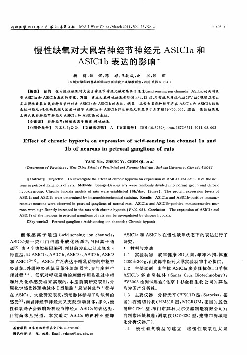
Ef e t o hr ni y o i n e pr s i n o c d s n i o h n l l n f c f c o c h p x a o x e s o fa i 。 e sng i n c a ne a d a 1 f ne r ns i e r s lg ng i n f r t o u o n p t o a a lo s o a s b
表 达 神 经元 ; 性 缺 氧 组 大 鼠岩 神 经 节 ASC a ASC b阳性 神 经 元 明显 多 于正 常 组 ( < O 0 ) 结 论 慢 性 缺 氧 能 慢 Il 和 I1 P . 5 上 调 大 鼠岩 神 经节 神 经元 AS C a和 A I 1 I1 S C b的表 达 。
[ src】 Obetv To iv siaet eefc f h o i h p xao x rs ino I aa d AS Cl ften u Ab ta t jcie n e t t h fe t rnc y o i ne p e so f g oc AS Cl n I bo h e —
YANG n。ZHENG Yu Yi 。CHE Qi t l N 。e a
( p rme t f P y ilg WetC ia S h o f P elnc l n r n i Me iie ih a n v ri De a t n h s o y, s h n c o l r ci i d Fo e s d cn .S c u n U ie s y.C e g u 6 0 4 ) o o o aa c t h n d 1 0 1
AS C1 n I b we e d t r n d b I a a d AS C1 r e e mie y i mmu o it c e c l t i i g Re ut AS Cl n I b p st e i n h so h mia an n . s sl s I a a d AS C1 - o i v mmu o i n— r a t e n u o s we eo s r e n p t o a a g i n fn r 1r t . AS C1 n I b p st e i e c i e r n r b e v d i e r s lg n l s o o ma a s v o I a a d AS Cl — o ii mmu o e c ie n u v n ra t e — v
免疫组化知识普及、二抗免疫组化试剂盒

Anti-Mouse, Anti-Rabbit
包装规格 种类 品名
描述
包含
15ml 60ml
HRP UV Quanto HRP ( DAB DAB )
UV Quanto HRP
DAB Quanto (Stable for Peroxide Block, UV-Block, Amplifier , HRP
免疫组化知识普及、二抗免疫组 化试剂盒
优宁维缪娓
直接法
• 直接法是将酶(如HRP)标记在特异性一抗上,然后用酶 标记抗体直接与相应抗原特异性结合,形成抗原-抗体HRP复合物,最后用酶底物显色。
• 优点:简单、步骤少、省时、特异性高。 • 缺点:敏感性差
间接法
• 我这边的讲的间接法就是酶标记在二抗上, 再与一抗反应,然后进行显色反应。
60ml 125ml
Storage: 2-8℃,18months
Permanent Fast permanently mounted red
Tablet Fast red aqueous mounted
DAB Quanto 1 2
DAB Quanto 1 DAB Plus 2
DAB Quanto 优势: Long stability 稳定性强,混合后可在室温保 存一周, 2-8 度保存2 周
Antibody
n 30min
n
20min
s
s
e
e
R i s MOM Polymer n 10min s e
R
i s DAB Quanto n 5min
s
e
R i s Counterstain n s e
IHC Staining-MOM Detection System
常用免疫组化试剂介绍
常用免疫组化试剂介绍概述免疫组化(IHC)是一门通过检测特定抗原在组织切片中的表达程度来揭示组织形态和细胞化学等方面信息的技术。
由于IHC是一种定性分析技术,所以该方法需要与定量技术(如分子生物学)结合使用,从而得到更准确的数据。
IHC涉及到多种试剂,其标记原理和表现形式各异,能够检测的抗原种类也不同。
本文将对常用的IHC试剂进行介绍,以此为指导,让大家更好地了解和应用IHC技术。
常用的免疫组化试剂抗原修复试剂抗原修复试剂(Antigen Retrieval)用于在组织样本中使细胞内蛋白与抗体亲和性和特异性增加。
这种试剂可通过热处理(如高压)或化学处理(如酶样蛋白和酸性处理)来复原被检测抗原的结构和功能。
常用的抗原修复试剂包括:EDTA、Tris、Citrate等。
原位杂交试剂原位杂交试剂(In Situ Hybridization)可检查DNA或RNA分子的特异性结合情况。
这种试剂可将RNA等分子与特定热稳定的蛋白质结合,用于检测基因突变等。
常用的原位杂交试剂包括:流式胶体金试剂、脉冲场凝胶电泳(PFGE)、双链RNA原位杂交等。
细胞计数试剂细胞计数试剂(Cell Counting)用于获得细胞数量或结构变化。
这种试剂通常用于确定某一类细胞的数量、细胞计数和分离、细胞生长和分裂的应用。
常用的细胞计数试剂包括:皮质醇、14C-labeled thymidine等。
细胞信号转导试剂细胞信号转导试剂(Cell Signaling)用于研究细胞分子级别的通讯系统,并采取措施以确定物质在细胞内的作用机理。
常用的细胞信号转导试剂包括:抗体、试剂原和阻断类似物等。
同型抑制剂同型抑制剂(Isotype Control)用于在IHC实验中作为实验设计的对照组,以检测所观察的抗原与探测剂结合的特异性表现。
常用的同型抑制剂包括:IgG1、IgG2a和IgG2b等。
情况控制试剂情况控制试剂(Condition Control)用于确定IHC试验的最佳条件,包括温度、缓冲液、酸碱程度等。
- 1、下载文档前请自行甄别文档内容的完整性,平台不提供额外的编辑、内容补充、找答案等附加服务。
- 2、"仅部分预览"的文档,不可在线预览部分如存在完整性等问题,可反馈申请退款(可完整预览的文档不适用该条件!)。
- 3、如文档侵犯您的权益,请联系客服反馈,我们会尽快为您处理(人工客服工作时间:9:00-18:30)。
大鼠ASIC1A ASIC1A 免疫组化试剂盒免疫组化试剂盒
该试剂盒以HRP 标记的链霉亲和素复合物(HRP Streptavidin Conjugate,HRP-SA)为基础,可用于检测细胞、组织内的特异性ASIC1A 抗原。
该试剂盒具有灵敏度高、特异性强、定性定位准确、背景清晰。
在所用的ASIC1A 一抗与相应靶抗原结合后,用生物化二抗与一抗特异性结合,最后加入HRP-SA,形成抗原—特异一抗—生物素化二抗—HRP-SA 复合物,显微镜下观察成像。
试剂盒所含试剂试剂盒所含试剂::
试剂A 通透液:0.1% Triton-X 100 10 mL(选用)
试剂B 封闭缓冲液(封闭用) 20 mL
试剂C (原装进口分装)已稀释的即用型ASIC1A 一抗(2.5ml)
试剂D (原装进口分装)生物素化羊抗兔IgG 1支
(浓度1.5 mg/mL,稀释比为1:300~1:500)50 μL+抗体稀释液20ml
试剂E HRP-SA 复合物1支(浓度1 μM,稀释比1:50~1:200)100 μL 试剂F DAB 显色液 5ml
用户自备试剂用户自备试剂::
1. 10mM TBS(pH7.2~7.4)
三羟基氨基甲烷1.21g
氯化钠7.6g
加蒸馏水800mL,浓盐酸调pH 值至7.2~7.4,最后定容至1000mL
TBS-T:TBS+Tween 20(0.05%体积比)
2.抗原修复液(依检测抗原不同而选择不同的修复液)
10mM pH6.0 柠檬酸缓冲液
柠檬酸0.38g
柠檬酸三钠2.45g
加蒸馏水900mL,浓盐酸调pH 值至6.0,最后定容至1000mL
或:0.5M EDTA 修复液(pH8.0)
EDTA·2H2O 186.1g
柠檬酸三钠2.45g
加蒸馏水700mL,用10mM NaOH 调pH 值至8.0,最后定容至1000Ml
3. 缓冲甘油封固剂10 mL
4. Tween 20 5 mL
石蜡包埋组织切片免疫染色石蜡包埋组织切片免疫染色
实验步骤实验步骤((建议方案建议方案):):
石蜡包埋组织切片3~4μm 厚度
1.烤片: 将待做切片置于切片架上,于60℃恒温烤箱中至少烤1 hr;
2.脱蜡: 切片放入盛有二甲苯的容器中脱蜡3次(即二甲苯Ⅰ、Ⅱ、Ⅲ),每次10 min;
3.水化: 切片经下行酒精水化,无水乙醇5min,95%乙醇2次(每次2min),85%乙醇2 min;75%乙醇2min,自来水冲洗,ddH2O 洗2×2min;
4.抗原修复: 根据抗体说明书推荐方法进行抗原修复,常采用高压、微波(温度达到98~100℃)或酶消化修复法,室温自然冷却,自来水冲洗,ddH2O 洗2×2min,TBS 洗涤(2×2min)(具体修复方法见附1)* 注:有些抗原勿需修复,直接进入第5步封闭。
5.封闭: 滴加试剂B,37℃湿盒孵育30 min;
6.加一抗:滴加用试剂C(即用型一抗),37℃湿盒孵育2 hr 或4℃过夜;
7.洗涤: TBS-T 洗涤(3×5 min);
8.封闭: 滴加试剂B,37℃湿盒孵育10 min;
9.加二抗: 滴加用抗体稀释液稀释的生物素化二抗(试剂D),37℃湿盒中孵育30 min;
10.洗涤: TBS-T 洗涤(3×5 min);
11.封闭: 滴加试剂Tween 20,37℃湿盒孵育封闭20 min;
12.加HRP-SA: 滴加用抗体稀释液稀释的试剂E(1:50~200,终浓度5~20 nM),37℃湿盒中孵育30 min;
13.洗涤: TBS-T 洗涤(3×5 min),TBS 洗涤(2×5 min);
14.显色:应用DAB 溶液(试剂F)显色;
15.复染:自来水充分冲洗,复染,脱水,透明;
16.封片: 待组织标本干后,用试剂缓冲甘油封固剂封片;
17.观察成像: 显微镜下观察成像。
注意事项注意事项::
1. 修复后缓冲液须自然冷却,自来水冲洗后方能把切片取出,骤冷有可能导致结晶或抗原封闭。
2. 缓冲液的量必须保证所有切片都能浸泡到,用过的柠檬酸缓冲液不能反复使用。
3. 若试剂为微量浓缩液,用前应低速离心,将内盖和管壁附着的溶液离到底部。
4. 封片前一定要换用TBS 充分洗涤,以便洗去组织上残留的Tween 20,否则会影响结果观察。
5. 如须复染细胞核,则在封片前复染或直接采用含有染核试剂的封片剂进行封片。
附1:
抗原修复方法抗原修复方法
常用抗原修复液:柠檬酸缓冲液(0.01M pH6.0)、EDTA 抗原修复液(pH8.0或9.0)等等。
一、酶消化修复法酶消化修复法
切片脱蜡水化处理,TBS 冲洗,在组织上滴加胃蛋白酶或胰蛋白酶,37℃孵育20~30min 后TBS 冲洗即可。
二、微波抗原修复法微波抗原修复法
微波盒中加入抗原修复液微波加热至沸腾,将脱蜡水化后的切片置于耐高温塑料切片架上,放入已沸腾的缓冲液中,中档或高档继续微波10~15min,取出微波盒冷却至室温后,自来水冲洗,取出切片。
因不同微波炉微波处理时间存在差异,须自行调整。
三、直接高压抗原修复法直接高压抗原修复法
取修复液于不锈钢高压锅中加热至沸腾,将组织切片置于耐高温切片架上,修复液沸腾后放入切片架,盖上锅盖,待喷气后计时1.5~2.5min 即可脱离热源,自然冷却至室温后自来水冲洗,取出切片。
此方法适用于较难检测或核抗原的修复。
四、隔水式高压抗原修复法隔水式高压抗原修复法
不锈钢高压锅中加自来水加热至沸腾,微波盒中加入修复液于微波炉中加热至沸腾,将切片放入微波盒中,再将微波盒放入高压锅中盖上锅盖,待喷气后计时4~8 min 即可关闭热源,自然冷却至室温后自来水冲洗,取出切片。
此方法适用于较难检测或核抗原的修复。
