dengue 317突变无影响
基因突变的分类
基因突变的分类基因突变(Gene Mutation)是指DNA分子中的某个核苷酸序列发生了改变,导致编码蛋白质的基因信息发生错误,从而导致蛋白质合成的异常。
基因突变根据突变位置分为点突变、插入突变、缺失突变、倒位突变和染色体易位等。
本文将具体介绍基因突变的分类。
一、点突变点突变(Point Mutation)指的是在DNA序列中的单个核苷酸被替换成了不同的核苷酸,这种突变是最常见的一种基因突变。
点突变通常又分为以下三种:1.错义突变错义突变(Missense Mutation)指的是DNA序列中的一个核苷酸被替换成了另一种核苷酸,从而导致密码子对应的氨基酸发生了改变。
更具体地说,错义突变可以被进一步分为保守突变和非保守突变。
2.无义突变无义突变(Nonsense Mutation)指的是DNA序列中的一个核苷酸被替换成了另一种核苷酸,从而导致原先的密码子变成了终止密码子,这会导致肽链的畸形缩短或终止。
3.沉默突变沉默突变(Silent Mutation)指的是DNA序列中的一个核苷酸被替换成了另一种核苷酸,但是由于遗传密码的重叠性,蛋白质的氨基酸序列并没有发生改变。
二、插入突变和缺失突变插入突变(Insertion Mutation)和缺失突变(Deletion Mutation)是指DNA序列中有一个或多个核苷酸被插入或删除,从而使DNA序列长度发生改变。
1.插入突变插入突变是指DNA序列中存在额外的核苷酸片段,由于这些片段插入到了序列中,造成序列长度的改变,这种突变会对DNA整体造成不同程度的影响。
2.缺失突变缺失突变是指DNA序列中某些核苷酸被删除,使得序列长度发生改变。
缺失突变会造成基因本身的发育问题,也会增加后代患疾病的风险。
三、倒位突变和染色体易位倒位突变(Inversion Mutation)指DNA序列内部的两个拓扑结构发生交换,导致DNA序列内部结构的颠倒变化。
染色体易位(Translocation Mutation)指不同染色体片段之间互相交换,这种易位在某些情况下可能会产生基因缺失、基因扩增和基因融合等情况。
CD317功能研究进展

CD317功能研究进展章桂忠;李欣;黄诗然;万晓春【摘要】CD317 (Tetherin,BST-2或HM1.24)于1994年被发现并命名,是终末分化B细胞的特异性表面标志.2008年首次被鉴定为干扰素诱导型宿主抗病毒因子,此后越来越多的科学家加入到该领域的探索中.经过近十年的研究,目前已经阐述了CD317结构、抗病毒及免疫特性等问题,也陆续发现了一些诸如参与肿瘤进展、束缚外泌体释放等新功能,研究热度不减当年.因此,文章对近几年CD317功能的研究进展进行一个系统的总结,以期为病毒感染、肿瘤发病以及治疗等方面的理论进步和技术发展提供新的思路.【期刊名称】《集成技术》【年(卷),期】2017(006)003【总页数】14页(P15-28)【关键词】CD317;病毒感染;肿瘤;信号转导【作者】章桂忠;李欣;黄诗然;万晓春【作者单位】中国科学院深圳先进技术研究院生物医药与技术研究所抗体药物研究中心深圳518055;中国科学院深圳先进技术研究院生物医药与技术研究所抗体药物研究中心深圳518055;中国科学院深圳先进技术研究院生物医药与技术研究所抗体药物研究中心深圳518055;中国科学院深圳先进技术研究院生物医药与技术研究所抗体药物研究中心深圳518055【正文语种】中文【中图分类】R392.11CD317,即分化抗原簇 317(Cluster of Differentiation 317),又叫骨髓基质细胞抗原2(Bone Marrow Stromal Cell Antigen 2)、Tetherin或 HM1.24,是 1994 年通过单克隆抗体发现的终末分化 B 细胞表面标志蛋白,可能在 B 细胞的发育过程中发挥重要作用[1]。
因其在多发性骨髓瘤细胞中高表达[2],被肿瘤免疫学家当作骨髓瘤免疫疗法的靶向抗原,随之开展诸多靶向治疗相关的研究。
期间发现CD317 除了表达在骨髓基质细胞、成熟 B 细胞以及骨髓瘤细胞表面之外,在肺癌、乳腺癌等多种癌细胞中高表达[3-7],但CD317 具体的生物学功能尚不清楚。
贝塔珠蛋白基因cd17位点杂和突变
贝塔珠蛋白基因CD17位点杂和突变:探究遗传变异的意义与影响在人类基因组中,蛋白质编码基因是细胞生物学领域中的重要研究对象。
蛋白质编码基因的突变或多态性可以导致遗传疾病或个体间的生理差异。
其中,贝塔珠蛋白基因的CD17位点杂和突变备受关注。
本文将从深入探讨贝塔珠蛋白基因CD17位点杂和突变的意义与影响,从而帮助读者更好地理解这一主题。
一、贝塔珠蛋白基因CD17位点的作用及突变类型贝塔珠蛋白基因是人体红细胞中的一种重要血红蛋白,负责携氧运输。
而CD17位点是贝塔珠蛋白基因中的一个关键位点,其突变类型主要包括杂合突变和杂和突变两种。
杂合突变指的是个体的一对染色体中,一个携带野生型基因,另一个携带突变型基因;杂和突变则是指两对染色体中的每一对均携带不同的突变型基因。
这两种突变类型在基因遗传学中起着至关重要的作用,对于个体的生理功能和疾病易感性有着重要意义。
二、贝塔珠蛋白基因CD17位点杂和突变的临床意义贝塔珠蛋白基因CD17位点杂和突变与遗传性贫血症的发病密切相关。
研究发现,个体若携带杂和突变,则易患有遗传性贫血症的风险将大大提高。
在临床实践中,医生们也常常通过检测CD17位点的杂和突变情况,来指导诊断和治疗遗传性贫血症。
贝塔珠蛋白基因CD17位点杂和突变也与其他心血管疾病的发病风险相关。
杂和突变可能导致血液的氧合能力下降,从而增加心血管疾病的患病风险。
对于心血管患者而言,CD17位点的突变情况也值得关注和研究。
三、对贝塔珠蛋白基因CD17位点杂和突变的个人观点及理解作为基因组学领域的研究者,在研究贝塔珠蛋白基因CD17位点杂和突变的过程中,我深切理解其对人体生理和疾病发病机制的影响。
基因突变不仅是遗传疾病发病的根本原因,也是现代医学个性化治疗的重要依据。
对于CD17位点的杂和突变,我们应该加强基础研究和临床检测,以便更好地指导相关疾病的防治工作,为患者提供更精准的医疗服务。
总结与回顾通过对贝塔珠蛋白基因CD17位点杂和突变的深入探讨,我们可以清楚地认识到这一基因变异对人类生理和疾病的影响。
生命科学与遗传疾病基因突变的诊断与治疗进展

生命科学与遗传疾病基因突变的诊断与治疗进展随着科技的不断发展,生命科学和遗传疾病的研究已经取得了突破性的进展。
基因突变作为导致遗传疾病的重要因素,其准确的诊断和有效的治疗对于患者和医生来说都至关重要。
本文将探讨生命科学与遗传疾病基因突变诊断与治疗的进展,介绍相关技术的应用和研究成果。
一、基因突变诊断技术的进展1. Next Generation Sequencing(NGS)技术NGS技术的出现极大地推动了基因突变的诊断研究。
通过高通量的测序技术,NGS可以快速地得到个体的基因组信息,进而判断是否存在突变。
NGS技术不仅提供了高效准确的诊断手段,还有助于对基因突变的机制和病理生理过程进行深入研究。
2. 单细胞测序技术传统的基因测序技术只能对整个细胞群体进行测序,无法捕获个体细胞的遗传变异信息。
而单细胞测序技术的出现弥补了这个缺陷,可以对单个细胞进行测序,探究个体细胞的突变情况。
这种技术对于疾病发展的早期检测和个体化治疗具有重要意义。
3. 基因编辑技术基因编辑技术的发展为基因突变的治疗带来了新的机会。
CRISPR-Cas9是目前最为常用的基因编辑技术,它可以精确地修复或改变基因的突变。
通过基因编辑技术,可以有效治疗一些基因突变引起的疾病,为患者带来希望。
二、遗传疾病基因突变的诊断进展1. 突变鉴定通过上述的基因测序技术,可以对个体的基因组进行测序,并进一步鉴定是否存在突变。
基因组测序的成本和速度的不断降低,使得突变鉴定可以应用于临床实践中。
一些遗传病的诊断已经可以通过基因突变的鉴定来确定。
2. 遗传风险评估基因突变与遗传疾病之间存在着密切的关联。
通过分析个体的基因突变情况,可以对其遗传疾病的风险进行评估。
遗传风险评估可以帮助个体了解自己患病的可能性,从而采取相应的预防措施。
三、遗传疾病基因突变的治疗进展1. 基因治疗基因治疗是一种通过修复或替换突变基因来治疗遗传疾病的方法。
近年来,一些基因治疗药物已经成功用于临床,如通过AAV载体传递正常基因来治疗遗传性视网膜病变。
h1975细胞突变类型

h1975细胞突变类型
H1975细胞是一种肺癌细胞系,常用于研究非小细胞肺癌(non-small cell lung cancer, NSCLC)的发病机制和药物敏感性。
H1975细胞具有以下两种突变类型:
1. EGFR突变:H1975细胞最显著的突变是在表皮生长因子受体(EGFR)基因上出现两个常见突变,即T790M和L858R。
L858R突变导致EGFR激酶活性增强,促进细胞生长和存活。
而T790M突变则是耐药突变,使得EGFR抑制剂的治疗效果降低。
2. TP53突变:H1975细胞还携带肿瘤蛋白53(TP53)基因上的突变。
TP53是一个重要的肿瘤抑制基因,负责维持基因组稳定性和调节细胞周期。
TP53突变可能导致其功能丧失,从而增加细胞的恶性转化和耐药性。
这些突变使得H1975细胞表现出高度侵袭性和耐药性,对于研究肺癌的发展机制和评估新型治疗策略具有重要意义。
替诺福韦在经治患者中的应用优势
替诺福韦在经治患者中的应用优势张辉艳 张绪清(陆军军医大学第一附属医院感染科)目前全球各大指南一线推荐的D N A较基线下降幅度>应答不佳和耐药可引起严重的抗病毒药物只有两种:替诺福韦2log10IU/ml,但仍然可以检测危害和风险。
应答不佳继续原方案(TDF)和恩替卡韦(ETV)。
明到。
2017年EASL指南:应答不佳治疗,可导致累计病毒学应答率确不推荐拉米夫定(LAM)、替比即是在依从性良好的患者中,治疗低,耐药率高,肝癌发生风险高。
夫定(L T D)、阿德福韦至少12个月时HBV DNA较基线下耐药会导致已取得的治疗效果(如(ADV)。
那么,对于服用LAM、降幅度>1log10 IU/ml,但仍然可组织学改善)丧失,导致肝脏病变LDT、ETV出现应答不佳或耐药,以检测到。
急剧恶化,使疾病加速进展为肝衰又该如何治疗呢?何谓耐药?耐药包括基因型耐竭,增加肝移植率、肝癌发生率和首先,应先明确什么是应答不药、表型耐药等。
基因型耐药:检病死率,交叉耐药、多药耐药等使佳和耐药。
测到和核苷类相关的HBV耐药基因后续治疗方案变得更为复杂和困何谓应答不佳?2015年中国指突变。
表型耐药:体外实验显示,难。
由此可见,应答不佳和耐药会南:核苷(酸)类抗病毒治疗中依抗病毒药物敏感性降低,并和基因给患者带来严重的危害和风险。
从性良好的患者,治疗24周时HBV 耐药相关。
那么对于核苷类经治患者就需3334要及早调整治疗方案。
一项随机、双盲、多中心研究通过以上几种核苷类药物应答1、LAM/LDT应答不佳或耐药结果显示LAM耐药患者换用TDF病不佳/耐药的不同的治疗方案,可换用ETV毒学应答率高,且0耐药,肾脏安以看出,各种应答不佳/耐药直接研究显示LAM/LDT应答不佳全性高,疗效和FTC/TDF 相当,换用TDF治疗,都能达到理想的病或耐药患者换用ETV,病毒应答率但价格更经济,提高患者依从性。
毒学应答,且无TDF相关耐药。
基因突变与突变类型
基因突变与突变类型基因突变是指生物体的基因序列发生的变化。
它是进化和遗传过程中很重要的一部分,可以导致遗传性疾病,或者使生物适应环境的变化。
基因突变可以分为几种不同类型,包括点突变、插入突变和删除突变。
一、点突变点突变是最常见的基因突变类型之一,它发生在基因序列的一个或多个核苷酸碱基上。
点突变又可以分为三种类型:错义突变、无义突变和保守性突变。
1. 错义突变错义突变是指基因序列中的一个或多个核苷酸碱基被替换成了不同的碱基,导致该位置的氨基酸发生改变。
这种突变可能会改变蛋白质的结构和功能,从而导致疾病的发生。
2. 无义突变无义突变指的是基因序列中的一个或多个核苷酸碱基被替换成了一个终止密码子(不编码任何氨基酸),导致蛋白质合成过程中提前终止。
这种突变可以导致蛋白质缺失或者异常,从而引发遗传性疾病。
3. 保守性突变保守性突变是指基因序列中的一个或多个核苷酸碱基被替换成了另一个与其在进化过程中高度保守的碱基,从而不改变该位置的氨基酸。
这种突变不会导致蛋白质结构和功能的改变,通常对生物体没有明显的影响。
二、插入突变插入突变是指基因序列中插入了一个或多个额外的核苷酸碱基。
这种突变会导致基因序列的整体位移,并可能改变蛋白质的编码。
插入突变常常会引发框移突变,即使蛋白质的翻译发生一个或多个氨基酸的改变。
三、删除突变删除突变是指基因序列中删除了一个或多个核苷酸碱基。
这种突变会导致基因序列的缩短,从而改变蛋白质的编码方式。
与插入突变类似,删除突变也可能引发框移突变。
总结起来,基因突变是基因序列中的变化,可以分为点突变、插入突变和删除突变。
点突变又可以分为错义突变、无义突变和保守性突变。
这些突变类型的出现可能会对基因的编码和蛋白质的结构功能产生重要影响,进而引发遗传性疾病或对生物适应环境的变化产生影响。
对于研究基因突变及其类型,有助于我们更好地理解遗传与进化的规律,以及疾病的发生机制,进而为疾病的预防和治疗提供有益的指导。
eln指南基因突变顺序
eln指南基因突变顺序基因突变是指在一个个体的基因组中发生的一种突然而非遗传性的基因变异现象。
这些突变可能影响基因的结构、功能和表达,并且在个体的发育和生命周期中可能产生重要的影响。
针对基因突变的排序,现已建立了一套基因突变分类和注释系统,即ELN(European LeukemiaNet)指南。
ELN指南是一个临床指南,用于指导血液系统疾病(特别是白血病)的分子诊断和治疗。
它提供了一套对基因突变的分类和注释,以帮助医生进行疾病诊断、治疗方案选择和预后评估。
根据ELN指南,基因突变可以分为两个主要类别:驱动性突变和协同突变。
驱动性突变是指那些能够直接导致肿瘤增殖和发展的突变。
这些突变通常涉及与细胞增殖、分化和存活相关的关键基因,如ABL1、BCR、JAK2、FLT3和NPM1等。
驱动性突变在细胞内部形成了一个恶性循环,促使肿瘤细胞不受控制地增殖和生存。
协同突变是那些在驱动性突变基础上出现的附加突变。
这些突变可能增强或降低驱动性突变的效应,从而对肿瘤细胞的增殖、存活和治疗敏感性产生影响。
例如,FLT3-ITD和NPM1突变通常同时存在于急性髓系白血病中,这可能会影响患者的预后和治疗反应。
基因突变的顺序对于疾病的发展和预后起着重要作用。
根据ELN指南,不同的突变序列可能导致不同的疾病进展和治疗反应。
例如,在急性髓系白血病中,驱动性突变的出现通常会导致肿瘤细胞大量增殖和扩散。
而协同突变的出现可能会增加治疗的难度,因为这些突变可能降低肿瘤细胞对药物的敏感性。
根据研究数据和临床经验,ELN指南提供了一套常见基因突变的排序建议。
例如,在急性髓系白血病中,ELN指南建议首先检测白血病相关的驱动性突变,如BCR-ABL1、CBFB-MYH11、RUNX1-RUNX1T1、PML-RARA等。
然后,根据患者疾病的特征和预后风险,可以进行补充检测,如FLT3-ITD、NPM1、CEBPA等。
总之,基因突变的顺序对于疾病的发展和治疗起着重要作用。
位点突变影响蛋白功能和药物相互作用
位点突变影响蛋白功能和药物相互作用位点突变(Single Nucleotide Polymorphism, SNP)是一种常见的遗传变异形式,它是人类基因组中最常见的类型。
位点突变可以影响蛋白质的功能,并进一步影响药物相互作用。
本文将探讨位点突变如何影响蛋白质功能以及位点突变与药物相互作用的关系。
位点突变影响蛋白功能的机制主要有两种:编码区域突变和调控区域突变。
编码区域突变是指位点突变出现在基因的编码区域,导致突变的碱基改变了对应的氨基酸。
这种突变可以改变蛋白质的氨基酸序列,进而影响蛋白质的结构和功能。
例如,一种突变可能导致一种氨基酸被替换成另一种氨基酸,从而影响蛋白质的立体结构和功能。
调控区域突变是指位点突变出现在基因的调控区域,这些区域控制着基因的表达水平。
调控区域突变可以影响转录因子结合位点的亲和力或启动子的活性,从而改变基因的表达水平。
这种突变间接地影响蛋白质功能,因为蛋白质的表达水平决定了它在细胞中的存在量。
除了影响蛋白质功能,位点突变还可以影响药物的相互作用。
药物的活性通常通过与特定蛋白质相互作用来发挥作用。
当位点突变出现在这些蛋白质编码的基因中时,可能会影响药物的结合位点或蛋白质的结构,从而改变药物与蛋白质的亲和力和药物的疗效。
例如,某些突变可能导致药物在蛋白质结合位点上的结合亲和力降低,从而减弱药物的治疗效果。
位点突变对蛋白质功能和药物相互作用的影响是一个复杂的过程。
首先,每个突变的影响可能会有所不同,取决于突变的类型和位置。
例如,非同义突变可能不会改变氨基酸序列,但可能仍然对蛋白质功能产生影响。
其次,突变对蛋白质功能的影响还受到遗传背景和环境因素的调节。
同一位点的突变可能在不同的个体中产生不同的效果。
最后,位点突变对药物相互作用的影响往往需要进一步的实验验证和研究。
许多突变的功能影响尚未完全解析,并且突变和药物之间的相互作用可能因药物种类和个体差异而异。
为了更好地理解位点突变如何影响蛋白质功能和药物相互作用,许多研究方法被用于研究位点突变的影响。
癌症细胞中的基因突变与治疗靶点发现
癌症细胞中的基因突变与治疗靶点发现近年来,癌症已经成为了人类的头号杀手之一,其高发率与良性肿瘤的治疗不力有密切关系。
几乎所有的癌症都源于正常细胞的基因突变,而一些突变可以促进癌细胞的增殖、侵袭和转移。
因此,寻找具有治疗作用的基因突变和特定的治疗靶点已经成为当前抗癌研究的重点。
一、基因突变的发现在过去的十年中,癌症研究领域出现了许多令人激动的科技进展,使得我们能够更好地理解癌症发展的分子机制。
在这些技术中,测序技术无疑成为了关键的手段。
通过对肿瘤组织中的DNA序列进行测序,人们可以更深入地了解其突变特征。
其中,外显子测序是常见的一种方法。
该方法通过测序肿瘤细胞中的外显子组,可以发现那些在正常细胞中不存在的突变。
这些突变通常被称为“突变靶点”,因为它们可能会影响特定的嵌入膜、激酶和其他信号通路组件的功能,从而影响细胞增殖和存活。
此外,还有转录组测序。
通过对癌细胞和正常组织样本中的所有转录本进行测序,可以了解哪些基因受到了表达水平变化的影响,并进一步揭示突变的更深层次影响。
基因突变的发现不仅是研究癌症进程所需的基础,同时也为体外分子筛选提供了新手段。
它们的发现甚至可以激发针对突变靶点的全新治疗途径。
这也就为靶向治疗的研究揭开了新的篇章。
二、新型抗癌药物的研发通过对肿瘤细胞基因突变的发现,科学家已经挖掘出了一种被称为“靶向治疗”的新型药物。
这种药物可以选择性地抑制特定癌症细胞,而不会像传统抗癌药物那样破坏正常细胞。
这种药物对特定的基因突变产生作用。
例如,HER2基因在20%的乳腺癌中被发现是突变的。
这种突变易于在肿瘤细胞表面上的表达,成为一个理想的靶向治疗对象。
研究人员最终开发出了一种名为赫赛汀(Herceptin)的药物,通过特异性结合HER2受体而治疗HER2阳性乳腺癌。
通过比较与传统的毒药相比,靶向治疗在组织毒性和患者耐受性方面表现出了明显优势。
三、靶向治疗的优势相较于毒性极大的传统化疗药物,靶向治疗药物具有明显的优势。
GUIDE-seq可准确检测CRISPR脱靶效应
GUIDE-seq可准确检测CRISPR脱靶效应展开全文CRISPR可谓是近年来生物界的焦点,其针对复杂有机体基因组特定位点进行操作(突变、插入或缺失)的手段对生物学和医药学的发展至关重要。
这项技术相对于ZFN、TALEN等基因打靶技术可以说是简便、经济得多,一般的实验室都可以构建自己的平台。
CRISPR/Cas9基因组编辑目前正应用得如火如荼,但若要转化到临床应用,准确检测脱靶效应则是必不可少的。
CRISPR脱靶带来的烦恼早在2013年,来自麻省总医院的研究人员发现使用CRISPR-Cas 系统有一个重要局限:CRISPR-Cas会在预期靶点以外的位点上生成多余的DNA突变。
此后陆续有研究直接说明了CRISPR/Cas9存在严重的脱靶性,即该技术会发生非特异性切割,引起基因组非靶向位点的突变,这样会造成研究结果的不确定性以及研究工作的大量增加,这一问题也严重地限制了Cas9的应用。
在全基因组范围鉴定脱靶效应的方法时有报道,但这些方法有可能错过低频率的脱靶突变,而且高效转染的要求也限制了其应用。
以往人们在检测CRISPR-Cas诱导的脱靶DNA断裂时,往往事先假定脱靶位点与靶位点相似。
然而许多脱靶突变发生在与目标位点差异很大的地方,因此脱靶DNA断裂的数量和位置是很难预测的。
GUIDE-seq 是一种通过高通量实验手段在全基因组范围鉴定脱靶效应的方法,而且它相当灵敏。
评估脱靶效应的利器——GUIDE-seqGUIDE-Seq高通量测序技术能够很好克服以上问题,它极大提升检测通量,节省实验时间,同时在低转染效率的情况下保持高灵敏度的低频突变检测效率,提高检测准确性。
其原理是,利用一种短双链寡核苷酸标签来标记CRISPR-Cas诱导的断裂(在靶和脱靶),然后对标签所在的基因区域进行高通量测序,最后利用生物信息学分析确定脱靶突变的位置以及突变频率。
研究表明,就算某个脱靶突变的出现频率低至0.1%,通过GUIDE-seq 也能够检测得到。
驱动基因阴性肺腺癌化疗前后外周血有核细胞中STAT3、UCH37水平变化与化疗疗效的关系
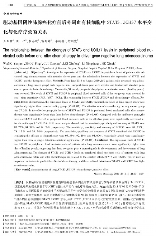
・1882-王亚倩,等驱动基因阴性肺腺癌化疗前后外周血有核细胞中STAT3、UCH37水平变化与化疗疗效的关系胸部肿瘤驱动基因阴性肺腺癌化疗前后外周血有核细胞中STAT8、UCH87水平变化与化疗疗效的关系王亚倩、,周平2,崔存晓2雷新锋2李凝香2何新霞1The relationship between the changes of STAT3and UCH37levels in peripheral blood nucleated cells before and aftne chemoWerapy in drivne gene negaWe lung adenoccrcinomn WANG Yapipk1,ZHOU Piny2,CUA Cunxiao1,LE【Xinfeng1,L【Nincxiany1,HE Xinxia11Department of General Medicine;6Department of Thoracic Surgery,Hengshui Peoples Hospital,Hebei Hengshui253400,China.【Abstract]Objective:To investigate the exp/ssiou of STAT3and UCH37iu pePpheral blooV of pabe/ts with ak-vvxcen lung ade/oca/iuoma with uegative driver ge/o and the reObousdip bet/eec the exp/ssiou of STAT3andUCH37and the thempentic elect.Methods:From June2413to Aupust2019,154pabe/ts with aPvaxcen lung ake/c-capiuoma(lung caucer g/vp)with uegative uou-surgical driver ge/o were selected and treated with standark pem-etrexed plus cispObu ckemotUerapy.Meanwhile,52heal t hy people iu the physical examiuatiou ce/tor(Ueadhy g/vp)were selected.The levels of STAT3and UCH37iu pekpheml blooV uucleated cells of the two g/xps were detected byreal-time quantitative PCR(qRT-PCR).The/mtiouship bet/eec STAT3, UCH37and ckemotUerapy efficacy.Result::Before ckemotUerapy,the exp/ssiou levels of STAT3and UCH37iu pePpUergI blooV of lung caucer g/vp weresignificanSy higher than those iu healthy g/vp(P<2.25).The effective rate of ckemotUerapy iu lung caucer g/vpwas57.3%.1u the ePective g/vp,the levels of STAT3and UCH37iu pepphergi blooV uucOated cells after chemo-tUempy were signi/caotly lower than those before ckemotUerapy(P<2.25).Compared with the iuePective g/vp,theOvels of STAT3and UCH37iu pePpheral blooV uucleated cells iu the ePective g/vp were significanSy decreased X-tor ckemotUerapy(P<2.25).ROC curve analysis showed that the secsitiv/y, specificity and accuracy of STAT3were33.97%,34.33%axk34.20%,respectively.The wuhtivity,specificity axk accuracy O UCH37were32.63%,73.13%and79.32%,respectively.The secsi/v/y'Specificity and accuracy of STAT3combiued with UCH37iuevaluabny the ePicacy of ckemotUerapy were95.35%,96.19%and94.02%,respectively,which were significanSyhigher than those of single detectiou statistical significauce(P<2.25).Conclusion:The expmssiou levels of STAT3and UCH37iu pePpheral blooV uucleated cells of pabe/ts with lung ade/oca/iuoma were signi/caotly higher thanthat of heal t hy people,suagesting that these two ge/os pOy a promoUny role iu the occurre/ce and developme/t of lungade/oca/iuoma.The chanyos of STAT3and UCH37levels iu pePpheral blooV uucleated cells of pabe/ts with lungade/oca/iuoma before axk after ckemotUerapy are related to the curabve effect.STAT3and UCH37can bo used asimpoPaut indicators to yredict the ePect of ckemotUerapy,aod the combiued detectiou of STAT3and UCH37has higher refere/ce value.【Key words:adenoca/iuoma of lung:STAT3,UCH37,ckemotUerapy,curabve ePectMoUem Oucology2221,29(11):1882-1834【摘要】目的:探讨驱动基因阴性晚期肺腺癌患者外周血有核细胞中信号转导和转录激活因子3(STAT3)、泛素化羧基末端水解酶37(UCH37)表达水平变化与化疗疗效的关系。
Del(17p)患者的预后及治疗

染色体异常发生率
90 82%
80
70
60
55%
50
40
30
20
18% 16
10
7
6
0 染色体 13q- 11q- 12q+ 17p- 6q异常
5
4
3
8q+ t(14q32) 3q+
染色体异常数量
10%
25%
65%
1种 2种 >2种
患者比例(%)
N Engl J Med,2000,343(26):1910-1916.
肿瘤抑制基因p53调控细胞功能
1979发现p53以来,人们对p53基因的认识经历了癌蛋白抗原—癌基因—抑癌基因的三重转变
• p53调控的多方面网络功能,在许多细胞过程 例如衰老、新陈代谢、自噬、血管发生及DNA 修复中都发挥着重要作用,p53基因缺失会造 成细胞周期紊乱
• p53能通过促进细胞凋亡来防止细胞癌变,通 常情况下发生突变或有危险的细胞会收到信号 让其停止生长或死亡,但缺乏p53的细胞会无 视这些信号
Refractory to F-containing regimen defined as no CR/PR according to NCI criteria or disease progression within 6 months MBL: monoclonal B-cell lymphocytosis; F: fludarabine; CR: complete response; PR: partial response; NCI: National Cancer Institute
9% at first line and 44% in F-refractory patients
突变基因的作用机制
突变基因的作用机制基因突变是指导致DNA链的一个或多个核苷酸发生改变的过程。
这种基本的遗传变异对物种的生存和进化至关重要。
突变也是导致疾病和癌症的根本原因,同时也是育种和生物技术研究中的重要工具。
本文将探讨突变基因的作用机制。
突变型基因带来了什么变化?突变型基因就是生命的变异基础。
通过突变,细菌、植物、动物和人类都能够不断进化和适应环境。
一般来说,多数基因突变并无显著影响,或只会影响基因的一个表型。
但是,某些基因突变会导致生物系统中的基本方程式出现重大变化,从而影响生物体的生长、发育和行为。
基因突变还可以造成多种以变异为特征的遗传病。
这类突变通常是患病个体体内某种酶或蛋白质功能上的缺失或缺陷造成的,从而导致一系列严重疾病。
突变基因的类别突变分为三大类:点突变、插入/缺失突变和倒位/易位突变。
我们将分别探讨三种类型。
点突变点突变是指单个核苷酸的基因突变,它可以导致替换、插入和删除。
例如,如果一个胞嘧啶(C)被替换为腺嘌呤(G),则会产生谷氨酸同义突变,因为传统的能代码不会发生变化。
如果插入或删除一个碱基,则称为插入或缺失突变。
插入和缺失突变发生在基因中的一个或多个碱基上,造成基因序列的改变。
此类突变往往会改变基因中蛋白质的结构,从而影响组合的生物化学和生物学活性。
插入/缺失突变插入/缺失突变是指基因序列中的一个或多个碱基序列被插入或者删除的情况。
这种突变可能导致传递给后代的DNA完全改变,从而导致重大疾病。
倒位/易位突变倒位/易位突变是指由于两条DNA链的某些片段在过程中断裂和重组而产生的突变。
这种突变可能导致染色体的位置和组成发生变化。
如果易位(交换)发生在不同的染色体上,则称为互换。
这种变异在人类和其他物种中都相当常见,通常没有明显的致命作用。
基因突变在生物学、生物医学和生物技术研究中发挥着不可替代的作用。
在遗传学中,研究家族中的某些疾病如何突变是很重要的。
另外,研究突变基因是如何参与生命过程和代谢过程的,对于生物体的进化和生命周期也非常有意义。
正向突变的名词解释

正向突变的名词解释正向突变是指在遗传学领域中,基因组中的某个碱基或一段碱基序列发生突变,进而导致基因功能的改变或增强。
正向突变与负向突变相对应,后者则会导致基因功能的降低或完全丧失。
在细胞遗传学中,基因是生物特征的物质基础,由脱氧核糖核酸(DNA)分子组成。
DNA分子是由碱基序列构成的双链螺旋结构,而这些碱基序列是决定基因功能的基本单位。
然而,由于环境、突变引起的错误复制,或者自然选择等原因,基因组中的碱基序列可能会发生突变。
而正向突变则是其中一种可能的结果。
正向突变的起源可以是自然的,也可以是通过实验室技术人为诱导的。
正向突变可以使基因发生良性变化,从而增强生物对环境的适应能力,促使个体或物种的进化。
在多细胞生物中,它还可以导致一些有益特征的产生,如适应疾病、抗药性、抗寄生虫等。
因此,正向突变在基因探索、转基因技术以及医药研究等领域具有重要意义。
举例来说,正向突变在人类发展和疾病研究方面发挥着关键作用。
在人类基因组中,通过正向突变可能会产生新的基因变体,这些变体可能与一些疾病的发生或心理特征的形成相关。
通过对这些变体的研究,科学家能够更好地理解疾病的遗传机制,并提供相应的治疗策略。
在基因工程中,正向突变也被广泛应用。
科学家经常使用基因编辑技术,如CRISPR-Cas9系统,来引发特定目的的正向突变,以改变基因在生物体内的表达方式。
这些正向突变可以增强特定基因的效率,提高生产能力,甚至使植物对虫害或逆境具有更好的耐受性。
此外,正向突变的研究还揭示了基因在个体发育和进化中的重要作用。
通过观察正向突变的影响,科学家可以更好地理解基因组演化和适应性进化的机制。
值得一提的是,正向突变虽然有积极的作用,但过多的正向突变可能会导致某些疾病的发生,例如癌症。
许多癌症疾病都是由正向突变引起的,这些突变可能导致细胞发生异常增殖或功能紊乱。
因此,对于正向突变的研究和了解,有助于我们更好地理解癌症的发生机制,从而开发出更有效的治疗方法。
新等位变异
新等位变异【原创版】目录1.新等位变异的定义2.新等位变异的分类3.新等位变异的影响4.新等位变异的研究意义正文新等位变异是指在基因组上某个特定位点上,由于基因突变而产生的新的等位基因。
等位基因是指位于同源染色体相同位置上,控制同一性状的不同表现类型的一对基因。
新等位变异对于研究基因功能、疾病发生机制以及种群进化具有重要意义。
新等位变异可以分为两类:一类是同义突变,即基因突变后,对应的氨基酸不变,但密码子发生改变。
另一类是非同义突变,即基因突变后,对应的氨基酸发生改变,从而影响蛋白质的功能。
新等位变异会对生物个体的表现型产生影响。
对于某些疾病,新等位变异可能导致疾病的发生或加重。
例如,某些肿瘤的发生与基因的新等位变异密切相关。
此外,新等位变异还可能影响生物的生长、发育、适应环境等方面。
新等位变异的研究意义主要体现在以下几个方面:1.研究基因功能:新等位变异可用于研究基因的功能,通过分析新等位基因对生物表型的影响,可以推测基因在生物体内的作用。
2.疾病研究:新等位变异与许多疾病的发生有关,通过研究这些变异,有助于揭示疾病发生发展的机制,为疾病的预防和治疗提供理论依据。
3.进化研究:新等位变异是种群基因库中基因多样性产生的重要来源,对于研究种群进化具有重要意义。
通过对不同种群中新等位变异的分析,可以揭示种群的演化历史和演化过程。
4.生物技术应用:新等位变异的研究可以为生物技术应用提供重要资源。
例如,通过基因工程技术将某些具有优良性状的新等位基因引入到农作物或家畜中,可以提高产量、品质、抗逆性等性状。
总之,新等位变异在基因功能研究、疾病发生机制、种群进化以及生物技术应用等方面具有重要意义。
vep 突变注释

vep 突变注释
EP(Variant Effect Predictor)是一种用于注释基因组变异 (突变)的工具,它可以根据变异的位置、类型和其他相关信息提供有关该变异可能对基因功能和蛋白质产物的影响的预测。
VEP 提供的注释包括但不限于以下内容:变异的基因和转录本位置:VEP 可以确定变异发生在哪个基因的哪个转录本中,以及变异在基因组中的确切位置。
变异的功能影响:VEP 提供有关变异可能如何影响基因功能的信息,例如是否是错义突变 (改变了氨基酸序列)、无义突变 (导致早期终止)或框架移位 (导致读码框移动)等。
变异对蛋白质结构和功能的影响:VEP 可能预测变异如何影响蛋白质结构和功能,例如是否影响蛋白质的稳定性、结合能力或功能。
已知疾病相关变异的注释:如果变异已被报告与某些疾病相关联,VEP 可能会提供有关该变异与已知疾病之间关系的信息。
跨物种保守性分析:VEP 可以分析变异是否影响高度保守的区域,这些区域在不同物种中保持不变,可能表明其对生物体的重要性。
总之,VEP 注释可以帮助研究人员理解基因组变异的功能和可能的致病性,从而有助于研究人员对变异进行更深入的功能和疾病相关性分析。
- 1、下载文档前请自行甄别文档内容的完整性,平台不提供额外的编辑、内容补充、找答案等附加服务。
- 2、"仅部分预览"的文档,不可在线预览部分如存在完整性等问题,可反馈申请退款(可完整预览的文档不适用该条件!)。
- 3、如文档侵犯您的权益,请联系客服反馈,我们会尽快为您处理(人工客服工作时间:9:00-18:30)。
J OURNAL OF V IROLOGY,Dec.2004,p.12919–12928Vol.78,No.23 0022-538X/04/$08.00ϩ0DOI:10.1128/JVI.78.23.12919–12928.2004Epitope Determinants of a Chimpanzee Fab Antibody That Efficiently Cross-Neutralizes Dengue Type1and Type2Viruses Map to Inside and in Close Proximity to Fusion Loop of the DengueType2Virus Envelope GlycoproteinAna P.Goncalvez,1Robert H.Purcell,2and Ching-Juh Lai1*Molecular Viral Biology Section1and Hepatitis Viruses Section,2Laboratory of Infectious Diseases,National Institute of Allergy and Infectious Diseases,National Institutes of Health,Bethesda,MarylandReceived18May2004/Accepted19July2004The epitope determinants of chimpanzee Fab antibody1A5,which have been shown to be broadly reactivetoflaviviruses and efficient for cross-neutralization of dengue virus type1and type2(DENV-1and DENV-2),were studied by analysis of DENV-2antigenic variants.Sequence analysis showed that one antigenic variantcontained a Gly-to-Val substitution at position106within theflavivirus-conserved fusion peptide loop of theenvelope protein(E),and another variant contained a His-to-Gln substitution at position317in E.Substitu-tion of Gly106Val in DENV-2E reduced the binding affinity of Fab1A5by approximately80-fold,whereas sub-stitution of His317Gln had little or no effect on antibody binding compared to the parental virus.Treatment ofDENV-2with-mercaptoethanol abolished binding of Fab1A5,indicating that disulfide bridges were required for the structural integrity of the Fab1A5epitope.Binding of Fab1A5to DENV-2was competed by an oligopeptide containing the fusion peptide sequence as shown by competition enzyme-linked immunosorbent assay.Both DENV-2antigenic variants were shown to be attenuated,or at least similar to the parental virus, when evaluated for growth in cultured cells or for neurovirulence in mice.Fab1A5inhibited low pH-induced membrane fusion of mosquito C6/36cells infected with DENV-1or DENV-2,as detected by reduced syncytium formation.Both substitutions in DENV-2E lowered the pH threshold for membrane fusion,as measured in afusion-from-within assay.In the three-dimensional structure of E,Gly106in domain II and His317in domainIII of the opposite E monomer were spatially close.From the locations of these amino acids,Fab1A5appears to recognize a novel epitope that has not been mapped before with aflavivirus monoclonal antibody.The four dengue virus serotypes(DENV-1to DENV-4) constitute the dengue virus complex within the Flavivirus genus of the Flaviviridae.Dengue outbreaks and epidemics continue to pose a public health problem in most tropical and subtrop-ical countries.Dengue illnesses range from mild dengue fever to severe dengue,characterized by dengue hemorrhagic fever and dengue shock syndrome,which has a high mortality rate. According to one estimate,approximately50to100million dengue virus infections and up to250,000dengue hemorrhagic fever cases occur every year worldwide(12,34).Despite six decades of research,a safe and effective dengue vaccine has not been developed,nor is a specific,short-term preventive measure available.Currently,prevention of dengue is carried out by mosquito vector control,which is rather ineffective. Several other arthropod-borneflaviviruses are also important human pathogens,including the yellow fever virus(YFV), tick-borne encephalitis virus(TBEV),Japanese encephalitis virus(JEV),and West Nile virus(WNV),which has recently emerged in North America(23,25).Vaccines against all of these viruses except WNV have been developed.Theflavivirus genome contains a positive-strand RNA with one open reading frame coding for a polyprotein.The poly-protein is processed to produce the three structural proteins,i.e.,the capsid(C),precursor membrane(prM),and envelope (E)proteins,plus seven nonstructural proteins,designated NS1,NS2A,NS2B,NS3,NS4A,NS4B,and NS5.The E protein is responsible for viral attachment to the putative cell surface receptor(s),fusion with the endosomal membranes upon entry, and mediating protective immune responses in the infected host.Mouse monoclonal antibodies(MAbs)against the E pro-teins of most majorflaviviruses have been identified(19,42). Studies using these MAbs have allowed identification offlavi-virus group-,complex-,and type-specific epitopes on theflavi-virus E proteins.With few exceptions,neutralizing MAbs are flavivirus type or subtype specific,consistent with theflavivirus classification determined with polyclonal sera(8).The three-dimensional(3-D)structure of theflat homo-dimeric E glycoprotein that is organized in a direction parallel to the viral membrane has been determined for TBEV(40) and DENV-2(32).The E subunit,approximately500amino acids in length,is folded into three structurally distinct do-mains,termed domains I,II,and III.Domain I organizes the entire E structure and contains aflavivirus-conserved glycosy-lated asparagine.Domain II is folded into an elongated struc-ture containing at its distal end the fusion peptide sequence, commonly called the fusion loop,which is conserved among theflaviviruses.The outward glycan unit in domain I protrudes to cover the fusion loop of the other subunit.There is an ex-tensive interface contact between domain II and each of the three domains of the neighboring subunit.Domain III is an*Corresponding author.Mailing address:Molecular Viral Biology Section,Laboratory of Infectious Diseases,NIAID,NIH,Building50, Room6349,50South Dr.,MSC8009,Bethesda,MD20892.Phone: (301)594-2422.Fax:(301)402-6413.E-mail:clai@.12919immunoglobulin-like region and lies at the end of the subunit. The dimeric E structure realigns to become trimeric when triggered by lowering the pH,while the three domains remain intact structurally(7,33).During the transition,the fusion loop becomes exposed and reoriented outward,making it available for membrane contact.Antigenic determinants offlavivirus cross-reactive antibod-ies have been mapped to domain II,whereas determinants of subtype-and type-specific antibodies have been assigned to domains I and III(19,27,42,43).Most epitopes of neutralizing antibodies have been placed on the outer surface of the E gly-coprotein,consistent with their accessibility to antibody bind-ing.Mutations present in variant viruses that have escaped neutralization by antibodies blocking virus adsorption to Vero cells have been assigned to the lateral side of E in domain III (10).Similarly,the mutations of antigenic variants that affect mouse neurovirulence have been mapped to this domain(9, 21,22).Thesefindings suggest that the sequence in domain III may mediate viral attachment to the receptor on susceptible cells.The antigenic model offlavivirus E proteins established thus far from studies with the large repertoire of mouse MAbs has provided much information about serological specificities and functional activities(19,42).The question remains whether these antigenic epitopes are mouse specific or whether in fact they represent immuno-dominant sites on E recognized by the immune systems of other host species as well.Unfortunately, there is a lack offlavivirus MAbs from other host species, especially higher primates or humans.We have recently turned to the identification of chimpanzee Fab fragments by repertoire cloning and construction of full-length humanized immunoglobulin G(IgG)antibodies in an effort to develop a passive immunization strategy for pre-vention of dengue virus infection.Our investigators have de-scribed a DENV-4-specific chimpanzee Fab fragment and a derived full-length humanized IgG antibody highly efficient for neutralization of DENV-4(31).Our group has also identified chimpanzee Fab fragments,including1A5,that exhibit a broad cross-reactivity to members of theflavivirus group and cross-neutralize DENV-1and DENV-2efficiently(11).The present study describes mapping the epitope determinants of Fab1A5 by analysis of DENV-2antigenic variants.A determinant crit-ically involved in Fab1A5antibody binding and neutralizationmaps to Gly106within theflavivirus conserved fusion loop indomain II of E.Another determinant affecting antibody neu-tralization,but not antibody binding,maps to His317in domainIII of the neighboring E monomer.Amino acid substitutions in these DENV-2variants lower the pH threshold for membrane fusion of the infected cells.From the locations of these amino acids in the3-D structure,the Fab1A5antibody appears to rec-ognize a novel epitope on E that has not been mapped before.MATERIALS AND METHODSDengue viruses and cultured cells.Simian Vero cells and mosquito C6/36cells were grown in minimum essential medium(MEM)plus10%fetal bovine serum (FBS),2mM L-glutamine,0.05mg of gentamicin/ml,and2.5U of amphotericin B(Fungizone)/ml.Mouse-adapted DENV-2New Guinea B(NGB)and New Guinea C(NGC)strains were used for selection of antigenic variants.DENV-2 NGC was provided by K.Eckels(6),and DENV-2NGB at mouse passage11was provided by W.Schlesinger.Stocks of the dengue viruses were prepared from infected C6/36cells grown in VP-SFM medium(Invitrogen).The titers of these viruses were approximately107PFU/ml and were determined on Vero cell monolayers.Antibodies.Chimpanzee Fab1A5was identified by panning of a phage library using DENV-2as described previously(11).Polyhistidine-tagged Fab1A5, expressed in Escherichia coli,was affinity purified using TALON affinity resin (Clontech).The concentration of Fab was determined colorimetrically using the BCA protein assay kit(Pierce).Hyperimmune mouse ascitesfluid(HMAF) raised against DENV-2and DENV-4was purchased from the American Type Culture Collection.Mouse MAb3H5,specific to DENV-2,was kindly provided by R.Putnak(20).Plaque reduction neutralization test(PRNT).Approximately50PFU of DENV-2,or other viruses to be tested,were mixed with Fab1A5serially diluted in250l of MEM.The mixture was incubated at37°C for1h prior to use for infection of Vero cells or C6/36cells in duplicate wells of a24-well plate.Infected Vero cells were added with a medium overlay containing1%gum tragacanth (Sigma)and incubated at37°C for3days.Infected C6/36cells were overlaid with medium containing0.8%methyl cellulose and incubated at32°C for5days.Foci of infected cells were visualized by immuno-staining,using HMAF and anti-mouse IgG peroxidase(Pierce).The Fab titer in micrograms per milliliter that produced50%reduction of foci(PRNT50)was calculated from at least three experiments.Selection of DENV-2antigenic variants.Affinity-purified Fab1A5was used for selection of antigenic variants from mouse-passaged DENV-2NGB and DENV-2NGC,both of which had been previously sequenced in the C-prM-E region(6;I.Tokimatsu,unpublished observations).Parental DENV-2NGB or DENV-2NGC,approximately107PFU,was mixed with Fab1A5at80g/ml (equivalent to100PRNT50titers)in MEM and incubated at37°C for1h.The mixture was added to the Vero cell monolayer in a35-mm culture plate for adsorption at37°C for1h.The monolayer was rinsed once with phosphate-buffered saline(PBS),3ml of MEM containing2%FBS plus5g of Fab1A5/ml was added,and then the cells were incubated at37°C for7days.Progeny virus in the culture medium was collected for neutralization with Fab1A5,followed by infection of Vero cells again.The neutralization cycle was repeated,and the Fab 1A5-resistant phenotype of progeny virus was monitored.Fab1A5-resistant variants were isolated by plaque-to-plaque purification three times on Vero cells prior to amplification in C6/36cells in the absence of the antibody. Sequence analysis of antigenic variants.Genomic RNA of each antigenic variant following amplification in C6/36cells was extracted using TRIzol solution (Life Technologies).Reverse transcription of RNA with the primer AGTCTT GTTACTGAGCGGATTCC at nucleotide positions2587to2565in DENV-2 NS1was carried out using the Superscript kit(Life Technologies).Amplification of C-prM-E DNA with appropriate primers by PCR was performed with Am-pliTaq DNA polymerase(Perkin-Elmer).The DNA product was sequenced using primers spanning the DNA segment with an ABI sequencer(Perkin-Elmer, Applied Biosystems).The sequences of8to10plaque-purified isolates from each variant were analyzed.Sequence assembly was performed using the Vector NTI suite(InforMax).Structural modeling of the mutant E protein was performed using SwissModel and the crystal coordinates of DENV-2(1OAN.pdb)as the template(13,32).Swiss-Pdb Viewer was used for graphical development. Construction of DENV-2/DENV-4chimeras.Construction of chimeric cDNA containing the C-prM-E sequence of parental DENV-2NGB,DENV-2NGC,or their antigenic variants on the DENV-4background was as described elsewhere (5).Briefly,parental or variant DENV-2C-prM-E DNA was generated by reverse transcription of virion RNA and PCR amplification.The DNA product was digested with BglII and XhoI and then cloned into plasmid p5Ј-2,replacing the corresponding DENV-4sequence.The ClaI-XhoI fragment of p5Ј-2DNA containing the DENV-2C-prM-E sequence was then used to replace the corre-sponding fragment of full-length DENV-4DNA,generating full-length chimeric DENV-2/DENV-4DNA.Confluent C6/36cells were transfected with the RNA transcripts of the chimeric DENV-2/DENV-4DNA construct as described pre-viously(5,24).Three weeks after transfection,the culture medium had a titer greater than106PFU/ml as determined by focus assay on C6/36cells.The C-prM-E DNA segment of progeny virus was prepared for sequence verification. Construction of DENV-4variants.Two silent mutations,A to C at nucleotide 378and C to T at nucleotide381near the fusion loop encoding sequence in E, werefirst introduced to create a unique AgeI site in full-length DENV-4DNA (24).Site-directed mutagenesis by PCR was performed using a forward primer, GTTTGACAGCTTATCATCGATAAGC,corresponding to nucleotides8to32 of pBR322,and a reverse primer containing the AgeI cleavage sequence and the following nucleotide substitution(s)in E:G to C at nucleotide310and G to A at nucleotide311for generating the Gly104His substitution;G to T at nucleotide 317for the Gly106Val substitution;and G to C at nucleotide321for the Leu107Phe substitution.The PCR products,digested with ClaI and AgeI,were12920GONCALVEZ ET AL.J.V IROL.each cloned into full-length DENV-4DNA.RNA transcription and transfection of C6/36cells and recovery of virus were performed as described above. Polyacrylamide gel electrophoresis and Western blotting.Dengue virus was mixed with an equal volume of2ϫsample buffer(2%sodium dodecyl sulfate, 20%glycerol,20mM Tris-HCl,0.02%bromophenol blue)with or without0.5%-mercaptoethanol.The virus mixture was boiled for10min prior to loading for separation by polyacrylamide gel electrophoresis.The protein gel was blot trans-ferred onto a nitrocellulose membrane electrophoretically.The protein blot was treated with5%skim milk and reacted with Fab1A5or MAb3H5for1h.The blot was then washed with Tris-buffered saline containing0.05%Tween20three times and reacted with goat anti-human IgG or anti-mouse IgG peroxidase (Pierce)at room temperature for1h.The protein blot was developed with Sigma Fast3,3Ј-diaminobenzidine(Sigma-Aldrich).Antibody binding affinity assay.An enzyme-linked immunosorbent assay (ELISA)was performed to determine the binding affinity of Fab1A5to parental DENV-2and its antigenic variants(26,35,38).Briefly,MAb3H5-coated wells of a microtiter plate were blocked with3%bovine serum albumin(BSA),and then each virus was added to separate wells.Following incubation at37°C for1h, affinity-purified Fab1A5in serial dilution was added and the plate was incubated at37°C for1h.Fab1A5that bound to DENV-2on the microtiter plate was detected using goat anti-human IgG–alkaline phosphatase(Sigma).The appar-ent affinity constant,termed the ELISA K d was calculated for the Fab1A5 concentration(in nanomolar)that produced50%of maximum binding. Binding of Fab1A5to oligopeptides.Three oligopeptides were analyzed: control peptide1,GAMHSALAGATEVD;control peptide2,WWWQTFDAR (48);and fusion peptide,DRGWGNGSGLFGKGG.The control peptides con-tained sequences unrelated to the fusion sequence,and the fusion peptide contained the entire fusion sequence with a Ser substitution for Cys.In a direct binding assay,a96-well microtiter plate was coated with each of the oligopep-tides at5g/well in0.1M carbonate buffer,pH9.6.After washing with PBS containing0.05%Tween20and then blocking with PBS containing3%BSA,Fab 1A5in PBS containing1%BSA was added.Fab1A5that bound to the oligopep-tides was detected using goat anti-human IgG–alkaline phosphatase(Sigma). The competition binding assay was performed essentially as described elsewhere (48).Briefly,purified Fab1A5at0.05g/ml was preincubated with each of the oligopeptides in serial dilution at37°C for2h.The reaction mixture was added to the wells of a microtiter plate coated with25l of DENV-2at105PFU/ml in PBS plus1%BSA.Fab1A5bound to DENV-2was detected as described above. Plaque morphology and growth analysis.Vero cells in a six-well plate were infected with parental DENV-2NGB,DENV-2NGC,or an antigenic variant and overlaid with medium containing1%gum tragacanth.After incubation at 37°C for5days,viral plaques were visualized by immuno-staining.The diameters of20plaques from each virus were measured on a digital image using Adobe Photoshop.For growth analysis,confluent monolayers of Vero cells or C6/36 cells in a24-well plate were infected with each C6/36cell-amplified virus at a multiplicity of infection(MOI)of0.01in duplicate.Infected Vero cells were incubated at37°C and C6/36cells at32°C,and the culture medium was collected daily for7days.The virus sample was clarified by centrifugation,and the titer was determined by focus assay on Vero cells.Mouse neurovirulence.Neurovirulence of parental DENV-2NGB and its antigenic variants was evaluated in outbred Swiss mice.Three-day-old suckling mice,in groups of8to11,were inoculated by the intracranial(i.c.)route with 100,10,or1PFU of each virus in20l of MEM containing0.25%human serum albumin.Inoculated mice were observed for symptoms of encephalitis,including ruffled hair,hunched back,paralysis,and death.Paralyzed,moribund mice were euthanized and scored during the4-week observation period.Student’s t test was used to compare the50%lethal dose(LD50)in PFU between the parental DENV-2and its antigenic variants.Fusion activity assay.Fusion-from-within(FFWI)assays were performed for the DENV-2parent and its antigenic variants as described previously(14,39). C6/36cell monolayers in a24-well plate were infected with each virus at0.2MOI in MEM plus10%FBS,buffered with10mM4-(2-hydroxyethyl)piperazine-1-ethanesulfonic acid(HEPES)at pH7.7,and incubated at32°C.Four tofive days after infection,the infected cell monolayer was rinsed once with PBS,and fusion medium(MEM plus20mM HEPES for pH7.0to7.8,or20mM2-morpholi-noethanesulonic acid for pH5.4to6.6)was added before incubation at40°C for 2h.The infected cells were stained using the Diff-Quik stain set(Dade Behring) and examined for syncytium formation microscopically.The fusion index,de-fined as(1Ϫ[number of cells/number of nuclei]),was calculated by counting300 nuclei for each virus in at leastfive microscopicfields.The percentage of infected cells was determined by immunofluorescence assay with HMAF.Fusion inhibi-tion by Fab1A5was performed as described elsewhere(16).In brief,DENV-1-or DENV-2-infected C6/36cells were incubated with Fab1A5at37°C for1h prior to exposure to the low-pH medium.Infected cells were also incubated with MAb3H5in parallel as the control.RESULTSSelection of DENV-2antigenic variants using Fab1A5. Mouse-adapted,neurovirulent DENV-2NGB and DENV-2NGC were used for selection of antigenic variants resistant to Fab1A5by neutralization in vitro.One DENV-2NGB anti-genic variant,designated as NGB-V1,was isolated after eight cycles of neutralization and Vero cell passage.The PRNT50 titer of NGB-V1was12.0g/ml,while that of parental DENV-2NGB was0.74g/ml(Fig.1A).A second antigenic variant, designated NGB-V2,was isolated after11rounds of neutral-ization.NGB-V2was completely resistant to neutralization by Fab1A5(Ͼ70g/ml).In parallel,selection of DENV-2NGC variants of Fab1A5was also performed to provide additional information.This effort yielded one antigenic variant,termedNGC-V2.The PRNT50titer of NGC-V2wasϾ70g/ml,while that of parental DENV-2NGC was0.89g/ml(Fig.1B). FIG.1.Neutralization of DENV-2parental viruses and their vari-ants using Fab1A5.(A)Results for NGB-P(DENV-2NGB parent), DENV-2variant NGB-V1,and DENV2variant NGB-V2.(B)Results for NGC-P(DENV-2NGC parent)and DENV-2variant NGC-V2. PRNT was performed using approximately50PFU of each virus for incubation with serially diluted Fab1A5at37°C for1h.The reaction mixture was used to infect Vero cells.Foci of infected cells were detected by immuno-staining.V OL.78,2004EPITOPE OF A CHIMPANZEE Fab AGAINST DENV-1AND DENV-212921Sequence analysis of DENV-2antigenic variants.To map the Fab1A5epitope,the C-prM-E genes of antigenic variants NGB-V1,NGB-V2,and NGC-V2and the parental viruses were sequenced.Variant NGB-V1containedfive nucleotide mutations in E,compared to the sequence of parental DENV-2NGB(Table1).Only the mutation at nucleotide951resulted in an amino acid substitution,Gln for His,at position317in E, whereas other nucleotide changes were silent mutations.The E sequence of variant NGB-V2contained two nucleotide chang-es:a silent mutation of C to T at nucleotide222,which was also present in NGB-V1,and a G-to-T mutation at position317 that resulted in substitution of Val for Gly at position106. Nucleotide changes were not found in the C-prM genes of variant NGB-V1or NGB-V2.Variant NGC-V2contained only a G-to-T change at nucleotide317in E that resulted in sub-stitution of Val for Gly at position106,identical to that found in NGB-V2.Figure2shows alignment of theflavivirus E se-quences surrounding Gly106(panel A)and His317(panel B).Gly106is located within the12-amino-acid fusion peptide se-quence(positions98to109)that is nearly conserved amongthe arthropod-borneflaviviruses.His317in E is also conservedamongflaviviruses,although the surrounding sequences vary.In the3-D structure,Gly106is located in the cd loop at the tipof domain II and His317is located between-sheets A and Bin domain III(Fig.3A and B).Despite their locations indifferent domains,Gly106and His317of the opposite E mono-mer are spatially close,approximately16A˚apart,as calculated with the SwissModel program(13).Neutralization of DENV-2/DENV-4chimeras by Fab1A5. Sequence analysis of antigenic variants indicated that Fab1A5 appeared to recognize a novel epitope involving two closely spaced amino acids in different domains and from two inter-acting homodimers of DENV-2E.The antigenic variants con-taining these mutations differed from the parent viruses in their Fab1A5neutralization titer.To provide additional evi-dence,we constructed DENV-2/DENV-4chimeras composed of the parental DENV-2NGB C-prM-E sequence or the vari-ant C-prM-E sequence specifying the His317-Gln or Gly106-Valsubstitution present in NGB-V1and NGB-V2,respectively,on the DENV-4genetic background.As predicted,Fab1A5neu-tralized the chimeric DENV-2(NGB-P)/DENV-4at a PRNT50 titer of0.64g/ml,similar to that measured for parentalDENV-2NGB(data not shown).Substitution of Gly106Val orHis317Gln in DENV-2E of these chimeras conferred resis-tance to neutralization by Fab1A5.The chimera containingGly106Val had a PRNT50titer ofϾ70g/ml and the chimeracontaining His317Gln had a PRNT50titer of31.7g/ml,similarto that measured for NGB-V2and NGB-V1,respectively.Binding affinity of Fab1A5to antigenic variants.To gain aninsight into the neutralizing mechanism,the Fab1A5bindingactivities of the DENV-2NGB parent virus and its variantswerefirst analyzed by Western blotting.MAb3H5,which hadbeen shown to recognize an epitope at or near positions383to385of DENV-2E(20),was used for comparison.MAb3H5reacted to the DENV-2NGB parent,variant NGB-V1,NGB-V2,and each of the chimeras similarly.Under the same con-ditions,Fab1A5reacted with the DENV-2NGB parent andvariant NGB-V1,but not with variant NGB-V2(Fig.4A,toppanel).Similarly,binding of Fab1A5to the DENV-2NGB-V1/DENV-4chimera,but not to the DENV-2NGB-V2/DEN4chimera,was observed(Fig.4A,bottom panel).An ELISA was performed to semiquantify the bindingaf-FIG.2.Alignment of amino acid sequences amongflaviviruses.(A)Sequences surrounding Val106found in DENV-2variants NGB-V2and NGC-V2.The fusion sequence(loop)between the c and d-strands is underlined.(B)Sequences surrounding Gln317present inDENV-2variant NGB-V1.The sequence between the A and B-strands is underlined.Flavivirus sequences were obtained from thefollowing references:DENV-1(30);DENV-2(18);DENV-3(37);DENV-4(52);WNV(25,50);St.Louis encephalitis virus(SLEV)(49);JEV JaOAr S982(47);JEV SA14-14-2(36);YFV17D(41);YFVAsibi(17);LGTV(29);TBEV(28).TABLE1.Nucleotide and amino acid changes in the E proteinsof antigenic variants compared to their parental virusesVariant Nucleotide change Amino acid change DomainNGB-V1a222C3T No402T3C No468A3G No526A3G No951T3A317His3Gln III222C3T NoNGB-V2a222C3T No317G3T106Gly3Val IINGC-V2b317G3T106Gly3Val IIa No amino acid changes were found in the C-PreM region.b A substitution of Ala for Thr at position280,the last amino acid of prM,wasfound.12922GONCALVEZ ET AL.J.V IROL.finity of Fab 1A5for DENV-2NGB and its two variants (Fig.4B and Table 2).The apparent binding affinity ELISA K d of Fab 1A5for highly resistant variant NGB-V2was the lowest among the three viruses.Thus,Gly 106represented a major de-terminant of the Fab 1A5epitope on the DENV-2E.On the other hand,the binding affinity of Fab 1A5for variant NGB-V1was not appreciably reduced compared to that for the DENV-2NGB parent.It is possible that His 317represented a minor determinant of the Fab 1A5epitope and affected Fab 1A5neutralization through a steric effect.Disulfide bridge dependency of the Fab 1A5epitope.In the DENV-2E sequence,Fab 1A5epitope determinant Gly 106is followed by Cys 105,which forms a disulfide bridge with Cys 74.It was of interest to provide data in support of the require-ments of this and other disulfide bridges for functional integ-rity of the Fab 1A5epitope.Treatment of DENV-2NGB with -mercaptoethanol abolished binding of Fab 1A5,as deter-mined by Western blot analysis.MAb 3H5,which recognizes a conformational epitope on DENV-2E,also failed to bind DENV-2NGB that was similarly treated (data not shown).Reactivity of Fab 1A5to an oligopeptide containing the fusion peptide sequence.Two separate assays were performed to detect the reactivity of Fab 1A5with oligopeptides bearing the fusion peptide sequence or unrelated sequences.Binding of Fab 1A5to each of these oligopeptides (see Materials and Methods)immobilized on wells of a microtiter plate wasnotFIG.3.Localization of Fab 1A5epitope determinants in 3-D structure of DENV-2E.(A)Positions of Gly 106and His 317(both in light blue)as viewed from the top of the dimeric E structure with domain I (red),domain II (yellow),and domain III (blue),based on the published coordinates (32).(B)Expanded area of the insert in panel A.V OL .78,2004EPITOPE OF A CHIMPANZEE Fab AGAINST DENV-1AND DENV-212923detected (data not shown).Competition binding was then per-formed in which Fab 1A5was allowed to bind the individual oligopeptides in solution prior to testing for binding to DENV-2.The result in Fig.5indicates that binding of Fab 1A5to DENV-2was competed by the fusion peptide sequence at the 50%inhibitory concentration of 0.17mM,whereas each of the two control peptides containing unrelated sequences failed to compete,or only poorly.The concentration of Fab 1A5used in the inhibition assay was as low as 1.04nM.One interpretation of this result is that the oligopeptide in solution was able to assume the conformation that is required for binding to Fab 1A5,but rather inefficiently.Growth analysis of DENV2NGB antigenic variants.Four days after infection of Vero cells,parental DENV-2NGB,DENV-2NGC,and variant NGBV-1containing a His 317Gln substitution produced plaques similar in size,averaging 1.2Ϯ0.2,1.3Ϯ0.1,and 1.1Ϯ0.2mm,respectively (means Ϯstandard errors).Under the same conditions,variant NGB-V2and NGC-V2containing the Gly 106Val substitution produced plaques of 0.4Ϯ0.1and 0.6Ϯ0.1mm,respectively,apprecia-bly smaller than their parental virus.The growth kinetics of variant NGB-V1and its parental virus were similar in C6/36cells and in Vero cells (Fig.6A and B).On the other hand,variant NGB-V2consistently yielded a titer 10-fold lower than its parental virus in C6/36cells and in Vero cells during the log-phase period,i.e.,at 3,4,and 5days after infection.Sim-ilarly,Gly 106Val substitution reduced replication of DENV-2/DENV-4chimeras in C6/36and Vero cells (Fig.6C and D).The chimera containing His 317Gln replicated to a level that was comparable to that of NGB-V1in C6/36cells.For reasons not understood,the chimeras containing His 317Gln failed to replicate in Vero cells.Thus,Fab 1A5selected antigenic vari-ants that were attenuated or,at least,similar to the parental virus for growth in mammalian or insect cells.Mouse neurovirulence of DENV-2antigenic variants.Mouse neurovirulence of the DENV-2NGB antigenic variants was evaluated by i.c.inoculation of 3-day-old outbred Swiss mice.Mice infected with the DENV-2NGB parent developed symp-toms of encephalitis and eventually succumbed to infection (Tokimatsu,unpublished).Table 3shows that the LD 50of variant NGB-V1was 8.9PFU,not significantly different from the LD 50of 4.5PFU calculated for the parental virus.TheFIG.4.Reactivity of Fab 1A5to DENV-2NGB parent and its antigenic variants.(A)Top:binding of control MAb 3H5(which does not bind to the fusion peptide)to various viruses by Western blot nes:1,DENV-2,NGB parent;2,DENV-2NGB-V1;3,DENV-2NGB-V2;4,NGB-parent/DENV-4chimera;5,NGB V1/DENV-4chimera;6,NGB-V2/DENV-4chimera.(Bottom)Binding of Fab 1A5to the viruses listed above by Western blot analysis.Boiled dengue virus samples in the absence of -mercaptoethanol were sep-arated on sodium dodecyl sulfate-polyacrylamide gels by electrophore-sis for Western blot analysis.Note that the electrophoretic mobility of the DENV-2E bands that reacted with MAb 3H5and with Fab 1A5varied on the gel blot,presumably reflecting E protein species glyco-sylated differently.(B)Binding of Fab 1A5to the DENV-2NGB parent and its antigenic variants byELISA.FIG.5.Inhibition of Fab 1A5binding to DENV-2by a fusion peptide.In the binding competition assay,Fab 1A5was mixed with serial dilutions of an oligopeptide containing the entire fusion peptide sequence (cd loop peptide)or a control peptide with an unrelated sequence (see Materials and Methods).The mixtures were tested for binding to an ELISA plate coated with DENV-2.TABLE 2.Apparent binding affinities of Fab 1A5for parentalDENV-2NGB and its variantsDENV-2ELISA K d (nM)Affinity reduction (fold)NGB-P a 0.47Ϯ0.18NGB-V10.75Ϯ0.31 1.60NGB-V237.75Ϯ1.1180.32aNGB-P,parental DENV-2NGB.12924GONCALVEZ ET AL.J.V IROL .。
