PDA固定RGD和bFGF,RGD和bFGF之间有协同协应。
朱军遗传学(第三版)习题答案之欧阳语创编

朱军遗传学(第三版)习题答案第三章遗传物质的分子基础1.半保留复制: DNA分子的复制,首先是从它的一端氢键逐渐断开,当双螺旋的一端已拆开为两条单链时,各自可以作为模板,进行氢键的结合,在复制酶系统下,逐步连接起来,各自形成一条新的互补链,与原来的模板单链互相盘旋在一起,两条分开的单链恢复成DNA双分子链结构。
这样,随着DNA分子双螺旋的完全拆开,就逐渐形成了两个新的DNA分子,与原来的完全一样。
这种复制方式成为半保留复制。
冈崎片段:在DNA复制叉中,后随链上合成的DNA不连续小片段称为冈崎片段。
转录:由DNA为模板合成RNA的过程。
RNA 的转录有三步:①RNA链的起始;②RNA链的延长;③RNA链的终止及新链的释放。
翻译:以RNA为模版合成蛋白质的过程即称为遗传信息的翻译过程。
小核RNA:是真核生物转录后加工过程中RNA的剪接体的主要成分,属于一种小分子RNA,可与蛋白质结合构成核酸剪接体。
不均一核RNA:在真核生物中,转录形成的欧阳语创编RNA中,含有大量非编码序列,大约只有25%RNA经加工成为mRNA,最后翻译为蛋白质。
因为这种未经加工的前体mRNA在分子大小上差别很大,所以称为不均一核RNA。
遗传密码:是核酸中核苷酸序列指定蛋白质中氨基酸序列的一种方式,是由三个核苷酸组成的三联体密码。
密码子不能重复利用,无逗号间隔,存在简并现象,具有有序性和通用性,还包含起始密码子和终止密码子。
简并:一个氨基酸由一个以上的三联体密码所决定的现象。
多聚核糖体:一条mRNA分子可以同时结合多个核糖体,形成一串核糖体,成为多聚核糖体。
中心法则:蛋白质合成过程,也就是遗传信息从DNA-mRNA-蛋白质的转录和翻译的过程,以及遗传信息从DNA到DNA的复制过程,这就是生物学的中心法则。
2.答:DNA作为生物的主要遗传物质的间接证据:(1)每个物种不论其大小功能如何,其DNA含量是恒定的。
(2)DNA在代谢上比较稳定。
临床免疫学检验第五版课后题重点(可打印修改)
第四章1.杂交瘤技术原理:以聚乙二醇(PEG)为细胞融合剂,使免疫后能产生抗体的小鼠脾细胞与能在体外长期繁殖的小鼠骨髓瘤细胞融合产生杂交瘤细胞,通过次黄嘌呤、氨基蝶呤和胸腺嘧啶核苷(HAT)选择性培养基的作用,只让融合成功的杂交瘤细胞生长,经反复的免疫学检测筛选和单个细胞培养(克隆化),最终获得机能产生所需单克隆抗体又能长期体外繁殖的杂交瘤细胞系。
将这种细胞扩大培养,接种于小鼠腹腔,可从小鼠腹水中得到高效价的单克隆抗体。
2.细胞株冻融的原则:快冻慢融。
目前均采用液氮保存杂交瘤细胞。
细胞放入液氮前需要逐步降温,如果冻存管放置于-80摄氏度低温冰箱过夜后再放入液氮中,可长期保存。
复苏细胞时,冻存管取出后,立即37摄氏度水浴,使之融化。
第五章1.凝集反应:是指细菌和红细胞或红细胞等颗粒性抗原或表面包被可溶性抗原(或抗体)的颗粒性载体与相应抗体(或抗原)特异性结合后,在适当电解质存在下,出现肉眼可见的凝集现象。
直接凝集反应:在适当电解质参与下,细菌、螺旋体和红细胞等颗粒性抗原直接与相应抗体结合后出现肉眼可见的凝集现象,称为~。
间接凝集反应:可溶性抗原(或抗体)先吸附于适当大小的颗粒性载体(如正常人O型红细胞、细菌、胶乳颗粒等)的表面,然后与相应抗体(抗原)作用,在适宜的电解质存在条件下出现特异性凝集现象,称为~。
其敏感度高于直接凝集反应和沉淀反应。
正向间接凝集反应:用可溶性抗原致敏载体以检测标本中的待检抗体。
反向间接凝集反应:用特异性抗体致敏载体以检测标本中的待检抗原。
间接凝集抑制反应:用抗原致敏的载体颗粒及相应的抗体作为诊断试剂,检测标本中是否存在与致敏抗原相同的抗原。
2.抗人球蛋白试验的类型及其原理。
原理:Coombs试验又称抗人球蛋白试验,用于检测红细胞不完全抗体,其原理是:不完全抗体多半是7S的IgG类抗体,能与相应的抗原牢固结合,但因其分子量较小,体积小,不能起到桥联作用,在一般条件下不出现可见反应,利用抗球蛋白抗体作为第二抗体,连接与红细胞表面抗原结合的特异抗体,可使红细胞凝集。
整合素RGD受体显像临床应用专家共识(2022版)
整合素RGD受体显像临床应用专家共识(2022版)整合素(integrin)是恶性肿瘤和心脑血管疾病等重大疾病的精准诊疗靶点,获得广泛关注已有30余年。
近10年来,整合素受体显像,特别是基于精氨酸-甘氨酸-天冬氨酸(RGD)多肽的分子影像发展迅速。
迄今,利用20余种RGD 多肽显像剂开展的百余项临床研究已被报道[1-5]。
无论是通过单光子发射计算机断层成像( SPECT),还是正电子发射断层显像(PET),整合素RGD受体显像的临床应用价值均日益清晰[4-5]。
而目前,国际、国内均未检索到针对整合素RGD受体显像的临床指南或共识。
为此,中华医学会核医学分会和中国生物物理学会分子影像学分会整合相关力量,回顾国内外临床研究最新进展,梳理、制定了《整合素RGD受体显像临床应用专家共识(2022版)》,旨在建立此类技术临床应用的指导原则。
01共识制订方法本共识专家组主要由参加我国自主创新Ⅰ类新药99m Tc-3PRGD2的Ⅲ期临床试验核医学专家组成。
在临床试验过程中,大家认识到建立一套共同遵守的临床指导原则的重要性和迫切性,因此决定共同编写此共识。
共识编写组以“integrin”“RGD”等关键词检索PubMed、中国知网、万方数据知识服务平台的临床研究文献,检索时间为建库至2021年12月31日。
为使推荐意见更权威,经专家组讨论后,本共识主要纳入两类证据:(1)高水平循证医学证据(1类),专家组一致同意,强力推荐(A);(2)证据水平稍低(2类),但专家组基本同意,中等推荐(B)。
其他等级证据暂不纳入。
02整合素及其受体整合素是细胞黏附分子家族的重要成员。
作为一类跨膜糖蛋白,由18种α亚单位和8种β亚单位组合,形成24种非共价结合的异二聚体,介导细胞与细胞、细胞与细胞外间质的黏附及信号传导,调节细胞的黏附、迁移、增殖和凋亡等功能[1]。
2.1 整合素受体亚族按与细胞外间质蛋白结合的特异性,整合素分为4个亚族(图1)[2]:(1)RGD受体亚族,含8种亚型;(2)胶原蛋白受体亚族,含4种亚型;(3)层粘连蛋白受体亚族,含4种亚型;(4)白细胞特异性受体亚族,含8种亚型。
精选最新第五代移动通信技术(5G)测试版题库288题(含答案)
2020年5G考试题库288题一、选择题1. NR下, RNA的组成方式有下面哪几种选择( )A) List ofcells;B) List ofRAN areas;C) List ofTAs;D) List ofPLMN。
2.5G中PUSCH支持的波形包括()A) DFT-S-OFDMB) CP-OFDMC) DFT-OFDMD) D.S-OFDM3.关于5GRLM下面描述正确的是()A) 至少有一个资源比Q_out好,就上报ISB) 至少有一个资源比Q_in好,就上报ISC) 所有资源都比Q_out差,就上报OOSD) 所有资源都比Q_in差,就上报OOS4.5G需求驱动力()A) 大视频B) 物联网C) VR/ARD) 自动驾驶5.5G核心网网元包括()A) AMFB) SMFC) UPFD) NEF6. 5G NR中有哪几种Raster ( )A) GlobalRasterB) ChannelRasterC) SS Raster7. 5G NR中ΔFGlobal 可以是( )A) 5kHzB) 15kHzC) 30kHzD) 60Khz8.PUSCH信道编码中数据信道采用的编码方式是( )A) LDPCB) RMC) PolarD) Turbo参考答案:A9.下列哪几种可以称作SpCell. ()A) PrimarycellB) MCGSecondary cellC) PSCellD) SCGSecondary cell10.NR中上行HARQ采用下列哪种方式()A) 异步B) 同步C) 自适应D) 非自适应11. EN-DC下可以建立哪些SRB ( )A) SRB0B) SRB1C) SRB2D) SRB312.下面关于BWP的描述正确的是()A) BWP可以支持更低的UE带宽能力B) BWP可以支持不同的numerologyC) BWP的灵活调度可以大大降低功耗D) 在某一个时刻,最多只能有一个激活的DLBWP和UL BWP13.MCGfailure 的触发原因有哪几种. ()A) upon T310expiryB) upon T312expiryC) uponrandom access problem indication from MCG MAC while neither T300, T301, T304norT311 is runningD) uponindication from MCG RLC that the maximum number of retransmissions has beenreached for an SRB or DRB14.MCGfailure 后,下面正确的是 ( )A) 安全模式已建立情况下,启动重建流程;B) 安全模式已建立情况下,回IDLE;C) 安全模式未建立情况下,启动重建流程;D) 安全模式未建立情况下,回IDLE;15.NR测量根据RS接收信号的类型可以分为:()A) 基于SSB的测量B) 基于CSI的测量C) 基于CSI-RS的测量D) 基于PSS的测量16.NR测量中测量上报的quantity可以为:()A) RSRPB) RSRQC) RSSID) SINR17. 5G NR中SSB组成()A) PSSB) SSSC) PBCHD) CSI-RS18.5G中PUSCH调制方式是()A) QPSKB) 16QAMC) 64QAMD) 256QAM19.以下哪种DCI Format必须与Format 1-0 size相等()。
南开大学微生物与生化药学答案

2. ①吸附 ②侵入 ③脱壳 ④病毒大分子的合成 ⑤装配与释放 注:此题分数较多,考试作答时应适当展开一些。参考沈萍《微生物》第二版
第 177 页。 3. ①迟缓期 细菌数量维持恒定或增加很少,细胞内的 RNA、蛋白质等物质 含量有所增加,细胞体积相对最大。
②对数生长期 以最大的速率生长和分裂,导致细菌数量呈对数增加,细胞 内各成分按比例有规律地增加,细菌代谢活性及酶活性高而稳定,细胞大小一致。 生产上广泛用作“种子”。
动鞭毛顺时针旋转,细菌做翻筋斗运动。如果引诱物结合到 MCP 上,则 CheA 的自身磷酸化被抑制,CheY 不能与鞭毛传动器结合,此时鞭毛逆时针旋转,细 菌则进行直线运动。CheY 的磷酸化由于 CheZ 蛋白的去磷酸化作用,只有很短 的 10s 左右,是翻筋斗时间不至于太长,以便随时适应环境变化。 7.见沈萍《微生物学》第二版 第三十页。 8.指受检者口服少量 13C 标记的尿素,30min 后呼出的气体用质谱仪检测是否含 有 13CO12,如果有,则表明,肠胃被幽门螺杆菌 HP 感染,因为 HP 具有人体不 具有的尿素酶,能分解尿素产生 13CO12。 9. 见沈萍《微生物学》第二版 第 249 页,负控诱导系统。 10.根据碳源、能源及电子供体性质的不同,微生物可分为:光能无机自养型、 光能有机异养型、化能无机自养型、化能有机异养型。 四.问答题 1.外毒素与内毒素的主要区别:
见沈萍《微生物》第二版第 412-413 页的抗病毒感染的免疫机制,可说明机 体内发的体液免疫和细胞免疫并无截然界限,二者是相辅相成的。
6. ①经典激活途径(CP) 由抗原抗体复合物结合与补体成分 C1,自 C1 至 C9 依次激活的途径成为经典激活途径。 ②替代途径(AP)又称旁路途径 由酵母多糖、LPS 等多种微生物及其产物 从 C3 和 B 因子开始激活的途径成为替代途径。 ③凝集素途径(lectin pathway) 由急性期蛋白与病原体结合从 C2 和 C4 开 始激活的途径。 图见沈萍《微生物》第二版第 399 页。 三条途径的中心均为 C3.经典途径由抗原 –抗体复合物活化 C1,作用于 C4 和 C2,产生经典途径的 C3 转化酶 C4b2a 并切割 C3 产生 C3b,进一步组成 C5 转 化酶 C4b2a3b,将 C5 切割为 C5a 和 C5b。Lectin 途径由肝产生的急性蛋白与病 原体的甘露糖残基结合后活化丝氨酸蛋白酯酶,后者与 C1 活性类似,作用于 C4 和 C2 引起与经典途径相同的反应过程。替代途径由体液中微量的 C3B 在病原体 等适当接触表面上与 B 因子结合后被 D 因子加工为旁路途径的 C3 转化酶 C3bBb, 切割 C3b 进一步组成 C5 转化酶 C3bBb3b。其后,都以同样的方式切割 C5,由 C5b 与后继成分装配成攻膜复合物,在菌膜或靶细胞膜上聚合成孔,造成内容物 泄露,导致细胞破裂死亡。 五.实验设计题 ①通过合成的方法得到 IL-2 基因。 ②将 IL-2 基因通过酶切、连接等分子生物学手段连接到带有 GST 或 His 标签的 载体上。 ③将质粒转化至大肠杆菌 BL21(DE3)中,涂板培养。 ④上述培养 12h 后,挑取单菌落接到含有 4mL 培养基的试管中 37℃过夜培养。 ⑤取 2mL 上述菌液 1:100 的比例接种至 200mL 培养瓶中同时加入相应的抗生 素,扩大至 OD 值约为 0.6。 ⑥扩大后将菌液放在 4℃冰箱冷却。 ⑦冷却后加入相应的诱导剂和适量的 DTT,在低温下诱导 24h,收菌。 ⑧在低温下用相应的亲和层析柱进行纯化,便可得到活性较高且稳定性较强的 IL-2 蛋白。
2021电信5G协优考试题库(多选、判断部分)

B、网络参数配置优化 C、覆盖增加技术应用 D、场景化产品解决方案 答案:ABCD 19.NR 测量根据 RS 接收信号的类型可以分为: A、基于 SSB 的测量 B、基于 CSI 的测量 C、基于 CSI-RS 的测量 D、基于 PSS 的测量 答案:AC 20.未来运营商可以采用的运营模式包括 A、超级管道 B、ICT 使能的基础租赁 C、批发 D、ICT 使能的业务超市 答案:ABD 21.核心网的演进分了 3 个阶段,在 5G 来临前,运营商要尽快完成核心网架构的 ready,包括 A、COREDC 改造 B、RegionalDC 的改造 C、CUPS D、NGC
9
B、TxBeamTracking C、RxBeamTracking D、Beamrecoveryrequest 答案:ABC 33.哪些波束需要基站进行维护,如下描述正确的是 A、广播波束 B、PRACH 波束 C、静态波束 D、动态波束 答案:BC 34.下列描述中,哪些是 mmWave 频段组网的缺点。 A、对射频硬件性能要求高 B、覆盖能力差 C、干扰更大 答案:AB 35.以下关于 AAU 的描述正确的是 A、AAU 的全称为 ActiveAntennaUnit B、AAU 集成了 RRU 和天线两个模块 C、AAU 具有简化天面、安装方便、加快建网的优点 D、AAU 架构更有利于天线校准精度,减少由于线缆连接而造成的不可控因素, 获得更好的波束赋型性能 答案:ABCD
11
D、around2GHz 答案:ABC 40.FR1 支持的子载波间隔是 A、15KHZ B、30KHZ C、60KHZ D、120KHZ 答案:ABC 41.支持远程倾角调整的好处有哪些? A、降低选站规划成本 B、节约协调成本 C、满足不同覆盖要求实现灵活组网 D、降低站点优化难度 答案:ABCD 42.NGEN-DC 场景部署描述正确的是 A、ng-eNB 作为主节点与 5G 核心网相连接 B、gNB 作为辅节点通过 Xn 接口与主节点相连接 C、gNB 通过 ng-eNB 或者直接向 5G 核心网发送用户面数据 D、LTE-NR 双连接 答案:ABCD 43.以下哪些事件会触发随机接入流程? A、失步状态下行数据到达
现代分子生物学第3版【第四章】课后习题答案

现代分子生物学第3版【第四章】课后习题答案第4章生物信息的传递(下)——从mRNA到蛋白质一、遗传密码有哪些特性?连续性,简并性,通用性和特殊性,摆动性。
二、有几种终止密码子?它们的序列和别名是什么?有三种终止密码子:UAA(赭石密码),UAG(琥珀密码),UGA(蛋白石密码)。
三、简述摆动学说1、根据摆动学说,在密码子与反密码子的配对中,前两对严格遵守碱基配对原则,第三对碱基有一定的自由度,可以“摆动”,使某些tRNA可以识别1个以上的密码子。
2、一个tRNA可以识别的密码子数量由反密码子的第一位碱基性质决定:反密码子第一位为A或C时可以识别1种密码子,为G或U时可以识别2种密码子,为I时可以识别3种密码子。
3、如果几个密码子同时编码一个氨基酸,凡是第一、二位碱基不同的密码子都对应于各自独立的tRNA。
四、tRNA在组成和结构上有哪些特点?1、所有tRNA的共同特征:①存在经过特殊修饰的碱基,②tRNA的3’端都以CCA-OH结束,该位点连接相应的氨基酸。
2、稀有碱基含量非常丰富,约有70余种。
3、二级结构为三叶草形,由4条手臂组成:受体臂,TψC臂,反密码子臂,D臂。
4、三级结构为L形:受体臂位于其中一个端点,反密码子臂位于另一个端点,即两个不同的功能基团处于最大程度的分离状态。
五、比较原核与真核的核糖体组成。
原核与真核的核糖体均由大小两个亚基组成。
核糖体大亚基小亚基蛋白质:RNA原核生物70s 50s(RNA:5s,23s)30s(RNA:16s)2:1真核生物80s 60s(RNA:5s,28s,5.8s)40s(RNA:18s)3:2六、什么是SD序列?其功能是什么?1、原核生物mRNA翻译起点上游存在一段富嘌呤区(5’-AGGAGGU-3’),能与核糖体小亚基16s RNA的3’端富嘧啶区互补结合,使mRNA与核糖体形成翻译起始前复合物。
2、作用:通过mRNA的SD序列,核糖体小亚基能够专一性识别和选择mRNA翻译起始位点,有助于翻译起始过程准确进行。
RGD肽抗肿瘤机制及进展

RGD肽抗肿瘤机制及进展管箫玉;李庆伟;王继红【摘要】近年来,癌症严重影响了人类的健康与生活。
研发一种能够靶向有效的治疗肿瘤细胞而又不引起相关副作用的抗肿瘤药物已成为人类面临的重大挑战。
随着研究的深入,已经证明精氨酸-甘氨酸-天冬氨酸( Arg-Gly-Asp,RGD)肽作为整合素与其配体相互作用的竞争性拮抗剂,有望成为有效的抗肿瘤治疗靶向药物。
整合素具有在肿瘤细胞表面高表达的特点,其与细胞外基质( ECM)蛋白相结合,能够介导肿瘤的侵袭和转移。
而整合素与ECM的识别位点就是RGD序列。
迄今为止在人体内已发现67种蛋白质中含有RGD模体序列,其中包含纤维蛋白原、玻连蛋白、层黏连蛋白等ECM蛋白。
这些ECM蛋白通过RGD序列位点与整合素结合,从而引起细胞与细胞外基质间的黏附、迁移、浸润和增殖等一系列生理行为。
而外源RGD肽可以竞争性拮抗ECM与整合素的结合,因此这些RGD肽在肿瘤的诊断与治疗上发挥着极其重要的作用。
本篇文章主要论述了RGD肽及其衍生物的抗肿瘤机制及其研究的进展。
【期刊名称】《吉林医药学院学报》【年(卷),期】2015(000)005【总页数】5页(P361-365)【关键词】RGD;整合素;肿瘤细胞【作者】管箫玉;李庆伟;王继红【作者单位】辽宁师范大学生命科学学院,辽宁大连 116029;辽宁师范大学生命科学学院,辽宁大连 116029; 辽宁师范大学七鳃鳗研究中心,辽宁大连116029;辽宁师范大学生命科学学院,辽宁大连 116029; 辽宁师范大学七鳃鳗研究中心,辽宁大连 116029【正文语种】中文【中图分类】R734.2如今,癌症的发病率和死亡率明显增高。
在肿瘤的侵袭和转移过程中,肿瘤细胞与细胞外基质的黏附是引起侵袭和转移的关键环节,整合素作为细胞黏附分子家族的重要成员,在这一过程中起到至关重要的作用。
整合素与配体间相互作用的识别位点为精氨酸-甘氨酸-天冬氨酸(Arg-Gly-Asp,RGD),因此RGD序列对肿瘤细胞与细胞外基质以及肿瘤细胞之间的黏附起了关键作用[1]。
某大学生物工程学院《细胞生物学》考试试卷(3221)
某大学生物工程学院《细胞生物学》课程试卷(含答案)__________学年第___学期考试类型:(闭卷)考试考试时间:90 分钟年级专业_____________学号_____________ 姓名_____________1、判断题(50分,每题5分)1. 真核细胞的snRNA、mRNA与tRNA,无论在细胞质还是在细胞核中,都是与相关的蛋白质结合在一起,即以各种RNP颗粒的形式存在的。
()答案:正确解析:与这些RNA结合的蛋白质具有核输出信号,从而介导出核转运。
2. 因为DNA的两条链是互补的,所以给定基因的mRNA能以任一链为模板合成。
()答案:错误解析:转录起始子的位置决定了转录进行的方向和以哪条DNA链作为模板链。
按相反方向进行转录会产生序列完全不同的(很可能是无意义的)mRNA链。
3. 146bp的DNA双螺旋盘绕组蛋白八聚体两圈整,并由组蛋白H1锁住核小体DNA的进出端。
()答案:错误解析:146bp的DNA双螺旋盘绕组蛋白八聚体1.75圈;组蛋白H1在核心颗粒外结合额外的20bp DNA,锁住核小体DNA的进出端。
4. 隐蔽mRNA在细胞质中之所以不能被核糖体识别,是因为它没有帽子结构。
()答案:错误解析:隐蔽mRNA在细胞质中之所以不能被核糖体识别主要是因为被其结合的蛋白质所保护。
5. 高尔基体是一种有极性的细胞器,它的顺面总是在它的凸面。
()答案:错误解析:高尔基体是一种有极性的细胞器,靠近细胞核的一面扁囊弯曲成凸面被称为顺面,但顺面并非总是在高尔基体的凸面,在细胞发育的某个阶段可能位于高尔基体的凹面。
6. mRNA最先与核糖体的大亚基结合形成起始复合物。
()答案:错误解析:mRNA最先与核糖体的小亚基结合形成起始复合物。
7. 经流式细胞仪分离出的细胞可继续培养。
()[上海交通大学2007研]答案:正确解析:只要染色过程不影响细胞活性,那么分离出来的细胞还可以继续培养。
8. 酵母细胞分裂时,同其他细胞一样,其纺锤体也位于细胞质中。
海南大学生物工程学院2021年《细胞生物学》考试试卷(964)
海南大学生物工程学院2021年《细胞生物学》课程试卷(含答案)__________学年第___学期考试类型:(闭卷)考试考试时间:90 分钟年级专业_____________学号_____________ 姓名_____________1、判断题(40分,每题5分)1. 几乎所有原核生物都有单个细胞组成,真核生物均为多个细胞组成。
()答案:错误解析:真核生物可以分为多细胞拆成真核生物与叶绿体真核生物。
2. 所有常染色质包含的基因,在任何时候都具有转录活性。
()答案:错误解析:3. 细菌DNA的复制受细胞分裂周期的限制,只能在S期进行。
()答案:错误解析:细菌DNA的复制不深受细胞分裂周期的限制,可以连续进行,真核细胞DNA只能在S进行复制。
4. 叶绿体基质中的类囊体是彼此独立的由单位膜封闭形成的扁平小囊。
()答案:错误解析:叶绿体中相邻基粒类囊体经网管状或扁平状的基质类囊体相连,使类囊体腔彼此相通,因而一个完整内的全部类囊体实际上是一个叶绿体连续的封闭膜囊。
5. 单细胞生物不存在细胞分化的现象。
()答案:错误解析:单细胞灵长类生物也存在细胞分化,如枯草杆菌的芽孢形成等,只不过单细胞生物的分化多为适应不同的生活环境。
6. 从能量转换的角度来看。
线粒体的内膜起主要作用。
()答案:正确解析:通过内膜上的电子传递和ATP合成酶的磷酸化的作用,将NADPH中的能量转化为 ATP中的活跃的化学能。
7. 细胞外被是指与细胞膜中的蛋白质或脂类分子共价结合的糖链。
()答案:正确解析:细胞外被又称糖萼,它是细胞膜的正常人结构组分,不仅对膜蛋白起保护积极作用,而且在细胞识别中起主要作用。
8. 单个核糖体的大小亚基总是结合在一起,核糖体之间从不交换亚基。
()答案:错误解析:在每一轮翻译后,核糖体的亚基之间核酸会进行互换。
当核糖体从一条mRNA链上释放下来后,它的两个亚基解体,进入一个含游离大亚基和踏入小亚基的库,并从那里催生翻译一条新mRNA时所需的核糖体。
RGD、作用
1.RGD存在于纤连蛋白和某些细胞外基质蛋白肽链中的“精氨酸(R)-甘氨酸(G)-天冬氨酸(D)”三肽序列。
可被一些整联蛋白所识别,并与之结合。
是细胞粘附、扩散和迁移的识别部位。
2.在新生血管生成过程中,内皮细胞间必须相互粘附并与细胞外基质粘附,以构建并扩充新生微血管,这些粘附事件可能是由内皮细胞上的粘附分子和ECM 分子介导,在内皮细胞迁移至血管周围间隙并与周围细胞装配成新血管时,ECM分子给内皮细胞提供必需的信号。
细胞粘附分子在血管生成过程中发挥重要作用。
细胞粘附分子指由细胞产生、存在于细胞表面,介导细胞与细胞或细胞与ECM 相互接触和结合的一类分子,它参与肿瘤生长和转移的每一步过程。
细胞粘附分子按其结构特点分为整合素家族、免疫球蛋白超家族、选择素家族、钙粘蛋白家族,此外还有一些尚未归类的粘附分子。
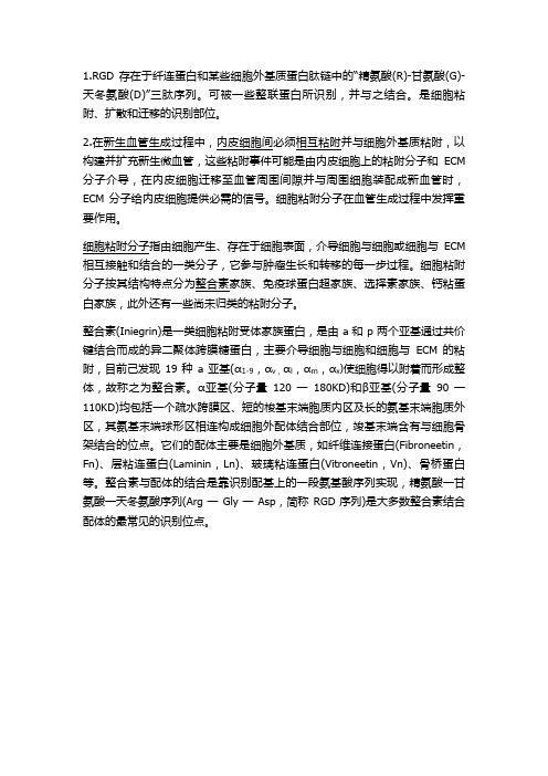
整合素(Iniegrin)是一类细胞粘附受体家族蛋白,是由a和p两个亚基通过共价键结合而成的异二聚体跨膜糖蛋白,主要介导细胞与细胞和细胞与ECM的粘附,目前己发现19种a亚基(α1-9,αv,αl,αm,αx)使细胞得以附着而形成整体,故称之为整合素。
α亚基(分子量120一180KD)和β亚基(分子量90一110KD)均包括一个疏水跨膜区、短的梭基末端胞质内区及长的氨基末端胞质外区,其氨基末端球形区相连构成细胞外配体结合部位,竣基末端含有与细胞骨架结合的位点。
它们的配体主要是细胞外基质,如纤维连接蛋白(Fibroneetin,Fn)、层粘连蛋白(Laminin,Ln)、玻璃粘连蛋白(Vitroneetin,Vn)、骨桥蛋白等。
整合素与配体的结合是靠识别配基上的一段氨基酸序列实现,精氨酸一甘氨酸一天冬氨酸序列(Arg一Gly一Asp,简称RGD序列)是大多数整合素结合配体的最常见的识别位点。
3.静脉注射iRGD,这是一种环状的能穿透肿瘤的肽(图中彩色的环),它们和肿瘤血管中的与血管形成有关的整联蛋白(图中蓝色和黄色的受体)结合。
纤维粘连蛋白与碱性成纤维细胞生长因子对成骨细胞黏附的协同刺激作用
纤维粘连蛋白与碱性成纤维细胞生长因子对成骨细胞黏附的协同刺激作用冯立;曹晓建;任永信【期刊名称】《中国修复重建外科杂志》【年(卷),期】2007(21)4【摘要】目的研究纤维粘连蛋白(fibronection,FN)与碱性成纤维细胞生长因子(basic fibroblast growth factor,bFGF)单独或联合应用时对成骨细胞在生物衍生骨支架上黏附的影响,并探索其潜在机制。
方法取第3代成骨细胞,以bFGF(0.1、1、10和100ng/ml)刺激,接种于FN(0.1、1、10和100μg/ml)或多聚赖氨酸(Polylysine)修饰的生物衍生骨支架上,MTT法检测组间细胞黏附效率差异,电镜观察细胞密度与形态。
GRGDS肽封闭后再次重复上述步骤。
结果单独应用FN与bFGF均可增加成骨细胞在生物衍生骨支架上的黏附效率(P<0.05),联合应用时,产生的效应显著增强,两者存在交互作用(P<0.01)。
而Polylysine与bFGF无此交互作用(P>0.05)。
电镜观察发现,两者联合应用时细胞密度、铺展效果均优于单独应用。
应用GRGDS肽后,FN联合bFGF产生的黏附促进作用被显著阻断。
结论bFGF与FN对成骨细胞在生物衍生骨三维支架上的黏附存在协同刺激作用,细胞黏附产生这一效应的机制很可能是由RGD-整合素α5β1途径来介导的,需深入研究探讨。
【总页数】6页(P390-395)【关键词】骨组织工程;纤维粘连蛋白;碱性成纤维细胞生长因子;生物衍生骨【作者】冯立;曹晓建;任永信【作者单位】南京医科大学第一附属医院骨科【正文语种】中文【中图分类】R318.17;Q813.1【相关文献】1.G蛋白偶联受体激酶结合蛋白1的SHD结构域对成骨细胞内局部黏附激酶活性的影响 [J], 于留钱;张宁;胡志毅;殷国勇2.碱性成纤维细胞生长因子对成骨细胞中转化生长因子-β1和碱性成纤维细胞生长因子mRNA表达的影响 [J], 曾晖;杜靖远;郑启新;刘勇;康斌;刘国平3.碱性成纤维细胞生长因子对成骨细胞和牙周膜成纤维细胞迁移、增殖的影响 [J], 谭震;宫苹;赵青4.碱性成纤维细胞生长因子对成骨细胞骨架蛋白作用的研究 [J], 夏露;王培志;王国平;梁星5.碱性成纤维细胞生长因子对成骨细胞与多孔生物材料黏附特性的影响 [J], 郑磊;王前;裴国献;马建标;王亦农因版权原因,仅展示原文概要,查看原文内容请购买。
院感基础知识考试题库(最全面的题库)
即墨市人民医院医院感染基础知识考试题库一、判断题:1、本次感染直接与上次住院有关,也属于医院感染. (√)2、污染-感染(IV类)切口指新鲜开放性创伤手术;手术进入急性炎症但未化脓区域者;被胃肠道内容物明显溢出污染者;术中无菌技术有明显缺陷(如开胸心脏按压)者。
(×)3、抗菌药物分为非限制使用、限制使用与特殊使用三类进行分级管理。
(√)4、手术病人常规预防性应用抗菌药物可有效预防手术切口感染的发生.(×)5、普通感冒、麻疹、水痘等病毒性疾病常规预防性应用抗菌药物可有效预防继发感染的发生。
(×)6、在盛装医疗废物前,应当对医疗废物包装物或容器进行认真检查,确保无破损,渗漏和其他缺陷。
(√)7、隔离的传染病病人产生的具有传染性的排泄物、分泌物、体液等应当严格消毒后排入污水处理系统.(√)8、抗菌药物包括各种抗生素、磺胺药、硝基咪唑类、喹诺酮类、呋喃类、抗结核、抗真菌等化学药物。
(√)9、正常准备皮肤、有效控制血糖、合理使用抗菌药物、手术期间给患者保暖是预防手术部位感染的措施.(√)10、术前预防使用抗菌药物,应静脉给药,1小时滴完,可放在大瓶液体内慢慢滴入。
(×)11、对乙类传染病中的传染性非典型肺炎,炭疽中的肺炭疽和人感染高致病性禽流感需要采取甲类传染病的预防,控制措施.(√)12、某些抗生素还具有抗肿瘤活性,用于肿瘤的化学治疗(√)15、抗生素是某些细菌、放线菌和真菌等微生物的次级代谢产物(√)16、保护性隔离措施:是指为预防高度易感病人受到来自其他病人、医务人员、探视者及病区环境中各种致病微生物的感染,而采取的隔离措施。
(√)17、消毒是指杀灭或清除传播媒介物上一切微生物,使其达到无活微生物存在的过程。
(×)18、灭菌是指杀灭或清除传播媒介物上的病原微生物,使其达到无害化的过程.(×)19、医务人员在诊疗、护理每一位病例后,均应认真洗手或对双手消毒,或更换使用一次性手套.(√)20、对传染病菌种、毒种和传染病检测样本的采集、保藏、携带、运输和使用可不实行分类管理,建立健全严格的管理制度.(×)21、应当定期更换置管穿刺点覆盖的敷料,无菌纱布为1次/日,无菌透明敷料1~2次/周(×)22、医务人员手部皮肤发生破损,在进行有可能接触病人血液、体液的诊疗和护理操作时不必须戴双层手套。
检验类之临床医学检验技术(师)综合提升练习题库
检验类之临床医学检验技术(师)综合提升练习题库单选题(共20题)1. 常用于POCT的技术,以下哪种是错误的A.免疫渗滤技术B.免疫层析技术C.化学发光技术D.微流控技术E.免疫比浊技术【答案】 C2. 电场中带电化合物的分子带净电荷多数取决于()。
A.电泳时的温度B.电泳时的电场强度C.电泳时通电时间的长短D.电泳时缓冲液的pH值E.电泳时缓冲液的离子强度【答案】 D3. 单向扩散试验呈双环的沉淀环是因为()。
A.含抗原性相同的两个组分B.两种抗原C.两种抗体D.抗体为单克隆抗体E.抗原为多态性抗原【答案】 A4. 编码ST的基因位于()A.质粒B.染色体C.质粒和染色体D.噬菌体E.以上都不对【答案】 A5. 与A族链球菌抗吞噬作用及分型作用密切相关的蛋白是A.M蛋白B.T蛋白C.R蛋白D.S蛋白E.链球菌溶血素O【答案】 A6. 用于急性心肌梗死的首选酶是()。
A.CK-MB.GGTC.LDHD.ALTE.HBDH【答案】 A7. 一名17岁的摔跤队队员因最近肩部有一大水疱前来就诊。
他说以前从未得过水痘,2年前接种过水痘疫苗。
他觉得身体很不舒服,水疱区所属的淋巴结肿大。
从损伤的皮肤处采取的碎片可看到有多核巨细胞。
A.无环鸟苷B.干扰素C.盐酸金刚烷胺D.indinavirE.丙氧鸟苷【答案】 A8. 脊髓灰质炎病毒感染按症状分不同类型,5%感染者病毒侵入血流,形成第一次毒血症的是()。
A.无症状感染型B.顿挫型C.无麻痹型D.麻痹型E.脊髓型【答案】 B9. 正常人每天需要维生素BA.5~10μgB.10~20μgC.20~25μgD.2~5μgE.25~30μg【答案】 D10. 检测mIg对B细胞计数通常应用的免疫技术为A.荧光抗体技术B.ELISAC.放射自显影D.发光免疫测定E.金免疫测定【答案】 A11. 细胞内液含量最多的阴离子是A.HCOB.HPOC.KD.ClE.Na【答案】 B12. PI特征是A.488nm,525nm,绿色B.488nm,575nm,橙色C.488nm,670nm,红色D.488nm,620nm,橙红色E.633nm,670nm,红色【答案】 D13. 慢性淋巴细胞白血病A.周围血中幼稚细胞、原始细胞>30%B.周围血有较多幼稚粒细胞伴嗜酸、嗜碱性、粒细胞增多C.周围血中幼红、幼粒细胞易见,骨髓出现"干抽"D.周围血和骨髓中淋巴细胞>60%E.周围血中易见盔形红细胞及破碎红细胞【答案】 D14. 精液中果糖含量可反映A.附睾的分泌功能B.前列腺的分泌功能C.精囊的分泌功能D.尿道球腺的分泌功能E.尿道旁腺的分泌功能【答案】 C15. 一个1岁女孩因阵发性严重咳嗽而入院。
532011[计算机辅助药物设计]天津大学考试题库及答案
![532011[计算机辅助药物设计]天津大学考试题库及答案](https://img.taocdn.com/s3/m/f436383d974bcf84b9d528ea81c758f5f61f29f0.png)
532011[计算机辅助药物设计]天津大学考试题库及答案计算机辅助药物设计复习题一、名词解释1、碎片连接法:是一种从头药物设计方法,它将受点区域划分为不同子区域,将不同的形状和性质分子碎片(如,单一官能团)对接上去,再将子区域的碎片连接起来,就得到一个完整的分子。
2、计算机辅助药物设计:用计算机处理并在屏幕上显示生物大分子和药物分子模型,特别是计算机辅助设计技术与合理药物设计过程的结合则称为计算机辅助药物设计。
3、锁和钥匙学说:酶与底物结构上的互补性,用锁与钥匙的关系来描述酶与底物的关系。
但此学说不能解释酶的逆反应。
4、拟肽药设计(模拟肽学):为了克服多肽药种种不利的性质,通过多种途径对肽从结构上进行改造或是结构替换。
5、先导化合物:通过各种途径、方法或手段获得的,具有一定生物活性的新的结构类型化学物。
活性不高、特异性差、副作用大、药代药动性质差。
6、速率学说:药物与受体相接触即产生生物效应,药物作用与其占有受体的速率成正比,而与其占有的多少无关。
7、药物潜伏化:利用药物代谢动力学知识,改变化合物的结构,从而改变其理化性质,调节分子在体内的代谢途径,可能使药效增强,毒副反应降低,生物利用度增加,该过程称为药物潜伏化。
8、生物电子等排:电子等排体指电子数目及排布情况相同的化合物或基团构成的电子等排体,它们具有相似的物理性质。
在生物领域里电子等排体表现为生物电子等排。
9、模板定位法:是一种从头药物设计方法,它是指在受体活性部位使用模板构建出一个性质互补的骨架,再根据其他作用性质把分子骨架转化为具体分子。
10、药效团模型方法:从一组活性化合物的结构出发,确定化合物共同的药效作用模式,构建一个药效基团之间特定的三维排列方式,并依此设计新的化合物。
11、拮抗剂:能使受体结合,阻碍内源性物质与受体结合而导致其生物作用的抑制,也称抑制剂。
只有较强的亲和力,无内在活性的药物。
12、毒性基团:毒性基团在一些药物中,如果药效基团所产生的生物效应为毒性反应(毒性、致癌性、致突变性),这些基团称为毒性基团。
- 1、下载文档前请自行甄别文档内容的完整性,平台不提供额外的编辑、内容补充、找答案等附加服务。
- 2、"仅部分预览"的文档,不可在线预览部分如存在完整性等问题,可反馈申请退款(可完整预览的文档不适用该条件!)。
- 3、如文档侵犯您的权益,请联系客服反馈,我们会尽快为您处理(人工客服工作时间:9:00-18:30)。
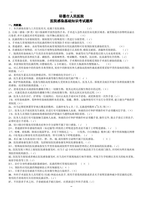
Polydopamine-mediated immobilization of multiple bioactive molecules for the development of functional vascular graft materialsYu Bin Lee a ,Young Min Shin a ,Ji-hye Lee a ,Indong Jun a ,Jae Kyeong Kang b ,Jong-Chul Park b ,Heungsoo Shin a ,c ,d ,*aDepartment of Bioengineering,College of Engineering,Hanyang University,17Haengdang-dong,Seongdong-gu,Seoul 133-791,Republic of KoreabDepartment of Medical Engineering,BK 21Project for Medical Science,Yonsei University College of Medicine,134Shinchon-dong,Seodaemun-ku,Seoul 120-752,Republic of Korea cInstitute for Bioengineering and Biopharmaceutical Research,Hanyang University,17Haengdang-dong,Seongdong-gu,Seoul 133-791,Republic of Korea dInstitute of Aging Society,Hanyang University,17Haengdang-dong,Seongdong-gu,Seoul 133-791,Republic of Koreaa r t i c l e i n f oArticle history:Received 29May 2012Accepted 5August 2012Available online 20August 2012Keywords:Human umbilical vein endothelial cell Polydopamine coating Surface modi fication EndothelializationDual factor immobilizationa b s t r a c tIn this study,we introduced a simple method for polydopamine-mediated immobilization of dual bioactive factors for the preparation of functionalized vascular graft materials.Polydopamine was deposited on elastic and biodegradable poly(lactic acid-co-3-caprolactone)(PLCL)films,and a cell adhesive RGD-containing peptide and basic fibroblast growth factor were subsequently immobilized by simple dipping.We used an enzyme-linked immunosorbent assay and fluorescamine assay to con firm that we had stably immobilized bioactive molecules on the polydopamine-coated PLCL film in a reaction time-dependent manner.When human umbilical vein endothelial cells (HUVEC)were cultured on the prepared substrates,the number of adherent cells and proliferation of HUVEC for up to 14days were greatest on the film immobilized with dual factors.On the other hand,the film immobilized with RGD peptide exhibited the highest migration speed compared to the other groups.The expression of cluster of differentiation 31and von Willebrand factor,which indicates maturation of endothelial cells,was highly stimulated in the dual factor-immobilized group,and passively adsorbed factors showed a negligible effect.The immobilization of bioactive molecules inspired by polydopamine was successful,and adhe-sion,migration,proliferation and differentiation of HUVEC were synergistically accelerated by the presence of multiple signaling factors.Collectively,our results have demonstrated that a simple coating with polydopamine enables the immobilization of multiple bioactive molecules for preparation of polymeric functionalized vascular graft materials.Ó2012Elsevier Ltd.All rights reserved.1.IntroductionCardiovascular diseases that usually result from malfunction in coronary arteries remain the leading cause of mortality worldwide.Severe arterial occlusion is treated by bypass surgery in which saphenous veins and mammary arteries are used as the preferred graft materials;however,this technique suffers from limited availability of suitable autologous tissue and donor site morbidity [1].Synthetic grafts including poly(tetra fluoroethylene),polylac-tide,polycaprolactone,and polyurethane are also routinely used for surgery [2].These polymeric grafts offer compliable mechanicalproperties,but often fail to provide long-term patency as a replacement for small-caliber blood vessels (<6mm in diameter).The reduced success rate is mainly due to the occurrence of the thrombogenic response and tissue thickening by intimal hyper-plasia (IH),which is prevented by the endothelium in normal blood vessels [3].Endothelialization on vascular grafts is a complex process that involves adhesion,migration,proliferation and differentiation of endothelial cells (EC),which are regulated by a myriad of signals.Extracellular matrix (ECM)proteins serve as non-soluble cues to modulate cell fate through cell signaling cascades [4].Collagen type I and fibronectin stimulate migration and proliferation of EC [5,6].In addition,vitronectin reduces the apoptosis rate through binding to a v integrin in the EC membrane [7].Numerous soluble growth factors are also implicated in new blood vessel formation and EC activities either by direct binding to cellular transmembrane receptors or to ECM proteins.In particular,vascular endothelial*Corresponding author.Department of Bioengineering,College of Engineering,Hanyang University,17Haengdang-dong,Seongdong-gu,Seoul 133-791,Republic of Korea.Tel.:þ82222202346;fax:þ82222982346.E-mail address:hshin@hanyang.ac.kr (H.Shin).Contents lists available at SciVerse ScienceDirectBiomaterialsjournal homepage:w ww.elsevi/locate/biomaterials0142-9612/$e see front matter Ó2012Elsevier Ltd.All rights reserved./10.1016/j.biomaterials.2012.08.011Biomaterials 33(2012)8343e 8352growth factor (VEGF)and basic fibroblast growth factor (bFGF)induce overexpression of their respective receptors within the EC membrane and regulate adhesion,migration,proliferation,and physical organization [8].To recapitulate these multi-step events,one effective engi-neering approach is to immobilize signaling bioactive molecules affecting EC function on synthetic vascular grafts.ECM proteins such as collagen,bovine serum albumin (BSA),fibronectin,and laminin have been immobilized on the surface of grafts,demon-strating improvement of adhesion,spreading,and proliferation of the EC [9].On the other hand,selected domains of ECM proteins have been used to functionalize the graft materials,which affect cell behaviors in the same way as ECM proteins [10].There have been reports that RGD (Arg-Gly-Asp)peptide recognized by integrin can enhance adhesion and migration of EC [11].None-theless,whereas immobilization of these molecules presents some success in enhanced endothelization,no ideal system has beenproposed.In particular,most studies have focused on one aspect of EC function,but few studies have included the multiple signals required for precise mimicry of the real endothelialization condi-tion (i.e.,combination of ECM proteins and growth factors).Although mainly delivered in the soluble form,growth factors such as VEGF,bFGF,and platelet-derived growth factor-BB have been covalently immobilized on the material surface.This has been shown to accelerate proliferation of HUVEC,implying that conju-gation of some growth factors preserves functional activity and stability in a long-term manner [12,13].The aim of this study was,therefore,to test our hypothesis that immobilization of dual factors (ECM components and growth factor)can synergistically modulate EC function.Various techniques are available for immobilization of biomol-ecules on polymeric biomaterials.Most vascular grafts are produced from inert hydrophobic polymers lacking functional moieties for chemical conjugation;therefore,selection of an appropriate functionalization method is critical.Plasma treatment and gamma ray irradiation use high energy sources to introduce reactive groups for subsequent immobilization of target biomole-cules such as fibronectin,collagen,gelatin,and RGD [14e 19].However,these methods have limitations in terms of penetration depth and possible cleavage in polymeric chains negatively affecting the mechanical property and long-term stability of the vascular graft [20].Recently,a facile surface modi fication method that can be applied to versatile solid materials from metal to synthetic polymers by simple dip-coating with dopamine solutionTable 1Sample conditions.Sample code ConditionDP Polydopamine-coated onto PLCL film DPr RGD-immobilized onto DP DPbf bFGF immobilized onto DPFig.1.Schematic diagram of polydopamine-mediated immobilization of bioactive molecules onto PLCL films that can be utilized as a functionalized vascular graft material.Y.B.Lee et al./Biomaterials 33(2012)8343e 83528344was reported [21].Under slightly basic conditions,dopamine can undergo oxidative polymerization,which creates a stable layer that is adherent to the surface of materials.Aside from the simplicity and cleanliness of the reaction environment,unlike with other chemical immobilization methods,post-modi fication of the poly-dopamine layer allows for the functionalization of bioactive molecules containing thiols or primary amines via imine formation and/or Micheal addition.Previous work has utilized the reactivity of polydopamine-coated substrate for immobilization of thiolated methoxy-poly(ethylene glycol)(PEG),animated methoxy-PEG,trypsin,BSA,concanavalin A-R Nase B,and antibody [22].In addi-tion,it was shown that growth factors such as VEGF and bone morphogenetic protein 2(BMP-2)immobilized on a titanium-based biomaterial surface following polydopamine coating enhanced proliferation of EC and differentiation of rat mesen-chymal stem cells,respectively [22].In this study,we developed a polydopamine-mediated immobi-lization platform together with a cell adhesive peptide,RGD,and an angiogenic growth factor,bFGF,on elastic biodegradable poly(lactide-co-caprolactone)(PLCL)substrates.We then investi-gated the effect of dual factors on regulation of HUVEC activity.Speci fically,we measured the immobilized amounts of RGD and bFGF on the polydopamine-coated PLCL film in a time-dependent manner and examined the synergistic or independent effects of each factor on adhesion,migration,proliferation and differentiation of HUVEC.2.Experimental 2.1.Materials3,4-Dihydroxyphenylamine was purchased from Sigma (St.Louis,MO,USA).Chloroform was purchased from Junsei (Tokyo,Japan).Hexa fluoroisopropanol (HFIP)was purchased from Wako (Osaka,Japan).Endothelial Cell Growth Medium-2(EGM-2)was obtained from Lonza (Basel,Switzerland).Tris e HCl was purchased from Shelton Scienti fic,Inc.(Peosta,IA,USA).BSA was purchased from Bioshop (Ontario,Canada).The fluorescent probes Hoechst 33,258,rhodamine-phalloidin,and Alexa Fluor 488rabbit anti-mouse IgG were purchased from Molecular Probes (Eugene,OR,USA).Other unspeci fied chemicals were purchased from Sigma.Distilled water (DW)was obtained using Milli-Q Plus System (Millipore,Billerrica,MA,USA).2.2.Preparation of PLCL films immobilized with bioactive molecule immobilization We synthesized poly(L -lactide-co-ε-caprolactone)(PLCL)by ring open poly-merization as previously described [23,24].To prepare PLCL film,PLCL was dissolved in chloroform (10wt%)and poured on a glass plate and dried at room temperature for 3days.For polydopamine coating,the PLCL film was submerged in 3,4-dihydroxyphenylamine (2mg/ml)dissolved in Tris e HCl buffer (10m M ,pH 8.5)and shaken on a rocker for 1h.After this process,the film was washed three times in DW.Immobilization of bioactive molecules was performed similarly to the poly-dopamine coating process.Brie fly,we prepared separate solutions of 1m M RGD peptide (GGGRGDS,Anygen,Gwangju,Korea)and 500ng/ml bFGF (Peprotech,Rocky Hill,NJ,USA)(dissolved in 10m M Tris e HCl buffer,pH 8.5).Polydopamine-coated PLCL film was cut into a circular shape (area :1.99cm 2),which was then incubated with 400m l of the prepared peptide or growth factor solution at 37 C for various times as indicated in Table 1.2.3.Quanti fication and stability of immobilized bioactive moleculesImmobilized RGD peptide on the surface was quanti fied by fluorescamine assay.After the immobilization process,the supernatant was obtained and reacted with fluorescamine solution (100m g/ml in acetone)in a 96-well plate (RGD sol-ution ¼75m l,fluorescamine solution ¼25m l).The reaction was performed for 1min at RT.The fluorescent intensity of each sample was observed (480nm of excitation,520nm of emission)using a plate reader (Spectramax M2,Molecular Device,CA,USA).Known concentrations of RGD peptide (20m g/ml e 625m g/ml)solution were also reacted with fluorescamine solution to obtain a standard curve.Similarly,reacted bFGF solution was collected and quanti fied through the bFGF enzyme-linked immunosorbent assay (ELISA)development kit (Peprotech,Rocky Hill,NJ,USA),following the manufacturer ’s guidelines.To calculate the detached amount of bioactive molecules,samples were sonicated in tris e HCl buffer (400m l)for 30min,and quanti fication was performed with the same ELISA development kit.The detached amount was calculated as the percentage compared to the immobilized amount for each incubation time.2.4.X-ray photoelectron spectrometer (XPS)The atomic composition of the bioactive surface was analyzed using an XPS.Analysis was performed with an ESCA LAB 200I (Thermo VG Scienti fic,MA,USA)spectrometer using a magnesium anode source.The changes in atomic composition by surface modi fication were investigated with peak fitting software.2.5.Culture of human umbilical vein endothelial cells (HUVEC)HUVEC were purchased from Lonza Group Ltd.(Basel,Switzerland).Sub-culture of HUVEC was performed in endothelial growth medium (EGM-2bulletkit,LonzaFig.2.Quanti fication of the immobilized bioactive molecules on polydopamine-coated PLCL film.(a)The amount of immobilized RGD peptide on the polydopamine-coated surface was measured by fluorescamine assay.(b)The amount of immobilized bFGF on the polydopamine-coated surface was measured by ELISA.(c)The percentages of detached bioactive molecules after application of sonication were calculated by normalization to the immobilized amount of bioactive molecules at each time point.Y.B.Lee et al./Biomaterials 33(2012)8343e 83528345Group Ltd.,Basel,Switzerland),and other conditions were the same as standard cell culture conditions(37 C,5%CO2,95%humidity).Every cell test was carried out using HUVEC passage number7.Other specific culture conditions(medium treat-ment,culture day)for each analysis are described later.The medium was changed every2days.2.6.Cell adhesion and immunofluorescent stainingFor cell adhesion study,circular-shapedfilms(1.99cm2)were placed inside the 24well polystyrene culture dish and HUVEC were seeded at the density of 1Â104cells/cm2with endothelial basal medium(EBM-2,Lonza Group Ltd.,Basel, Switzerland)supplemented with FBS(5%).For the positive control group,HUVEC were seeded at the same density on the tissue culture plate under the same media condition,but we also added bFGF(10ng/ml).In order to preventfilmsfloating from the surface,samples were overlaid with stainless rings(24well size)and HUEVEC were cultured for24h.For immunostaining of cluster of differentiation31 (CD31)expression,HUVEC were seeded at the higher density of4Â104cells/cm2 with EBM-2.After24h,the media was replenished with EBM-2supplemented with FBS(5%),and additionally cultured for3days(n¼5).The cells(for adhesion test and CD31staining)were thenfixed with4%paraformaldehyde for20min, gently washed with PBS,and treated with cytoskeletal(CSK)buffer(pH6.8,50m M NaCl,150m M sucrose,3m M MgCl2,50m M trizma-base,0.5%Triton X-100)for cell permeabilization(20min,4 C).Then,the samples were treated with blocking buffer(5%BSA in PBS)for1h at37 C,followed by treatment with anti-paxillin solution(1:200,BD science,NJ,USA)for1h at37 C.For CD31staining,CD31 antibody(Cell Signaling Technology Inc.,Danvers,MA,USA)solution(1:200)was used instead of anti-paxillin solution.The samples were incubated with Alexaflour 488rabbit anti-mouse IgG(1:100),rhodamine-phalloidin(1:200)and Hoechst 33,258(1:10,000)-containing solution to visualize focal contact or CD31expression and cell skeletal structure(1h,37 C).After incubation,cells were gently washed with PBS and mounted with Vectashield mounting medium(Vector Laboratory, UK).Immunofluorescence images were taken using a confocal microscope(Nikon, Tokyo,Japan).2.7.Cell migration assayThe migratory behavior of HUVEC was observed using a wound healing model. Briefly,a square silicon-insert with two rectangular wells(Ibidi,Munich,Germany) was laid on the PLCLfilms to create a cell-free gap(500m m).In each rectangular well,HUVEC(2Â105cells/cm2)were seeded with EBM-2supplemented with FBS (5%)and incubated for24h to achieve confluent cell adhesion.After this process, samples were placed on an inverted microscope(Olympus Optical Co.Ltd.,Tokyo,Japan)with a micro culture system(37 C,5%CO2,95%humidity).Then,the insert was removed,and cell migration to the gap was observed with a change-coupled device camera(Electric Biomedical Co.Ltd.,Osaka,Japan),and images were ob-tained every5min for up to12h.From these images,the migratory speed was calculated and statistically analyzed.2.8.HUVEC proliferationTo observe the effect of bioactive molecules on proliferation of HUVEC,we performed cell counting for up to14days.HUVEC were seeded at1Â104cells/cm2 on the circular-shapedfilms with EBM-2and cultured for24h.Then,we added5% FBS to the EBM-2media and the cells were cultured for14days with the media change in every2day.Sample images were taken at1,3,7,10,and14days,andfive spots(center,right,left,top and bottom)for each sample group(n¼5)were captured for cell counting(magnification,40x).2.9.Reverse transcription-polymerase chain reaction(RT-PCR)We used RT-PCR to analyze the expressions of von Willebrand factor1(vWF-1), CD31,and GAPDH as a housekeeping gene.The circular-shapedfilms were placed inside the24well tissue culture dish and HUVEC were seeded at4Â104cells/cm2 with EBM-2.After24h,the media was changed with EBM-2supplemented with FBS (5%)and cultured for3days.In the positive control group,bFGF(10ng/ml)was additionally supplemented.RNA was obtained from these samples and reverse-transcribed to cDNA.200ng of cDNA was used for each PCR reaction.The cDNA was denatured at95 C for5min and then95 C for30s,and annealing was per-formed for45s at56 C for CD31,54 C for vWF-1,and55 C for GAPDH(GAPDH:fw. 50-ACCACAGTCCATGCCATCAC-30rev.50-TCCACCACCCTGTTGCTGTA-30,CD31:fw.50-GAGTCCTGCTGACCCTTCTG-30rev.50-CACTCCTCCCACCAACACCT-30,vWF-1:fw.50-ATGATTCCTGFFAGATTTGC-30rev.50-AGACTCTTTGGTCCCCCTGT-30).Elongation was performed at72 C for60s,and the reaction was repeated for35cycles.Thefinal elongation was performed for5min at72 C.The RT-PCR product was loaded in a1.5%agarose gel with buffer(7m l)and separated by gel electrophoresis.Separated bands were stained with ethidium bromide,and images were obtained using the Gel Logic100Imaging System(Kodak,Rochester,NY,USA)UV illumination tool.The band intensity was measured with image software(Image J),and statistical quan-titative analysis was performed.2.10.Western blottingSample preparation for Western blotting was performed in the same way as for RT-PCR.After3days of culture,cells were lysed with400m l of RIPA buffer,andtotal Fig.3.Chemical composition analysis.(a)XPS spectra of the substrates,and(b)high resolution spectra of the carbon peak(C1s).Y.B.Lee et al./Biomaterials33(2012)8343e83528346protein was measured using the BCA protein assay kit (Pierce,Rockford,IL,USA).Cell lysates were loaded on SDS-PAGE gels to observe the b -actin and CD31bands separated through electrophoresis.Then,the proteins were transferred onto a nitrocellulose membrane (0.45m m pore size,Invitrogen,Carlsbad,Ca,USA).The membrane was treated with blocking buffer for 1h at 37 C and then with diluted primary antibody solution (1:100)overnight at 4 C for b -actin (Applied Biological Materials Inc.,Richmond,BC,USA)and CD31detection.After reaction with primary antibodies,the membrane was washed with PBS-T (0.01%Tween-20in PBS)for 15min and then incubated with anti-mouse secondary antibody (Applied Biological Materials Inc.,Richmond,BC,USA)for 2h at 25 C.We used the ECL plus Western blotting detection system (Amersham Bioscience,Buckinghamshire,MO,USA)to visualize the protein bands.The bands intensity was measured with Image J,and statistical quantitative analysis was performed.2.11.Statistical analysisAll quantitative data were obtained from triplicate samples,and the results are reported as mean Æstandard deviation (SD).Statistical signi ficance was distin-guished through one-way ANOVA,Tukey ’s HSD test,and Student ’s t -test (p <0.05).3.Results and discussionLimited endothelialization on synthetic vascular graft mate-rials often causes undesirable complications such as in flamma-tory reactions,thrombogenic responses,and intimal hyperplasia.Surface modi fication of vascular grafts may be a promising approach;however,an ideal material that canprecisely emulate complex endothelialization processes has never been proposed.Our ultimate goal is to develop vascular grafts that can provide multiple signals to facilitate endotheli-alization in a stable and long-term manner.Given that,in this study,we investigated how EC function is affected by the coordinated effects of a cell adhesive peptide and an angiogenic growth factor-immobilized onto polydopamine-coated PLCL films.Fig.1shows the scheme of our strategy,and sample codes are presented in Table 1.Fig.4.Adhesion morphology and spreading of HUVEC on the substrates.Immuno fluorescent images of adherent HUVEC on substrates,which were stained for f-actin (red),paxillin (green),and nuclei (blue),and the number of adherent cells following in vitro culture for 1day (scale bar ¼100m m).“*”indicates signi ficance as compared to the DPbf group,and “x ”indicates signi ficance as compared to the DPrbf group (p <0.05).(For interpretation of the references to colour in this figure legend,the reader is referred to the web version of this article.)Y.B.Lee et al./Biomaterials 33(2012)8343e 835283473.1.Quanti fication and stability of immobilized bioactive moleculesWe coated polydopamine on PLCL films,which were subse-quently immobilized with RGD-containing peptide and bFGF by simple dipping.As shown in Fig.2a and b,the amount of immo-bilized peptide was recorded as 66.60Æ1.97mg/cm 2after 24h of incubation.Since the concentration of bFGF was much less than that of peptide,the complete immobilization of bFGF was more rapidly achieved.The amount of immobilized bFGF was 8.32Æ0.27ng/cm 2for 10min of incubation,which did not change despite prolonged incubation.The calculated values of immobilized factors suggest that the polydopamine layer coated on PLCL films enables effective immobilization of peptides and growth factors.Previously,peptide or growth factors have been immobilized on the surfaces of biomaterial substrates via physical adsorption or chemical reaction,demonstrating bioactivity of the immobilized molecules [25e 28].However,the previous immobilization processes left signi ficant room for improvement since they oftenrequired a long process time,extensive labor,complicated expertise and uncontrolled immobilization ef ficiency.Our results suggest that we could more easily immobilize bioactive molecules with relatively high ef ficiency within a few hours.Considering that bFGF at concentrations greater than 10ng/ml can modulate proliferative properties of HUVEC,the immobilized amount of bioactive mole-cules is expected to be suf ficient to induce speci fic cell behaviors [29].We then tested the stability of immobilized bioactive mole-cules by applying sonication.As shown in Fig.2c,the detached amount following employment of sonication was less than 2%and 0.2%for RGD peptide and bFGF immobilized substrates,respec-tively,suggesting that the immobilization onto polydopamine-coated PLCL films supported stable binding of bioactive molecules.3.2.X-ray photoelectron spectrometry (XPS)Surface chemical composition of functionalized substrates was investigated with XPS.As shown in Fig.3a,three separate peaksFig.5.Migration of HUVEC on the substrates.Images of migrated cells at 12h.The top left image shows the initial condition (0h)with a 500-m m defect in the middle of the cell monolayer.The lower graph presents the migration speed of HUVEC for 12h “*”indicates signi ficance as compared to the DP group (p <0.05).Y.B.Lee et al./Biomaterials 33(2012)8343e 83528348corresponding to C1s (288eV),N1s (399eV),and O1s (533eV)were observed as the main elements.Notably,the intensity of the N1s peak increased in samples immobilized with peptide and/or growth factor compared to the DP.All groups had C e H (284.6eV),C e O (286.6eV),C ]O (288.6eV),C e N (286.0eV)peaks from high resolution spectra of C1s,as shown in Fig.3b,and the DP group had the lowest ratio of C e N bonding (0.028).In contrast,the DPrbf had the C e N peak with the highest ratio (0.155),which is directly related to the presence of a large number of amino acids.In previous research on the immobilization of RGD using diamine-PEG as a spacer on polydopamine-coated poly(methyl methacrylate),the nitrogen composition of the RGD-immobilized surface was 7.4%,which is higher than that of the polydopamine-coated surface (7.2%)[30].Another study that immobilized VEGF on polydopamine-coated substrate presented a higher value for the nitrogen ratio in the VEGF-immobilized group (13.5%)in compar-ison to the polydopamine-coated group (9.1%)[31].Therefore,the change in the C e N peak suggests that RGD and bFGF were successively immobilized on the polydopamine-coated PLCL film.3.3.Cell adhesion study and immuno fluorescent stainingAs shown in Fig.4,HUVEC were adherent and well-dispersed on all samples.Overall,the morphology of EC on the surface appeared to be similar among all groups,and it was clear that co-immobilized factors produced a synergistic effect on cell adhesion by increasing the number of adherent cells;DPrbf presented the highest cell number (465Æ23),and DPbf,P,DPr,DP recorded sequentially lower cell numbers (360Æ9,351Æ24,245Æ17,148Æ18,respectively).Although DPr had a relatively lower value than the other groups,the combination of RGD peptide and bFGF synergis-tically affected the adhesion of HUVEC.Collectively,adhesion and spreading were enhanced by polydopamine coating,which were further promoted features by immobilization of bioactive factors,particularly in the case of dual factors.Adhesion is the initialprocess in endothelialization and is closely implicated in later stages of cell behavior,including migration,proliferation,and differentiation.Therefore,regulating cell adhesion is crucial for constructing a healthy endothelium [32].There are some reports that cell adhesion is enhanced on polydopamine-coated surfaces [33,34].In addition to that,other reports have observed enhanced EC adhesion by introducing RGD or bFGF through plasma treat-ment,secondary conjugation,or chemical immobilization [28,35,36].Considering these previous reports,we suggest that enhanced hydrophilicity of the polydopamine-coated surface and the effects of RGD peptide and bFGF could synergistically in fluence HUVEC adhesion.3.4.Cell migration assayCell migration plays an important role in endothelialization,organization of tightened endothelium surface and angiogenesis [11].Therefore,we investigated the effect of immobilized mole-cules on migratory behavior of HUVEC using a simple wound healing assay model.As shown in Fig.5,the wound was effec-tively generated by removal of a square silicon-insert,leaving a cell-free defect with a pre-determined size (500m m).Time-lapse monitoring for longer than 12h showed that migrating HUVEC partially covered the defects.The migration speed of HUVEC on the PLCL film immobilized with bioactive molecules was signi ficantly greater than that on the film coated with pol-ydopamine;the migration speed was 20.64Æ2.04m m/h on DP,which signi ficantly increased to 31.74Æ 3.43,25.2Æ 2.42,30.06Æ2.68,and 26.4Æ2.43m m/h for DPr,DPbf,DPrbf,and P,respectively.Interestingly,the migration speed of HUVEC on DPbf and P was signi ficantly lower than that on DPr and DPrbf,indi-cating that cell adhesive RGD-containing peptide dominantly in fluenced migration of HUVEC,and no synergistic effects of bFGF were observed.In addition,we concluded that bFGF can be physically adsorbed onto the PLCL film,which affected theFig.6.Proliferation of HUVEC on the substrates.(a)Images of proliferated cells at 3and 14days (scale bar ¼200m m),and (b)the number of proliferating HUVEC on the substrates.Immobilized molecules in fluenced the proliferation of HUVEC for 14days and co-immobilized factors produced a synergistic effect.“*”indicates signi ficance as compared to the DPrbf group at each time point (p <0.05).Y.B.Lee et al./Biomaterials 33(2012)8343e 83528349。