斑节对虾 虾蜕皮阶段的区分
虾蟹养殖学第1-9章思考题(10水产)
《虾蟹养殖学》第1~9章思考题日照长度:表示每天太阳日照时数日照时数:在某一段时间内,太阳照射地面的总时数。
补偿点:在池塘中的某一点光合作用与呼吸作用相等的点。
/一定的光强度下,植物中光合强度与呼吸强度相等,吸放二氧化碳的量相同。
补偿深度:日间在池塘的某一深度,其浮游植物光合作产生氧的量恰好等于浮游生物呼吸作用的耗氧量,此深度称为补偿深度。
增氧层:补偿深度以上的水层。
耗氧层:补偿深度以下的水层。
氧盈:超过溶解氧饱和度100%以上的值称为氧盈。
OS=DOt-SOt氧债:(OD)是指好气性微生物、有机物的中间产物及无机还原物在缺氧条件下理论耗氧值受到抑制的哪一部分耗氧量。
是理论耗氧量与实际耗氧量之差:OD=TOC-AOC潜在性酸性土壤日投饵率:每天的投饵量占养殖对象体重的百分比。
全长:额剑前端至尾节末端的长度(虾);体长:眼柄基部或额角基部眼眶缘至尾节末端的长度(虾);甲宽:头胸甲的最宽处的长度,最宽处如为齿时则自齿的基部量起(蟹);甲长:头胸甲前缘至后缘中线的长度(蟹);开放式纳精囊:无甲壳、骨片等形成的囊状结构,仅在第四、五对步足间腹甲上由甲壳皱褶、凸起及刚毛等甲壳衍生物形成一区域用于接纳精荚,精荚多粘附其上。
此种交接器者仅见于南半球产的对虾种类,如南美白对虾、南美蓝对虾等。
对虾以外的虾类多属于此类交接器类型。
封闭式纳精囊:具一袋状或囊状的纳精囊,交配时精荚的豆状体即贮于其中。
大多数的对虾属种类具封闭式的纳精囊。
触角腺:系甲壳纲软甲亚纲中之端足目、糠虾目、燐虾目、十足目的排泄器官,开口于第二触角基部。
常呈青绿色,又称绿腺。
相当于其他软甲亚纲和切甲亚纲开口于第一小颚附近的小颚腺。
在系统上也与环节动物的体节器相同。
始于薄壁之体腔囊,经具括约肌的漏斗和复杂曲折的输尿管、排泄管,开口于上述部位,输尿管有时互相交叉,在管壁的连接处产生连络口,形成网状的迷路。
生物学最小型: 水生动物首次达到性成熟的最小体长。
对虾的蜕壳过程及补钙的重要性
对虾的蜕壳过程及补钙的重要性一蜕壳前期:主要特征表现在1、外骨骼钙质溶解全部汇聚于肝胰脏和血淋巴中。
2、蜕皮膜形成,此阶段全部被老皮覆盖。
3、通过鳃、外壳、前肠和中肠吸收大量的水分导致身体膨胀。
4、通过鳃来吸收水体中的钙质来调节身体内外的渗透压。
5、老皮出现裂缝,为蜕壳做前期的准备工作。
二蜕壳期此时对虾不摄食,头胸部膨胀,远动频率加快,腹部进行弯曲和伸直,剧烈的抽动促使旧壳分离。
三蜕壳后期1、肝胰脏和血淋巴供给钙质和糖原和成几丁质和外骨骼,此时,外骨骼还未完全钙化无法抵御敌害生物捕食和同类残食。
比较脆弱。
2、新蜕壳的对虾会躲在安静的水底保持相对静止,开始慢慢吃食,此时的对虾基本可以自我保护。
四蜕壳间期1、新壳坚硬,可以抵御敌害生物和捕食2、血钙降低,活动和摄食恢复正常3、体质恢复正常,免疫、营养、生长达到最佳。
所以,对虾的一生大多数的身体机能都是伴随着对虾蜕壳的不同时期而发生变化,会在蜕壳结束后的特定时间内快速成长。
南美白对虾蜕壳期补钙的重要性:蜕壳期是虾体最虚弱的时候,基本上不进食,得不到营养的补充。
用“速补钙”2亩/瓶+“活力爽”2亩/瓶+“解毒应激VCE”2亩/包全池泼洒防止蜕壳期的应激,同时起到很好的补钙作用。
南美白对虾自身的能量全部用来生命的升级,没有多余的能量来维持机体的正常运转,此时虾体储存的钙质流失严重,若外界没有及时补充,南美白对虾吸收不到水体提供的钙,容易引起对虾蜕壳不遂,蜕壳后硬壳时间过长,若此时遭受细菌侵袭或者出现应激,极容易批量死亡。
所以,我们要通过人为的方式来补充水体中的钙质,泼洒“钙镁速补”2亩/包,对虾通过呼吸和身体的渗透吸收水体中的钙和能量得以己用。
这样既能保证南美白对虾蜕壳的顺利进行,又能促进养虾的成功率。
感谢您的阅读,祝您生活愉快。
对虾的幼体发育分为几期?
对虾的幼体发育分为几期?对虾有一个复杂的幼体发育阶段,在此其间对虾幼体要多次蜕皮,并在形态上及生活习性上发生很大变化。
对虾的幼体发育分为无节幼体期、蚤状幼体期、糠虾期以及仔虾期等阶段。
无节幼体期:幼体体形为倒梨形,有三对附肢,体前端中部有一眼点,尾部有一对尾棘。
无节幼体消化道不与体外相通,不摄食,靠体内的卵黄营养生存。
幼体靠三对附肢的间歇性划水在水中作间歇性游动,当附肢不划动时,幼体下沉,游动时则上浮,幼体具趋光性。
无节幼体的发育分为六期,经六次蜕皮变态为溞状幼体。
各期无节幼体可由尾棘数目来区分,第一至六期无节幼体尾棘数目分别是一对、二对、三对、四对、六对及七对,而第一、二期无节幼体可由附肢刚毛由光滑变为羽状来区分。
溞状幼体期:幼体己明显分节,有头胸甲,腹部明显拉长,出现复眼,附肢增多。
溞状幼体口与肛门已与外界相通,开始进食。
摄食方式主要为滤食,后期具捕食能力。
游动时靠附肢作类似蝶泳式的划水在水中游泳。
溞状幼体经三次蜕皮变态为糠虾幼体。
第一期溞状幼体没有额剑;复眼没有眼柄;尾部没有尾肢。
第二期糔状幼体出现额剑;复眼大、有眼柄;腹部后端甲壳下已有尾肢雏形。
第三期溞状幼体尾节增大,尾肢生出。
糠虾幼体期:幼体腹部发达,各部附肢齐全。
摄食以捕食为主,也有滤食能力。
游动时头朝下,尾朝上,形成独特的倒立游泳姿势。
糠虾幼体蜕皮三次变态为仔虾,第一期糠.虾幼体步足爪状;游泳足刚刚长出,芽状,小而不明显。
第二期糠虾幼体步足出现螯状结构;游泳足增长,分为二节。
第三期糠虾幼体步足、游泳足继续长大,游泳足片状。
仔虾期:幼体进一步发育、生长,体形与幼虾基本相似。
主要形态变化有:额剑上、下缘小齿增多,游泳足前端生出刚毛,平衡囊及交接器逐渐形成。
随着发育、生长,仔虾由营浮游生活转入水底栖生活,食物也渐渐由浮游生物为主转向底栖生物为主。
仔虾活动从游泳足划水游动、步足爬行,有时作弹跳活动,对虾在仔虾阶段需蜕皮14-20余次,各期仔虾区别主要以额剑刺齿多少及尾节形态等,生产上多以仔虾日龄来表示。
海水养殖斑节对虾的生长规律与养殖周期
海水养殖斑节对虾的生长规律与养殖周期斑节对虾(Penaeus vannamei)是一种重要的养殖物种,其高产高效的特点使其成为全球最主要的海水养殖物种之一。
了解斑节对虾的生长规律和养殖周期对于提高养殖效益至关重要。
本文将介绍斑节对虾的生长规律、养殖周期以及影响其生长的因素。
斑节对虾的生长规律是指虾苗从孵化到成虾的整个生命阶段中不同阶段的生长变化情况。
一般来说,斑节对虾的生长可以分为幼虾期、生长期和成熟期三个阶段。
幼虾期是指虾苗孵化出来后至体长约2厘米的阶段。
在这个阶段,虾苗的生长速度非常快,每天可以增长约0.2-0.4厘米。
幼虾期的主要任务是提供合适的饵料、保持适宜的水质和水温,以促进虾苗的健康生长。
生长期是指体长大于2厘米但小于10厘米时的阶段。
在这个阶段,斑节对虾的生长速度逐渐减缓,但仍然相对较快,大约每天增长0.2-0.5厘米。
此时,虾体开始逐渐形成外壳,并逐渐增强对外界环境的适应能力。
养殖者需要提供适宜的饵料、养护水质、维持适宜的温度和盐度,以确保虾的健康生长。
成熟期是指虾体长达到成熟时的阶段。
斑节对虾一般在体长10-14厘米左右时进入成熟期。
在这个阶段,虾体的生长速度几乎停止,但体重仍然会有增加。
成熟期的主要任务是提供适宜的环境条件,促进虾体内繁殖细胞的形成和储存,保证虾的繁殖能力。
养殖周期是指从虾苗放养到收获的整个时间周期。
根据斑节对虾的生长规律,一般养殖周期大约为4-7个月左右。
具体的养殖周期会受到多种因素的影响,包括养殖水域的水质和温度、饵料的品质和投喂量、虾苗的质量等等。
影响斑节对虾生长的因素有很多。
首先,饵料的品质和投喂量直接影响虾的生长速度和体重增加。
合理选择和投喂高质量的饵料是促进斑节对虾健康生长的关键。
其次,水质和温度对虾的生长也有重要影响。
适宜的水质和温度能提供良好的生长环境,帮助虾体吸收养分,促进生长。
同时,水质的酸碱度、氧含量、盐度等也要保持在适宜的范围内。
此外,斑节对虾的生长也与养殖密度、水域的容积和通风等因素有关。
蜕壳,是养虾的生死关!如何保证虾顺利蜕壳呢?技巧都在这
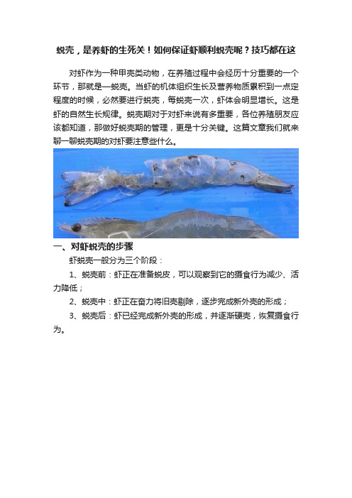
蜕壳,是养虾的生死关!如何保证虾顺利蜕壳呢?技巧都在这对虾作为一种甲壳类动物,在养殖过程中会经历十分重要的一个环节,那就是—蜕壳。
当虾的机体组织生长及营养物质累积到一点定程度的时候,必然要进行蜕壳,每蜕壳一次,虾体会明显增长。
这是虾的自然生长规律。
蜕壳期对于对虾来说有多重要,各位养殖朋友应该都知道,那做好蜕壳期的管理,更是十分关键。
这篇文章我们就来聊一聊蜕壳期的对虾要注意些什么。
一、对虾蜕壳的步骤虾蜕壳一般分为三个阶段:1、蜕壳前:虾正在准备蜕皮,可以观察到它的摄食行为减少、活力降低;2、蜕壳中:虾正在奋力将旧壳剔除,逐步完成新外壳的形成;3、蜕壳后:虾已经完成新外壳的形成,并逐渐硬壳,恢复摄食行为。
二、蜕壳时间1、当在28℃时,仔虾蜕壳间隔时间分别是30-40小时;2、当在28℃时,1-5g幼虾蜕壳间隔时间4-6天;3、当在28℃时,15克以上成虾的蜕壳间隔时间15天。
也就是说,虾越大,蜕一次壳需要完成积累营养的时间越长。
三、影响虾蜕壳的因素1、营养因素这是虾难以蜕壳的主要原因之一,营养不良的虾没有足够的物质来填满它的外壳,或者根本无法蜕壳,这对于养殖来说,都是十分不利的。
为了让虾活得好,虾必须吃足够的饲料,蛋白质含量在32-48%之间。
2、疾病在养虾的过程中,一些疾病,如真菌、藻类、寄生虫,也会导致虾蜕壳缓慢或者无法蜕壳。
3、养殖环境在蜕壳过程中,虾对氧气的需求是原来的两倍,所以当你看到有迹象表明虾准备蜕壳时,你需要多开启增氧机来补充溶解氧。
一般来说,在虾的脱壳过程中,溶解氧保持在5-6mg/L。
其次,虾塘中的盐度越高,池塘中的溶解矿物质含量就越高,反之亦然。
其次还有碱度、硬度等相关因素会影响到对虾的蜕壳。
四、保障对虾蜕壳顺利的措施1、一般来说,初一十五(月圆月缺),虾会集中蜕壳。
另外,我们也要定期采样,并对虾蜕壳情况与时间做好记录,这样,我们可以准确预测出虾蜕壳周期会在什么时候发生。
2、需要稳定水质,特别是盐度,以使其不会急剧变化,以防止虾受到冲击。
斑节对虾的生长与发育特征
斑节对虾的生长与发育特征斑节对虾(Penaeus monodon),又称黑对虾、巨型对虾,是一种重要的经济水产养殖物种。
斑节对虾的生长与发育特征对于养殖业的发展具有重要的意义。
本文将深入探讨斑节对虾的生长与发育特征。
斑节对虾的生长特征主要包括生长速度、生长形态和生长季节等方面。
首先,斑节对虾是一种快速生长的物种,其生长速度与温度、饵料、饵料种类和养殖技术等因素密切相关。
一般而言,斑节对虾在水温28~32℃时生长最为迅速,每月可以达到3~5厘米的生长速度。
其次,斑节对虾的生长形态特点明显,小型幼体为透明无色,随着生长逐渐变为灰色或黑色,携带有明显的黑色斑纹。
最后,斑节对虾的生长季节主要与养殖水温和养殖环境密切相关。
对于斑节对虾的养殖,选择合适的养殖季节对于提高养殖效益具有重要意义。
斑节对虾的发育特征包括发育周期、发育阶段和性成熟等方面。
首先,斑节对虾的发育周期在28~32℃水温条件下一般为4~5个月。
整个发育过程可分为孵化、幼体、稚虾和成虾阶段。
孵化阶段为眼点阶段,幼体阶段为后续虾期,稚虾阶段为入池虾,成虾阶段为性成熟的虾。
其次,斑节对虾的发育阶段存在明显的形态和生物学特征。
从孵化到成虾的过程中,体色逐渐由透明变为黑色,并逐渐形成明显的斑纹。
最后,斑节对虾的性成熟期在18~24个月左右。
性成熟的斑节对虾体型较大,具有明显的性别差异,雄虾较小,雌虾较大,雌虾体内含有大量的卵巢。
斑节对虾的生长与发育特征对养殖业的发展具有重要的意义。
首先,深入了解斑节对虾的生长规律可以为养殖者提供科学合理的养殖方案。
通过掌握斑节对虾的生长季节、生长速度和生长形态等特征,养殖者可以在合适的时机进行饵料投喂、增氧通风、换水换肥等操作,提高养殖效益。
其次,研究斑节对虾的发育特征有助于提高养殖的可控性和稳定性。
通过精确把握发育周期和发育阶段,养殖者可以合理规划虾苗引种和策略性的饲养管理,以提高成活率和生长率,降低经济风险。
最后,了解斑节对虾的性成熟规律有助于实施科学的养殖繁育计划。
想要对虾长得快,关键是蜕壳
技术学堂TECHNOLOGY FORUM090·《当代水产》 2018·6想要对虾长得快,关键是蜕壳五月,春夏交际,刚刚经过料峭春寒,气候逐渐温暖适宜,对虾的生长速度会明显的飙升上来。
而想要对虾长得快,自然首先得让对虾正常蜕壳硬壳,那么甲壳健康调理就成了这个时期最主要关注的问题。
对虾养殖什么最重要?可以毫不夸张的说,蜕壳过程决定了对虾的生死。
蜕壳时间偏短说明对虾蜕壳非常紧急匆忙,说明缺乏安全、安静蜕壳的区域(一般密度过大所致),不能很好的把褪下来的甲壳充分利用掉(池底能捞出大量较为完整的虾壳);蜕壳时间偏长(蜕壳障碍或蜕壳不遂)对虾体质会大幅下滑,最终诱发纤毛虫、褐斑、肝萎缩以及慢大等问题,严重的还会直接耗尽体能出现偷死;蜕壳周期偏短对虾会出现软壳、肌肉白浊、抽筋(弯弓);蜕壳周期偏长对虾则会出现老壳、毛壳(纤毛虫)、黄壳、黑壳。
蜕壳后硬壳过快(总硬度偏高、重金属离子偏高),对虾常会出现甲壳偏硬、黑壳、甲壳表面粗糙;蜕壳之后硬壳过慢不仅会引发纤毛虫、褐斑、分节、黄身、红身等症状,严重的还会有畸形和自残出现。
至于无法硬壳的自然就没有存活的可能了,软壳一久就很容易被其他虾吃掉,关键是这种软壳死亡很难发现,这才是名副其实的“偷死”,而养虾最终的产量低,这就是主要的原因之一。
这些还仅仅是蜕壳过程常出现的问题,若是环境其他因子还有问题,那出现的甲壳异常就更多了。
多数研究认为对虾一生需要蜕壳40余次。
而我们养殖对应的只是对虾生命中间的一个阶段而已,所以不需要那么多次。
对虾苗期蜕壳周期较短,相对频繁(短则半天或者1d1次,长则3~4d1次),越到后期蜕壳周期越长(短则1周,长则半个月甚至二十多d),所以满打满算下来,养殖期间对虾大致会蜕壳25~30次左右。
既然对虾养殖期间会蜕壳如此多次,而每一次蜕壳事关对虾生死,那么养殖者就必须详细的了解对虾蜕壳的整个过程以及蜕壳过程中须要掌握的注意事项。
虾蟹类的生长

包括2个总科:对虾总科、樱虾总科。
02
特征
二、枝鳃亚目特征与种类
樱虾总科 Sergestoidea
芒虾科 Aristeidae 深海芒虾科 Benthesicymidae
管鞭虾科 Solenoceridae
分类
萤虾科 Luciferidae 樱虾科 Sergestidae 对虾总科 Penaeoidea
第3步足至少是指节超过鳞片。额角后脊上有小凹点…...… ………………………………………..长毛对虾P. penicillatus
3.第3步足伸不到第二触角鳞片末端…..中国对虾P. chinensis
常见种检索
雌体灰青色,雄体色发黄。额角上缘具7-9齿,下缘为3-5齿。头胸甲无肝脊。第三步足伸不到第二触角鳞片的末端。是中国增养殖品种之一。主要产于渤海。
(三星虾)Acetes intermedius
中国毛虾 Acetes chinensis(虾皮)
三、腹胚亚目种类特征
01
猬虾下目 Stenopodidea
02
真虾下目 Caridea
03
螯虾下目 Astacidea
04
海蛄虾下目 Thalassinidea
中国对虾P. chinensis (中国明对虾Fenneropenaeus chinensis)
日本对虾P. japonicus
( 日本囊对虾 Marsupenaeus japonicus )
额角上缘具8-10齿,下缘具1-2齿。具额胃脊,额角侧沟长,伸至头胸甲后缘附近。额角后脊的中央沟长度大于头胸甲长的1/2。尾节具3对活动刺。体表具鲜明的横斑,为土黄色、橙色环带相间,带淡蓝色,尾肢具棕色横带,末半蓝绿色。出水后能经较长时间不死,便于运输。
- 1、下载文档前请自行甄别文档内容的完整性,平台不提供额外的编辑、内容补充、找答案等附加服务。
- 2、"仅部分预览"的文档,不可在线预览部分如存在完整性等问题,可反馈申请退款(可完整预览的文档不适用该条件!)。
- 3、如文档侵犯您的权益,请联系客服反馈,我们会尽快为您处理(人工客服工作时间:9:00-18:30)。
Index of molt staging in the black tiger shrimp (Penaeus monodon)Waraporn Promwikorn1, Pornpimol Kirirat2and Pinij Thaweethamsewee2AbstractPromwikorn, W., Kirirat, P. and Thaweethamsewee, P.Index of molt staging in the black tiger shrimp (Penaeus monodon) Songklanakarin J. Sci. Technol., 2004, 26(5) : 765-772Molting is a phenomenon of shedding the old cuticle and re-generating the new one found in crustaceans including shrimps and many other species of invertebrates. The molting cycle is a dynamic process, composed of pre-ecdysis (premolt, D stage), ecdysis (E stage), postecdysis (postmolt, A-B stages), and intermolting (C stage) stages. In healthy shrimp, molting cycles are repeated several times through shrimp life in order to increase body size and mass (growth). In this paper we gather the knowledges and important findings related to the molting cycle of crustaceans from the past until present, and highlight the physical evidence of cuticular tissue that we used for molt staging in the black tiger shrimp, Penaeus monodon. At the end of this paper we summarize the easily observed criteria used for molt staging in the black tiger shrimp. Key words:molting stages, Penaeus monodon, setae, setal cone1Ph.D.(Cell and Molecular Biology), 2M.Sc.(Anatomy), Department of Anatomy, Faculty of Science, Prince of Songkla University, Hat Yai, Songkhla, 90112 Thailand.Corresponding e-mail: waraporn.p@psu.ac.thReceived, 9 January 2004 Accepted, 8 April 2004Vol. 26 No. 5 Sep.-Oct. 2004766Promwikorn, W., et al.∫∑§—¥¬àÕ«√“¿√≥åæ√À¡«‘°√ æ√æ‘¡≈ §’√√µπå·≈– æ‘π® ∑«’∏√√¡‡ «’¥—™π’∫ß™’√–¬–°“√≈Õ°§√“∫¢Õß°ÿß°ÿ≈“¥” (Penaeus monodon)«. ߢ≈“π§√‘π∑√å«∑∑. 2547 26(5) : 765-772°“√≈Õ°§√“∫§◊Õ°‘®°√√¡∑’°ß ≈—¥‡Õ“‡ª≈◊Õ°‡°à“∑’ÀÕÀÿ¡µ—«ÕÕ°·≈– √â“߇ª≈◊Õ°…À¡à¢π¡“·∑π∑’°“√≈Õ°§√“∫‡ªìπ°√–∫«π°“√∑’¡°“√‡ª≈’¬π·ª≈ߢÕß√à“ß°“¬‡ªìπ«ß®√Õ¬à“ßµàÕ‡π◊Õß ´÷ß “¡“√∂·∫à߬àÕ¬‡ªìπ√–¬–µà“ßÊ4 √–¬– ‰¥â·°à√–¬–°àÕπ°“√≈Õ°§√“∫ √–¬–≈Õ°§√“∫ √–¬–À≈—ß°“√≈Õ°§√“∫ ·≈–√–¬–√–À«à“ß«ß®√°“√≈Õ°§√“∫∑ÿ°Ê «ß®√°“√≈Õ°§√“∫¢Õß°ÿß∑’Õ¬Ÿ…π ¿“«–ª°µ‘°ÿß®–¡’¢π“¥‡æ‘¡¢÷π∑ÿ°§√—ß ¥—ßπ—π®÷ß°≈à“«‰¥â«“°“√≈Õ°§√“∫¡’º≈‚¥¬µ√ßµàÕ°“√‡®√‘≠‡µ‘∫‚µ¢Õß°ÿß°ÿ≈“¥” °“√»÷°…“§√—ß𒉥âª√–¡«≈§«“¡√Ÿ·≈–ß“π«‘®¬∑’ ”§—≠∑’‡°’¬«¢âÕß°—∫°“√≈Õ°§√“∫¢Õß —µ«å…πµ√–°Ÿ≈ªŸ·≈–°ÿßµ—ß·µàÕ¥’µ®π∂÷ßªí®®ÿ∫π‚¥¬‡πâπ∑’°“√Õ∏‘∫“¬≈—°…≥–∑“ß°“¬¿“æ¢Õ߇ª≈◊Õ°·≈–‡π◊Õ‡¬◊Õº‘«Àπ—ß∑’‡ª≈’¬π·ª≈ßµ≈Õ¥«ß®√°“√≈Õ°§√“∫¢Õß°ÿß°ÿ≈“¥” ·≈–…πµÕπ∑⓬‰¥â √ÿª‡°≥±å ”§—≠∑’…™â‡ªìπ¥—™π’∫ß™’√–¬–°“√≈Õ°§√“∫¢Õß°ÿß°ÿ≈“¥”¿“§«‘™“°“¬«‘¿“§»“ µ√å§≥–«‘∑¬“»“ µ√å¡À“«‘∑¬“≈—¬ ߢ≈“π§√‘π∑√åÕ”‡¿ÕÀ“¥…À≠à®—ßÀ«—¥ ߢ≈“ 90112Molting, a phenomenon of shedding the old cuticle and re-generating the new one, occurs in many species of invertebrates, including crustaceans. It is an essential process for post-larval growth of crustaceans. In healthy animals, molting cycles are repeated several times in order to allow growth throughout crustacean life. Through the molting cycle, the animals show dramatic changes in many aspects, including body structure, biology and behavior. The regulatory mechanism of the molting process has long been studied in various species of crustaceans, mainly in crabs. The studies related to the molting cycle have so far paid attention to molt-inhibiting hormone from the X-organs located at the eye-stalks (Hubschman and Armstrong, 1972; Rao, 1973; Freeman and Costlow, 1979; Keller and Schmid, 1979; Bellon-Humbert and Van Herp, 1988; Sithigorngul et al., 1999; Watson, 2001), and molt-stimulating ecdysteroid hormones secreted by the Y-organs (Carlisle, 1957; Hubschman and Armstrong, 1972; Charmantier and Trilles, 1973; 1979; Keller and Willig, 1976; Chaix et al., 1976; Spaziani, 1999; Chang, 2001), although changes in the structures and biochemistry of hepato-pancreas, haemolymph and integument related to molting cycle have also been reported in many species (for example see Watanabe and Kono, 1997; Watanabe et al., 2000; Sousa et al., 2001; Fernandez et al., 2001; Pratoomchart et al., 2002a;b). These findings accumulate more knowledge about the regulatory mechanism of the molting. However, the direct evidence in the black tiger shrimps (Penaeus monondon), which is well known as an important agricultural export product of Thailand, has not been yet elucidated. The objective of this study is to gather background knowledge about molting stages and criteria used to determine molting stages in crustaceans, focus-ing especially on physical evidence used to determine molting stages in the black tiger shrimp. We hope that this paper will be useful for further researches which aim to promote shrimp growth, which is at the heart of shrimp farming.Molting stages and criteria used to determine molting stages in crustaceans.Each molting cycle of crustaceans is com-posed of different stages including preecdysis (premolt, D stage), ecdysis (E stage), postecdysis (postmolt, A-B stages), and intermolting (C stage) stages (Drach, 1939; Skinner, 1962). Various criteria are in use for staging the crustacean molt cycle. These include the degree of hardness ofVol. 26 No. 5 Sep.-Oct. 2004Promwikorn, W., et al.767the exoskeleton, changes in matrices of the setae (Drach, 1939), the growth of re-generating limb buds (Skinner, 1962; Stevenson et al., 1968; Stevenson and Henry, 1971; Hopskin, 1982), the progressive development of gastroliths in the digestive organs (McWhinnie, 1962; Hopkins, 1977; Rao et al., 1977), histological changes of organs and tissues (Travis, 1957; Skinner 1962; Stevenson, 1968; Stevenson et al., 1968; Benhalima et al., 1998), development of setae (Lyle and MacDonald, 1983; Moriyasu and Mallet, 1986), and a combination of the above methods (O’Halloran and O’Dor, 1988; Musgrove, 2000). The premolt stage is a period of biological preparation of organs and tissues all over the animal body for shedding its cuticle (exoskeleton). It is divided into four sub-stages; D1, D2, D3 and D4 depend-ing on the degree of retraction of the epidermal tissue from the cuticle (Drach, 1939). After the D4 stage, the animals will spend a few seconds for shedding their old cuticle during ecdysis (E stage). The animals will subsequently enter postmolt stage where they will spend some time for building up and hardening the new cuticle. Postmolt is divided into an A and a B stage. The A stage follows immediately after the ecdysis. The main task of the A stage is the retraction of the contents of the new setae. The A stage is divided into A1 and A2 sub-stages. The B stage is the period for mineralisation of the cuticle, and is divided into the B1 and B2 sub-stages, the first is defined by the retraction of the contents of the setae to the region where cones will form inside, the latter is defined by the formation of setal cones. At the end of the B2 stage the formation of the setal cones and chemical change in the preecdysial layer of the newly-synthesized cuticle are completed (Drach,1939). The animals subsequently enter the C stage, in which four sub-stages are distinguished; C1, C2, C3 and C4. Stages C1 and C2 are defined by an increase of integument rigidity, but there are no marked criteria to divide these. At stage C3 the integument reaches its final stage of rigidity, after which in stage C4 the synthesis of the membranous layer of the procuticle is completed. Thus C3-C4 stages are so-called intermolting stages, as the formation of the new cuticle is finished, and the animals await the signals from ecdysteroid hormones from the Y-organs located at the mouth region to trigger re-entering the premolt of the next cycle (Carlisle, 1957; Hubschman and Armstrong, 1972; Charmantier and Trilles, 1973; 1979; Chaix et al., 1976; Keller and Willig, 1976). While the animal body is not under limitation of the cuticle during the period from late premolt to postmolt, they are increasing up body size and mass, hence growing.Molt staging in the black tiger shrimp.Molt staging in penaeid shrimps was pre-viously described by numbers of researchers based on various criteria (Schafer 1968; Longmuir 1983; Smith and Dall 1985; Yashiro, 1991). In this paper we reveal physical characteristics that we used to determine molting stages in the black tiger shrimp, Penaeus monodon, based on the criteria described by Drach, 1963 and a number of other researchers (for review see Skinner, 1985; Stevenson, 1985). From our direct observations, we found that in the intermolt stage (C stage, Figure a), setal cones (SC) were fully developed, and each was arranged in a neat line at the base of the setae. These two structures form a mature cuticle. The epidermal tissue (E), which is located under (interior to) the cuticle, was spreading throughout the area under the cuticle and between pairs of setal cones. When the premolt stage began, the process of continuous retraction of the epidermal tissue away from the cuticle started. The degree of retraction of the epidermal tissue determined the sub-stages of the premolt. Once the retraction begins, the shrimp is designated as stage D0. At the end of D0 stage, a clear straight margin of the epidermal tissue was observed at the base of the setal cones (Figure b). The D1 stage (Figure c) was defined by the presence of a narrow clear zone between the setal cones and the epidermal tissue. The clear zone implies the formation of a new cuticle. In the D2 stage (Figure d), the width of the clear zone between the setal cone and the epidermal tissue increased, and the edge of the epidermal tissue now had a wave-like pattern. This implies the continuousVol. 26 No. 5 Sep.-Oct. 2004768Promwikorn, W., et al.Determination of molting stage. Uropods of the black tiger shrimps were examined and photographed under a light microscope (O lympus BX51) connected to a digital camera (Olympus DP11). The criteria used for molt staging followed Drach's staging. The images of physical chracteristics indicate intermolt stage (a), premolt stage (b-f) and postmolt stage (g,h). Bar = 50 µm. EE = epidermal edge, I = indent pattern of the epidermis, L = white layer at the edge of the epidermis, S = setae, SC = seta cone, SCn = newly-formed setal cones, Sn = newly-formed seta, = wavy edge of epidermis, * = clear zone between cuticle and epidermis.Vol. 26 No. 5 Sep.-Oct. 2004Promwikorn, W., et al.769synthesis of new cuticle. Small and quite thin fiber-like projections between each setal cone and epidermal tissue were observed. These projections would later become new setae. In the next premolt stage, the D3 stage (Figure e), the clear zone between the setal cones and the epidermal tissue was clearly widened. A thin white layer at the edge of the epidermal tissue together with a highly wavy edge of the epidermal tissue could then be observed. The fiber-like projections connecting each setal cone with the epidermal tissue were still seen. At the final, the D4 stage of premolt (Figure f), the clear zone between the setal cones and the edge of the epidermal tissue was extremely clear and dominant. The typical characteristics of the epidermal tissue were clearly marked. As the white layer at its edge was reflecting the light, and formed sharp and serrate notches, young setae were clearly seen projecting from the impressions between the sharp notches. The pigments in the epidermal tissue were obviously indent and arranged in a paralleled-band fashion. The setal cones then started to deform. After ecdysis the shrimps immediately entered the post molt A stage (Figure g), in which the new cuticle and the setae were very soft and delicate. The setal cone could then not yet be seen. Only a few hours after ecdysis, the shrimps entered post molt B stage (Figure h) where the cuticle was hardening by mineralisation of inorganic compounds such as calcium and phosphorus (Roer and Dillaman, 1984; Cameron, 1985; Machado et al., 1990; Compere et al., 1993; Ziegler, 1997; Pratoomchart et al., 2002a). In the B stage, young setal cones were developing. Different sizes of setal cones were thus observed at the base of the setae. When the development of the setal cones was completed, the shrimps entered C stage (Figure a). In late C stage (C 3-4) when the synthesis of all layers of new cuticle was complete, the shrimps were designated as in the intermolt stage.Concluding RemarksThe criteria used for molt staging in the black tiger shrimp included:1)The degree of retraction of the epidermal tissue from the cuticle.2)The width of the clear zone between the setal cones and the epidermis.3)The characteristics of the epidermis.4)The presence of newly-formed setae.5)The formation of setal conesAll of above criteria make up easily observed physical characteristics of each stage as follows:1)Intermolt stage (C stage): mature setal cones and full-spread epidermis2)Premolt stage:D0-a clear margin of the epidermal tissueat the base of setal conesD1-a clear narrow zone between thesetal cones and the epidermisD2-a wider clear zone and a wavy edgeof the epidermisD3-a wider clear zone, highly wavy edgeof the epidermis and a white thinlayer at the edge of the epidermis D4-an obvious wider clear zone, serratededge of the epidermis, light-reflect-ing white layer at the edge of theepidermis, and paralleled-bandfashion of the epidermis.3)Postmolt stage:A-soft and delicate setae, absence ofsetal conesB-presence of the young setal cones Further application.The above conclusions can serve as standard criteria for upcoming experiments. For example, examination of physical and biological changes of organs and tissues through the molting cycle, effect of diseases on molting cycle progression, role of environment on molting cycle, etc. The forthcoming results will lead to a clearer under-standing of the regulatory mechanism of the molt-ing cycle, specially of Penaeus monodon.AcknowledgementsWe specially thank Professor Boonsirm Withyachumnarnkul (Dept. of Anatomy and Centexshrimp, Mahidol University) and Dr. Ausa Chand-umpai (Dept. of Biochemistry, Faculty of Science, PSU) for critical suggestions. This work was funded by the Ministry of University Affairs, Thailand Research Fund (TRF), and the Dept. of Anatomy, Faculty of Science, PSU.ReferencesBellon-Humbert, C. and Van Herp, F. (1988). L ocal-isation of serotonin-like immunoreactivity inthe eyestalk of the prawn Palaemon serratus(Crustacea, Decapoda, Natantia). J. Morphol.196: 307-320.Benhalima, K., Moriyasu, M. and Hebert, M. (1998).A technique for identifying the early-premoltstage in the male snow crab Chionoecetes opilio(brachyura: Majidae) in Baie des Chaleurs,southern gulf of St. Lawrence. Can. J. Zool. 76:609-617.Cameron, J.N. (1985). Post-moult calcification in the blue crab (Callinectes sapidus): Relationshipsbetween apparent net H+ excretion, calcium andbicarbonate. J. Exp. Biol. 119: 275-285. Carlisle, D.B. (1957) On the hormonal inhibition of moulting in Decapod Crustacea II. The terminalanecdysis in crabs. J. Mar. Bilo. Assoc. U.K. 36:291-307.Chaix, J-C., Trilles, J-P. and Vernet, G. (1976). De-generescence de l’ organe Y chez les malespuberes d’ Acanthonyx lunulatus (Risso)(Crustacea, Decapoda, Oxyrhyncha). C.R. Acad.Sc. Paris 283: 523-525.Charmantier, G. and Trilles, J-P. (1973). Degener-escence de la glande de mue chez les malespuberes de Sphaeroma serratum (Crustace,Isopode). C.R. Acad. Sc. Paris 276: 581-583. Charmantier, G. and Trilles, J-P. (1979). L a degener-escence de l’organe y chez Sphaeroma serratum(Fabricius, 1787) (Isopoda, Flabllifera): Etudeultrastructrsale. Crustaceana 36: 29-38. Chang, E.S., Chang, S.A., Mulder, E.P. (2001).Hormones in the lives of crustaceans: Anoverview. Amer. Zool. 41: pere, P., Morgan, J.A. and Goffinet, G. (1993).Ultrastructure location of calcium and mag-nesium during mineralization of the cuticle ofthe shore crab, as determined by the K-pyro-antimonate method and X-ray microanalysis.Cell Tissue Res. 274: 567-577.Drach, P. (1939). Mue et cycle d’intermue chez les crustaces Decapodes. Annls. Inst. Oceanogr. 19:103-391.Fernandez-Gimenez, A.V., Garcia-Carren, F.L., Navarrete del Toro, M.A. and Fenucci, J.L.(2001). Digestive proteinases of Artemesialonginans (Decapoda, Penaeidae) and relation-ship with molting. Comp. Biochem. Physiol.130: 331-338.Freeman, J.A. and Costlow, J.D. (1979). Hormonal control of apolysis in barnacle tissue epidermisin vitro. J. Exp. Zool. 210: 333-346. Hopkins, P.M. (1977). Control of gastrolith deposition in crayfish. Amer. Zool. 17: 900.Hopskin, P.M. (1982). Growth and regeneration patterns in the fiddler crab, Uca pugilator. Biol. Bull. 163:301-319.Hubschman, J.H. and Armstrong, P.W. (1972). Influence of ecdysterone on molting in Palaemonetes. Gen.Comp. Endocrinol. 18: 345-438.Keller, R. and Schmid, E. (1979). In vitro secretion of ecdysteroids by Y-organs and lack of secretionby mandibular organs of the crayfish followingmolt induction. J. Comp. Physiol. 130: 347-353. Keller, R. and Willig, A. (1976). Experimental evidence of the molt controlling function of the Y-organof a macruran decapod, Orconectes limosus.J. Comp. Physiol. 108: 271-278.Longmuir, E. (1983). Setal development, moult-staging and ecdysis in the banana prawn Penaeusmerguiensis. Mar. Biol. 77: 183-190.Lyle, W.G. and MacDonald, C.D. (1983). Molt stage determination in the Hawaiian spiny lobsterPanulirus marginatus. J. Crust. Biol. 3: 208-216. Machado, J. Ferreira, K.G., Ferreira, H.G. and Fernandes, P. L. (1990). The acid-base balancemantle epithelium Anodonta cygnea. J. Exp.Zool. 150: 159-169.McWhinnie, M.A. (1962). Gastrolith growth and calcium shifts in the freshwater crayfish,Orconectes virilis. Comp. Biochem. Physiol. 7:1-4.Moriyasu, M. and Mallet, P. (1986). Molt stages of the snow crab Chionoecetes opilio by observationof morphogenesis of setae on the maxilla. J.Crustacean Biol. 6: 709-718.Musgrove, R.J. (2000). Molt staging in the southern rock lobster Jasus edwardsii. J. Crustacean Biol.20: 44-53.O’Halloran, M.J. and O’Dor, R.K. (1988). Molt cycle of male snow crabs, Chionoecetes opilio, fromobservations of external features, setal changes,and feeding behavior. J. Crustacean Biol. 8:164-176.Pratoomchart, B., Sawangwong, P., Guedes, R., De lurdes Reis, M. and Machado, J. (2002b). Cuticleultrastructures changes in the crab Scylla serrataover the molt cycle. J. Exp. Zool. 293: 414-426. Pratoomchart, B., Sawangwong, P., Pakkong, P. and Machado, J. (2002a). Organic and inorganiccompound variations in haemolymph, epidermaltissue and cuticle over the molt cycle in Scyllaserrata (Decapoda). Comp. Biochem. Physiol.131A: 243-255.Rao, K.R., Fingerman, S.W. and Fingerman, M. (1973).Effects of exogenous ecdysones on the moltcycles of fourth and fifth stage Americanlobsters, Homarus americanus. Comp. Biochem,Physiol. 44A: 1105-1120.Rao, K.R., Mohrherr, C.J., Reinschmidt, D., and Fingerman, M. (1977). Gastrolith growth duringproecdysis in the crayfish Faxonella clypeata(hay, 1899) (Decapoda astacoida). Crustaceana.32: 256-264.Roer, R. and Dillaman, R. (1984). The structure and calcification of the crustacean cuticle. Amer.Zool. 24: 893-909.Schafer, H.J. (1968). The determination of some stages of the molting cycle of Penaeus duorarum, bymicroscopic examination of the setae of theendopodites of pleopods. FAO Fish. Rep. 57:381-391.Skinner, D.M. (1962). The structure and metabolism ofa crustacean integumentary tissue during a moltcycle. Biol. Bull. 123: 635-647.Skinner, D.M. (1985). Molting and regeneration. In: Bliss, D.E. and Mantel, L.H. The biology ofcrustacea, Academic Press, New York, V ol. 9:43-128.Sithigorngul, W., Jaideechoey, S, Saraithongkum, W., Longyant, S. and Sithigorngul, P. (1999). Puri-fication and characterization of an isoform ofcrustacean hyperglycemic hormone from theeyestalk of Macrobrachium rosenbergii. J. Exp.Zool. 284: 217-224.Smith, D.M. and Dall, W. (1985). Moult staging the tiger prawn Penaeus esculentus Haswell. In:Rothlisberg P.C., Hill, B.J. and Staples, D.J. eds.,Second Australian national Prawn Seminar,Cleveland, Queensland, Australia. pp 85-93. Sousa, L.G. and Petriella, A.M. (2001). Changes in the hepatopancreas histology of Palaemonelesargentmus (Crustacea, Caridea) during moult.Biocell. 25: 275-281.Spaziani, E., Mattson, M. and Wang, W.L. (1999).Signaling pathways for ecdysteroid hormonesynthesis in crustacean Y-organs. Amer. Zool. 39:496-512.Stevenson, J.R. (1968). Metecdysial molt staginh and changes in the cuticle in the crayfish Orconectessariborni (Faxon). Crustaceana 14: 169-177. Stevenson, J.R. (1985). Dynamics of the integument.In: Bliss, D.E. and Mantel, L.H. The biology ofcrustacea. Academic Press, New York, V ol. 9:1-41.Stevenson, J.R. and Henry, B.A. (1971). Correlation between the molt cycle and limb regenerationin the crayfish Orconectes sariborni (Hagen)(decapoda, Astacidea). Crustaceana. 20: 301-307.Stevenson, J.R., Guckert, R.H. and Cohen, J.D. (1968).Lack of correlation of some proecdysial growthand developmental processes in the crayfish.Biol. Bull. (Woods Hole, Mass.) 134: 160-175.Travis, D.F. (1957). The molting cycle of the spiny lobster, Panulirus argus Latreille. II. Preecdysialhistological and histochemical changes in thehepatopancreas and integumental tissue. Biol.Bull. (Woods Hole, Mass). 108: 88-112. Watanabe, T. and Kono, M. (1997). Isolation of a DNA encoding a chitinase family protein from cuti-cular tissues of the Kuruma prawn Penaeusjaponicus. Zoolog. Sci. 14: 65-68. Watanabe, T., Persson, P, End, H. and Kono, M. (2000).Molecular analysis of two genes, DD9A and B,which are expressed during the postmolt stagein the decapod crustacean Penaeus japonicus.Comp. Biochem. Physiol. 125: 127-136.Watson, R.D., L ee, K.J., Qiu, S., L uo, M., Umphrey,H.R., Roer, R.D. and Spaziani, E. (2001).Molecular cloning, expression, and tissue distri-bution of Crustacean molt-inhibiting hormone.Amer. Zool. 41: 407-417.Yasiro, R. (1991). Methods for molt and ovary staging in marine shrimp. Thai Fisheries Gazette. 43(6):451-454. (in Thai).Ziegler, A. (1997). Ultrastructural changes of the anterior and posterior sternal integument ofthe terrestrial isopod Porcellio scarba Latr.(Crustacea) during the moult cycle. Tiss. Cell.29: 63-76.。