Stains-for-Developing-TLC-Plates(薄层层析显色剂)
TLC薄层层析技术经验交流
展开方法
展开剂浸入薄层下端高度不应超过 0.5cm,当心将样品带被展开剂浸泡 硬板可以进行近水平、上行、下行、双向、多次展开等 展开槽有直立式、平卧式、双槽式、夹心式或水平式
Note:
a.展开槽应密闭。溶剂蒸气、液相( 展开剂)、固定相 ( 吸附剂) 一起构成复杂的三维层析过程
Note:
a.展开剂浑浊不清,应分液澄清后
b.正丁醇和丙醇对斑点扩散影响较小
c.丙酮可混溶溶剂,降低剂粘度,加快展速
d.由于混溶性和硅胶耐酸能力的限制,水和酸的使 用是有限度的。
nBuOH/aceton e/AcOH/H2O
VB1及其磷酸酯
nPrOH/NH3H2O (con.)/H2O =20/20/1
b.展缸预先饱和以减弱边缘效应,如果薄层板也同时 预饱和效果更佳。
c.点样带宽时(常见制备板)。先用大极性溶剂展2cm 以浓集样品,干燥薄层板,再用所需展开剂展开。
展开剂的选择原则
① 使各成分间有较好的分离; ② Rf在0.2~0.8为有效;监控点应在0.4~0.6之间 ③ 不与待测组分发生化学反应; ④ 沸点适中,黏度较小; ⑤ 展开后组分斑点圆且集中; ⑥ 混合溶剂最好新鲜配制;
Note: a. 点样量 c.留样对照
b.检测时间 d. 重要板留存
32
检测时机
反应物加完应立即检测;反应中出现变化(温度,颜色, 气体、沉淀)立即检测 反应物加完后半小时内应检测,然后适时跟踪检测 快速反应的体系(如硝化、还原),在短时间内需要检测 过夜反应,过夜前后应检测
33
官能团转化
极性增大:
红、桃红或棕色斑点
氨基酸、多肽
香草醛/硫酸
香草醛1g溶于硫酸lOOml。喷后于120℃加热 高级醇、酚、甾类及精
最全的TLC经验薄层层析显色剂
最全的TLC经验薄层层析显色剂
一、TLC经验简介
TLC(Thin Layer Chromatography,薄层层析)是一种常用的分离技术,它是一种物理分离方法,用于分离从未接触过的化学混合物。
它主要利用不溶于固定相的物质在一定的有机溶剂/挥发溶剂系统中的互相溶解性,从而将混合物分离成不同组分。
TLC可以大量分离未知物质、识别未知物质、鉴定物质的纯度、测定物质的比例、比较不同批次的物质、测定固体液体及气体的组成等。
二、TLC实验步骤:
1.准备薄层层析膜:TLC膜一般用玻璃或石英板,平板制成,一般涂有活性炭、白土、石英砂、铝粉等。
2.准备支持介质:需要选择的是盐酸和醇溶液,如85%乙醇-盐酸、苯甲酸-乙酸-乙醇和砒霜-乙醇等。
3.准备液体样品:需要用适当的溶剂溶解样品,然后将溶解后的液体样品用滤纸过滤,去除杂质后,备用。
4.加载樣品:将液体样品加入到提前准备的膜上。
5.洗膜:在膜上加入支持介质,移动膜直至支持介质溶解样品,使样品在膜上分离成几个不同的区域。
6.显色:用显色剂溶液浸泡膜,以使样品呈现出特定颜色。
薄层色谱法PPT参考课件

14
16
四、操作方法
①薄层板的制备
市售薄层板(预制板) 临用前一般应在110℃活化30分钟。聚酰胺薄膜不 许活化。预制薄层板分常规薄层板及高效板两种,由于商品供应来源不一, 需注意并记录生产厂家及生产批号,必要时需测定板效,以保证较好的重 现性。在质量保证的前提下,应尽量选用预制薄层极,以提高时效和色谱 的重现性。 自制薄层板 除另有规定外,将1份固定相(如硅胶G)和3份左右的水(或 加有粘合剂的水溶液,如0.2%-0.5%的CMC-Na水溶液,或规定浓度的改 性剂溶液)在研钵中沿同一个方向充分研磨混合,去除表面的气泡后,倒 入涂布器中,在玻板上平稳地移动涂布器,使硅胶浆均匀地涂布,薄层厚 度一般为0.2~0.3mm,涂布好的薄层板于室温下水平台上晾干,再于 110℃加热活化约30分钟,置干燥器中备用。使用前应在反射光和透射光下 检查板面及纹理是否均匀,如不均匀,有气泡或有麻点、有破损或已污染 (如灰尘、纤维)者应弃去不用。
365nm紫外光检视例图
254nm紫外光检视例图 15
⑥薄层色谱扫描仪
系指用一定波长的光对薄层板上有吸收的斑点,或经激发后能发射出荧 光的斑点,进行扫描,将扫描得到的谱图和积分数据用于物质定性或定 量的分析仪器。 薄层扫描仪不仅可以进行原位定量,薄层扫描全图谱对定性鉴别也能提 供有用的信息。
薄层色谱扫描仪
2
薄层色谱法是快速分离和定性分析少量物 质的一种很重要的实验技术,也用于跟踪反 应进程。 薄层色谱不需要特殊设备,操作简单,试 样和展开剂用量少,展开速度快。特别适合 于挥发性较小或较高温易发生变化的物质的 分离。 薄层色谱经常被用于探索柱层色谱分离条 件和监测柱层色谱过程 。 薄层色谱在药品食品检验、化工化学、临 床医学、农药残留检测等领域得到广泛应用。
@薄层色谱法(TLC)讲稿(xin).
温湿度的控制
温湿度对薄层影响都很大。不冻结的前
提下,通常温度越低分离越好,较难的 分离需在低温下分离,例如人参皂苷。 湿度的影响,估计主要是影响薄层板的 吸附能力,导致选择性(容量因子)的 变化,湿度应根据实际情况确定。温度 控制使用空调器或冰柜,湿度控制是通 过在另一展开槽放置相应浓度的硫酸。
薄层板示意图
前沿
原点
I0
I2 I1
比移值 :
组分移动距离
Rf= 溶剂前沿移动距离
相对比移值 :
Rx=
样品Rf值 参照物的Rf值
2.仪器与材料
薄层板 点样器 展开容器 显色设备 色谱检视装置
2.1薄层板
2.1.1薄层板分类
预制薄层板和自制薄层板(制备方法)
普通薄层板和高效薄层板(分离效果)
2.2点样器
毛细管 微量注射器 半自动及全自动点样器
2.3展开容器
专用平底或双槽展开缸、盖能密 闭
特殊水平展开缸
2.4显色设备
喷雾显色:玻璃喷雾瓶或专用喷
雾器,用压缩气体可使试剂呈均 匀细雾状喷出
浸渍显色
蒸气熏蒸显色
2.5色谱检视装置
可见紫外三用仪 装置可见紫外光源及相应滤光片
的暗箱 拍摄图像的设备
③香草醛/硫酸 检出物:高级醇、酚、甾类及精油。 溶液:香草醛1g溶于硫酸lOOml。 方法:基苦基偕肼 ’ 检出物:醇类、萜烯、羰基、酯与醚类。 溶液:本品15mg溶于氯仿25ml中。 方法:喷后于110oC加热5~lOmin。 结果:紫色背景呈黄色斑点。
薄层色谱法(TLC)的规 范化技术要求
1.什么是薄层色谱法?
系将适宜的固定相(吸附剂或载体)涂布于 玻璃板、塑料或铝基上,使成一均匀薄 层,待点样、展开后,与适宜的对照物 按同法在同板上所得的色谱图对比,并 可用薄层扫描仪进行扫描,用以进行药 品的鉴别、杂质检查或含量测定的方法。
薄层色谱扫描法测定板蓝根颗粒中亮氨酸的含量

瑞士卡玛公 司扫描仪 : C A MA G T L C S C A N N E R 3 ; 半 自动
点样器 : C A M A G L I N O MA T 5 ; H H - 4型数显恒温水浴锅 , 国华 电 器有限公司 ; N e w C l a s s i c M S电子天平 ( 0 . 0 l m g ) , 梅特勒公司。 板蓝根颗粒 ( 含糖 型 , 每袋装 1 0 g , 相 当于饮 片 1 4 g , 马鞍
摘要 : 目的 建立板蓝根颗粒 中亮氨酸含量 的测定方法 。方法 采用单 波长薄层 扫描 法 , 硅胶 G高效预制 板 , 正丁醇一 冰 醋酸一 水( 1 9 : 5 : 5 ) 为展开剂 , 测 定波长 5 0 9 n m。结果 亮氨酸点样量在 0 . 1~ 0 . 9 g 之间与吸收度积分值呈 良好 的线性关 系, 平均 回
2 方 法 与 结 果
2 . 1 薄层色谱条件及 扫描 方法 取对 照品溶 液 5 L点 于硅
胶 G高效预制板 上 , 展开剂 为正 丁醇一 冰醋酸一水 ( 1 9 : 5 : 5 ) ; 上行展开 , 展距 9 . 5 c m; 显 色剂 为茚 三酮 试液 。显色条 件 为
ห้องสมุดไป่ตู้
和精氨酸单组份 的含量 J ,没有测定其 有效 成分亮氨 酸的方
n e r o f s i n g l e w a v e l e n g t h w a s s e l e c t e d t o d e t e c t l e u c i n e wi t h s i l i c a g e l a t h i n l a y e r . T h e s a mp l e w a s s e p a r a t e d b y u s i n g b u t a n o l — g l a c i a l a c e t i —
薄层层析TLC通用显色剂
薄层层析通用显色剂1通用试剂(1)重络酸钾-硫酸:检查一般有机物.喷洒剂:5克重络酸钾溶于100毫升40%硫酸中.薄层检查:喷洒后加热到150℃至班点出现(2)荧光素-溴:检查不饱和化合物喷洒剂:0.1克荧光素溶于100毫升乙醇中溴试剂:5%的溴的四氯化碳溶液喷洒后处理:喷洒荧光素溶液后,放置存有溴溶液的缸内,可于紫外线分析灯下检查荧光,荧光素与溴化和成曙红(Eosin)(无萤光),而不饱和化合物则成溴加成物,保留了原有荧光;若点样较多,则呈黄色斑点,底板呈红色.(3)碘:检查一般有机物.方法:a层析谱放密闭缸内或瓷盘内,缸内预先放有碘结晶少许,大部分有机化合物呈棕色斑点。
b层析谱放碘蒸气中5分钟(或喷5%碘的氯仿溶液)取出置空气中待过量的碘蒸气全部挥发后,喷1%淀粉的水溶液,斑点转成蓝色。
(4)硫酸:通用喷洒剂:5%的浓硫酸乙醇溶液,或15%浓硫酸正丁醇溶液,或浓硫酸-醋酸(1:1)喷洒后处理:空气中干燥15分钟,再热至110℃直至出现颜色或荧光。
(5)硝酸银-氢氧化铵(Tollen-Zaffaroni)试剂:检查还原性物质。
溶液I:0.1%N硝酸银;溶液II:5N氢氧化铵喷洒剂:I和II以1:5混合(临用前混合)喷洒后处理:105℃加热5~10分钟,至深黑色斑点出现.(6)磷钼酸或磷钨酸,硅钨酸:检查还原性物质,类脂体,生物碱,甾体喷洒剂:5~10%磷钼酸或磷钨酸或硅钨酸乙醇溶液喷洒后处理:120℃加热至斑点出现.沉淀试剂:1克硅钨酸溶于20毫升水中,加10%盐酸至强碱性.2生物碱(7)硫酸:检查生物碱及含碘化合物喷洒剂:0.1克硫酸?混悬于4毫升水中,加入1克三氯醋酸,加热至沸,逐滴加入浓硫酸至澄清.喷洒后处理:110℃加热数分钟至斑点出现.(8)碘化铋钾(Drage ndorff)试剂:检查生物碱及其他含氟化合物.溶液I:0.85克次硝酸铋溶于10毫升冰醋酸40毫升水中溶剂II:8克碘化钾溶于20毫升水中.制备液I+II,等体积混合.可用于棕色瓶中保存较长时间,一般制备液可作沉淀试剂用.喷洒液:制备液1毫升与2毫升醋酸,10毫升水混合即得(9).碘化汞钾(Mayer)试剂:检查生物碱.制备液:13.55克氯化汞和49.8克碘化钾各溶于20毫升水中,等体积混合并用水稀释至1000毫升.喷洒液:制备液加1/10体积的17%盐酸.喷洒后处理:观察斑点,并于紫外线荧光分析灯下检出.(10)?酸纳-浓硫酸(Mandelin)试剂:检查生物碱.1%>酸纳的浓硫酸溶液.与多种生物碱呈不同颜色.(11)碘-碘化钾(Wagner)试剂:检查生物碱.1克碘及10克碘化钾,溶于50毫升水中,加热,加2毫升醋酸,再用水稀释至100毫升.可作纸层板显色剂,液可作沉淀试剂.3酚类,鞣质(12)三氯化铁:检查酚类及??酸.喷洒剂:1~5%三氯化铁的水溶液或乙醇溶液.并加盐酸少许.??酸呈红色斑点,酚类称蓝色或绿色斑点.(13)铁氰化钾-三氯化钾:检查酚类,芳香胺类及还原性物质.喷洒剂:1%铁氰化钾水溶液,2%三氯化铁水溶液.临用前等体积混合.喷洒后处理:喷洒后酚性物质呈蓝色斑点.再喷2N盐酸,能使颜色加深,纸谱可用稀盐酸洗去喷洒液.(14)4-胺基安替比林-铁氰化钾(Emerson反应):检查酚类.喷洒剂:I.2%4-氨基安替比林乙醇溶液;II.8%铁氰化钾水溶液.或用0.9%4-氨基安替比林和5.4%铁氰化钾水溶液方法:先喷洒I,再喷洒II,即显色,或再放入密闭缸中,缸内放25%氢氧化铵,即产生橙色至深红色.(15)对氨基苯磺酸,重氮盐(Pauly试剂):检查酚类,芳香胺类及能偶合的杂环化合物.喷洒剂:4.5克对氨基苯磺酸,加热溶于45毫升12N盐酸中,用水稀释至500毫升,取10毫升稀释液用冰冷却,加10毫升冷4.5%亚硝酸钠水溶液,0℃放15分钟(此试剂于0℃可保存3天),用前加等体积1%碳酸钠水溶液.一般重氮化试剂,也可用联苯胺,对硝基苯胺等.(16)对甲苯磺酸:检查甾体,黄酮,鞣质.喷洒剂:20%对甲苯磺酸氯仿溶液.喷洒后处理:100℃加热数分钟,紫外线分析灯下检查荧光斑点.4含氧杂环及蒽醌类*(17)三氯化铝:检查黄酮体.喷洒1%三氯化铝乙醇液于紫外线荧光分析灯下检示,呈黄色荧光(18)碱式醋酸铅:检查黄酮体.喷洒剂:饱和碱式醋酸铅(或饱和醋酸铅)水溶液.于紫外线荧光分析灯下检查荧光斑点.(19)醋酸镁:检查蒽醌甙,甙元及黄酮体喷洒剂:0.5%醋酸镁甲醇溶液方法:90℃加热5分钟,呈红色至紫色斑点.(20)氢氧化钾:检查香豆素,蒽醌甙及甙元喷洒剂:5~10%氢氧化钾的甲醇溶液.于日光及紫外线荧光分析灯下检示斑点.5萜类,甾体(21)三氯化锑(Carr-Price试剂):检查甾体,萜类,皂类.喷洒剂:25克三氯化锑溶于75克氯仿中(亦可以用氯仿或四氯化碳的饱和溶液).喷洒后处理:100℃加热5分钟,于紫外线荧光分析灯下检示荧光.(22)五氯化锑:检查甾体,萜类,皂甙.喷洒剂:五氯化锑-氯仿或四氯化碳(1:4),用前新鲜配制.喷洒后处理:120℃加热至斑点出现,并于紫外线荧光分析灯下检示.(23)香兰醛-硫酸:检查高级醇类,酚类,甾体,萜类,芳香油.喷洒剂:1克香兰素溶于100毫升浓硫酸,或0.5克香兰醛溶于100毫升硫酸-乙醇(4:1)中.喷洒后处理:室温或120℃加热观察显色斑点.(24)4-二甲氨基苯甲醛,醋酸,磷酸(E.P.试剂):检查?Azulene及?前体Proazulene.喷洒剂:0.25克4-二甲氨基苯甲醛,溶于50毫升醋酸5克85%磷酸和20毫升水的混合液中(棕色瓶中保存数月):烃室温即成蓝紫色斑点,>前体于80℃加热10分钟出现蓝紫色斑点.(25)氯胺T-三氯醋酸:检查强心甙.喷洒剂:I.3%氯仿T水溶液新鲜制备.II.25%三氯醋酸乙醇溶液(能保存数天).10毫升I加40毫升II,用前混合.喷洒后处理:110℃加热7分钟,紫外线荧光分析灯下检示呈蓝色或黄色荧光. (26)亚硝酸基铁氰化钠-氢氧化钠(Legal试剂):检查不饱和内酯;甲基酮或活性次甲基,常用于强心甙喷洒剂:1克亚硝基铁氰化钠溶于100毫升2N氢氧化钠-乙醇(1:1)的水溶液.显红色或紫色斑点.(27)3,5-二硝基苯甲酸(Legal试剂):检查强心甙,α,β-不饱和内酯.喷洒剂:1克3,5-二硝基苯甲酸溶于50毫升甲醇,加入1N氢氧化钾50毫升.强心甙呈紫红色斑点.6糖类(28)邻苯二甲基苯胺:检查还原糖.喷洒剂:0.93克苯氨,1.66克邻苯二甲酸溶于100毫升水饱和的正丁醇中.喷洒后处理:105℃加热10分钟.(29)2,3,5-Triphenyl-tetrazo lium chloride(T.T.C.):检查还原糖及其他还原物质。
薄层色谱法

特点
快,需十至几十分钟,同时展开多个 试样。
试样预处理简单,对试样限制少。 仪器简单,操作方便。 分离能力较强。 灵敏度较高。
发展简史
1938年N.A.Izmailor和M.S.Schraiber首 次在显微镜载玻片上涂布的氧化铝薄层 用微量圆环技术分离了多种植物酊剂中 的成分。
50年代J.G.Kirchner及ler以硅胶 为吸附剂,煅石膏为粘合剂涂布于玻璃 载板上制成硅胶薄层,成功地分离了挥 发油 。
Ri,s值
Ri,s= b/a
前沿
与组分、色谱条件、参考物质有
关。
参比物i
Ri,s值可以大于1,也可以小于1。 被测物s
a
重现性和可比性均比Rf值好,能
b
消除系统误差(参考物质与组分 原点 ×
在完全相同的条件下展开)
(b)
分配系数
K= Cs /Cm
在分离达到平衡时,组分在固定相的浓度Cs和 流动相的浓度Cm之比。
3.薄层色谱法的误差来源有哪些?
1、 已知A,B两物质在某薄层色谱系统中的 分配系数分别为100和120,哪一个的Rf值小 些?
答:因为在薄层色谱中,组分的分配系数与 比移值成反比关系,所以,分配系数为120的 组分Rf值小些。
2、 在薄层色谱中,以硅胶为固定相,氯仿为流 动相时,试样中某组分Rf值太大,若改用氯仿-甲 醇(2∶1)时,则试样中该组分的Rf值会变得更 大,还是变小?为什么?
– 点样量应适中,过载会引起斑点拖尾,分离 度变差,以最小检测量的几倍~几十倍为宜。
– 手工点样工具:定容玻璃毛细管(1 –5ul), 微量注射器。
展开
展开方式:上行展开
展开过程:
展开缸预先用展开剂饱和,平衡系统, 薄层板浸入展开剂展开(距边缘0.51cm),挥干展开剂
薄层层析法在医药中的应用
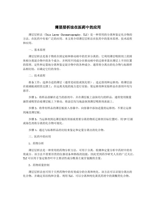
薄层层析法在医药中的应用薄层层析法(Thin Layer Chromatography,TLC)是一种常用的分离和鉴定化合物的方法,在医药中有着广泛的应用。
本文将介绍薄层层析法在医药中的基本原理、技术流程和应用。
一、基本原理薄层层析法是基于物质在固定相和移动相中的差异分离的。
它利用薄层吸附剂上的固体相分离混合物中的各个成分,并利用不同成分在移动相中的迁移率落在薄层上不同位置的原理,定性和定量地分离和鉴定混合物中的各种成分。
通常将分离出的化合物与标准样品相比较,以确定它们的身份。
二、技术流程准备工作:选择合适的薄层(通常是硅胶或氧化铝)、选定溶剂和运移剂;将薄层涂在玻璃板或铝箔支撑上;在远离光线的地方进行实验,使运移剂和实验样品在溶剂中均匀悬浮。
步骤1:将样品溶解在适当的溶剂中,并在薄层板上涂抹均匀的样品。
通常使用微量滴管或吸管沿着薄层板上下移动,将涂层均匀地涂抹到薄层吸附剂表面上。
步骤2:将带有样品的薄层板放入容器中,向容器中添加适量的运移剂,不要让运移剂淹没薄层板。
步骤3:当运移剂到达薄层板的顶部或需要分离的物质迁移到目标位置时,用UV灯源或染色剂将分离的化合物可视化。
步骤4:通过与标准样品的比较来鉴定和定量分离出的化合物。
三、医药中的应用1、药物分析薄层层析法是一种常用的药物分析方法,可用于分离、检测和定量分析中药材中的有效成分。
该方法不需要昂贵的仪器设备和熟练的技能,因此受到药学研究人员的广泛关注。
TLC可以用于鉴定银杏叶中主要活性成分酰基左旋甘氨酸的含量。
2、药物质量控制薄层层析法也可用于天然药物中的有效成分的分离和纯化。
该方法可以识别分离出的化合物,并确定其结构和含量。
利用TLC,可以分离和纯化黄芪药材中的黄酮类化合物。
薄层层析法可以用于毒物检测和识别。
该方法可以分离毒物和其代谢产物,并将其与标准毒物库中的物质进行比较。
TLC可以用于检测尿液中的吗啡、可卡因和芬太尼等毒物。
薄层层析法具有简便,快速,准确,无需昂贵的仪器和熟练的技能等优点,在医药中有着广泛的应用。
薄板层析
薄层色谱法(英语:Thin layer chromatography,简称TLC,又称为薄层层析)是一种用于分离混合物的色谱技术。
[1]在化学分析特别是对于有机化合物的分析中,薄层色谱是极为重要的分离方法。
薄层色谱在覆盖有很薄一层吸附剂的玻璃板、塑料片或铝箔上进行。
吸附剂又称为薄层色谱固定相:常为硅胶、氧化铝或纤维素。
操作时先将混合物样品用毛细管点于板上,而后在密闭的层析缸中,用单一或混合溶剂作为流动相,通过毛细作用缓慢地将混合物试样中的成份由下而上带到板的顶端。
由于样品中各组分对于吸附剂的作用力不同,且在洗脱溶剂的溶解度也不同,导致各组分的上升速度有差异而最终在板上形成不同的斑点,达到分离混合物的目的。
[2]薄层色谱可用于:▪监测反应进程▪在已经有对照样品的条件下鉴定化合物▪测定物质的纯度下列为使用薄层色谱的实例:▪分析神经酰胺与脂肪酸▪检测在食物和水中的农药或杀虫剂▪在法医的工作中,分析纤维的染料成份▪化验放射性药物的放化纯度▪鉴定药用植物和它们的组成[3]高效薄层色谱是对经典薄层色谱的改进法之一,该法中色谱的灵敏度和分辨力都有很大的提高,可以准确地检出极微量的物质。
[编辑]薄层板的制作TLC板通常可在市场上直接购买,如硅胶G板或聚酰胺板等,根据其固定相标准颗粒大小范围而分为不同规格,通常颗粒越细分离效果越好。
将吸附相(如硅胶)与少量惰性粘合剂(硫酸钙)和水混合形成的浆状物,均匀地铺于以玻璃片、厚铝箔或塑料制成的载板上。
铺过固定相的板先晾干,然后在烤炉内于110℃加热三十分钟进行活化。
用于分析鉴定时吸附剂厚度一般为0.1–0.25 毫米,而用于制备时(见下文)则为0.5–2.0 毫米。
[4] [编辑]薄层层析技术展开一个TLC板,一个紫色的斑点被分离为一个红色斑点与一个蓝色斑点10种精油通过薄层色谱展开后,再使用香草醛试剂显色后的效果。
薄层层析的操作过程类似于纸层析,且原理方面也相似于柱色谱,因而与纸色谱相比具有很多优势,例如它分离效果好,灵敏快速,对于固定相的选择更多,且TLC 的结果还可作为柱色谱的参考。
TLC显色方法英文版
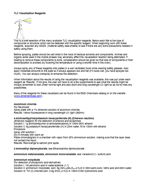
TLC Visualization ReagentsThis is a brief selection of the many available TLC visualization reagents. Below each title is the type of compounds or structure which can be detected with the specific reagent. When beginning work with these reagents, acquired any MSDS (material safety data sheets) to see if there are any extra precautions needed in safely using them.Before spraying, plates should be well dried in the hood of residual solvents and components. Amines and organic acids used in the mobile phases may adversely affect the visualization reaction being attempted. If heating to remove these components is done, consideration should be given so that loss of components or their decomposition is avoided (by lowering the temperature or using a shorter time in the oven).Always spray any of these reagents onto plates in a well ventilated hood while wearing safety glasses. Also apply moderate amounts to the plate so it always appears dull and flat (if it looks wet, you have sprayed too much). You can always overspray to enhance the detection.When information about the results of using the visualization reagents was available, this was put under each reagent as ‘Results’. If not give, the user will have to do a few experiments to see what the results might be. Always remember to look under normal light and also short and long wavelength UV light so as not to miss any possibilities.Many of the reagents for these visualizers can be found in the EMD Chemicals catalog or on the website ().Aluminium chlorideFor flavonoidsSpray plate with a 1% ethanolic solution of aluminum chloride.Results: Yellow fluorescence in long wavelength UV light (360nm)4-Aminoantipyrine/potassium hexacyanferrate (III) (Emerson reaction)(Emerson reagent) for the detection of phenols and arylaminesSolution I: 1g aminoantipyrine (4-aminophenazone) in 100ml 80% ethanolSolution II: 4g potassium hexacyanoferrate (III) in 20ml water, fill to 100ml with ethanolProcedure:Spray with solution IDry 5 minutes with warm airPlace chromatogram in a chamber with vapor from 25% ammonium solution, making sure that the layer does not contact the liquid.Results: Red-orange to salmon pink spots2-Aminoethyl diphenylborate, see Ethanolamine diphenylborateAmmonium metavanadate, ammonium monovanadate, see Vanadium(V) / sulfuric acidAmmonium molydbateFor detection of phosphoric acid derivativesSolution I: 1M perchloric acid in water/acetone (1:1)Solution II: ammonium molybdate soln: 5g (NH4)6Mo7O24.4 H2O in 35ml semi-conc. Nitric acid and 65ml water. Solution III: Tin (II) chloride soln: 0.5g SnCl2.2 H2O in 100ml 0.5M hydrochloric acid:Dry developed chromatogram and heat to 60CHydrolyse di- and triphosphates by spraying perchloric acid (solution I) onto the warm plate. After spraying 2 times, dry plate slowly at 50C. Amidophosphates might not be decomposed.In any case, spray the still warm plate with ammonium molybdate solution (solution II)Then spray the still wet plate with tin (II) chloride solution (solution III)Results: Phosphates appear as blue to blue-green spots. Polyphosphates can also be detected by dipping the plates in a solution of ammonium molybdate (1g) dissolved in water (8ml) and perchloric acid (3ml, ca. 70%), filled up to 100ml with acetone. Then phosphates appear as yellow-green spots on a blue background. Also see Molybdenum blue reaction according to Dittmer and Lester.Aniline phthalateFor the detection of reducing sugarsDry the developed chromatogramSpray with 0.93g aniline and 1.66g o-phthalic acid dissolved in 100ml n-butanol saturated with water.Briefly dry with hot air, then heat to 105C for 10 minutesResults: Substance spots show different colors on an almost colorless background. Some spots give fluorescence at 365nm.p-Anisaldehyde – sulfuric acidFor detection of phenols, sugars, steroids, and terpenesSpray with a solution of freshly prepared 0.5ml p-anisaldehyde in 50ml glacial acetic acid and 1ml 97% sulfuric acid.and heat to 105°C until maximum visualization of spots. The background might be brightened by water vapor. Results: Lichen constituents, phenols, terpenes, sugars, and steroids turn violet, blue, red, grey or green.For detection of sugarsSpray with a solution of freshly prepared 1ml p-anisaldehyde, 1ml 97% sulfuric acid in 18ml ethanol and heat at 110°C.Results: Sugar phenylhydrazones produce green-yellow spots in 3 min. Sugars will produce blue, green, violet spots in 10min. Also detects digitalis glycosides.p-Anisidine HydrochlorideFor detection of carbohydrates / sugarsMix a solution of 3% p-anisidine hydrochloride in n-butanolSpray and heat at 100°C for 2-10min.Results: Aldohexoses are seen as green-brown spots, ketohexoses as yellow spots, aldopentoses as green spots, and uronic acids as red spots.Anisidine phthalateFor detection of carbohydrates and reducing sugarsSpray with a solution of 1.23 g p-anisidine and 1.66g phthalic acid in 100ml 95% ethanol.Results: Hexoses, green; pentoses, red-violet – sensitivity 0.5ug; methylpentoses, yellow-green; uronic acids, brown – sensitivity 0.1-0.2ug.Antimony (III) chlorideFor detection of flavonoidsSpray with a 10% solution of antimony (III) chloride in chloroformResults: Fluorescing spots in long wavelength light (360nm).Antimony (III) chlorideFor detection of vitamins A & D, carotenoids, steroids, sapogenins, steroid glycosides, terpenesSpray with a solution of 25g antimony (III) chloride in 75ml chloroform (generally a saturated solution of antiomony (III) chloride in chloroform or carbon tetrachloride is used).Heat 10min at 100C, view under long wavelength light (360nm).Bromine / Carbon tetrachlorideFor detection of organothiophosphorous pesticidesPlace chromatogram in a chamber with a 10% bromine and tetrachloride without contact with the liquid.Bromocresol greenFor detection of organic acidsDip chromatogram in a solution of 0.1g bromocresol green in 500ml ethanol and 5ml 0.1M NaOHResults: Acids yield yellow spots on a blue background.Bromthymol blueFor detection of lipids and phospholipidsReagent: 0.1% bromthymol blue in 10% aqueous ethanol made just alkaline with NH4OHSpray dried plate.Results: Compounds above produce blue-green colors; sensitivity 0.1-1µg.Chloranil reagentFor detection of phenolsSpray with a solution of 1% tetrachloro-p-benzoquinone in tolueneChlorine / o-tolidineFor detection of of compounds forming chloroamines, e.g., urea derivatives, carbamated, antibioticsSolution I: 160mg o-tolidine in 30ml glacial acetic acid, filled to 500ml with distilled water, plus 1g KI solution Solution II: saturated solution of o-tolidine in 2% acetic acid/0.85% KI solution (1:1, v/v)Procedure APlace chromatogram 15-20min in a chlorine atmosphere (e.g., Potassium permanganate +10% Hydrochloric acid)Leave 5 minutes at ambient temperature until the chlorine is evaporated completely (spray corner of plate to insure no blue color is seen, showing complete absence of chlorine).Spray with solution IProcedure BSpray with 2% potassium hypochlorite solution in waterLeave 1-1.5hr at ambient temperatureSpray with solution IICopper sulfate / phosphoric acidUsed as a charring reagent for polymer bound TLC plates (the newer hard layer plates)Spray with a solution of 10% copper (II) sulfate in 10% phosphoric acidHeat 5-30min at 110°CResults: View frequently (every 5-10min) to see if colored or fluorescent spots (at 254 and 360nm) can be seen. Charring can be continued until spots are brown, grey or black.Chromosulfuric acidSee under Potassium dichromate / sulfuric acidDDQ Reagent (Dichlorodicyanobenzoquinone)For detection of phenolsSpray with a solution of 2% 2,3-dichloro-5,6-dicyano-1,4-benzoquinone in tolueneDichlorofluoresceinFor the detection of sweeteners saccharine & cyclamateSpray with a 0.2% solution of dichlorofluorescein in 96% ethanolDry with warm air; if necessary, spray with waterView under 360nm UV lightDichlorofluorescein / fluorescein sodium saltFor detection of N-substituted barbituratesSpray with a 0.1% ethanolic solution of dichlorofluoresceinThen spray with a 0.1% ethanolic solution of fluorescein sodium salt2,6-Dichloroquinone -4- chloroimideFor detection of antioxidants, phenols, primary and secondary aliphatic amines, secondary and tertiary aromatic amines, aromatic hydrocarbons, pharmaceuticals, phenoxyacetic acid herbicides, etcSpray with a freshly prepared 0.5-2% solution of 2,6-dichloroquinone-4-chloroimide in ethanol (reagent stable for 3 weeks if refrigerated).Heat 10min at 110C; treat with ammonia vaporp-DimethylaminobenzaldehydeFor detection of sulfonamidesSpray with a solution of 1% p-dimethylaminobenzaldehyde in 5% hydrochloric acid; add 5% ethanolDetects sulfonamidesp-Dimethylaminobenzaldehyde / hydrochloric acid reagent (Ehrlich’s reagent)For detection of amines, indole derivativesSpray with a solution of 1% p-dimethylaminobenzaldehyde in conc. hydrochloric acid/methanol (2:2)Heat plates for 20min at 50C2,4-DinitrophenylhydrazineFor detection of aldehydes and ketonesSpray plate with solution of 0.4 g 2,4-DNPH in 100ml 2N hydrochloric acid, add 1ml ethanolResults: Yellow-red spots will be seen.DiphenylamineFor detection of glycosides, glycolipidsReagent: 10ml 10% diphenylamine in ethanol, 100ml HCl and 80ml glacial acetic acidSpray lightly, cover plate with another glass plate, heat 30-40min at 110°C until positive areas appear Results: Glycolipids produce blue spots.s-DiphenylcarbazoneFor detection of barbituratesSpary with a solution of 0.1% s-diphenylcarbazone in 95% ethanolResults: Barbiturates will produce purple spots2,2’-DiphenylpicrylhydrazylFor detection of aldehydes and ketonesReagent: dissolve 15mg of 2,2’-DPPH in 25ml chloroformSpray, heat 5-10min at 110°C;Results: Yellow spots on a purple background will be seen.DithizoneFor detection of heavy metal ionsDissolve 20mg dithizone in 100ml acetone, store in a brown bottle in a refrigeratorProcedure:Spray with dithizone solutionSpray with 25% ammonia solutionDittmer and LesterSee Molybdenum blueDragendorff reagentFor detection of nitrogen compounds, alkaloids, antiarrhythmic drugs, surfactantsSolution 1) 1.7g basic bismuth nitrate and 20g tartaric acid in 80ml waterSolution 2) 16 g potassium iodide in 40ml waterStock solution (stable for several weeks in a refrigerator):Mix equal volumes of solutions 1 and 2Procedure:Spray with a solution of 10g tartaric acid, 50ml water and 5ml stock solutionEthanolamine diphenylborate (flavone reagent according to Neu)For detection of flavonoidsSpray with a 1% solution of ethanolamine diphenylborate in methanolSpray with a 5% ethanolic solution of polyyethylene glycol for fluorescence stabilizationIrradiate 2 minutes with intense 365nm UV lightView under 365nm UV lightErhlich’s reagentSee p-DimethylaminobenzaldehydeEmerson reagentSee 4-aminoantipyrine/potassium hexa-cyanoferrate (III)Fast Blue B reagentFor detection of cannabinoids, phenols, tanning agents, amines with can be coupledSpray with a solution of 0.5g Fast Blue B (tetraazotized di-o-anisidine) in acetone/water (9:1, v/v), always prepared freshThen overspray with 0.1M sodium hydroxide solutionResults: Cannabinoids turn dark red/purple in colorFerric Chloride / sulfuric acidUsed as a charring reagent for polymer bound TLC plates (the newer hard layer plates)Spray with a solution of 2g FeCl3 in 83ml n-butanol and 15ml conc. sulfuric acid.Heat 5-30min at 110°CResults: View frequently (every 5-10min) to see if colored or fluorescent spots (at 254 and 360nm) can be seen. Charring can be continued until spots are brown, grey or black.Flavone reagent according to NeuSee ethanolamine diphenyl borateFluorescamineFor detection of primary and secondary amines, peptides, sulfonamides, e.g., nitrosoamines after photolysis Spray plate with a solution of 0.1mg/ml 4-phenyl-spiro[furan-2(3H),1-phthalan]-3,3-dione in acetone prepared fresh dailyFor stabilization of fluorescence at 366nm spray with 10g triethylamine, brought to 100ml with dichloromethane. Fluorescent IndicatorFor detection of compounds which absorb UV lightSome TLC plates when manufactured have an inorganic fluorescent indicator added to the slurry poured to make the final plates. This type of indicator will not dissolve or elute off. They are activated at 254nm or 360nm (see recommendations of the manufacturer for that type of plate).Results: When activated the fluorescent indicator will turn a green or white (depending on the indicator added) and the compounds appear as dark spots or shadows against this background. If viewing at other than the activation wavelength, the compounds might also have some fluorescence of their own, so various colors against a dark background would be seen.Formaldehyde / sulfuric acidFor detection of alkaloids, aromatic hydrocarbons, e.g., antihypertensive drugsSpray with a solution of 37% formaldehyde in conc. sulfuric acid (1:10) immediately after taking the plate from the developing chamber. Heating is not necessary.Results: various colored spots.Formaldehyde / phosphoric acidFor detection of steroid alkaloids, steroid sapogenins and phenothiazine derivativesSpray with a solution of 0.03g formaldehyde in 100ml of 85% phosphoric acid with stirring at room temperature. The reagent is stable for several weeks.Furfural / sulfuric acidFor detection of carbamate estersSpray solution I: 1% solution of furfural in acetoneSpray solution II: 10% solution of sulfuric acid in acetoneSpray plate with I, then II.Gentian Violet – BromineFor detection of lipidsSpray 0.1% gentian violet (crystal violet) in methanol onto plate and place in a tank containing bromine vapor. Results: lipids produce blue spots on a yellow background.Gibb’s reagentFor detection of phenols. For further applications see 2,6-dichloroquinone-4-chloroimideSpray with a solution of 3% 2,6-dibromo-N-chloro-p-benzoquinone imine in toluene or methanol. Hydroxylamine / iron (III) chlorideFor detection of amides, lactones, carboxylic acid esters and anhydridesSolution 1) Mix 1 vol part of 7g hydroxylammonium chloride in 100ml methanol w 1 vol part of a solution of 7.2 g potassium hydroxide in 100ml methanol. Filter from precipitated potassium chloride.Solution 2) 2% solution of iron (III) chloride in 1% aqueous hydrochloride acidSpray air dried plate first with solution 1, then with solution 2Iodine containing compoundsDetection by decomposition under UVDry plates at 100CAfter cooling spray with a small amount of 50% acetic acidIrradiate some minutes with unfiltered UV light.Results: Iodine compounds show weakly violet to brown spots. The color can be enhanced by spraying with 10% acetic acid and irradiation with UV light (sudden appearance of blue spots).Iodine vaporRelatively unspecific universal reagent for many organic compoundsCharge chamber with some crystals of iodinePlace developed, dried chromatogram in iodine vaporResults: spots turn tan-brown in colorIodoplatinateFor detection of organic nitrogen compounds, alkaloids, e.g., cocaine metabolitesSpray with a freshly prepared mixture of 3ml hexachloroplatinic (IV) acid solution (10%) in 97ml/min water and 100ml aqueous potassium iodide solution.Note - use 5% ethanol or methanol in water to prepare these solutions for the new polymer bound TLC plates. Iron (III) chloride / potassium hexacyanoferrate / sodium arsenate (according to Patterson & Clements) For detection of iodine compounds, e..g., thyroid gland hormonesSolution I: 2.7% iron (III) chloride hexahydrate in 2N hydrochloric acidSolution II: 3.5% potassium hexacyanoferrate in waterSolution III: dissolve 3.8g arsenic trioxide in 25ml 2N sodium hydroxide solution heating slightly, cool to 5C, and add 50ml 2N sulfuric acid, fill to 200ml with waterImmediately before use mix 5ml solution I, 5ml solution II and 1ml solution III.Spray onto the dry layer and dry carefully (temp below 50C)Cover with glass plate and leave 15min in the darkResults: Iodine containing compound show light blue spots on a yellowish background.Lead tetraacetate / 2,7-dichlorofluoresceinFor detection of vicinal diols, glycosides and phenols, e.g., sugar acidsSolution I: 2% (w/v) lead tetraacetate in glacial acetic acidSolution II: 1% (w/v) 2,7-dichlorofluorescein in ethanolMix 5ml of each solution 1 and 2, fill to 200ml with dry toluene. This reagent solution is stable for only about 2 hours.Manganese / salicylaldehydeFor detection of organothiophosphorus pesticidesSolution I: dissolve 100mg manganese chloride (MnCl2.4H2O) in 100ml 80% alcoholSolution II: dissolve 1.3g 2-hydrozine quinoline in the lowest possible volume of hot ethanol. Dissolve 1 g salicyaldehyde in 5ml ethanol and add 1-2 drops glacial acetic acid. Combine both solutions and reflux 30 minutes. The crystals of salicyl-2-aldehyde –2-quinolinehydrazone precipitated during the cooling are recrystallized from ethanol. For solution dissolve 50mg of the salicylate derivative in 100ml ethanolSpray with a mixture of equal volumes of solutions 1 and 2.Mandelin’s reagentSee Vanadium(V) / sulfuric acidMercury (II) chloride / diphenylcarbazoneFor detection of barbituratesSolution I: 2% ethanolic mercury (II) chlorideSolution II: 0.2% ethanolic diphenlycarbazoneMix freshly before use in equal partsResults: Pink spots on a violet backgroundMercury (II) chloride / dithizoneFor detection of barbituratesSpray with a freshly prepared 1:1 mixture of 1-2% mercury (II) chloride in ethanol and 0.1-0.2% dithizone in ethanol.View under 360nm UV light4-Methoxybenzaldehyde / sulfuric acid / ethanolFor detection erythromycin and metabolitesSpray with 4-methocybenzaldehyde/sulfuric acid/ethanol (1:1:9)Heat 1 minute at 100CMethyl yellowFor detection of chlorinated insecticides and antimicrobial compoundsSpray dried plate with a solution of 0.1g methyl yellow (N,N-dimethyl-4-phenylazoaniline) in 70ml ethanol, add 25ml water and fill to 100ml with ethanol.Dry at ambient temperatureIrradiate 5 min with UV light without a filterResults: Red spots on a yellow backgroundMolybdatophosphoric acidSee under Phosphomolybdic acidMolybdenum blue reaction according to Dittmer and LesterFor detection of phospholipids and phosphoric acid derivativesSolution I: Boil 40.11g MoO3 in 1 liter 25N sulfuric acid for 3-4 hours until the molybdenum oxide is completely dissolved. Let the light yellow solution slowly cool to ambient temperature overnight. The solution will turn light blue.Solution II: Boil 1.78 g molybdenum powder and 500ml of solution I for 15min, cool and decant from the remaining residue.For preparation of the spray reagent, add equal volumes of solutions I and II to 4.5 volume parts water. A dark green solution is formed.Solutions I and II are stable for several months when stored in the dark. The spray reagent has to be prepared weekly.NinhydrinFor detection of amino acids, amines, amino sugars.Spray with a solution of 0.2g ninhydrin in 100ml ethanol and heat to 110C until spots appear.Results: reddish spots appearNinhydrin / cadmium acetateFor detection of amino acids and heterocyclic aminesDissolve 1g ninhydrin and 2.5g cadmium acetate in 10ml glacial acetic acid and fill to 500ml with ethanol. Spray and heat 20min at 120CResults: Red, pink, or purple spots are seen.Ninhydrin / pyridine / glacial acetic acidFor detection of peptidesSpray with a 1% ninhydrin in pyridine/glacial acetic acid (5:1, v/v)Heat 5 min at 100CNitric acid / ethanolFor detection of amines and alkaloidsSpray with a solution of 50 drops 65% nitric acid in 100ml ethanol (higher acid concentrations are also possible). If necessary, heat to 120C for some time.Orcinol (Bials reagent)For detection of glycosides, glycolipidsReagent: dissolve 0.1g orcinol in 40.7ml conc. HCl, add 1ml 1% ferric (111) chloride, and dilute to 10mlSpray and heat at 80°C for 90 minutes.Results: Glycolipids produce violet spots.Patterson and ClementsSee under Iron (III) chloride/potassium hexacyanoferrate/sodium arsenateParaffin oilFor enhancement of fluorescence spots – more stable and greater intensity1% paraffin oil in hexaneSpray evenly over the TLC plateResults: spots should be more stable (no fading with time) for scanning and are of greater intensitym-PhenylenediamineFor detection of reducing sugarsSpray with a solution of 3.6g m-phenylenediamine dihydrochloride in 100ml 70% ethanol and heat briefly at 105°CResults: Intensely fluorescence colors in UV light (wavelength not specified, so check 254 and 366nm).o-Phenylenediamine – trichloroacetic acidFor detection of alpha-keto acidsSpray with a solution of 0.05g 1,2-phenylenediamine in 100ml 10% aqueous trichloroactic acid and heat plate at 100C for no more than 2 minutes.Results: Green fluorescence spots in long wavelength UV light.p-Phenylenediamine – phthalic acidFor detection of conjugated 3-ketosteroidsSpray with a solution of 0.9 p-phenylenediamine and 1.6g phthalic acid in 100ml 1-butanol saturated with water and heat plate at 100-110°CResults: Yellow to orange spotsPhenylhydrazine sulfonateFor detection of some antimicrobial compoundsSolution I: dissolve 3.5g phenylhydrazine 4-sulfonic acid hemihydrate in 10ml water and 20ml 1N NaOH solution Solution II: mix 30ml 1N sodium hydroxide solution with 40ml acetoneThe spray reagents have to be prepared fresh each time.Procedure:Wet chromatogram evenly with spray solution 1After air drying the plate, shake spray solution 2 and spray plate.Phosphoric acidFor detection of sterols, steroids, and bile acidsSpray heavily until the layer appears transparent with a solution of 85% phosphoric acid with water (1:1, v/v) Then heat 10-15minutes at 120°CResults: Sterols, steroids and bile acids and bile acids produce various colors under visible and UV light. Phosphoric acid – bromineFor detection of digitalis glycosidesSpray solution I: 10% aqueous phosphoric acid solutionSpray solution II: Mix 2ml saturated aqueous potassium bromide, 2ml saturated solution aqueous potassium bromate and 2ml 25% hydrochloric acid.Procedure: Spray plate with I and heat 12 min at 120°C.Results: Digitalis glycosides of the series B, D, and E show blue fluorescence in long wavelength UV light Procedure continued: Heat the plate again at 120°C and spray lightly with IIResults: Glycosides of the series A show orange, of the series C show grey-green to grey-blue fluorescence in UV light.Phosphomolydbic acidFor detection of reducing substances, e.g, alcohols, bile acids, lipids, fatty acids, steroidsAlso used as a charring reagent for polymer bound TLC plates (the newer hard layer plates)Spray with a solution of 250mg molybdatophosphoric acid in 50ml ethanolHeat to 120C until spots appear (oven or heat gun)If necessary, treat with ammonia vapors to remove some background coloration.The reagent solution is stable for only 10 days even in the dark.Results when using as a charring reagent: View frequently (every 5-10min) to see if colored or fluorescent spots (at 254 and 360nm) can be seen. Charring can be continued until spots are brown, grey or black. Phosphotungstic acidFor detection of cholesterol and its esters, reducing compounds, lipids, sterols, and steroidsSpray with 20% phosphotungstic acid in ethanol, heat at 110°C for 5-15min or until maximum visualization of the spots occursResults: Cholesterol, esters will produce red spots.Pinacryptol yellowFor detection of sweetners, surfactants, alkyl- and arylsulfonic acidsDissolve 100mg pinacryptol yellow in 100ml hot water or ethanol (or some combination for polymer bound plates)Spray with reagent solutionResults: Yellow to orange fluorescence spots under long wavelength UV light (366nm)Potassium dichromate / sulfuric acid (chromosulfuric acid) - see belowPotassium permanganate / sulfuric acid – see belowRhodamine BFor detection of a wide variety of compoundsDry the developed chromatogram and spray with 0.025-0.25% ethanolic rhodamine B reagent.Results: In most cases red-violet zones with an intense fluorescence at 365nm develop on a pink background. RP phases are less suited, because in this case the environment of the spots also forms an intense color. By placing the sprayed chromatograms into an ammonia atmosphere, the detection sensitivity can be improved. Rhodamine 6 GFor detection of lipidsSpray plate with a solution of 1mg rhodamine 6 G in 100ml acetoneInspect at long wavelength UV.Silver nitrate / hydrogen peroxideFor detection of halogenated hydrocarbonsSpray plate with a solution of 0.1g silver nitrate in 1ml water, add 10ml 2-phenoxyethanol, fill to 200ml with acetone and add 1 drop hydrogen peroxide (30% solution)Irradiate with unfiltered UV light until optimal contrast is obtained. For alumina plates, about 50 minutes, for silica gel plates about 15 minutesResults: dark spots are formed.Luckow [Fensenius Z. Anal. Chem., 294, 288, (1979)] uses this combination without the addition of hydrogen peroxide for the detection of pesticide ioxynil (3,5-diiodo-4-hydroxybenzonitrile).Sodium azideFor detection of antibiotics (penicillins and cephlosporins)Solution I: 0.5% solution of soluble starchSolution II: 3.5% sodium azide in 0.1N iodine solutionProcedure: spray with 1), dry, spray with 2)Results: Detects penicillin, sensitivity 0.2µg. Penicillin and penicilloic acids are also detected by starch-iodine-iodine reagents, as are cephalosporins.Sodium 1,2-napthaquinone-4-sulfonate (NZS reagent)For detection of thiazide drugs, basic drugs with primary amino groupsSolution I: 0.1N NaOHSolution II: saturated solution of reagent in 1:1 ethanol: waterSpray with I, then II.Results: Thiazide drugs appear as orange spots within 15min; basic drugs with primary amino groups also react but barbiturates do not.Sodium nitrite / hydrochloric acidFor detection of indoles and thiazolesSpray plate with a freshly made solution of 1g sodium nitrite in 100ml hydrochloric acid, 1mol/L and heat at 100°C.Results: Indoles turn red and thiazole derivatives turn light green.Sodium nitroprusside / hydrogen peroxideFor detection of guanidine, urea, thiourea and derivatives, creatine and creatinine.Spray with a solution made up of 2ml 5% aqueous sodium nitroprusside, 1ml 10% aqueous sodium hydroxide and 5ml 3% aqueous hydrogen peroxide and dilute with 15ml water. This solution can be stored several days in the refrigerator.Sodium nitroprussate / potassium hexacyanoferrate (III)For the detection of aliphatic nitrogen compounds, cyanamide, guanidine, urea, thiourea and derivatives, creatine, and creatinine.Spray with a solution of 1 volume part each of 10% aqueous sodium hydroxide, 10% sodium nitroprussate, and 10% potassium hexacyanoferrate (III) with 3 vol. parts water. Let the solution stand at least 20min at ambient temperature before use. Stored in the refrigerator, it is stable for several weeks.Procedure: For use, mix the reagent solution with an equal part of acetone and spray.Stannic ChlorideSee under Tin (IV) chlorideTetracyanoethylene - TCNE reagentFor detection of aromatic hydrocarbons and heterocycles, aromatic amines, and phenolsSpray with a solution of 0.5-1.0g tetracyanoethylene in dichloromethane or toluene.Results: Aromatic hydrocarbons show various colors, some of them for a brief time. Also try heating at 100°C for a short time.TetranitrodiphenylFor detection of cardiac glycosidesSpray solution I: Saturated solution of 2,3’,4,4’-tetranitrodiphenyl in tolueneSpray solution II: 10% potassium hydroxide solution in 50% aqueous methanolSpray with I, dry at room temperature, then spray with II.Results: Blue spots are observed.Tetrazolium blueFor detection of corticosteroids and other reducing compounds.Spray with a freshly prepared 1:1 mix of a) 0.5% methanolic tetrazolium blue solution and 6M NaOH in water or methanol/water (1:1).Results: violet spots are observed at room temperature or with slight warming.Thymol / sulfuric acidFor detection of sugarsSpray with a solution of 0.5g thymol in 95ml ethanol, and add 5ml 97% sulfuric acid with caution.Heat 15-20min at 120°CResults: Sugars show pink spots.Tin (IV) chlorideFor detection of triterpenes, sterols, steroids, phenols, and polyphenolsSpray with a solution of 10ml tin (IV) chloride in 160ml equal volumes of chloroform and glacial acetic acid. Heat the layer for 5-10min at 100°C and inspect in visible and long wavelength UV light.o-Tolidine, diazotizedFor detection of phenolsTolidine solution – fill up 5g o-tolidine and 14ml conc. hydrochloric acid to 100ml waterNitrate solution – 10% aqueous sodium nitrate solution prepared freshSpray solution – mix 20ml tolidine solution and 20ml nitrate solution at 0°C stirring constantly.The spray solution is stable for about 2-3 hours.After spraying it can take several hours until colored spots are formed.p-Toluenesulfonic acidFor detection of steroids, flavonoids and catechinsSpray with a solution of 20% p-toluenesulfonic acid in chloroform and heat a few minutes at 100°C.Inspect under long wavelength UV light.Trichloroacetic acidFor detection of steroids, digitalis glycosides, veratrum alkaloids and vitamin DSpray solution I: 25% solution of trichloroacetic acid in chloroform.Spray solution II – for vitamin D – 1% trichloroacetic acid in chloroform.。
最全的TLC经验-薄层层析-显色剂【范本模板】

最全的TLC经验薄层色谱(TLC)是一种非常有用的跟踪反应的手段,还可以用于柱色谱分离中合适溶剂的选择。
薄层色谱常用的固定相有氧化铝或硅胶,它们是极性很大(标准)或者是非极性的(反相)。
流动相则是一种极性待选的溶剂。
在5.301中以及大多数实验室实验中,都将使用标准硅胶板.将溶液中的反应混合物点在薄板上,然后利用毛细作用使溶剂(或混合溶剂)沿板向上移动进行展开.根据混合物中组分的极性,不同化合物将会在薄板上移动不同的距离。
极性强的化合物会“粘”在极性的硅胶上,在薄板上移动的距离比较短。
而非极性的物质将会在流动的溶剂相中保留较长的时间从而在板上移动较大的距离。
化合物移动的距离大小用Rf值来表达。
这是一个位于0~1之间的数值,它的定义为:化合物距离基线(最先点样时已经确定)的距离除以溶剂的前锋距离基线的距离。
薄层色谱(TLC)实验步骤:1) 切割薄板。
通常,买来的硅胶板都是方形的玻璃板,必需用钻石头玻璃刀按照模板的形状进行切割。
在切割玻璃之前,用尺子和铅笔在薄板的硅胶面上轻轻地标出基线的位置(注意不要损坏硅胶面)。
借助锋利的玻璃切割刀和一把引导尺,你便可方便地进行玻璃切割。
当整块玻璃被切割后,你就可以进一步将其分成若干独立的小块了.(开始的时候,也许你会感到有一些难度,但经过一些训练以后,你便会熟练地掌握该项技术。
)2)选取合适的溶剂体系。
化合物在薄板上移动距离的多少取决于所选取的溶剂不同。
在戊烷和己烷等非极性溶剂中,大多数极性物质不会移动,但是非极性化合物会在薄板上移动一定距离.相反,极性溶剂通常会将非极性的化合物推到溶剂的前段而将极性化合物推离基线。
一个好的溶剂体系应该使混合物中所有的化合物都离开基线,但并不使所有化合物都到达溶剂前端,Rf值最好在0.15~0。
85之间。
虽然这个条件不一定都能满足,但这应该作为薄层色谱分析的目标(在柱色谱中,合适的溶剂应该满足Rf在0.2~0。
3之间)。
那么,应该选取哪些溶剂呢?一些标准溶剂和他们的相对极性(从LLP中摘录)列于如下:强极性溶剂:甲醇〉乙醇〉异丙醇中等极性溶剂:乙氰〉乙酸乙酯〉氯仿>二氯甲烷〉乙醚〉甲苯非极性溶剂:环己烷,石油醚,己烷,戊烷常用混合溶剂:乙酸乙酯/己烷:常用浓度0~30%.但有时较难在旋转蒸发仪上完全除去溶剂。
薄层色谱(TLC)详细介绍及制作

薄层色谱,或称薄层层析(thin-layer chromatography),是以涂布于支持板上的支持物作为固定相,以合适的溶剂为流动相,对混合样品进行分离、鉴定和定量的一种层析分离技术。
这是一种快速分离诸如脂肪酸、类固醇、氨基酸、核苷酸、生物碱及其他多种物质的特别有效的层析方法,从50年代发展起来至今,仍被广泛采用。
一、基本原理薄层层析是把支持物均匀涂布于支持板(常用玻璃板,也可用涤纶布等)上形成薄层,然后用相应的溶剂进行展开。
薄层层析可根据作为固定相的支持物不同,分为薄层吸附层析(吸附剂)、薄层分配层析(纤维素)、薄层离子交换层析(离子交换剂)、薄层凝胶层析(分子筛凝胶)等。
一般实验中应用较多的是以吸附剂为固定相的薄层吸附层析。
吸附是表面的一个重要性质。
任何两个相都可以形成表面,吸附就是其中一个相的物质或溶解于其中的溶质在此表面上的密集现象。
在固体与气体之间、固体与液体之间、吸附液体与气体之间的表面上,都可能发生吸附现象。
物质分子之所以能在固体表面停留,这是因为固体表面的分子(离子或原子)和固体内部分子所受的吸引力不相等。
在固体内部,分子之间相互作用的力是对称的,其力场互相抵消。
而处于固体表面的分子所受的力是不对称的,向内的一面受到固体内部分子的作用力大,而表面层所受的作用力小,因而气体或溶质分子在运动中遇到固体表面时受到这种剩余力的影响,就会被吸引而停留下来。
吸附过程是可逆的,被吸附物在一定条件下可以解吸出来。
在单位时间内被吸附于吸附剂的某一表面积上的分子和同一单位时间内离开此表面的分子之间可以建立动态平衡,称为吸附平衡。
吸附层析过程就是不断地产生平衡与不平衡、吸附与解吸的动态平衡过程。
例如用硅胶和氧化铝作支持剂,其主要原理是吸附力与分配系数的不同,使混合物得以分离。
当溶剂沿着吸附剂移动时,带着样品中的各组分一起移动,同时发生连续吸附与解吸作用以及反复分配作用。
由于各组分在溶剂中的溶解度不同,以及吸附剂对它们的吸附能力的差异,最终将混合物分离成一系列斑点。
薄层色谱法教学流程
薄层色谱法教学流程英文回答:Thin layer chromatography (TLC) is a widely used technique in analytical chemistry for separating and analyzing mixtures of compounds. It is a quick and inexpensive method that can provide valuable information about the composition of a sample. The teaching process for TLC can be divided into several steps.First, the instructor should introduce the basic principles of TLC to the students. This includes explaining the concept of chromatography, the different components of a TLC plate (such as the stationary phase and the mobile phase), and the factors that influence the separation of compounds.Next, the instructor should demonstrate the preparation of the TLC plate. This involves applying a thin layer of the stationary phase (usually silica gel or alumina) onto aglass or plastic plate. The plate is then dried and activated before use. The instructor should explain the importance of a uniform and evenly distributed stationary phase for accurate results.After the plate is prepared, the instructor can demonstrate the spotting technique. This involves applying a small amount of the sample mixture onto the plate using a capillary tube or a micropipette. The instructor should emphasize the need for precise and controlled spotting to ensure accurate separation.Once the samples are spotted, the TLC plate is placedin a developing chamber. The chamber is filled with a suitable solvent, which acts as the mobile phase. The instructor should explain the concept of solvent selection and its impact on the separation of compounds. The plate is then covered to prevent evaporation and left undisturbedfor a specific period of time.After the development, the TLC plate is removed from the chamber and allowed to dry. The instructor can thendemonstrate the visualization of the separated compounds. This can be done by exposing the plate to UV light, spraying it with a suitable reagent, or by using a visualization chamber. The instructor should explain the principles behind each visualization method and the importance of accurate interpretation of the results.Finally, the instructor should guide the students in analyzing and interpreting the TLC results. This involves measuring the Rf values (retention factor) for each separated compound and comparing them to known standards. The instructor should explain how to calculate the Rf values and how they can be used to identify unknown compounds.中文回答:薄层色谱法(TLC)是一种在分析化学中广泛应用的技术,用于分离和分析化合物混合物。
薄层色谱法制板流程
薄层色谱法制板流程英文回答:Thin-layer chromatography (TLC) is a common technique used in analytical chemistry for the separation and identification of compounds. The process involves the use of a thin layer of stationary phase, typically coated on a glass or plastic plate, and a mobile phase that moves over the stationary phase. The compounds in the sample are separated based on their different affinities for the stationary and mobile phases.The first step in the TLC plate preparation process is to choose the appropriate stationary phase material. Common materials include silica gel, alumina, and cellulose. These materials have different polarities and can be selected based on the desired separation characteristics. For example, silica gel is commonly used for separating nonpolar compounds, while cellulose is suitable for polar compounds.Once the stationary phase material is selected, it is mixed with a binder, such as a cellulose derivative or a polymer, to form a slurry. The slurry is then spread evenly onto a glass or plastic plate using a spreader or a pipette. The thickness of the stationary phase layer is typically around 0.1-0.25 mm, although it can vary depending on the application.After the stationary phase is applied, the plate isleft to dry in a fume hood or oven to remove any residual solvent. This ensures that the stationary phase adheres firmly to the plate and does not move during the chromatographic process.Once the TLC plate is prepared, it is ready for use. A small spot of the sample to be analyzed is applied to the plate using a capillary tube or a microsyringe. The plateis then placed in a developing chamber, which contains a suitable mobile phase. The mobile phase can be a single solvent or a mixture of solvents, depending on the compounds to be separated.As the mobile phase moves up the plate, the compounds in the sample will interact with the stationary phase. Compounds with higher affinity for the stationary phasewill move more slowly, while those with higher affinity for the mobile phase will move faster. This differential migration leads to the separation of the compounds on the TLC plate.The separation can be visualized by the use of a visualization technique, such as UV light, iodine vapor, or a specific staining reagent. The compounds appear as spots on the plate, and their Rf values (retention factor) can be calculated by measuring the distance traveled by the compound divided by the distance traveled by the mobile phase.In conclusion, the process of TLC plate preparation involves selecting the appropriate stationary phase material, applying it to a glass or plastic plate, and allowing it to dry. The prepared plate is then used for the separation and identification of compounds using a mobilephase. TLC is a versatile technique that is widely used in various fields, including pharmaceuticals, forensics, and environmental analysis.中文回答:薄层色谱法(Thin-layer chromatography,TLC)是一种常用的分析化学技术,用于化合物的分离和鉴定。
薄层色谱TLC点板教程市公开课获奖课件省名师示范课获奖课件
1)让溶剂向上展开约90%旳薄 板长度。
2)从展开池中取出薄板而且 立即用铅笔标注出溶剂到达 旳前沿位置。
3)让薄板上旳溶剂挥发掉, 将薄板干燥后喷以显色剂, 或在紫外灯下显色,用铅笔 标出全部显色点或有紫外活 性旳点。
单一溶剂旳极性从小到大顺序为:
❖ 石油醚<汽油<庚烷<己烷<二硫化碳<二甲苯 <甲苯<氯丙烷<苯<溴乙烷<溴化苯<二氯乙 烷<三氯甲烷<异丙醚<硝基甲烷<乙酸丁酯< 乙醚<乙酸乙酯<正戊烷<正丁醇<苯酚<甲乙 醇<叔丁醇<四氢呋喃<二氧六环<丙酮<乙醇 <乙腈<甲醇<氮氮二甲基甲酰胺<水
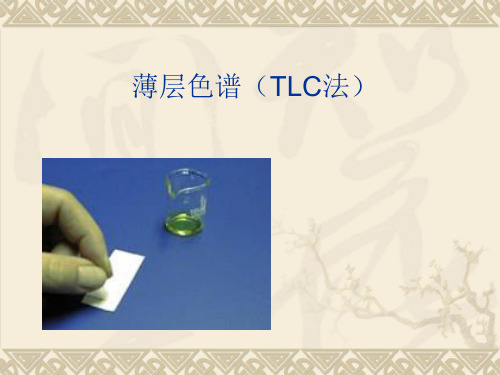
薄层色谱(TLC法)
❖ 定义、原理
薄层色谱(Thin Layer Chromatography)简称TLC,又 称薄层层析,属于固-液吸附色谱。样品在薄层板上旳吸 附剂(固定相)和溶剂(移动相)之间进行分离。因为多 种化合物旳吸附能力各不相同,在展开剂上移时,它们进 行不同程度旳解吸,从而到达分离旳目旳。
❖2.展开: ➢ 展开方式可分为三类:上行展开和下行展开;单次展开和屡
次展开;单向展开和多向展开 ➢ 常用旳是上行展开,就是使展开剂从下往上展开:当样点上
旳溶剂充分挥干后,将薄层置于盛有合适展开剂旳标本缸、 大量筒或方形玻缸中,使展开剂浸入薄层旳高度约为0.5cm。 ➢ 注意事项:先悬空饱和、再入液展开;样点不能泡在展开剂 中;薄层浸入时不能歪斜进入。
前沿线
色斑
起始线
❖ 3.显迹:用合适旳措施或者合适旳显色剂处理薄层,使 其上可能已经分离旳各成份斑点显示出来,以以便计算各 个斑点旳比移值,从而提供定性与定量旳根据。措施有:
tlc适宜的rf值 -回复

tlc适宜的rf值-回复TLC适宜的RF值指的是薄层层析法(Thin Layer Chromatography,TLC)实验中,具体化合物在薄层板上的迁移距离与溶剂前端移动距离之比。
在TLC实验中,RF值的测定是非常重要的,它可以用来确定化合物的迁移性质,帮助鉴别和定量分析样品中的化合物。
本文将一步一步回答关于TLC 适宜的RF值的各种问题。
第一步:理解TLC实验的基本原理和步骤在进行TLC实验之前,我们需要先了解该实验的基本原理和步骤。
薄层层析法是一种色层分离分析技术,它利用薄层板(通常是硅胶或铝箔)作为固定相,涂布在玻璃底板上,称为薄层板。
将涂有样品的点称为起点,然后将薄层板放入含有溶剂的容器中,让溶剂从底部渗透上升,溶剂前端移动到顶端。
在渗透过程中,样品中的化合物会在薄层板上分离出不同的斑点,这些斑点就是要鉴别和定量分析的化合物。
第二步:明确RF值的定义和计算公式RF值是指具体化合物在薄层板上的迁移距离与溶剂前端移动距离之比。
计算RF值的公式为:RF值= 化合物的迁移距离/ 溶剂前端移动距离通过测量化合物的迁移距离和溶剂前端移动距离,即可计算得到RF值。
第三步:确定适宜的RF值范围在进行TLC实验时,RF值的适宜范围是由实验数据和相关文献确定的。
一般来说,适宜的RF值范围应该在0.2至0.8之间。
如果RF值太小,说明化合物的迁移性不好,可能与固定相之间的相互作用太强,需要调整溶剂体系或固定相的选择。
如果RF值太大,说明化合物的迁移速度太快,可能与固定相之间的相互作用太弱,也需要调整溶剂体系或固定相的选择。
第四步:优化溶剂体系和固定相的选择为了使TLC实验得到准确和可靠的结果,我们需要对溶剂体系和固定相进行优化选择。
这涉及到溶剂选择、溶剂混合比例和固定相种类的选择。
一般来说,优化的溶剂选择应考虑溶剂的挥发性、溶解度和极性。
选择合适的溶剂体系可以提高化合物的分离效果,同时也能够调整化合物的迁移速度,从而获得适宜的RF值。
TLC Stains薄层色谱显色方法
TLC Stains薄层色谱显色方法TLC Stains薄层色谱显色方法The plate can be stained with iodine. This can be achieved rapidly, by shaking the plate in a bottle containing silica and a few crystals of iodine. The iodine will stain any compound that reacts with it and so is especially good for visualizing unsaturated compounds. Most spots show up within a few seconds, but the stain is not usually permanent.UV lightThe plate can be viewed under a ultraviolet lamp to show any uv-active spots.Dipping Solutions:The plate can be treated with one of the reagents listed below and then heated to stain the spots. The reagent can be sprayed onto the plates, butthis technique is quite hazardous and it is more effective for them to be dipped in the reagent. To do this, first let the tlc solvent evaporate, then holding the edge of the plate with tweezers, immerse the plate as completely as possible in the stain and remove it quickly. Rest the edge of the plate on a paper towel to absorb the excess stain before heating carefully on a hot plate or with a heat gun, until the spots show. This method is always permanent and should be done last. When glass plates are used the spots can sometimes be seen more clearly from the glass side of the plate. p-AnisaldehydePreparation: anisaldehyde (15 g) in ethanol (250 ml) + conc. sulfuric acid (2.5 ml).Good general reagent, gives a range of colors. CarbohydrateBromocresol green: carboxylic acidsPreparation: 0.3% in 1:4 water-methanoladd 8 drops of 30% NaOH/100 mLcarboxylic acids stain yellow-green on a blue background.Ceric Ammonium Sulfate SprayPreparation: 1% cerium (IV) ammonium sulfate (CAS) in 50% phosphoric acid.Vinca alkaloids (Aspidospermas).Preparation: 400ml 10% H2SO4 (aq.) , 10g ammonium molybdate , 4g cerric ammonium sulfatepeptidesCeric Sulfate (Ce(SO4)2)Preparation: 15% aqueous sulfuric acid saturated with ceric sulfate.Fairly general, gives a range of colors.Distilled Water SpraySpots turn translucent or opaque while background of plates turns clear.2,4-DNPPreparation: 2,4-dinitrophenylhydrazine (12 g) + conc. sulfuric acid (60 ml) + water (80 ml) + ethanol (200 ml). Mainly for aldehydes and ketones, gives orange spots.Dragendorff ReagentPreparation: Solution A: 1.7 g basic bismuth nitrate in 100 mlwater/acetic acid (4:1). Solution B: 40 g potassium iodide in 100 ml of water. Mix reagents together as follows: 5 ml A + 5 ml B + 20 ml acetic acid + 70 ml water. Spray plates, orange spots develop. Spots intensify if sprayed later with HCl, or 50% water-phosphoric acid.Good for phenols.Ferric Chloride SprayPreparation: 1% Ferric (III) Chloride in Methanol/water (1:1).Good for phenols.IodoplatinatePreparation: 10ml 5% PtCl2 20ml H2SO4 50ml H2OalkaloidsNinhydrinPreparation:200 mg ninhydrin 95 ml butanol 5 ml 10% AcOH Comments:Good for amines.PDAB-NaNO2 Reagent - Ehrlichs, Van UrksPreparation: Solution A: 0.1%p-dimethylaminobenzaldehyde (PDAB) in conc. HCl.Solution B: 0.1% NaNO2 (nitrite) in water. Spray plates with PDAB(solution A) first - warm -heat gun, then spray with NaNO2.Indoles give blue colors if the 2-position of the indole ring is unsubstituted.Permanganate (KMnO4)Preparation: potassium permanganate (3 g) + potassium carbonate (20 g) + 5% aqueous, NaOH (5 ml) + water (300 ml). Mainly for unsaturated compounds and alcohols, gives yellow spots.Preparation: phosphomolybdic acid (12 g)( in ethanol (250 ml).Good general reagent, gives blue-green spots.Sulfanilic Acid Reagent (Diazotized), Pauly's ReagentPreparation:Solution A: 0.5% sulfanilic acid in 2% HCl.Solution B: 0.5% NaNO2 (nitrite) in waterSolution C: 0.5% NaOH in 50% ethanolMix equal volumes of A and B and spray TLC plates. Warm sprayed plate with a heat gun if necessary. Spray plates with solution C.phenolic compounds turn orange or yellow with this reagent.Sulfuric AcidPreparation: 5% sulfuric acid in methanol.This reagent is usually sprayed on the TLC.VanillinPreparation: vanillin (15 g) in ethanol (250 ml) + conc. sulfuric acid (2.5 ml).Good general reagent, gives a range of colors. OthersCeric Ammonium Nitrate AmmoniaVanadium Oxinate Ferric chlorideSilver-Iodine BenedictFormaldehyde-Phosphoric Acid Cobal chlorideAlizarine Lead tetracetateFerric chloride-Potassium Ferrocyanide N,N-dimethyl-p-phenyldiamineVanillin-KOH Ferrous sulfocyanatep-Nitrophenyl diazonium fluoroborate Diphenylamine-Zinc chlorideMorine 3,5-Dinitrobenzoic Acidp-aminobenzoic Acid Kedde2,3,5-Triphenyltetrazolium Magnesium acetatep-Anisidine-Phthalic acid Ferric HydroxamateAntrone Antimonium ChlorideNaphthoresorcinol Libermann-BurchardCupric Acetate-Potassium Ferrocyanide Sodium Nitroprussiate-Sodium Hydroxide。
- 1、下载文档前请自行甄别文档内容的完整性,平台不提供额外的编辑、内容补充、找答案等附加服务。
- 2、"仅部分预览"的文档,不可在线预览部分如存在完整性等问题,可反馈申请退款(可完整预览的文档不适用该条件!)。
- 3、如文档侵犯您的权益,请联系客服反馈,我们会尽快为您处理(人工客服工作时间:9:00-18:30)。
Stains for Developing TLC PlatesOnce a TLC has been developed, it is frequently necessary to aid in the visualization of the components of a reaction mixture. This is true primarily because most organic compounds are colorless. Frequently, the organic compounds of interest contain a chromophore which may be visualized by employing either a short or a long wave UV lamp. These lamps may be found as part of a standard organic chemistry research or teaching lab. Typical examples of functional groups which may be visualized through this method are aromatic groups, α,β-unsaturated carbonyls, and any other compounds containing extensively π-conjugated systems. While exposing these TLC plates to UV light, you will notice that the silica gel will fluoresce, while any organic molecule which absorbs UV light will appear as a dark blue spot. Circling these spots gently with a dull pencil will permit an initial method for visualization. Fortunately, there are a number of permanent or semi-permanent methods for visualization which will not only allow one to see these compounds but also provide a method for determining what functional groups are contained within the molecule. This method is referred to as staining the TLC plate, and experience will allow you to determine what functional groups will appear as what color upon visualization. Following is a listing of the most commonly employed stains, the kind of compounds for which they're usually employed, and a typical recipe.A Note on TLC PlatesAlthough it should be obvious, be sure that the kind of TLC plate you are using is compatible with the stain or the conditions for its development. For instance, the inexpensive plates using a plastic polymer backing cannot be used for stains requiring heat for development. Glass backing is fine for this, but the silica gel is typically not tightly bonded to the glass, and tends to be inadvertantly scraped off very easily; thus, these are not suitable for storage following development. In our group, we use aluminum-backed plates, which are less expensive than glass, are heat-impervious, the silica gel is very tightly bound to the backing, and are so thin that, if desired, a particularly spectacular plate can be taped into your lab notebook.The Stain ListIodineThe staining of a TLC plate with iodine vapor is among the oldest methods for the visualization of organic compounds. It is based upon the observation that iodine has a high affinity for both unsaturated and aromatic compounds.RecipeA chamber may be assembled as follows: To 100 mL wide mouth jar (with cap) is added a piece of filter paper and few crystals of iodine. Iodine has a high vapor pressure for a solid and the chamber will rapidly become saturated with iodine vapor. Insert your TLC plate and allow it to remain within the chamber until it develops alight brown color over the entire plate. Commonly, if your compound has an affinity for iodine, it will appear as a dark brown spot on a lighter brown background. Carefully remove the TLC plate at this point and gently circle the spots with a dull pencil. The iodine will not remain on the TLC plate for long periods of time so circling these spots is necessary if one wishes to refer to these TLC's at a later date.Ultraviolet LightGood for visualizing any compounds which are UV-active, particularly those with extended conjugation, aromatic rings, etc. Spot(s) must be lightly traced with a pencil while visible, since when the UV light is removed, the spots disappear.Ceric Ammonium SulfateSpecifically developed for vinca alkaloids (aspidospermas).RecipePrepare a 1% solution of of cerium (IV) ammonium sulfate in 50% phosphoric acid.Cerium SulfateGeneral stain, particularly effective for alkaloids.RecipePrepare an aqueous solution of 10% cerium (IV) sulfate and 15% sulfuric acid.Ferric ChlorideExcellent for phenols.RecipePrepare a solution of 1% ferric (III) chloride in 50% aqueous methanol.Morin HydrateGeneral stain (morin is a hydroxy flavone), is fluorescently active.RecipePrepare a 0.1% solution of morin hydrate (by weight) in methanol.NinhydrinExcellent for amino acidsRecipeDissolve 1.5g ninhydrin in 100mL of n-butanol and then add 3.0mL acetic acid.Dinitrophenylhydrazine (DNP)Developed mainly for aldehydes and ketones; forms the corresponding hydrazones, which are usually yellow to orange and thus easily visualized.RecipeDissolve 12g of 2,4-dinitrophenylhydrazine, 60mL of conc. sulfuric acid, and 80mL of water in 200mL of 95% ethanol.VanillinVery good general stain, giving a range of colors for different spots.RecipePrepare a solution of 15g vanillin in 250mL ethanol and 2.5mL conc. sulfuric acid.Potassium PermanganateThis particular stain is excellent for functional groups which are sensitive to oxidation. Alkenes and alkynes will appear readily on a TLC plate following immersion into the stain and will appear as a bright yellow spot on a bright purple background. Alcohols, amines, sulfides, mercaptans and other oxidizable functional groups may also be visualized, however it will be necessary to gently heat the TLC plate following immersion into the stain. These spots will appear as either yellow or light brown on a light purple or pink background. Again it would be advantageous to circle such spots following visualization as eventually the TLC will take on a light brown color upon standing for prolonged periods of time.RecipeDissolve 1.5g of KMnO4, 10g K2CO3, and 1.25mL 10% NaOH in 200mL water. A typical lifetime for this stain is approximately 3 months.Bromocresol Green StainThis particular stain is excellent for functional groups whose pKa is approximately 5.0 and lower. Thus, this stain provides an excellent means of selectively visualizing carboxylic acids. These will appear as bright yellow spots on either a dark or light blue background and typically, it is not necessary to heat the TLC plate following immersion. This TLC visualization method has a fairly long lifetime (usually weeks) thus, it is not often necessary to circle such spots following activation by staining.RecipeTo 100 ml of absolute ethanol is added 0.04 g of bromocresol green. Then a 0.1 M solution of aqueous NaOH is added dropwise until a blue color just appears in solution (the solution should be colorless prior to addition). Ideally, these stains may be stored in 100 mL wide mouth jars. The lifetime of such a solution typically depends upon solvent evaporation. Thus, it would be advantageous to tightly seal such jars in-between uses.Cerium Molybdate Stain (Hanessian's Stain)This stain is a highly sensitive, multipurpose (multifunctional group stain). One word of caution, very minor constituents may appear as significant impurities by employing this stain. To ensure accurate results when employing this stain, it is necessary to heat the treated TLC plate vigorously (a heat gun works well). Thus, this may not be a stain to employ if your sample is somewhat volatile. The TLC plate itself will appear as either light blue or light green upon treatment, while the color of the spots may vary (although they usually appear as a dark blue spot). Typically, functional groups will not be distinguishable based upon the color of their spots; however, it would be worth while to make a list of potential colors of various functional groups as you experience variations in colors. This may permit future correlations which may prove beneficial when performing similar chemistry on related substrates.RecipeTo 235 mL of distilled water was added 12 g of ammonium molybdate, 0.5 g of ceric ammonium molybdate, and 15 mL of concentrated sulfuric acid. Storage is possible in a 250 mL wide mouth jar. This stain has a long shelf-life so long as solvent evaporation is limited. It may also prove worth while to surround the jar with aluminum foil as the stain may be somewhat photo-sensitive and exposure to direct light may shorten the shelf-life of this reagent. It is worth while to also mention that it would be beneficial to circle the observed spots with a dull pencil following heating as this stain will eventually fade on the TLC plate after a few days.p-Anisaldehyde Stain #1This stain is an excellent multipurpose visualization method for examining TLC plates. It is sensitive to most functional groups, especially those which are strongly and weakly nucleophilic. It tends to be insensitive to alkenes, alkynes, and aromatic compounds unless other functional groups are present in the molecules which are being analyzed. It tends to stain the TLC plate itself, upon mild heating, to a light pink color, while other functional groups tend to vary with respect to coloration. It is recommended that a record is kept of which functional group stains which color for future reference, although these types of comparisons may be misleading when attempting to ascertain which functional groups are present in a molecule (especially in complex molecules). The shelf-life of this stain tends to be quite long except when exposed to direct light or solvent is allowed to evaporate. It is recommended that the stain be stored in a 100 mL wide mouth jar wrapped with aluminum foil to ensure a long life time.RecipeTo 135 mL of absolute ethanol was added 5 mL of concentrated sulfuric acid, 1.5 mL of glacial acetic acid and 3.7 mL of p-anisaldehyde. The solution is then stirred vigorously to ensure homogeneity. The resulting staining solution is ideally stored in a 100 mL wide mouth jar covered with aluminum foil.p-Anisaldehyde Stain #2A more specialized stain than #1 (above), used for terpenes, cineoles, withanolides, acronycine, etc. As above, heating with a heat gun must be employed to effect visualization.RecipePrepare solution as follows: anisaldehyde:HClO4:acetone:water (1:10:20:80)Phosphomolybdic Acid (PMA) StainPhosphomolybdic acid stain is a good "universal" stain which is fairly sensitive to low concentrated solutions. It will stain most functional groups, however it does not distinguish between different functional groups based upon the coloration of the spots on the TLC plate. Most often, TLC's treated with this stain will appear as a light green color, while compounds of interest will appear as much darker green spots. It is necessary to heat TLC plates treated with this solution in order to activate the stain for visualization. The shelf life of these solutions are typically quite long, provided solvent evaporation is kept to a minimum.RecipeDissolve 10 g of phosphomolybdic acid in 100 mL of absolute ethanol.Occasionally, if you find it necessary to develop or investigate other staining techniques, the following references may be helpful:•Handbook of Thin-Layer Chromatography J. Sherman and B. Fried, Eds., Marcel Dekker, New York, NY, 1991.•Thin-Layer Chromatography 2nd ed. E. Stahl, Springer-Verlag, New York, NY, 1969.•T hin-Layer Chromatography Reagents and Detection Methods, Vol. 1a: Physical and Chemical Detection Methods: Fundamentals, Reagents I H. H. Jork, W. Funk, W. Fischer, and H. Wimmer, VHC, Weinheim, Germany, 1990.•Thin-Layer Chromatography: Techniques of Chemistry, Vol. XIV, 2nd ed. J. G. Kirchner and E. S. Perry, Eds., John Wiley and Sons, 1978.。