2015 EAU 泌尿系结石治疗的热点与争议
体外冲击波碎石治疗泌尿系统结石进展

体外冲击波碎石治疗泌尿系统结石进展ESWL对98%的病人是有效和安全的,但是,对于ESWL能否减少事实上的复发率和长期的并发症等问题---争论依然存在。
通过15年的经验,我们可以认为ESWL对几乎所有的尿路结石病例是基本的治疗,加上内窥镜法是非常安全有效的。
肾脏里长结石,这要在以前是件相当棘手的事情。
它的棘手之处在于若不取出结石,不仅腰腹部疼痛,而且可能影响肾功能,甚至导致肾衰、尿毒症。
要想取出结石,就要开膛破肚动手术,虽说有点小题大做,可又没有别的好办法。
现在,这类患者再也不用忍受如此大的痛苦了,体外冲击波碎石术的出现,彻底免除了肾结石患者的开刀之苦。
用“开山碎石”来形象地形容这种治疗手段,是很贴切的。
1体外冲击波碎石术(ESWL)的历史和发展1983年,在米兰,一个并不出名的德国的年轻的泌尿学者Christian Chaussy,展示了具有创新技术概念的肾结石体外碎石机器,当时,尽管有很多泌尿学者依然怀疑,但是在过去十五年中,在第一次ESWL临床应用后的十五年中,ESWL已经获得了世界范围内的治疗各种形态尿路结石的首要的选择。
ESWL现在就象手机和电脑一样被广泛接受。
体外冲击波碎石术(ESWL)的发展经历了四个阶段。
第一阶段:从Hasler's开始的理论性研究并造出第一台碎石装置在动物上进行了实验性的体外碎石研究。
然后,将这个设备于上世纪八十年代初期用于临床人类的治疗。
从1983年起,碎石机的产品开始多样化,到1985年,在世界范围内,体外冲击波碎石术(ESWL)得到了广泛的推广,并形成了许多碎石治疗中心。
第一代碎石机是 Dornier HM3,第二代的碎石机产生了很多种类,不再需要水浴法,聚焦面积的进一步缩小使治疗不再需要镇痛。
第三代碎石机有了更为有效的定位系统(放射和超声波),并且治疗全程有计算机控制的监视器。
第四代碎石机已经成为袖珍型机器,可以方便地携带,主要是由Storz公司开发,将被用于男科学。
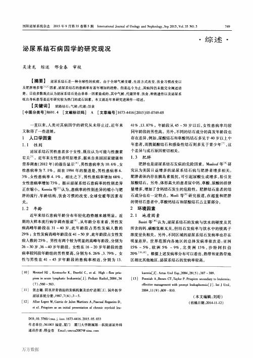
泌尿系结石病因学的研究现况
of Urology and Nephrology,Sep 2015,V01.35 NO.5
749
・综述・
泌尿系结石病因学的研究现况
吴清龙综述邢金春审校
【捅要】
泌尿系结石是一种全球性的疾病。由于全球气候变暖、生活方式改变、饮食习惯改变以
3.3单基因遗传的尿石症
近期对单基因型尿石症的遗传基础和病理生理学研究 强调,在肾小管和非肾小管上皮的转运蛋白、通道蛋白、受体 蛋白具有重要研究意义。虽然这种孟德尔性状在成年和小 儿尿结石患者中仅分别占2%和10%¨…,但是他们比常见的 多基因型结石更有特点:更严格的结石形成表型,合并有进 展性的肾功能损害。例如Dent病、原发性高草酸尿症、APRT 缺乏症等都与肾功能衰竭有关联。对于单基因型尿石症的 早期诊断是预防的关键。所以这些致病基因的识别和致病 变异序列的认识对于疾病的及早诊断和治疗起到非常关键 的作用。
3遗传因素
3.1
特发性草酸钙结石病的基因多态性
在家族相关性方面的研究上,既往已经有很多nick等¨刮报道,625例草 酸钙尿石症患者的一级亲属与其他亲属对照,患病率明显较 高,约高出20%。Coe等m1也报道,9例患有高钙尿症和复 发性尿石症的患者一级亲属患病率(43%)较一般人群明显 升高。最近,Curhan等’1副报道,通过对37999例健康男性的 队列研究发现,有尿石症家族史的人群患肾结石的相对危险 因素为2.57。综合各方面报道结果,肾结石患者的一级亲属 或较远亲属患尿结石的遗传度估计为15%一65%L1 9’…。在 孪生子研究方面,多个研究已证实,在肾结石遗传单基因方 面,同卵双生双胞胎比异卵双生双胞胎有更高的关联性。估 计遗传度为52%~56%旧1’”。。在连锁基因和候选基因研究 方面,由于大部分。肾结石是含钙结石,而且高钙尿症是最常 见的尿石症代谢危险因素,所以在寻找特发性草酸钙尿石症 的易感基因的任务上,重点放在了钙代谢相关基因。目前已 经有研究机构选择性繁殖出最高水平尿钙的SD大鼠,这些 大鼠经过了30代以上的培育后生产出了表型类似的钙调节 异常大鼠B 3|。这里所描述的钙调节异常,是指肠道钙离子超 吸收,骨钙吸收增加以及肾小球钙重吸收受损旧“。更详细的
【课题申报】泌尿系统结石的新治疗方法评估

泌尿系统结石的新治疗方法评估课题申报书一、课题来源及研究背景泌尿系统结石是一种常见的泌尿系统疾病,其发生率逐年增加。
目前,临床常用的治疗方法包括药物治疗、体外冲击波碎石术、内镜下碎石术等。
然而,传统的治疗方法存在一定的局限性,如药物治疗疗效欠佳,体外冲击波碎石术并发症较多,内镜下碎石术创伤较大等。
因此,寻求更为安全、有效的新治疗方法势在必行。
二、研究目的和意义本课题旨在评估泌尿系统结石的新治疗方法,并探讨其在临床上的应用前景。
通过对新治疗方法的疗效、安全性、费用效益和患者的生活质量等方面进行评估,为临床治疗提供更可靠的依据。
结合目前临床实践的经验和实际需求,本课题的研究对改善患者的生活质量、减轻经济负担具有重要的现实意义和社会意义。
三、研究内容和技术路线本课题计划通过临床试验和实验研究,评估泌尿系统结石的新治疗方法。
具体研究内容和技术路线如下:1. 临床试验部分:(1) 选取符合研究条件的患者作为研究对象,随机分组,治疗组采用新治疗方法,对照组采用传统治疗方法。
(2) 对两组患者进行治疗,并对治疗效果、并发症发生率等指标进行观察和比较。
(3) 通过统计分析,评估新治疗方法的疗效和安全性。
2. 实验研究部分:(1) 提取患有泌尿系统结石的动物模型,分为治疗组和对照组。
(2) 分别对两组动物进行新治疗方法和传统治疗方法的处理。
(3) 观察结石大小、数量和分布等指标的变化,并进行比较分析。
四、预期成果和应用前景通过本课题的研究,预期将获得以下成果:1. 对泌尿系统结石的新治疗方法进行评估,获取其疗效和安全性等方面的数据。
2. 就治疗效果、并发症发生率等指标进行比较,验证新治疗方法的优势和可行性。
3. 探讨新治疗方法在临床上的应用前景,为进一步推广应用提供可靠的依据。
4. 完善泌尿系统结石的治疗方法,提高患者的生活质量,减轻经济负担。
五、研究计划和进度安排1. 第一年:(1) 收集泌尿系统结石的临床资料,筛选符合研究条件的患者。
泌尿系结石治疗的现状和新进展

泌尿系结石治疗的现状和新进展泌尿系结石是一种常见的疾病,在全球范围内都有较高的发病率。
结石主要由尿液中的矿物质结晶所形成,可以在泌尿系的任何部位出现,例如肾脏、输尿管、膀胱等。
泌尿系结石不仅会引起剧烈的疼痛和不适,还可能导致感染、肾功能损害等严重后果。
因此,及时有效的治疗对患者的健康至关重要。
目前,泌尿系结石的治疗方法主要包括保守治疗、药物治疗和手术治疗。
保守治疗主要针对非症状性或小的结石,通过饮水增加尿液产量,促进结石的排出。
药物治疗则是通过使用特定的药物来溶解结石或减少结石的形成。
手术治疗是对于较大、影响泌尿系统功能、不能通过保守治疗或药物治疗有效缓解症状的患者而言的一种常见选择。
在泌尿系结石的手术治疗中,腹腔镜手术和尿道内微创手术是常用的方法。
腹腔镜手术通过股动脉或腹壁的切口引入腹腔镜,直接观察并处理结石,在减少创伤的同时提高手术效果。
这种手术技术已经在临床实践中得到广泛应用,取得了良好的疗效。
尿道内微创手术则通过尿道引入的微创器械对结石进行取石和治疗,无需股动脉切口,减轻了患者的创伤和恢复期,并提高了手术的成功率。
近年来,泌尿系结石治疗领域出现了许多新的进展。
其中,激光技术被广泛应用于结石碎石术中,通过运用低能量激光对结石进行精确切割和碾压,从而将结石分解为较小的碎片,便于排出体外。
这种技术具有高效、安全的特点,并且可以广泛适用于不同部位的结石。
另外,经皮穿刺肾镜技术也是近年来的一项重要进展。
这种技术通过在患者腰部进行穿刺,引入肾镜进行结石治疗。
相较于传统的手术方式,经皮穿刺肾镜技术具有更小的创伤和更高的手术成功率。
特别是对于复杂的大型肾结石,经皮穿刺肾镜技术能够有效地清除结石,减少并发症的风险。
此外,生物技术的进步也为泌尿系结石治疗带来了新的希望。
研究人员正在探索使用纳米技术来制备具有较高活性的溶石剂。
这些溶石剂可以更好地作用于结石表面,起到更好的治疗效果。
同时,基因治疗也正在被研究用于治疗泌尿系结石,通过改变特定基因的表达,减少结石形成的风险。
【课题申报】泌尿系统结石的新外科治疗技术

泌尿系统结石的新外科治疗技术《泌尿系统结石的新外科治疗技术》课题申报摘要:近年来,泌尿系统结石发病率不断增加,给患者身心健康带来巨大困扰。
传统的泌尿系统结石治疗方法存在创伤大、术后痛苦和并发症发生率高等问题,亟待寻找新的外科治疗技术。
本研究旨在探究泌尿系统结石的新外科治疗技术,提高手术疗效、降低手术风险和术后并发症。
一、研究背景泌尿系统结石是一种多发的疾病,其高发生率和复发率给患者带来极大的痛苦和经济负担。
传统的治疗方法包括开放手术和内窥镜技术,但存在显著的局限性,如创伤大、术后疼痛、并发症发生率高等。
因此,急需寻找新的外科治疗技术,以提高治疗效果和减少患者的痛苦。
二、研究目的本研究旨在探究泌尿系统结石的新外科治疗技术,提高手术疗效、降低手术风险和术后并发症。
三、研究内容和方法3.1 研究内容本研究主要包括以下内容:(1) 剖析传统治疗方法的优缺点(2) 探讨目前新兴的泌尿系统结石治疗技术(3) 测试并评估新外科治疗技术的疗效和安全性(4) 探究新外科治疗技术在泌尿系统结石的临床应用3.2 研究方法(1) 文献综述:对泌尿系统结石相关的文献进行探讨和分析。
(2) 实验室研究:通过实验室模型和动物实验评估新外科治疗技术的疗效和安全性。
(3) 临床试验:选择一定数量的泌尿系统结石患者,应用新外科治疗技术进行临床试验,并进行观察和分析。
(4) 数据分析:采用统计学方法对实验结果进行分析和总结。
四、研究意义和预期成果本研究的意义在于寻找对泌尿系统结石患者更加安全和有效的治疗方法,降低治疗风险和术后并发症。
预期成果包括:(1) 对传统治疗方法的优缺点进行深入剖析,为新外科治疗技术的研究提供理论基础。
(2) 探索新兴的泌尿系统结石治疗技术,为患者提供更加安全和有效的治疗选择。
(3) 通过实验室研究和临床试验,评估新外科治疗技术的疗效和安全性。
(4) 提供对泌尿系统结石患者进行新外科治疗技术的指导和优化。
五、研究计划本研究计划为期3年,具体安排如下:第一年:文献综述和理论基础研究第二年:实验室研究和动物实验第三年:临床试验和数据分析六、经费预算本研究的经费预算总计XXX万元,具体包括:实验材料及设备费XXX万元,人员费用(包括酬金和差旅费)XXX万元,实验室场地租赁费XXX万元。
泌尿系统结石症出现的损害纠纷
泌尿系统结石症出现的损害纠纷一、临床上出现诊断失误所造成的医疗损害1. 泌尿系统结石症漏诊的常见原因有以下几种:(1)有一部分病人临床上缺乏典型的肾绞痛症状,特别在女性,往往诊断为尿路感染、肾炎而长期误诊和误治。
(2)当结石影与骨骼重叠、检查前对肠道清理准备不满意或为透X线的“阴性结石”时,容易出现漏诊。
(3)在检查时如果只行静脉尿路造影而未摄尿路的各种位置的平片,可因造影剂的掩盖而不能发现结石。
(4)因阅片不仔细或满足于已看到的一处结石而可漏掉他处结石。
另外,结石引起巨大肾积水,可误诊为腹部肿块,双侧上尿路结石伴肾功能不全可误诊为慢性肾炎尿毒症。
此外,造影检查时,可由于患侧肾功能损害而使肾输尿管不能清楚地显示,结石合并的先天性尿路畸形、狭窄、肿瘤与息肉、囊肿等病变常可能导致漏诊。
2. 临床上出现误诊的几种情况泌尿系结石是外科常见病、多发病,可因并发症的出现或症状不典型等原因而发生误诊。
据临床上不完全统计误诊率有时可达10%左右。
(1)临床上被误诊为慢性肾盂肾炎双肾与输尿管结石可致尿路梗阻,肾盂内压增高,肾小管缺血性萎缩及功能不全,因而尿内出现较多的红细胞和蛋白,血中非蛋白氮升高和一氧化碳结合力降低,并有贫血、浮肿等临床表现,故被误诊为慢性肾盂肾炎的比较多。
由此可见,误诊不仅由于缺乏典型的尿路结石的临床表现,而且与尿路结石所致尿流梗阻能引起慢性肾功能不全缺乏认识有关。
(2)临床上被误诊为肾结核尿路结石继发的脓肾可因忽视病史等原因而误诊。
例如有的病例由于没有腰痛,有肾外结核病史,同时又出现尿急等膀胱刺激征,故拟诊肾结核。
肾盂积水合并感染后缺乏典型化脓感染的表现,又见切口长期不愈合,故以为是结核性脓肾。
例如有的病人曾有多发性血尿,如对产生血尿的原因进行多方面分析,则应想到尿石的可能,并作进一步检查,就不致于造成临床上的延误诊断。
(3)临床上被误诊为胆石症、胆囊炎、胆管结石例如患者临床上具有引起右上腹疼痛和压痛的囊性包块、发热和WBC数增高等表现,颇似胆石症、胆囊炎。
中西医结合的泌尿系统结石-泌尿系统论文-医学论文

中西医结合的泌尿系统结石-泌尿系统论文-医学论文——文章均为WORD文档,下载后可直接编辑使用亦可打印——摘要:目的比较泌尿系统结石行常规治疗和中西医结合治疗的效果,探究取得疗效更好的治疗方法。
方法选取本院在2016年1月至2017年7月期间收治的泌尿系统结石患者80例,把全部患者平均随机进行分组,每组患者40例,保证两组的可对比性。
对照组40例患者以常规西医方式进行治疗,观察组40例患者在西医治疗的基础上以中医治疗方式进行辅助治疗。
对两组治疗效果进行观察,观察指标包括总有效率、不良反应发生率、排石时间以及尿红细胞数量。
结果经过治疗后比较两组患者的治疗总有效率情况:观察组显著高于对照组,在不良反应发生率方面,对照组显著高于观察组,经比较,有显著的统计学差异(P0.05)。
观察组患者的平均排石时间:(7.66±2.37)d,对照组:(16.22±1.14)d,观察组患者的平均尿红细胞数:(6.55±1.96)个/ml,对照组:(16.00±1.00)个/ml。
上述指标比较,有显著的统计学差异,P值都小于0.05。
结论应用中西医结合的治疗方法治疗泌尿系统结石,效果显著。
关键词:泌尿系统结石;西医治疗;中西医结合治疗;疗效对比0引言泌尿系结石所包含的疾病很多,主要有肾结石、输尿管结石、尿道结石等等,发病率较高,给患者带来较大痛苦[1]。
这种疾病具体是指在泌尿系统中由于尿液浓缩而聚集成的块样聚集物,通常突然给患者造成剧烈腰痛,临床表现可见:尿频、尿急、尿痛等等,在泌尿外科已经作为十分常见的疾病被引起重视。
如果疾病没有得到及时的治疗有可能转变为癌,需要引起广大患者的重视[2]。
当前,由于人们的物质生活水平不断地提高,泌尿系统结石疾病的患病率也呈现出逐年升高的趋势,而找到治疗疾病有效的方法是比较重要的,以不断地提升患者的身体健康水平与生活质量。
本研究将中西医结合治疗的措施用于治疗泌尿系统结石疾病,对采用常规西医疗法和中西医结合疗法进行治疗的效果进行比较。
2015年EAU男性LUTS指南解读
排尿期LUTS: 5α还原疗指南推荐
推荐 5α还原酶抑制剂用于前列腺体积增大(>40mL)的中重度男性LUTS患者
LE
GR
1b
A
第二十四页,共34页。
Gravas S et al. EAU Guidelines 2015
5α还原酶抑制剂的疗效
不应用于白内障手术前 • 射精异常
ACEi = 血管紧张素转换酶抑制剂 ARBs = 血管紧张素受体阻滞剂 PDE-5 = 磷酸二酯酶-5抑制剂
第十九页,共34页。
Gravas S et al. EAU Guidelines 2015
2015 EAU指南:症状是LUTS进展的表现之一
• 有关LUTS的进展:部分患者的LUTS症状长期持续存在并进展
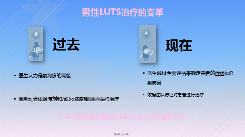
男性LUTS治疗的变革
过去
现在
▪ 医生认为是前列腺的问题 ▪ 使用α1受体阻滞剂和/或5α还原酶抑制剂进行治疗
▪ 医生通过全面评估来确定患者的症状和识 别病因
▪ 依据症状特征对患者进行治疗
从以前列腺为基础的方案到以症状为基础的方案
第一页,共34页。
2011年起,EAU指南发生转变: 从BPH指南转变为男性LUTS指南
5α还原酶抑制剂需要至少6-12月的治疗才能观察到临床疗效;因此有必要进行长期治疗 治疗2-4年后,IPSS评分下降约15-30%,前列腺缩小约18-28% 症状的改善(IPSS评分降低)取决于开始治疗时的前列腺体积 前列腺体积<40mL患者的疗效与安慰剂相当 5α还原酶抑制剂控制症状的疗效(IPSS)取决于:
总体症状评分的变化
坦索罗辛可有效控制LUTS症状长达66个月
且对不同体积的患者均有显著效果
• 共包括2003EAU公布的12项研究的汇总分析,2561例患者
泌尿系结石如何治疗

泌尿系结石如何治疗资阳市雁江区人民医院四川资阳 641300在常见泌尿系统疾病当中,泌尿系结石具有发病率比较高、病因比较复杂的特点。
有关报道显示,泌尿系结石高达5%~15%的发病率,而泌尿系结石的复发率几乎能够达到50%。
对于泌尿系结石的患者来说,反复的肾绞痛对身心造成了严重的伤害,还会因为结石对泌尿道造成梗阻,从而会引发肾功能衰竭。
这使得泌尿系结石引发的问题给患者和社会都造成了比较大的负担。
近年来,在结石治疗当中,体外冲击波碎石、腹腔镜取石、经皮肾镜碎石、取石术、钬激光碎石等都取得了比较良好的效果。
与传统的手术相比,这些方法创伤性比较小,但是这些方法有着比较昂贵的价格,还有不可避免的固有并发症问题。
对泌尿系结石进行治疗时,相比较开放性手术治疗方法,体外冲击波碎石法有着比较不凡的表现。
对肾结石治疗中应用最多的技术是激光碎石。
但是由于泌尿系结石患者结石成分、危险因素等不同,再加上泌尿系结石有着比较复杂的成因,为了对泌尿系结石最大限度地进行清理,可以根据患者的具体情况来制定具体的治疗手段。
1、体外冲击波碎石法在对泌尿系结石进行治疗时,体外冲击波碎石法是一种常规治疗方法,在体外利用冲击波把肾及输尿管的结石粉碎成粉末状,之后随着尿液自然排出体外。
从体外冲击波碎石术在临床上广泛应用以来,促使泌尿结石的外科治疗发生了根本的变革。
该方法具有无需麻醉、副作用小、易排除、疗效可靠、没有严重并发症等特点。
对患者进行碎石前需要对患者进行检查,比如,血尿常规、肾功能、心电图等。
对泌尿系畸形进行排除,结石下段没有梗阻,对尿路感染者需要进行控制。
当前体外冲击波碎石法对严重骨骼畸形、孕妇等禁忌治疗。
对于铸型结石、巨大结石、多发结石应采用经皮肾镜技术和体外冲击波碎石联合疗法,或者开放手术和体外冲击波碎石联合疗法。
对于输尿管中上段小于1厘米的结石,病史比较短并且是初发病采用体外冲击波碎石效果比较好,但是当遇到肾积水比较严重,患者疼痛比较剧烈等情况,可以采用输尿管镜置双“J”管加上体外冲击波碎石法。
尿石症诊断治疗指南(一)
尿石症诊断治疗指南〔一〕2007一、背景泌尿系结石是泌尿外科的常见病之一,在泌尿外科住院病人中占居首位。
欧美国家的流行病学资料显示,5%~10%的人在其一生中至少发生1次泌尿系结石,欧洲泌尿系结石年新发病率约为100~400/10万人。
我国泌尿系结石发病率为1%~5%,南方高达5%~10%;年新发病率约为150~200/10万人,此中25%的患者需住院治疗。
近年来,我国泌尿系结石的发病率有增加趋势,是世界上3大结石高发区之一。
近年来,随着泌尿系结石病因研究的深入,结石的代谢危险因素越来越为泌尿外科工作者所重视。
体外冲击波碎石术(extracorporeal shock wave lithotripsv,ESWL)、经皮肾镜取石术(percutaneous nephrolithotripsy,PNL)、输尿管肾镜取石术(ureterorenoscope lithotripsy,URL)、腹腔镜取石术(1aparoscope lithotomy)的陆续呈现,使泌尿系结石的治疗逐渐向微创标的目的开展。
此外,结石复发的预防工作已经成为了泌尿外科工作者存眷的重点。
然而,迄今为止国内对于泌尿系结石的治疗和预防并无统一的尺度,各地不同病院之间对于结石治疗方案和预防办法的选择仍然存在着必然的差距。
基于这些原因,我们复习了近年来国表里的文献,依据循证医学的原那么编纂了这本指南,但愿以此对于尺度国内关于泌尿系结石的治疗和预防、减少结石的复发等方面起到必然的指导作用。
按照循证医学的原那么,我们将指南引用的文献资料按其科学可信性分为不同等级的证据和不同级此外“保举〞。
证据级别(LE)和“保举〞等级(GR)的尺度见表Ⅶ-1、表Ⅶ-2。
需要强调的是,由于经济和技术程度的差别,不同地域在治疗方法和诊断技术上差别较大,我们强调要选择对患者来说最适宜、损伤最小、并发症风险最小的诊疗方法,但并不是说其他的方法是不克不及用的,如果某一治疗方法不保举使用,我们将会出格说明。
EAU《指南》要点解读:特殊肾结石的管理

EAU《指南》要点解读:特殊肾结石的管理一、残留结石的管理残留肾结石的临床问题与发展的风险有关:·新结石(非均质成核);·持续的UTI;·碎石片移动引起有/无阻塞症状及其它症状。
建议LE GR1b A识别相关生化危险因素,为结石患者或结石残余碎片患者提供适当的预防措施[178,325,326]。
4 C随访结石患者或结石残余碎片患者,需定期监测疾病进展。
感染性结石治疗后残留碎片的复发风险高于其他结石。
不管何种结石成分,5年内有21-59%的残留结石患者需要治疗。
结石碎片> 5 mm的结石与比其小的碎片更需要干预[178,324,327]。
有证据表明碎片> 2 mm结石更有可能生长,尽管这与一年内随访的再次干预率增加不相关。
治疗积极清除残留结石的指征和程序的选择与结石的治疗标准相同,包括重复的SWL 。
如果不需要干预,根据结石分析,患者风险组和代谢评估可能有助于防止结石残留片段的再生长。
证据摘要LE1b对于下盏的残留结石,可以同时进行倒转治疗,在强利尿下+机械振机动可能促进结石清除[270]。
建议LE GR1a A经冲击波碎石术和输尿管镜检查后,在存在残留碎片的情况下,使用α阻滞剂提供药物排石治疗可以改善碎片清除率。
表:处理残留结石碎片的建议残片(最大有症状残留结无症状残留结LE GR直径)石石< 4-5 mm去石定期随访(依4 C赖于风险因素)> 5 mm去石 4 C二、怀孕期间尿结石治疗及相关问题怀孕的尿路结石患者的临床管理很复杂,需要病人,放射科医师,产科医师和泌尿科医师密切协作。
如果无自发排石,或者如果发生并发症(例如诱导早产),需要放置输尿管支架或经皮肾造瘘,遗憾的是,通常孕妇对这些临时治疗有较差的耐受性相关,并且在怀孕期间,由于潜在的结石迅速生长,还需要多次更换输尿管支架这样的干预。
因此输尿管镜已成这些情况的合理的替代方案。
虽然可行,但在怀孕期间,逆行内窥镜和经皮去除肾结石,仍然虽患者自我决定,且只能在有经验的中心进行。
泌尿系结石的治疗新进展

泌尿系结石的治疗新进展引言:泌尿系结石是一种常见的疾病,给患者带来了很大的痛苦和不便。
多年来,医学界对于结石的治疗方法一直在不断进步和完善。
本文将介绍泌尿系结石治疗领域的新进展,包括非手术治疗、微创手术以及药物治疗等方面。
一、非手术治疗新进展1. 超声碎石技术超声碎石技术是一种非侵入性的治疗方法,通过特定频率和强度的超声波将结石震碎成较小的碎片,从而帮助患者排出体外。
近年来,随着超声技术的不断改进和完善,准确性和安全性得到了极大提高。
此外,超声碎石技术还具有操作简便、恢复快等优点。
2. 体外冲击波碎石(ESWL)ESWL是一种利用冲击波将结石震碎为较小颗粒后排出体外的方法。
传统ESWL存在能量传输不均匀、治疗效果不稳定等问题,而现代ESWL通过引入成像技术和计算机辅助技术,提高了精确性和治疗效果。
此外,ESWL还可以针对不同类型的结石进行定制化治疗,从而提高了成功率。
二、微创手术新进展1. 内窥镜下腔镜碎石术(URS)URS是一种通过尿道将内窥镜引入体内,在视觉指导下使用激光或钳夹等器械将结石碎取或拔除的微创手术方法。
近年来,URS技术得到了进一步发展,并在细镜直径缩小、器械精细化等方面有了新的突破。
这些改进使得URS操作更加精准、安全,大大减少了并发症的发生率。
2. 经皮肾镜取石术(PCNL)PCNL是一种通过穿刺经皮径路将肾内的结石切割或抽取出来的手术方法。
近年来,随着导丝技术和气腹技术的改良,PCNL手术变得更加安全有效。
同时,增强型CT扫描的应用,可以帮助医生准确评估肾内结石的大小和位置,提高手术成功率。
三、药物治疗新进展1. 草酸盐代替硫酸镁溶液目前,用于促进结石排出的主要药物是硫酸镁溶液。
然而,硫酸镁溶液有一定副作用,并且不适用于所有患者。
近期的研究表明,草酸盐可能是一种有效的替代药物。
草酸盐不仅能够促进结石排出,还具有较少的副作用。
2. 碱化药物碱化尿液可以减少尿液中结石形成的风险。
最新: EAU 尿石症介入治疗的最佳实践指南解读

最新:EAU 尿石症介入治疗的最佳实践指南解读文章内容:摘要:2022年8月,欧洲泌尿外科学会(European Association of Urology,EAU) 更新了尿石症介入治疗的最佳实践指南(以下简称指南)。
对上尿路结石的管理提供了最新的指导,并对外科治疗中有争议领域进行了讨论。
本文针对指南中更新的内容及临床意义进行总结和解读。
主要内容:1、背景目前泌尿系结石发病率越来越高,且泌尿系结石的治疗造成患者经济负担较重[1-3] 。
当前外科手术仍是泌尿系结石治疗的主要手段,包括冲击波碎石术(shock wave lithotripsy,SWL),输尿管硬/软镜(ureteroscopy,URS)和经皮肾镜取石术(percutaneous nephrolithotomy,PCNL)。
开放手术取石仅用于病情特殊患者,所占比例较低[4] 。
近几年来随着一次性输尿管软镜、负压吸引软镜鞘、高功率激光和微型经皮肾通道等技术的应用,泌尿系结石的手术治疗发生了巨大的变化。
随之也出现了多方面的争议,例如:输尿管镜术后能否不留置支架管,经皮肾镜碎石术后能否完全“无管化”,直径> 2 cm 的结石能否优先选择输尿管软镜等。
2022年8 月,欧洲泌尿外科学会(European Association of Urology,EAU)更新了尿石症介入治疗的最佳实践指南(以下简称指南)[5] ,EAU 对截至到2021 年5 月份的新文献数据进行了全面总结和综述,对尿石症的管理提供了最新的指导,对一些较重要的争议领域进行了讨论,并为指南的每一项建议提供强度评级。
此外,还指出尿石症诊断治疗期间患者和医生的辐射暴露应遵循尽可能低的合理可行原则。
本文对指南中更新的内容及临床意义进行总结和解读。
2、结石手术围术期注意事项2.1 围术期预防感染在泌尿系结石手术的相关并发症中,泌尿外科医生通常对感染最为小心,所以在围术期应用大量抗生素预防感染,出现抗生素滥用情况。
【课题申报】泌尿系统结石的新治疗方法

泌尿系统结石的新治疗方法课题名称:泌尿系统结石的新治疗方法1. 研究背景与意义泌尿系统结石是一种常见的泌尿系统疾病,严重影响了患者的生活质量和健康。
传统的结石治疗方法存在局限性,如手术创伤大、复发率高、费用高昂等问题。
因此,寻找一种新的、高效的治疗方法对于改善泌尿系统结石患者的生活质量具有重要意义。
2. 研究目标本课题旨在探索和验证一种新的治疗方法,以提高泌尿系统结石的治疗效果和减少患者的痛苦。
具体目标包括:(1)分析泌尿系统结石的成因及其发生机制;(2)研究和开发一种新的治疗方法,针对泌尿系统结石进行治疗;(3)验证新治疗方法的安全性和有效性;(4)对新治疗方法的优势和特点进行总结和评估。
3. 研究内容与方法(1)成因与发生机制分析:通过文献综述和临床病例分析,了解泌尿系统结石的成因和发生机制,为新治疗方法的研究提供理论依据。
(2)新治疗方法的研发:基于成因和发生机制的分析,开展实验室研究,设计并制备适用于泌尿系统结石治疗的新型治疗药物或介入材料。
(3)安全性和有效性验证:通过动物模型实验和临床实验,验证新治疗方法的安全性和有效性,评估其对泌尿系统结石的治疗效果。
(4)优势和特点总结与评估:总结和评估新治疗方法的优势和特点,包括疗效、手术创伤、复发率、费用等方面。
4. 预期成果与应用价值(1)预期成果:研究完成后,将获得一种新的治疗方法,该方法将在泌尿系统结石的治疗中发挥重要作用,并为医生提供指导和决策依据。
(2)应用价值:新治疗方法具有突破传统治疗局限性的优势,不仅可提高患者的生活质量和治疗效果,还能减少手术创伤和复发率,从而降低医疗费用,具有广泛的应用前景。
5. 研究计划与进度安排(1)第一年:- 文献综述和病例分析,了解泌尿系统结石的成因和发生机制;- 开展实验室研究,设计制备新型治疗药物或介入材料;- 制定动物实验方案,验证新治疗方法的安全性和有效性。
(2)第二年:- 进行动物实验,评估新治疗方法在泌尿系统结石治疗中的效果;- 收集和分析实验数据,进行统计学处理;- 准备论文稿件,撰写研究成果的中期报告。
【课题申报】泌尿系统结石患者的治疗效果评估

泌尿系统结石患者的治疗效果评估《泌尿系统结石患者的治疗效果评估》一、项目背景与意义泌尿系统结石是一种常见的疾病,其发病率逐年上升,给患者的生活质量和健康带来了严重影响。
治疗泌尿系统结石的方法有很多,包括药物治疗、外科手术、尿道镜取石等。
然而,现有的治疗方法在临床应用中存在很大的争议,治疗效果也因此差异较大。
因此,本课题旨在对泌尿系统结石患者经不同治疗方法后的治疗效果进行评估,为临床选择最佳的治疗方案提供科学依据。
二、研究目标和内容1. 研究目标评估泌尿系统结石患者不同治疗方法的治疗效果,比较其优劣,并推荐最佳治疗方案。
2. 研究内容(1)收集病例资料:从我院泌尿科2019年1月至2022年12月收治的泌尿系统结石患者中,随机选取病例进行收集资料,包括患者基本情况、病史、临床表现、诊断结果等。
(2)分组和治疗:将收集的病例根据不同的治疗方法进行分组,包括药物治疗组、外科手术组、尿道镜取石组等。
(3)治疗效果评估:统计患者治疗前后的症状缓解情况、结石的消失与复发情况,根据其变化情况评估不同治疗方法的疗效。
(4)数据分析与结果展示:使用SPSS软件对数据进行统计分析和比较,并进行图表展示,比较不同治疗方法在治疗效果上的差异。
三、研究方法1. 研究设计本研究采用前瞻性队列研究设计,即选择一定的泌尿系统结石患者进行治疗,随访观察一定的时间,收集数据并进行统计分析。
2. 样本选择与采集采用方便抽样的方法,从我院泌尿科2019年1月至2022年12月收治的泌尿系统结石患者中,随机选取一定数量的样本进行病例资料收集和治疗。
3. 数据分析与结果展示采用SPSS软件对所收集的数据进行统计分析,包括描述性统计、相关性分析、T检验等。
并使用图表等形式展示数据和结果。
四、预期成果1. 通过对泌尿系统结石患者不同治疗方法的治疗效果评估,比较各种治疗方法的优劣差异,为临床选择最佳治疗方案提供科学依据。
2. 提供相关病例资料和治疗效果数据,为泌尿系统结石患者的临床治疗和研究提供参考。
尿石症热点问题“随意谈”解析
步解决。RIRS碎石器械的不断改进、腔内碎石达到 雾霾化水平、负压吸引鞘的研发和推广,将使RIRS
取代PCNL成为可能。
兴、高小峰等11位国内知名尿石症专家以及吴开 俊、齐琳、孙西钊等3位大师级点评专家在陈志强、 郭小林教授主持下就若干个热点问题进行了热烈讨 论,大家各抒己见,精彩纷呈。
一、ESWL的前景黯淡还是光明
微创治疗手段,但是取石效率仍较低、器械较易损
DOI:10.3760/cma.j.issn.1000-6702.2016.1
1.002
富,必将对尿石症的临床诊治起到极大的促进作用。
(收稿日期:2016-09-01)
作者单位:510120广州,中山大学孙逸仙纪念医院泌尿外科(许 可慰);华中科技大学同济医学院附属同济医院泌尿外科(叶章群) 通信作者:叶章群,Email:zhangqun—ye@163.com
致也是微创。 四、尿石症超微创治疗的方向
专家们指出,目前各种尿路结石微创手术发展 迅猛,在结石的治疗中占据了主导地位,ESWL常常 被有意无意地忽视;然而ESWL才是近乎无创的治 疗手段。国内外尿石症的诊治指南中,ESWL都有
重要的一席之地,特别是直径<2 cm的肾结石、直
径<1cm的上段输尿管结石应首选ESWL。造成我 国ESWL发展相对落后的直接原因是由于当前国内 ESWL定价过低,每次ESWL收费仅600元左右,连 碎石机的成本都难以回收,这极大地限制了ESWL 的进一步研发和广泛应用。但专家们依然坚信,随 着医改深入,医疗定价回归价值体现,必然迎来第二 次ESWL时代的到来。
史堡婆星处型盘查!!!!生!!旦筮!!鲞筮!!塑堡h也』型翌!:盟坚!坚些!!Q!鱼:!!!:!Z:№:!!
.专家论坛.
- 1、下载文档前请自行甄别文档内容的完整性,平台不提供额外的编辑、内容补充、找答案等附加服务。
- 2、"仅部分预览"的文档,不可在线预览部分如存在完整性等问题,可反馈申请退款(可完整预览的文档不适用该条件!)。
- 3、如文档侵犯您的权益,请联系客服反馈,我们会尽快为您处理(人工客服工作时间:9:00-18:30)。
Platinum Priority –EditorialReferring to the articles published on pp.x–y of this issueContemporary Management of Stone Disease:The New EAU Urolithiasis Guidelines for 2015Matthew Bultitude a ,*,Daron Smith b ,Kay Thomas aaStone Unit,Guy’s and St.Thomas’NHS Foundation Trust,London,UK;b Stone /EndoUrology Unit,University College Hospital,London,UKIn this month’s issue of European Urology ,two major components of the European Association of Urology guidelines are summarised regarding the diagnosis and management and the interventional treatment of urolith-iasis [1,2].The low level of evidence for many of the reference statements demonstrates the paucity of high-quality randomised trials available in stone disease,making this expert consensus all the more important for guiding current practice.The diagnostic guidelines are clear.Although ultraso-nography is the primary diagnostic tool,it remains user dependent,with a range of sensitivities from 19%to 93%.Noncontrast computed tomography scan (NCCT)is the standard for investigation of acute flank pain,with dose-reduction techniques offering high sensitivity (97%)and specificity (95%).Radiation exposure reference ranges are given,demonstrating that low-dose NCCT achieves similar doses to kidney,ureter,and bladder x-ray (0.97–1.9vs 0.5–1.0mSv).Computed tomography (CT)scanning also allows assessment of Hounsfield units (HU),which may be helpful in planning treatment;the guidelines suggest that >1000HU are less likely to be fragmented with shockwave lithotripsy (SWL).An aspect not addressed is the potential for dual-energy CT to assess stone composition.This type of technology may be helpful in the future not only for directing surgical therapy but also for identifying stones suitable for medical dissolution with alkalinising agents.Diagnostic imaging in pregnancy is a challenging situation,and ultrasound remains the imaging method of choice,with magnetic resonance imaging as a second-line option,but these updated guidelines now include low-dose CT scanning as the final option in selected cases.Nonsteroidal anti-inflammatory drugs are clearly super-ior to opiate medication for the acute stone episode [3],and prescription of these drugs should be first line if no contraindication exists.Readers,however,should be aware of evolving literature regarding cardiovascular side effects,as regulatory authorities have recommended that systemic diclofenac should be contraindicated in patients with ischaemic heart disease,peripheral arterial disease,cere-brovascular disease,and congestive heart failure due to the risk of thrombotic events [4].Naproxen and ibuprofen may have lower risk.Medical expulsive therapy (MET)has become widely adopted since publication of the meta-analysis of trials in 2006[5],and this is reflected in these guidelines,which advocate MET to facilitate spontaneous passage and to reduce painful episodes.A recent large multicentre randomised trial of 1167patients [6],however,may change future guidance on MET.This trial showed no benefit from MET with tamsulosin or nifedipine versus placebo for stone passage,analgesic use,or time to stone passage.Although this is a single trial,it had more patients than the meta-analysis data,and significant weight needs to be given to this paper when deciding whether to continue to offer MET for ureteric stones.An issue discussed in both guidelines [1,2]is the management of asymptomatic calyceal stones.The evi-dence base remains poor,with the risk of symptomatic episode or need for intervention quoted as 10–25%per year.Although observation of tiny stones seems sensible for most,many would not observe stones of up to 15mm in size,as suggested in the guideline;clearly,treatment needs to be tailored to the individual patient.Active treatment ofE U R O P E A N U R O L O G Y X X X (2015)X X X –X X Xa v a i l ab l e a t ww w.sc i e n c ed i re c t.c o mj o u r n a l h o m e p a g e :w w w.e u r o p e a n u r o l o g y.c omDOIs of original articles:/10.1016/j.eururo.2015.07.041,/10.1016/j.eururo.2015.07.040.*Corresponding author.Stone Unit,2nd Floor,Tower Wing,Guy’s Hospital,Guy’s and St.Thomas’NHS Foundation Trust,Great Maze Pond,London SE19RT,UK.Tel.+442071889099.E-mail address:matthew.bultitude@ (M.Bultitude)./10.1016/j.eururo.2015.08.0100302-2838/#2015European Association of Urology.Published by Elsevier B.V.All rights reserved.smaller stones for patients with solitary kidneys or frequent travellers may be appropriate,whereas observation of quite large stones is certainly possible in the elderly or comorbid and in those who have easy access to medical treatment.Management of these stones in the presence of comorbid-ities poses challenges,as patients may be less able to tolerate intervention in the future if they experience stone growth,symptoms,or obstruction.Although the only randomised trial in this area (SWL vs observation)showed no benefit [7],21%of the observation group required ‘‘additional treatment’’at a median of only 2.2yr.More studies are required to further direct the management of these patients,including the optimal frequency and duration of follow-up.In an era of concern regarding antibiotic resistance,it is good to see clear guidance on the use of prophylactic antibiotics in stone surgery.This update states that single-dose administration is sufficient for ureteroscopy (URS)and percutaneous nephrolithotomy (PNL)procedures.Although there is evidence for longer courses [8],the guidelines are pragmatic to minimise overuse of antibiotics and can be tailored for specific patients with recurrent infections.There is no benefit to routine antibiotic prophylaxis in SWL unless there is a potential infective source such as infection stone or nephrostomy tube.Decision making in choosing active treatment is aided by a treatment algorithm to help stratify choices among SWL,URS,and PNL.It is interesting that miniaturisation of instruments has led to PNL taking on smaller and smaller stones.At the same time,durability and digitalisation of flexible ureteroscopes has led to surgeons tackling larger and larger stones,with published series demonstrating efficacy in stones 2–3cm.Forming a treatment algorithm is now almost impossible,but what is clear is that rates of SWL have been declining worldwide,and the emphasis of this guideline reflects this shift,although SWL remains a vital component of a stone surgeon’s armamentarium with appropriate patient selection.Ureteral access sheaths (UAS)have clear benefits,as outlined;however,care has to be taken to prevent ureteral injury,as detailed by Traxer and Thomas [9].The decision of whether to use UAS may depend on the choice of whether to dust the stone or fragment and remove it.The guideline makes a bold statement that dusting should be limited to large renal stones.Little evidence exists to promote either strategy,and it could be argued that larger stones are best being removed because of the sheer stone volume,whereas dusting small stones is acceptable.What is clear is that if there is no prior stone analysis,then one should be obtained for every patient to guide future decision making.The use of stents and nephrostomy tubes after URS and PNL,respectively,remains controversial.Despite the guidance suggesting that tubeless or totally tubeless procedures are safe after uncomplicated PNL,many surgeons will remain cautious and tend to favour placement to maximise postoperative access and options (including second-look PNL).Following URS,the evidence suggests that stenting is not required after uncomplicated proce-dures;however,this depends on a number of factors,and strategies to minimise indwelling time,such as leaving the tether attached to allow easy early removal (‘‘stent on a string’’),are useful in cases of uncertainty.In conclusion,these publications provide a timely well-referenced guide for the treating clinician;however,these are only guidelines,and many aspects will differ in clinical practice.A lot of the emphasis in treating stones must be given to individual patient factors as well as local and timely availability of services.Conflicts of interest:Matthew Bultitude has worked as a consultant forBoston Scientific.Daron Smith has received an honorarium from Lilly forlectures.Kay Thomas has nothing to disclose.References[1]Tu¨rk C,Petr ˇı´k A,Sarica K,et al.EAU guidelines on diagnosis and conservative management of urolithiasis.Eur Urol.In press.http:///10.1016/j.eururo.2015.07.040[2]Tu¨rk C,Petr ˇı´k A,Sarica K,et al.EAU guidelines on interventional treatment for urolithiasis.Eur Urol.In press./10.1016/j.eururo.2015.07.041[3]Holdgate A,Pollock T.Nonsteroidal anti-inflammatory drugs(NSAIDs)versus opioids for acute renal colic.Cochrane DatabaseSyst Rev 2005:CD004137.[4]Coxib and Traditional NSAID Trialists’(CNT)Collaboration.Vascularand upper gastrointestinal effects of non-steroidal anti-inflammato-ry drugs:meta-analyses of individual participant data from random-ised ncet 2013;382:769–79.[5]Hollingsworth JM,Rogers MAM,Kaufman SR,et al.Medical therapyto facilitate urinary stone passage:a ncet2006;368:1171–9.[6]Pickard R,Starr K,MacLennan G,et al.Medical expulsive therapy inadults with ureteric colic:a multicentre,randomised,placebo-con-trolled ncet 2015;386:341–9.[7]Keeley Jr FX,Tilling K,Elves A,et al.Preliminary results of arandomized controlled trial of prophylactic shock wave lithotripsyfor small asymptomatic renal calyceal stones.BJU Int 2001;87:1–8.[8]Mariappan P,Smith G,Moussa SA,Tolley D.One week of ciprofloxa-cin before percutaneous nephrolithotomy significantly reduces up-per tract infection and urosepsis:a prospective controlled study.BJUInt 2006;98:1075–9.[9]Traxer O,Thomas A.Prospective evaluation and classification ofureteral wall injuries resulting from insertion of a ureteral accesssheath during retrograde intrarenal surgery.J Urol 2013;189:580–4.E U R O P E A N U R O L O G Y X X X (2015)X X X –X X X2。
