超声引导下区域阻滞
超声引导下神经阻滞

01
包括手术麻醉、疼痛治疗、分娩镇痛等。
超声引导下神经阻滞可以提高麻醉效果,减
02
少麻醉药物的使用,降低麻醉风险。
超声引导下神经阻滞可以提高麻醉操作的准
03
确性,减少麻醉并发症的发生。
超声引导下神经阻滞可以提高麻醉操作的效
04
率,缩短手术时间,降低医疗成本。
疼痛治疗
超声引导下神经 阻滞可应用于各 种慢性疼痛的治 疗,如腰痛、关
节痛等。
超声引导下神经 阻滞可提高治疗 的准确性,减少
副作用。
超声引导下神经 阻滞可应用于手 术前后的疼痛控
制。
超声引导下神经 阻滞可应用于癌 症患者的疼痛治
疗。
其他应用
01
04
精神疾病治疗:用于治疗 某些精神疾病,如抑郁症、 焦虑症等
03
神经外科手术:用于辅助 神经外科手术,提高手术 准确性和安全性
减少损伤:超声引导 下神经阻滞可以减少 对周围组织的损伤, 降低并发症风险。
实时调整:超声引导 下神经阻滞可以实时 调整阻滞位置和深度, 提高阻滞效果。
01
02
03
04
缺点
操作难度较大,需要熟练掌握超 声技术和神经阻滞技术
设备成本较高,需要购买专业的 超声设备和神经阻滞设备
操作时间较长,需要花费更多的时 间进行超声引导和神经阻滞操作
存在一定的风险,如神经损伤、 出血等并发症
发展前景
技术进步:随着超声技术的不断 发展,神经阻滞的准确性和成功
率将不断提高。
应用领域拓展:超声引导下神经 阻滞技术有望在更多领域得到应 用,如疼痛管理、康复治疗等。
降低风险:超声引导下神经阻滞 技术可以降低传统神经阻滞手术
超声引导下胸椎旁神经阻滞在胸腔镜手术中的应用

超声引导下胸椎旁神经阻滞在胸腔镜手术中的应用胸腔镜手术是一种通过胸腔镜技术进行的微创手术,能够减少组织损伤、恢复快、并发症少的优势,因此在临床上得到了广泛的应用。
术中对患者的镇痛管理一直是一个非常重要的问题。
传统的经皮性静脉药物输注容易出现药物的浓度波动大,作用不稳定的情况,而且产生的镇痛效果也有限。
超声引导下胸椎旁神经阻滞在胸腔镜手术中的应用成为了一种较为理想的镇痛方式。
本文将详细介绍这一新颖的镇痛方式在胸腔镜手术中的应用及其效果。
一、超声引导下胸椎旁神经阻滞的技术原理超声引导下胸椎旁神经阻滞是利用超声波来引导锥体间隙内药物的精确定位,通过神经阻滞来达到术后镇痛的目的。
具体操作方法为,患者取下一侧衣服,坐位或仰卧位,暴露出锥体间隙的皮肤,采用无菌操作技术,用超声探头在胸骨柱的侧面扫查,识别出肋骨、椎弓和胸横韧带等解剖结构,确认锥体间隙的位置。
然后,将局部麻醉药浸润皮肤和软组织,用7.5-10MHz的线性探头放置在背侧上缘的第八个肋间隙处,逐层扫查,最终定位到锥体间隙,确认阻滞区域。
确定好位置后,使用22G的局部麻醉针,在超声引导下穿刺到达锥体间隙,确保不穿过锥体间隙,然后注入药物,使神经传导受阻,达到镇痛的目的。
1.减少手术时患者的疼痛和焦虑感:胸腔镜手术通常需要在患者的胸腔内进行操作,因此术中患者疼痛的程度会比较大,加之对手术的恐惧和焦虑,会影响手术效果。
而采用超声引导下胸椎旁神经阻滞,可以明显减少患者的术中疼痛和焦虑感,有利于手术的顺利进行。
2.术后镇痛效果好,减轻术后并发症:术后疼痛是胸腔镜手术患者非常常见的问题,严重影响患者的术后恢复和康复。
而超声引导下胸椎旁神经阻滞具有准确、定位清晰的优势,能够提供较长时间的术后镇痛效果,可以有效减轻术后疼痛,促进患者的早期恢复,并且减少术后并发症的发生。
3.减少对全身镇痛药物的使用:采用超声引导下胸椎旁神经阻滞,可以减少对全身镇痛药物的使用,避免了因药物浓度波动大、作用不稳定等问题而导致的不良反应。
超声引导下外周神经阻滞

故追踪针有很大难度 优势:最大程度避免损伤重要器官 仍需联合使用神经刺激器
超声引导下外周神经阻滞
超声引导下腰丛阻滞
探头选择:(2-5 MHz)的凸阵探头,景深> 8cm
超声引导下的腰丛神经阻滞的目的在于:在 影像下识别横突和腰大肌以及两者至皮肤表 面的距离,这样可以让操作者在进针前判断 腰丛神经的深度
超声引导下外周神经阻滞
无 菌 手 套 法
超声引导下外周神经阻滞
无 菌 灯 罩 法
超声引导下外周神经阻滞
神 经 刺 激 仪
超声引导下外神经阻滞
主要内容
超声引导下外周神经阻滞的背 景
设备和条件的准备 超声引导下上肢外周神经阻滞 超声引导下下肢外周神经阻滞
超声引导下外周神经阻滞
超声引导下上肢神经阻滞
臀下区入路-坐骨神经
超声引导下外周神经阻滞
超声引导下臀区坐骨神经阻滞
探头:低频凸阵探头,10cm神经阻滞针 适应症:膝部、胫腓骨、踝关节和足部的外
科手术 局麻药:15~20ml 坐骨神经比较深,在横轴位可能呈扁平状 推荐使用神经刺激器确定阻滞针与神经的接
触 尽量使局麻药呈环形包绕坐骨神经
超声引导下外周神经阻滞
超声引导下下肢外周 神经阻滞
腰丛 股神经 隐神经 股外侧皮神经 坐骨神经(臀区、臀下、转子下、腘
窝、前路)
超声引导下外周神经阻滞
腰丛
腰丛由L1-L4前支和T12、L5前支的一部分 组成
50%的人群腰丛包含T12前支的一部分和/ 或L5前支的一部分
上部三支神经:髂腹下神经、髂腹股沟神经 及生殖股神经;
MED = 正中神经
超声引导下上肢神经阻滞
超声引导下臂丛神经阻滞技术操作规范

超声引导下臂丛神经阻滞技术规范1范围本标准规定了超声引导下臂丛神经阻滞的适应症、禁忌症、准备工作、操作方法和注意事项。
2术语和定义下列术语和定义适用于本文件。
臂丛神经阻滞麻醉BrachiaIp1exusb1ockanesthesia将局部麻醉药注入臂丛神经干周围使其所支配的区域产生神经传导阻滞的麻醉方法称为臂丛神经阻滞麻醉。
是临床上常用的麻醉方法之一。
臂丛神经臂丛由第5~8颈神经前支和第♦•胸神经前支大部分组成。
经斜角肌间隙穿出,行于锁骨下动脉后上方,经锁骨后方进入腋窝。
臂丛五个根的纤维先合成上、中、下三干,由三干发支围绕腋动脉形成内侧束、外侧束和后束,由束发出分支主要分布于上肢和部分胸、背浅层肌。
主要的分支有:胸背神经、胸长神经、腋神经、肌皮神经、正中神经、楼神经、尺神经。
超声引导下神经阻滞临床医师通过超声显像观察外周神经及其周围结构,并在超声实时、动态引导下穿刺到目标神经周围,精准地实施神经阻滞,避免了对周围重要组织结构的损伤,减少了并发症的发生几率,提高了阻滞效率。
3适应症主要适用于主要适用于肩部及上肢手术的麻醉,也可用于临床疼痛治疗。
4禁忌症4.1 精神高度紧张,不合作者。
4.2 双上肢同时进行手术者。
4.3 穿刺局部或全身感染者。
4.4 休克、衰竭或濒危病人。
4.5 局麻药过敏者。
4.6 凝血功能障碍。
5准备工作5.1 核对患者姓名、床号、签署知情同意书。
5.2 操作前了解病史,亚阅气管镜报告(特别是封堵右肺时注意右肺上叶开口与隆突位置关系,肺癌是否累及左、右主支气管)。
5.3 检查喉镜、气管导管、支气管封堵器(检查气囊是否漏气,管路是否堵塞,套囊是否易脱落)等器械。
5.4 牙垫、胶布、听诊器、吸引器等物品是否齐全、完好,准备纤维支气管镜备用。
5.5 术者戴口罩、帽子、洗手。
6操作方法6.1 摆放体位病人取仰卧,头转向对侧,肩下垂,患肢贴向躯体。
术者面向病人。
嘱咐病人不能任意转动头部。
超声引导下腰方肌阻滞

拔出穿刺针:注射 5 完成后,将穿刺针 拔出,用无菌纱布 覆盖穿刺点
观察效果:观察患 6 者疼痛缓解情况, 评估阻滞效果
3 注意事项
操作技巧
超声引导:实时 观察穿刺针的位 置和深度,确保
准确穿刺
穿刺角度:根据 患者体型和超声 图像调整穿刺角 度,提高成功率
麻醉药选择:根 据患者病情和需 求选择合适的麻
08
观察:观察患者反应,确保 阻滞效果和安全性
药物注射
确定穿刺点:在超 1 声引导下确定穿刺 点,通常在腰椎横 突和肋骨之间
消毒皮肤:在穿刺 2 点周围进行消毒, 防止感染
穿刺针进入:将穿 3 刺针插入皮肤,在 超声引导下进入腰 方肌
注射药物:在超声 4 引导下将药物注射 到腰方肌内,达到 阻滞效果
进行阻滞
穿刺技术
01
定位:确定穿刺点,通常在腰 椎横突和髂嵴连线的中点
03
麻醉:局部麻醉,减轻穿刺 过程中的疼痛
05
确认:确认穿刺针进入腰方肌, 避免损伤其他组织
07
拔针:穿刺完成后,拔出穿刺 针,压迫穿刺点止血
02,在超声 引导下进行穿刺
06
注射:将药物注射到腰方肌 内,达到阻滞效果
04
缓解腰背肌紧 张:减轻腰背 肌紧张,缓解
腰背痛症状
超声引导的优势
01
实时可视化:超声引导可以实时观察穿刺针的位置和深度,提高穿刺准确性。
02
减少损伤:超声引导可以避免损伤神经、血管等重要组织,降低并发症风险。
03
提高成功率:超声引导可以提高穿刺成功率,减少重复穿刺次数。
04
实时调整:超声引导可以实时调整穿刺角度和深度,提高穿刺效果。
超声引导下腰方肌阻 滞
超声引导下的神经阻滞治疗你了解吗

超声引导下的神经阻滞治疗你了解吗超声技术具有强大功能,在医疗领域具有较高应用价值,最常见的是临床超声检查,对疾病进行诊断。
超声引导下的神经阻滞治疗明显优势,是一项新的技术,有效利用超声技术,保证治疗过程顺利进行。
那么什么是超声引导下的神经阻滞治疗呢?下面我们一起来学习下。
以往受到技术条件限制,在开展神经阻滞时,要利用体表标志才能找到神经。
超声技术的应用改善了这种情况,改变了神经阻滞方式,医生利用超声成像技术直接观察神经及周围结构,在超声引导下直接穿刺到目标神经周围,保证神经阻滞的准确性。
目前超声引导下的神经阻滞术已经被广泛运用。
超声超声是一种频率高于20000Hz的声波,具有方向性好、反射能力强等优点,可以获得比较集中的声能,相比较于空气中传播,水中传播距离要更远,被广泛应用于各个领域,主要包括工业、医学等,实际效果显著。
之所以被称为超声波,是因为人类听不到,已经超过了人的听觉上限。
超声技术在医学方面有着较高应用价值,超声诊断学在上世纪初开始运用,随着技术的成熟,已经成为了现代医学诊断主要手段。
在进行超声检查时,医生会采用专业技术设备,将探头放在患者身上进行移动扫查,超声检查被用于多个科室,例如心脏科、消化科等。
借助于超声检查,可以生成图像,通过分析来进行诊断。
目前使用的设备操作起来非常方便,具有很高的灵活性,例如便携式扫描仪。
相比较于其他检查方式,超声检查的价格更低,大多数患者都可以接受,因此应用范围很广。
关于超声检查是否对人体会产生损伤,这是人们一直探讨的一个问题,但实践证明并不会对人体健康产生影响,是比较安全的,因此不必过于担心。
超声引导下的神经阻滞超声引导下的神经阻滞核心技术是超声,可以起到引导作用,清楚了解神经走向,保证神经阻滞针穿刺到准确位置上,对患者实施麻醉,阻滞冲动传导,保证局部区域处于麻醉状态。
神经阻滞只需要注射一处,就可以起到良好阵痛效果。
超声引导下的神经阻滞优势分析一是可以实现精准定位。
2、2009北医-哈佛麻醉论坛超声引导下区域神经阻滞讲稿

UltrasoundGuided Regional Anesthesia Heather Ballard MD, Phil Hess MD, Yunping Li MD Department of Anesthesia, Beth Israel Deaconess Medical Center Harvard Medical School, Boston, MA Table of Contents Introduction...........................................................................................................................................3 Interscalene Block...............................................................................................................................4 Supraclavicular Block.........................................................................................................................6 Infraclavicular block...........................................................................................................................8 Axillary Nerve Block..........................................................................................................................10 Femoral Nerve Block……………………………………………………………………….………………………12 Popliteal Nerve Block.......................................................................................................................14 Sciatic Nerve Block............................................................................................................................16 Transversus Abdominus Plane (TAP) block.............................................................................18 References............................................................................................................................................20 Introduction Though few randomized controlled studies exist for ultrasound-guided nerve blocks, its use is gaining wide popularity. Conventional nerve blocks rely on surface landmarks; these techniques may result in reduced accuracy, greater time, and greater patient discomfort in patients with difficult anatomy. There are several advantages to using ultrasound including: visualization of nerve and surrounding structures (vessels, bone, pleura,), needle placement, local anesthetic injection, and potential decreased needle attempts.Ultrasound is a technology that uses sound waves traveling through various mediums to produce images. Based upon how well waves are able to penetrate the material and reflect back to the transducer, the degree of brightness, or echogenicity, is displayed. Bright white images with a high degree of reflection such as the diaphragm, gallstone, and pericardium are deemed hyperechoic. Black images such as blood vessels and fluid-filled structures that have little reflection are called anechoic. Hypoechoic images are tissues with reflections in between the other two classes, e.g. solid organs. The nerve root in the interscalene and supraclavicular regions is typically hypoechoic; nerves below the clavicle and in the lower extremity are predominantly hyperechoic and have a honeycomb appearance in cross-section. In this pattern, the surrounding hyperechogenicity reflects the amount of connective tissue where the central hypoechoic portion is due to neural tissue.While performing peripheral nerve blocks, it is important to ensure local anesthetic is not inadvertently injected into vascular structures. On ultrasound, small peripheral veins and arteries are difficult to distinguish from nerve roots. The Color Doppler is a feature that can be utilized to differentiate vascular structures that have flow from nerves that have no flow. This technology detects motion using the Doppler effect, where the frequency of sounds waves change when reflected off a moving target (red blood cell). When the source is moving towards the transducer, the vascular structure is displayed in red. Conversely, the vascular structure is displayed in blue if the source is moving away from the transducer. Doppler detection of flow is best when the transducer is parallel to blood flow.One of the most difficult aspects of performing nerve blocks is visualizing an in-plane view of the block needle. When the needle tip and shaft do not readily appear, three transducer movements can be used to visualize the tip: alignment, rotation, and tilting. Alignment is the act of sliding the transducer longitudinally along the course of the nerve or needle. Rotation is the clockwise and counterclockwise rotation of the transducer to visualize the target anatomy or needle. Tilting refers to angling the transducer so it is less than perpendicular with the patient’s skin to obtain better visualization (often called rocking back and forth). A combination of these maneuvers may be required for optimal viewing.This block summarizes seven of the most frequently utilized peripheral nerve blocks: interscalene, supraclavicular, infraclavicular, axillary, femoral, sciatic, and popliteal. The transversus abdominus plane block, a superficial anterior abdominal wall block is also included. This packet is not meant to be an all-inclusive description of nerve blocks, but more as a convenient handbook resource.Interscalene Block Anatomy and IndicationsThe interscalene nerve block is used to provide anesthesia to the shoulder and arm. The local anesthetic works at the level of the brachial plexus roots; these nerves are found in theinterscalene groove at the level of the cricoid cartilage (C6.) The interscalene groove is found between the anterior and middle scalene muscles, deep to the sternocleidomastoid muscle.Ultrasound scanning techniqueTo optimize picture quality, the patient should be placed supine with the head turned 45 degrees to the opposite side. After the skin and transducer are prepped in the usual fashion, the transducer is placed on the neck at the level of the cricoid cartilage. The nerve roots are viewed in the transverse view; the appropriate depth of view is 3-4 cm.Figure 1: Transverse view ofinterscalene block anatomySCM: sternocleidomastoid muscleIJV: internal jugular veinCA: carotid arteryASM: anterior scalene muscleMSM: middle scalene musclePicture modified from Reg Anesth Pain Med 1998; 23: 77-80Figure 2: Surface landmarks ofinterscalene blockNotice how the transducer is placedcephalad to the clavicle, lateral to thecricoid cartilage. The head is turned 45degrees to the opposite side.CL: claviclePicture reprinted with permission fromra.caThe nerve roots appear as hypoechoic round or oval structures between the anterior and middle scalene muscles. The internal jugular and carotid artery are located medially andsternocleidomastoid muscle is superficial. If visualization is difficult, start scanning in the supraclavicular region where the brachial plexus appears as a “bunch of grapes” next to the subclavian artery and move the transducer cephalad.After forming a skin wheal of local anesthetic, a 5cm 22G needle is inserted lateral to the transducer. The needle is inserted parallel to the long axis of the transducer in order to visualize the needle as it is advanced in real-time.Local anesthetic injectionWith correct needle position, local anesthetic is seen as hypoechoic fluid in the interscalene groove. Between 15 to 40 mL of local anesthetic are utilized for this nerve block.Intramuscular injection, seen by an increase of echogenicity within the muscle, should be avoided. Figure 3: Ultrasound view of theinterscalene nerve blockSCM: sternocleidomastoidmuscleASM: anterior scalene muscleMSM: middle scalene muscleCA: carotid arteryIJV: internal jugular veinArrowheads: nerve rootsPicture reprinted with permissionfrom ra.caFigure 4: Ultrasound view ofneedle placementASM: anterior scalene muscleMSM: middle scalene muscleArrowheads: nerve rootsArrows: needle shaftPicture reprinted with permissionfrom ra.caSupraclavicular Block Anatomy and IndicationsThe supraclavicular block is used for surgical procedures and postoperative pain control for procedures of the arm, forearm, and hand. The brachial plexus is blocked at the level of the trunks at this location. The brachial plexus lies superior to the first rib and lateral to thesubclavian vein and anterior scalene. The subclavian artery usually lies medial and anterior in close proximity to the brachial plexus.Ultrasound scanning techniqueAfter preparing the skin and transducer in the usual fashion, the patient is placed in the supine position with the head slightly turned to the opposite side. The transducer is placed in the supraclavicular fossa in the coronal plane. The appropriate depth of the field is 2-3cm. The nerve trunks appear as hypoechoic ovals in the transverse view.Figure 5: Anatomy of supraclavicular nerve block SA: subclavian artery SV: subclavian vein CL: clavicle FR: first rib 1: anterior scalene muscle 2: middle scalene muscle Picture reprinted with permission from ra.caFigure 6: Surface landmarks ofsupraclavicular nerve blockNote how the head is slight turned tothe opposite side. The transducer isplaced in the supraclavicular fossa.Picture reprinted with permissionfrom ra.caThe brachial plexus is typically easy to locate at this position due to its consistent location lateral and posterior to the pulsatile subclavian artery and superior to the first rib. The subclavian vein is found medially to the artery and nerve.After forming a skin wheal of local anesthetic, a 5cm, 22G needle is inserted lateral to the transducer. The needle is advanced along the long axis of the transducer in order to view the needle in real time as it moves towards the nerve roots.Local Anesthetic injectionThe goal is to see local anesthetic as hypoechoic fluid around the nerve trunks during injection. If anesthesia of the distal limb is intended, place most of the local anesthetic above the first rib and next to the subclavian artery. The usual volume of local anesthetic is 25 to 40 ml. It is important to use color Doppler to differentiate arteries from nerves as the suprascapular and transverse cervical arteries are often seen when imaging the supraclavicular region. Figure 7: Ultrasound view of thesupraclavicular nerve blockSA: subclavian arteryArrowheads: brachial plexustrunks/divisionsPicture reprinted with permissionfrom ra.caFigure 8: Ultrasound view ofneedle placementSA: subclavian arteryArrowheads: brachial plexusArrows: needle shaftPicture reprinted with permissionfrom ra.caInfraclavicular block Anatomy and IndicationsThe infraclavicular block is used to provide anesthesia and postoperative analgesia to the forearm, arm, and hand. The cords of the brachial plexus lie anterior to the scapula andposterior to the pectoralis major and minor muscles. The brachial plexus surround the axillary artery with the lateral cord superior and lateral to the artery, the posterior cord posterior and the medial cord posterior and medial.Ultrasound scanning techniqueThe patient is placed in the supine position with the affected arm placed next to the patient’s side. The transducer is placed caudad to the clavicle medial to the coracoid process. In most patients, the depth is between 3-5cm. Using this technique, the brachial plexus and axillary vessels are seen in transverse view.The brachial plexus cords appear as hyperechoic circular structures with the lateral cord cephalad to the artery (9-12 o’clock), the posterior cord posterior to the artery (6-9 o’clock), andFigure 9: Anatomy of infraclavicularnerve blockAA: axillary arteryAV: axillary veinCL: clavicleCP: coracoid processBP: brachial plexus cordsPMIM: pectoralis minor musclePicture reprinted with permissionfrom ra.caFigure 10: Surface landmarks ofinfraclavicular blockNotice how the transducer is placedcaudad to the clavicle with theneedle approaching from superior toinferiorPicture reprinted with permissionfrom ra.cathe medial cord caudad to the artery (3-6 o’clock.) The artery is differentiated from the vein by because it is pulsatile and unable to be compressed.After the patient and ultrasound probe have been prepared, the needle is inserted cephalad to the transducer at a 45-60 degree angle parallel to the long axis.Local Anesthetic injectionLocal anesthetic (30-40mL) is injected around all 3 brachial plexus cords resulting in a U-shape spread of fluid around the axillary artery. It may be easier to inject the local anesthetic posterior to the artery at 6 o’clock first (posterior cord) and then withdraw to 9’ o’clock (lateral cord.)Figure 11: Ultrasound view ofinfraclavicular nerve blockPMM: pectoralis major musclePMIM: pectoralis minor muscleAA: axillary arteryAV: axillary vein Arrowhead: brachial plexus cordsFigure 12: Ultrasound view ofneedle placementAA: axillary arteryLA: local anestheticArrows: needle shaftPicture reprinted with permissionfrom ra.caAxillary Nerve Block Anatomy and IndicationsThe axillary nerve block is used to provide anesthesia for surgery and post-operative pain for procedures of the elbow, forearm, wrist, and hand. This block uses local anesthetic to block transmission of brachial plexus nerves at the level of the terminal branches. The median, ulnar, and radial nerves are reliably blocked by this technique; the musculocutaneous nerve is often spared because it branches off from the brachial plexus high in the axilla.Ultrasound scanning techniqueThe patient is placed in the supine position with the affected arm abducted 90 degrees. After prepping the skin and preparing the transducer, the transducer is placed in the transverse plane along the axillary crease.Figure 13: Anatomy of axillary artery nerve blockNote how the branches of thebrachial plexus surround the axillary artery at this location.Picture reprinted with permission from ra.caFigure 14: Surface anatomy of axillary nerve blockNote how the patient’s arm is abducted 90 degrees. Thetransducer is located in the axillary crease.Picture reprinted with permission from ra.caThe pulsatile, noncompressible, hypoechoic axillary artery is the reference point for this nerve block. In performing an anatomical survey, the triceps, biceps, and coracobrachialis muscles are seen surrounding the artery. The humerus is deep to these muscles. Outside of the muscle layers, the median, ulnar, and radial nerves are located around the axillary artery. Thesenerves appear as oval-shaped hypoechoic nodules with hyperechoic rims and are distinguished by their honeycomb appearance. The musculocutaneous nerve most commonly lies in a tissue plane between the biceps and coracobrachialis muscles.Local Anesthetic injectionThe needle is inserted at a shallow angle, parallel to the long axis of the transducer. As the needle is placed in-line with the ultrasound beam, the needle's path can be visualized as it approaches the target nerves. The musculocutaneous nerve is blocked separately within the coracobrachialis muscle. It is very important to avoid intravascular injection by determining the presence of auxiliary vessels by using color Doppler. As nerves can occupy variable locations around the axillary artery, the identity of each nerve can be determined by nerve stimulation. After careful aspiration, 10-15ml of local anesthetic in 3-5ml increments is injected at each nerve location. Circumferential local anesthetic spread around each nerve suggests proper needle placement within the facial sheath.Figure 15: Ultrasound landmarks of axillary block AA = axillary arteryCB = coracobrachialis muscle H = humerus M = median nerve MC = musculocutaneous nerve R = radial nerveFigure 16: Ultrasound view of needle insertionArrows = block needle AA = axillary artery H = humerusM = median nerve R = radial nerve U = ulnar nervePicture reprinted with permission from ra.caFemoral Nerve BlockAnatomy and IndicationsThe femoral nerve is often blocked for surgical procedures and postoperative pain control for the anterior thigh and knee surgery; this block is often combined with the sciatic nerve block for lower extremity anesthesia. The femoral nerve is typically blocked in the inguinal region where it is located lateral to the femoral artery, deep to the fascia iliaca, and superficial to the iliopsoas muscle.Ultrasound scanning techniqueBefore prepping the skin and transducer in the usual fashion, the patient is placed in the supine position with the leg in neutral position. The transducer is placed lengthwise along the inguinal crease. The field depth is typically 1-3 cm.The femoral nerve is typically found within a triangular hyperechoic region, lateral to thepulsatile femoral artery, superficial to the iliopsoas muscle, and deep to the fascia iliaca. The femoral vein is located medial to the femoral artery. If more than one artery is visualized with color Doppler, move the transducer cephalad to visualize the artery before the profunda femoris Figure 17: Transverse view of femoral nerve blockFL: fascia lata FI: fascia iliaca FS: femoral sheath FN: femoral nerve FA: femoral artery FV: femoral veinIPM: iliopsoas muscle PECT: pectineus musclePicture reprinted with permission from ra.caFigure 18: Surface landmarks of femoral nerve blockNote how the transducer is placed in the inguinal crease. The needle approach is from lateral to medial.Picture reprinted with permission from ra.caInguinal lymph nodes may also be confused for the femoral nerve, but can be distinguishedfrom the nerve by scanning proximally and distally. The nerve can be visualized throughout the scan due to long continuous anatomy where the discrete lymph node disappears.Local anesthetic injectionAfter negative aspiration and test dose, 20-30 ml of local anesthetic is injected. A hypoechoic ring of local anesthetic should appear around the hyperechoic nerve structures. Scan proximally and distally to visualize the local anesthetic spread around the nerve.Figure 19: Ultrasound view of the femoral nerve blockFA: femoral artery FV: femoral vein FN: femoral nerve IPM: iliopsoas muscleReprinted with permission from ra.caFigure 20: Ultrasound view of needle placementFA: femoral artery FV: femoral vein Arrows: needle shaftReprinted with permission from ra.caPopliteal Nerve Block Anatomy and IndicationsThe sciatic nerve block at the popliteal fossa is indicated for anesthesia and post-operative pain control for lower limb surgery including distal tibia, fibula, ankle and foot. The sciatic nerve at this location is located laterally to the popliteal artery and bordered superolaterally by the biceps femoris muscle and superomedially by the semimembranosus and semitendonosus muscles. The sciatic nerve branches into the tibial and common peroneal (fibular) nerves.Ultrasound Scanning TechniqueAfter positioning the patient prone, the skin and transducer are prepped in the usual fashion. The transducer is placed in the transverse plane above the popliteal crease. The typical depth is 2 to 3 cm. By scanning cephalad and caudad, the branching of the sciatic nerve into the tibial and peroneal branches can be visualized. The sciatic nerve is hyperechoic and located lateral to the popliteal artery.Figure 21: Anatomy of sciatic nerve blockNotice that the sciatic nerve divides into the common fibular (peroneal) nerve and tibial nerve in the popliteal fossa. Thelocation of this divide is variable in patients.Picture modified from O’Rahilly, et al, Basic Human Anatomy 2009; 15:1Figure 22: Surface landmarks of popliteal nerve blockNotice that the transducer is placed cephalad to the popliteal creaseReprinted with permission from ra.caThe sciatic nerve is hyperechoic located superficial to the femur and lateral to the pulsatile popliteal artery. In order to improve the view of the nerve, it may be necessary to angle the transducer caudad.Local anesthetic injectionAfter forming a skin wheal of local anesthetic, a 5-8cm 22G needle is inserted lateral to thetransducer and advanced along the long axis of the transducer. 20-30 ml of local anesthetic are utilized for this block; the “donut sign,” circumferential spread of local anesthetic around the nerve, is utilized to judge adequacy of spread.Figure 23: Ultrasound view of the popliteal nerve blockPA: popliteal artery PV: popliteal veinArrowhead: sciatic nerveReprinted with permission from ra.caFigure 24: Ultrasound view of needle placementLA: local anesthetic Arrows: needle shaft Arrowhead: sciatic nerveReprinted with permission from ra.caSciatic Nerve Block Anatomy and IndicationsThe sciatic nerve originates the lumbosacral plexus, L4-S3; it provides sensation to theposterior thigh and majority of the leg and foot. This nerve block is performed in combination with the femoral nerve block for knee and hip surgery or alone for leg and ankle surgery. In the gluteal region, the sciatic nerve lies deep to the gluteus maximus and superficial to superior and inferior gemellus muscles.Ultrasound Scanning TechniqueThe patient is positioned in the lateral decubitus position with the operative side uppermost; the knee and hip of the operative leg are flexed. After the skin and transducer are prepared, the transducer is placed on a line halfway between the greater trochanter and sacral hiatus; the field depth is usually 4cm or greater. In the transverse axis, the sciatic nerve appears hyperechoic with a wide flat shape.Figure 25: Gluteal region anatomyThe red dotted box is the target for the gluteal approach sciatic nerve block. Note how the sciatic nerve liessuperficial to the ischial bone and lateral to the ischial spine.Reprinted with permission from ra.caFigure 26: Surface anatomy of gluteal regionGT: greater trochanterPSIS: posterior superior iliac spine SH: sacral hiatusReprinted with permission from ra.ca Color Doppler can be used to visualize the pudendal artery and vein, which both lie medially to the sciatic nerve, next to the ischial spine. The sciatic nerve can be difficult to identify due to the its depth especially in patients with large amounts of adipose tissue.Local Anesthetic InjectionAfter preparing the skin and transducer, the needle is inserted on the lateral end of the ultrasound transducer. The needle is advanced in the same plane of the ultrasound beam in order to visualize the needle as it advances towards the sciatic nerve. Once the needle tip is visualized in close proximity to the nerve, 15-20 mL of local anesthetic is injected. The goal is to surround the sciatic nerve circumferentially in local anesthetic; this may require a second needle insertion site. Figure 28: Sciatic nerve block needle placementGMM: gluteus maximus muscle IB: ischial boneArrows: needle shaft Arrowhead: sciatic nerve Reprinted with permission from ra.caFigure 27: Color Doppler scan of Sciatic Nerve Gluteal Region GMM: gluteus maximus muscle IB: ischial boneArrows: pudendal vein and artery Arrowhead: sciatic nerveReprinted with permission from ra.caTransversus Abdominus Plane (TAP) block Anatomy and IndicationsThe TAP block is used to block the T10 to L1 nerve roots that provide sensation to the anterior abdominal wall. These nerves are known to travel between the transversus abdominus muscle and internal oblique at the Triangle of Petit formed posteriorly by the latissimus dorsi muscle, anteriorly by the external oblique and inferiorly by the iliac crest. The TAP block has been used successfully for postoperative pain control for hysterectomy, colectomy, prostatectomy, and Cesarean delivery.Ultrasound scanning techniquePrior to preparing the transducer and skin, the patient is placed in the supine position. The transducer is placed on the lateral abdominal wall at the Triangle of Petit. The appropriate depth is variable from 2 to more than 6 cm.. Figure 29: Anatomy of Transversus Abdominus Plane blockNotice the transverse position of the transducer in order to see the block needle in plane.Reprinted from Reference #6Figure 30: Surface landmarks of TAP blockThe patient is placed in the supine position. An assistant may benecessary to retract excess tissue of morbidly obese patientsReprinted from Reference #6The nerves are not usually seen during this block; rather, the local anesthetic is injectedbetween the internal oblique and transversus abdominus muscle planes and assumed to spread to the nerve roots. From shallow to deep, the different planes encountered are skin, subcutaneous tissue and fat, external oblique, internal oblique, transversus abdominus, peritoneum, and intraabdominal cavity. The intraabdominal cavity can be distinguished by visualizing peristalsis of abdominal contents.After forming a wheal of local anesthetic, a 5cm 22G needle is inserted superior to thetransducer and advanced parallel to the long axis of the transducer in order to visualize the needle in real-time.Local anesthetic injectionBetween 15-20 mL of local anesthetic are utilized for each side of this block. With correct needle position, there is hypoechoic fluid separating the fascial layer between the internal oblique and transversus abdominus muscle layers.Figure 31: Ultrasound view of TAP blockEOAM: external oblique muscle IOAM: internal oblique muscleTAM: transversus abdominus muscleReprinted from Reference #6Figure 32: Ultrasound view of needle localizationEOAM: external oblique muscle IOAM: internal oblique muscleTAM: transversus abdominus muscle Arrows: needle shaftReprinted from Reference #6References 1. Chan, Vincent, et al. Ultrasound for Regional Anesthesia. ra.ca2. Marhofer, et al. Ultrasound guidance in regional anaesthesia. British Journal ofAnaesthesia. 94 (1): 7-17 (2005)3. Sites, BD, et al. Artifacts and pitfall errors associated with ultrasound-guided regionalanesthesia. Part II: a pictorial approach to understanding and avoidance. Reg AnesthPain Med. 32 (5): 419-33 (2007)4. Sites, BD, et al. Artifacts and pitfall errors associated with ultrasound-guided regionalanesthesia. Part I: understanding the basic principles of ultrasound physics and machine operations. Reg Anesth Pain Med. 32 (5): 412-8 (2007)5. De Andres, Sala-Blanch, X. Ultrasound in the practice of brachial plexus anesthesia.Reg Anesth Pain Med. 27 (1): 77-89 (2002)6. El-Dawlaty, AA, et al. Ultrasound-guided transversus abdominus plane block: descriptionof a new technique and comparison with conventional systemic analgesia duringlaparoscopic cholecystectomy. British Journal of Anaesthesia (2009), Apr 17 (Epubahead of print)7. Marhofer, et al. Ultrasound-Guided Regional Anesthesia: Current Concepts and FutureTrends. International Anesthesia Research Society. 104 (5): 1265-9 (2007) 20 。
超声引导下神经阻滞

实时监测:超声引导下神经阻滞可以实时监测神经阻滞的效果,减少并发症的发生。
02
精确定位:超声引导下神经阻滞可以精确定位神经,减少对周围组织的损伤。
01
缩短手术时间
实时监测:超声引导下神经阻滞可以实时监测神经位置,提高手术精度
提高成功率:超声引导下神经阻滞可以提高手术成功率,缩短手术时间
减少损伤:超声引导下神经阻滞可以减少对周围组织的损伤,缩短手术时间
超声引导下神经阻滞
演讲人
目录
超声引导下神经阻滞概述
01
超声引导下神经阻滞的优势
02
超声引导下神经阻滞的应用
03超声引导下神经阻滞的培 Nhomakorabea和推广04
1
超声引导下神经阻滞概述
技术原理
01
超声引导:通过超声波成像技术,实时监测神经位置和状态
02
神经阻滞:通过注射药物,阻断神经传导,达到镇痛效果
03
实时监测:通过超声波成像技术,实时监测药物注射位置和效果
减少损伤:超声引导下神经阻滞可以减少对周围组织的损伤,提高精确度。
提高成功率:超声引导下神经阻滞可以提高阻滞的成功率,减少并发症的发生。
降低风险:超声引导下神经阻滞可以降低神经损伤的风险,提高精确度。
减少并发症
提高成功率:超声引导下神经阻滞可以提高神经阻滞的成功率,减少并发症的发生。
04
降低风险:超声引导下神经阻滞可以降低神经损伤的风险,减少并发症的发生。
效果评估:通过培训和推广,提高医疗质量和安全,降低医疗事故发生率
培训方式:理论授课、实际操作、模拟演练等
谢谢
随着科技的进步,超声引导下神经阻滞技术将更加智能化、精准化。
超声引导下神经阻滞技术将与其他医疗技术相结合,提高手术效果。
超声引导下区域阻滞

超声指引下地区阻滞1.超声指引下地区阻滞/镇痛管理的专家共鸣/指南中华医学会麻醉学会地区麻醉学组最近几年来,超声在地区阻滞中的应用日趋宽泛。
现有的文件主要集中于超声指引下肌间沟、锁骨上、锁骨下、腋路臂丛神经、坐骨神经及股神经阻滞,对超声指引下腰丛、腹腔神经丛及星状神经节阻滞也有报导。
已经证明,使用超声指引可显然降低成人、小孩及临产孕妇神经轴阻滞的难度。
传统的外周神经阻滞技术没有可视化指引,主要依靠体表解剖标记来定位神经,有可能针尖或注药地点不理想而致使阻滞失败;在解剖定位困难的病人,频频穿刺和操作时间的延伸致使病人不用要的痛苦,并使操作者产生挫败感。
在地区阻滞中使用超声指引,可清楚看到神经构造及神经四周的血管、肌肉、骨骼及内脏构造;进针过程中可供给穿刺针行进的及时影像,以便在进针同时随时调整进针方向和进针深度,以更好地凑近目标构造;注药时能够看到药液扩散,甄别无心识的血管内注射和无心识的神经内注射;别的,有凭证表示,与神经刺激器对比,使用超声指引可缩短感觉阻滞的起效时间,提升阻滞成功率,减少穿刺次数,减少神经损害。
超声指引下地区阻滞技术的基础是超声图像的获取和组织构造的辨别。
在平时地区阻滞工作中娴熟使用超声,需要娴熟掌握超声成像的基根源理和超声仪器的使用方法,熟习扫描部位的解剖构造,并能选择适合的扫描技术获取更好的超声影像,且娴熟掌握进针技术,使穿刺针能顺利抵达目标构造。
一、介绍操作者需掌握的超声知识超声仪的基本构造各种超声探头成像特色超声仪各功能键的使用认识医学领域超声波的常用频次及不一样超声频次与穿透性和成像质量的关系超声波与组织接触后发生的声学反响及生物学效应理解高回声、低回声及无回声的含义及人体不一样组织、构造表此刻超声图上的回声特色熟习脂肪、肌肉、骨骼、血管、神经、肌腱等常有组织的超声影像学特色认识超声及时成像、血流多普勒和能量多普勒成像的基根源理常有伪像的辨别清楚。
(四)进针技术依据穿刺方向与探头长轴的关系分为平面内(in-plane )、平面外out-of-plane)两种进针技术。
超声引导下的神经阻滞
超声引导下的神经阻滞温州医学院附属第二医院麻醉科325027 徐旭仲李挺传统上神经阻滞需要借助于局部解剖的体表标志、动脉搏动、针刺感觉异常及神经刺激器探查定位技术寻找神经。
但是,超声技术正使神经阻滞的方式发生根本性变革,麻醉医师已经能够通过超声成像技术直接观察神经及周围的结构,在实时的超声引导下直接穿刺到目标神经周围,实施精确地神经阻滞。
还可通过超声观察局麻药的注射过程,从而保证局麻药均匀的扩散到神经周围。
一、超声技术的基础知识在进行超声引导神经阻滞前,我们需要了解超声的基础物理知识。
从临床观点考虑,其中有两个重要的概念,即穿透性与分辨率。
任何形式的波,包括声波及超声波,都有特定的波长与频率。
频率与分辨率相关,波长与穿透性相关。
临床应用的超声频率在2.5-20MHz之间,高频率超声(>10MHz)可较好的显示神经结构,但只有当神经结构位于表浅的位置(如斜角肌间隙的臂丛神经)才能通过高频超声看到神经。
另外,当频率提高时,波长便降低,因此分辨率(频率)提高时,穿透性(波长)便降低,这时高频超声不能显像深部的神经。
在临床上为了能够清楚的观察斜角肌间隙、锁骨上区域及腋窝的臂丛神经,我们一般选择探头频率在8MHz以上,最好在12-14MHz。
而对于锁骨下、喙突区神经,其频率在6~10MHz之间较为适合。
这种低频可获得更好的穿透性,并能更精确的进行神经定位。
深部神经的超声引导应与神经区域的局部解剖学相结合。
超声的多普勒技术可以清楚地区分血管及血管中的血流速度,从而提高对于局部解剖的观察。
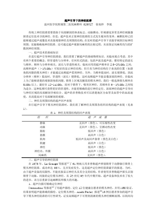
二、神经及周围结构的超声回声表现在行超声引导下臂丛神经阻滞时,我们需了解神经及周围各组织结构的超声表现(见表1)。
表1:神经及周围结构的回声表现组织超声成像静脉无回声(黑色),可压缩性改变动脉无回声(黑色),呈搏动性改变脂肪低回声(黑色)筋膜高回声(白色)肌肉低回声及高回声条带(黑色及白色)肌腱高回声(白色)神经低回声(黑色)神经内、外膜高回声(白色)局麻药无回声(黑色)。
超声引导区域阻滞专家共识

超声引导区域阻滞专家共识引言:在医疗技术的发展中,超声引导区域阻滞已成为一种常见而有效的麻醉和疼痛管理方法。
随着这一技术的不断应用和研究深入,专家们对超声引导区域阻滞的标准和操作规范也越发重视。
本文将介绍一份超声引导区域阻滞专家共识,以帮助医务人员更好地掌握和应用这一技术。
一、引言随着医疗技术的不断进步,超声引导区域阻滞在手术和疼痛管理中得到了广泛应用。
超声引导区域阻滞通过将局部麻醉药精确注射至神经或感觉分布区域,以达到麻醉和止痛的目的。
相比传统的触诊或神经刺激技术,超声引导区域阻滞具有更高的准确性和安全性。
二、超声引导区域阻滞的优势1. 准确性:超声引导区域阻滞可以清晰显示目标组织、神经和血管的位置,帮助医务人员准确注射麻醉药物,降低误刺和神经损伤的风险。
2. 安全性:超声引导区域阻滞可以避开主要血管和神经结构,减少术后并发症的发生率,如血肿、感染和神经损伤等。
3. 效果持久:超声引导区域阻滞在术中和术后提供良好的镇痛效果,减少术后疼痛的发生,改善患者的手术体验。
三、超声引导区域阻滞的适应症超声引导区域阻滞适用于以下情况:1. 手术麻醉:超声引导区域阻滞可作为主要或辅助麻醉技术,适用于各类手术,包括关节置换、外科修复和矫形手术等。
2. 疼痛管理:超声引导区域阻滞可用于控制急性或慢性疼痛,如神经性疼痛、肌肉骨骼疼痛和癌症相关疼痛等。
3. 康复治疗:超声引导区域阻滞可用于康复治疗,如神经肌肉电刺激和物理治疗。
四、超声引导区域阻滞的操作技巧超声引导区域阻滞的操作主要包括以下步骤:1. 选择适当的探头和频率:根据患者的体型和需要阻滞区域的深度,选择合适的超声探头和频率。
2. 定位目标神经或组织:使用超声技术清晰显示目标神经或组织的位置和形态。
3. 注射麻醉药物:在清晰显示目标区域后,将麻醉药物精确注射至目标神经或组织。
4. 动态观察:在注射麻醉药物的过程中,可以通过超声指导实时观察药物的扩散情况和阻滞效果。
超声引导下神经阻滞PPT课件

肌间沟臂丛神经阻滞
➢ 采用短轴平面内技术 ➢ 在超声探头的外侧部位皮肤
处穿刺,经中斜角肌推进
➢ 先深后浅,注射局部麻醉药
10-15 ml,可观察到局部麻 醉药的扩散
臂丛神经阻滞--锁骨上入路
• 臂丛中的腰麻? • 打外围,绕神经 • 显肋骨,避胸膜
锁骨上臂丛神经阻滞
➢ 采用短轴平面内技术,从探头 外侧穿刺
--效果更佳
前路坐骨神经阻滞
➢ 位置较深,需用低频凸阵探头 ➢ 将探头在股骨小转子水平横置于
股骨前内侧
➢ 短轴平面内技术,探头外侧进针 ➢ 在股动脉、股骨小转子之间,把
针尖推进至坐骨神经表面,注射 20ml局麻药
病例介绍
患者信息: 女性,37岁,因“车祸伤后昏迷3小时”入院 既往病史: 3年前因“二尖瓣狭窄”行“球囊扩张术” 诊断资料: 1.多发伤:脑损伤,双肺挫裂伤,左股骨远端粉碎性骨 折,骨盆粉碎性骨折 2.失血性休克 手术方式:左股骨远端骨折切开复位内固定术
--注入少量药物,看药液扩散 --建立液体空间,调整进针方向 • 平面外技术 --可以注入生理盐水,判断针头位置 • 避免同一神经通路二次损伤
临床常用神经阻滞经验分享
✓颈丛神经
✓臂丛神经
--肌间沟入路
上肢部分
--锁骨上入路 --肋锁间隙入路 --腋窝入路
✓肌皮神经
✓肋间臂神经
✓肘部神经
✓ 股外侧皮神经 ✓ 股神经 ✓ 隐神经 ✓ 闭孔神经 下肢部分 ✓ 坐骨神经
--臀下入路 --腘窝入路
--前路
上肢部分—颈丛阻滞
• 胸锁乳突肌中点外侧,C4N根水平 • 注药观察药液包绕情况 • 膈N主要来源于C4,深丛阻滞注意 • 5ml局麻药即可
超声引导区域阻滞镇痛的专家共识(2020版)

超声引导区域阻滞的实施
C. 长轴平面内进针后放置导管(详见附录2,图11)。理论上,此 技术结合了上述两种方法的优点,同时避免了缺点。超声下可视神 经长轴、针体/针尖及导管。然而,实际工作中难以做到保持神经穿 剌针及导管在同一超声平面内。
D. 导管固定技术。置入导管后,在皮肤导管出口处喷洒粘合剂或敷 贴,再使用胶布将导管固定于皮肤,并用透明防水敷料覆盖。放置 标签注明阻滞种类、置管日期及时间。也可使用(皮下隧道)技术 固定导管,可减少感染和导管被意外拨出的可能。
超声引导区域阻滞的实施
1. 人员准备 1)操作者应熟练掌握区域阻滞/镇痛的相关临床知识,准确掌握其 适应证和禁忌证。应根据患者病情和手术种类充分评估区域阻滞/镇 痛的风险和收益,选择最适宜的区域阻滞入路和阻滞用药。
超声引导区域阻滞的实施
2)操作者应熟练掌握超声基础知识。推荐操作者需掌握的超声基础知识包括: ① 超声仪的基本结构; ② 各类超声探头成像特点; ③ 超声仪各功能键的使用; ④ 了解医学领域超声波的常用频率及不同超声频率与穿透和成像质量的关系;
标结构呈90°入射时,超声束可被完全反射并被探头接收,此时图像最清晰。
超声引导区域阻滞的实施
② 进针技术 ▪ 根据穿剌方向与探头长轴关系分平面内(in-plane)、平面外(out-
of-plane)两种进针技术。平面内技术是指穿剌方向与探头长轴一 致,在超声影像上可看到针的全长;平面外技术是指穿剌方向与 探头长轴垂直,在超声影像上,穿剌针表现为一个高回声的点, 但不能区分针尖与针体
内部与表面的回声一致。
超声引导区域阻滞的实施
② 焦点的调节 ▪ 选择适宜的焦点数,并调节聚焦深度,使聚焦深度与目标结构深度一致。 ③ 合理使用多普勒功能 ▪ 利用多普勒效应帮助鉴别血管及药物扩散方向。 ④ 正确存储和导出图像 ▪ 能够对静态影像及动态视频进行存储及记录,并能将其归档。
超声引导区域阻滞镇痛专家共识护理课件

区域阻滞镇痛是一种局部镇痛方法,通过注射局麻药或镇痛药至手术断,失去传导痛觉的能力。这样,患者在 手术过程中或手术后的一段时间内,手术部位疼痛感会显著减轻或消失。
区域阻滞镇痛的优势与局限性
总结词
区域阻滞镇痛具有操作简便、起效快、镇痛效果好等 优势,但也存在一定的局限性,如操作技术要求高、 可能存在并发症等。
详细描述
区域阻滞镇痛相比其他镇痛方法,具有操作简便、起效 快、镇痛效果好等优势。由于是局部镇痛,对全身影响 较小,因此对于一些老年、幼儿、身体虚弱的患者尤其 适用。此外,区域阻滞镇痛还可以减少术后疼痛对呼吸 功能的影响,有利于术后恢复。然而,该方法也存在一 定的局限性,如操作技术要求高,需要经验丰富的医生 进行操作;同时,由于是局部麻醉,可能会引起局部组 织损伤、感染等并发症。
用药管理
根据医生指导,准确给药,并视察 患者对药物的反应。
区域阻滞镇痛患者的术后护理与管理
术后疼痛管理
评估患者疼痛程度,遵医嘱给予 适当的镇痛药物或物理治疗,确
保患者舒适。
术后视察
密切视察患者术后情况,特别是 区域阻滞镇痛部位的反应,及时
发现并处理并发症。
康复指点
根据患者情况,指点患者进行适 当的康复训练,促进术后恢复。
挑战
区域阻滞镇痛技术的发展仍面临一些挑战,如操作难度大、并发症风险高等,需要进一步研究和改进 。
感谢您的观看
THANKS
03
阿片类药物
如吗啡、芬太尼等,具有 较强的镇痛作用,适用于 多种疼痛的治疗。
非甾体抗炎药
如布洛芬、吲哚美辛等, 具有消炎、镇痛作用,常 用于轻至中度疼痛的治疗 。
局部麻醉药
如利多卡因、布比卡因等 ,能够阻断神经传导,到 达镇痛效果,常用于手术 麻醉及术后镇痛。
超声引导下腹横肌平面阻滞用于剖宫产术后镇痛
超声引导下腹横肌平面阻滞用于剖宫产术后镇痛
剖宫产术后的镇痛一直是临床医生和产妇所关注的一个重要问题。
有效的术后镇痛除
了可以减轻产妇的疼痛感受,还可以避免并发症的发生,提高产后恢复质量和产妇满意度。
在目前的临床中,超声引导下腹横肌平面阻滞已广泛应用于剖宫产术后镇痛中。
腹横肌平面阻滞是一种针对腰椎神经前支和副神经前支的区域性阻滞技术,随着超声
诊断技术的不断发展,超声引导下的腹横肌平面阻滞已经成为了一种安全有效的术后镇痛
方法。
其优点在于可使阻滞药物准确定位于腹横肌平面,降低了周围组织受损的风险,同
时能够监测阻滞药物的注射位置和分布情况,进一步提高了镇痛效果和安全性。
剖宫产术后超声引导下腹横肌平面阻滞可有效减轻患者镇痛需求和疼痛程度,降低了
术后镇痛药物的用量和副作用。
常规使用的阻滞药物有罗哌卡因、布比卡因、利多卡因等。
在给予患者镇痛的同时,还能减轻患者的血压升高和兴奋状态,具有一定的镇静作用,同
时还能明显降低并发症、提高患者安全指数。
但是在使用超声引导下腹横肌平面阻滞时,也需要考虑到术者的经验和注意事项。
首先,术者必须熟悉超声诊断技术和解剖结构,确保阻滞药物准确注入腹横肌平面内,而不
是误注到其他受损组织中。
同时,应该注意注射压力和注射速度,避免过度快速注射造成
患者不适和过敏反应。
在术后镇痛中,超声引导下腹横肌平面阻滞已经得到了广泛的应用,并且在临床中证
实了显著的镇痛效果。
还需要进一步加强在临床上使用的规范性和安全性,对于患者和医
护人员都有较为重要的意义。
超声引导下的胸椎旁阻滞PPT

优点
01
02
03
04
可视化操作
超声引导可以实时显示进针过 程,减少损伤周围血管和神经
的风险。
准确定位
通过超声图像,可以清晰地看 到胸椎旁的解剖结构,确保针
头准确放置在目标位置。
减少并发症
由于可视化操作和准确定位, 患者术后并发症的发生率可能
降低。
适用于各种患者
对于肥胖、脊柱畸形或其他影 响传统定位的患者,超声引导
心血管功能影响
胸椎旁阻滞可能会引起低血压和 心动过缓等心血管功能不良反应
,需要谨慎使用。
04
CATALOGUE
超声引导下胸椎旁阻滞的实施
操作前的准备
患者评估
对患者进行全面的病史和体格检查,了解是否存 在禁忌症,如局部感染、凝血障碍等。
设备准备
确保超声设备和麻醉相关物品准备齐全,包括超 声探头、麻醉药物、注射器等。
适应症
适用于多种胸部手术的术后镇痛,如 开胸手术、胸腔镜手术等。此外,还 可用于治疗慢性胸壁疼痛和胸膜疼痛 。
02
CATA
01
02
03
超声波的物理特性
超声波具有良好的方向性 、穿透性和反射性,能够 清晰地显示人体内部结构 。
实时成像
通过高频探头,将超声波 转换为实时动态图像,能 够清晰地显示目标区域的 结构和毗邻关系。
慢性胸痛
对于慢性胸痛患者,胸椎旁阻滞可 以阻断疼痛信号的传递,缓解疼痛 ,改善生活质量。
胸椎旁阻滞的禁忌症
严重凝血功能障碍
胸椎旁阻滞需要穿刺胸膜,如果患者存在严重凝血功能障碍,会 增加出血和血肿的风险。
严重心肺功能不全
胸椎旁阻滞可能会引起呼吸抑制和低血压等不良反应,对于严重心 肺功能不全的患者应谨慎使用。
- 1、下载文档前请自行甄别文档内容的完整性,平台不提供额外的编辑、内容补充、找答案等附加服务。
- 2、"仅部分预览"的文档,不可在线预览部分如存在完整性等问题,可反馈申请退款(可完整预览的文档不适用该条件!)。
- 3、如文档侵犯您的权益,请联系客服反馈,我们会尽快为您处理(人工客服工作时间:9:00-18:30)。
超声引导下区域阻滞/ 镇痛管理的专家共识/ 指南中华医学会麻醉学会区域麻醉学组近年来,超声在区域阻滞中的应用日益广泛。
现有的文献主要集中于超声引导下肌间沟、锁骨上、锁骨下、腋路臂丛神经、坐骨神经及股神经阻滞,对超声引导下腰丛、腹腔神经丛及星状神经节阻滞也有报道。
已经证实,使用超声引导可明显降低成人、儿童及临产孕妇神经轴阻滞的难度。
传统的外周神经阻滞技术没有可视化引导,主要依赖体表解剖标志来定位神经,有可能针尖或注药位置不理想而导致阻滞失败;在解剖定位困难的病人,反复穿刺和操作时间的延长导致病人不必要的疼痛,并使操作者产生挫败感。
在区域阻滞中使用超声引导,可清晰看到神经结构及神经周围的血管、肌肉、骨骼及内脏结构;进针过程中可提供穿刺针行进的实时影像,以便在进针同时随时调整进针方向和进针深度,以更好地接近目标结构;注药时可以看到药液扩散,甄别无意识的血管内注射和无意识的神经内注射;此外,有证据表明,与神经刺激器相比,使用超声引导可缩短感觉阻滞的起效时间,提高阻滞成功率,减少穿刺次数,减少神经损伤。
超声引导下区域阻滞技术的基础是超声图像的获取和组织结构的辨识。
在日常区域阻滞工作中熟练使用超声,需要熟练掌握超声成像的基本原理和超声仪器的使用方法,熟悉扫描部位的解剖结构,并能选择适宜的扫描技术获得更好的超声影像,且熟练掌握进针技术,使穿刺针能顺利到达目标结构。
一、推荐操作者需掌握的超声知识1. 超声仪的基本结构2. 各类超声探头成像特点3. 超声仪各功能键的使用4. 了解医学领域超声波的常用频率及不同超声频率与穿透性和成像质量的关系5. 超声波与组织接触后发生的声学反应及生物学效应6. 理解高回声、低回声及无回声的含义及人体不同组织、结构表现在超声图上的回声特点7. 熟悉脂肪、肌肉、骨骼、血管、神经、肌腱等常见组织的超声影像学特点8. 了解超声实时成像、血流多普勒和能量多普勒成像的基本原理9. 常见伪像的识别10. 能够对静态影像及动态视频进行存储及记录,并能将其归档二、推荐操作者需掌握的操作技能(一)超声仪器常用的参数设置1. 图像深度的调节选择适宜的深度可更好地显示目标结构。
适宜的深度是指将目标结构置于超声图像的正中或使深度比目标结构深1cm。
2. 增益的调节即时间/ 距离补偿增益。
超声在穿过组织时会发生衰减,调节增益补偿衰减,能够使组织结构内部与表面的回声一致。
3. 焦点的调节选择适宜的焦点数,并调节聚焦深度,使聚焦深度与目标结构深度一致。
4. 合理使用多普勒功能利用多普勒效应帮助鉴别血管及药物扩散方向。
(二)探头的选择探头既是超声波的发出装置,也是超声波的接收装置。
探头内的压电晶体发出超声波,超声波碰到物体后反射回来,由探头接收并将反射回来的超声波转换成电压信号,通过超声仪处理后形成影像。
根据探头内压电晶体的排列方式,探头可分为线阵探头,凸阵探头,扇形探头等,线阵探头获取的超声影像为方形,而凸阵探头和扇形探头获取的超声影像为扇形。
根据探头发出的超声波频率,可分为低频探头与高频探头,低频探头穿透性好,分辨率低,而高频探头穿透性差,但分辨率高。
因此,目标结构较表浅应选择高频线阵探头,而目标结构位置较深时应选择低频凸阵探头。
(三)扫描技术即探头的运动方式,可总结为英文单词“ PART”。
P:pressure 加压,利用不同组织结构在不同压力下的不同表现加以区别,如:静脉可被压闭而动脉不能。
A:Alignment ,沿皮肤表面滑动探头。
一般用于追溯某结构的走行。
R:Rotation ,旋转探头,以获得目标结构的横断面或纵切面。
T:Tilting ,倾斜探头,改变探头与皮肤的夹角即改变超声的入射角度。
超声束与目标结构呈90°入射时,超声束可被完全反射并被探头接收,此时图像最清晰。
(四)进针技术根据穿刺方向与探头长轴的关系分为平面内(in-plane )、平面外(out-of-plane )两种进针技术。
平面内技术是指穿刺方向与探头长轴一致,在超声影像上可看到针的全长;平面外技术是指穿刺方向与探头长轴垂直,在超声影像上,穿刺针表现为一个高回声的点,但不能区分针尖与针体。
穿刺时可根据个人习惯选择进针技术。
对操作风险较高的部位如锁骨上臂丛神经阻滞,应选择平面内技术,实时观察针尖位置,避免损伤临近组织。
(五)导管技术1. 短轴平面内进针后放置导管此法的优点:短轴易确认靶神经位置,同时,超声下可显示针体及针尖,便于穿刺针准确定位神经。
此法的缺点:首先,始终保持针体在超声平面内有一定难度,当定位深部神经时,超声下针尖的辨认更为困难;另外,由于穿刺针垂直于神经,导管穿过针尖后,可能与神经交叉,造成置管成功率下降。
因此,推荐置管长度为超出针尖2〜3cm。
2. 短轴平面外进针后放置导管类似传统神经刺激器定位技术,理论上导管易于靠近神经,因此,导管通过针尖后可适当增加放置长度。
推荐置管长度为超出针尖3〜8cm。
此法缺点是无法观察前进的针尖,理论上可能增加意外碰触神经、血管、腹膜及胸膜等重要结构的机率。
然而,由于穿刺针与神经平行,因此,穿刺到神经的可能性较小。
实际操作中可联合观察组织运动及“水定位”技术确定针尖位置。
3. 长轴平面内进针后放置导管理论上,此技术结合了上述两种方法的优点,同时避免了缺点。
超声下可视神经长轴、针体/ 针尖及导管。
然而,实际工作中难以做到保持神经、穿刺针及导管在同一超声平面内。
(六)适应症和禁忌症同传统区域阻滞,详见《区域阻滞并发症手册》。
根据病情和手术种类选择合适的区域阻滞入路和阻滞用药。
具体实施过程详见《超声引导下外周神经阻滞操作手册》。
(七)无菌技术穿刺部位常规消毒铺单。
注意探头及其缆线均应保持无菌,尤其在进行椎管内阻滞和连续外周神经阻滞置管时,更应严格无菌。
可选择无菌贴膜和无菌保护套。
穿刺时要使用无菌耦合剂以避免穿刺部位感染。
三、提高超声引导下区域阻滞/ 镇痛的安全性尽管在超声引导下操作,但仍不能避免局麻药全身毒性反应、神经及重要脏器的损伤,推荐如下:(一)对于初学者或无法清晰辨认神经的情况下易发生神经内注射,建议:1. 联合神经刺激器定位。
2. 避免在患者全麻下或深度镇静下操作。
(二)如神经周围存在小血管或血管丰富,建议使用彩色多普勒以区分血管及神经结构,避免血管内注药。
(三)危险区域操作(如锁骨上臂丛神经阻滞)时采用平面内技术。
(四)超声引导技术可明显减少区域神经阻滞局麻药用量,使用局麻药最小有效容量以减少局麻药全身毒性反应。
四、临床常用超声引导下区域阻滞/ 镇痛技术(一)颈部颈丛颈丛神经来源于C1至C4,分为深丛和浅丛。
颈深丛神经出椎间孔经横突结节间沟下行,向外分布于颈部肌肉及其它深部组织。
膈神经是颈深丛最重要的分支,支配膈肌,其感觉纤维也分布于胸膜、心包及膈下面的部分腹膜。
右膈神经的感觉纤维尚分布到肝、胆囊和肝外胆道等。
颈丛神经经胸锁乳突肌深部由内侧向外侧走行, 于Erb's 点(胸锁乳突肌后缘中点)穿出颈筋膜浅层形成颈浅丛,分为升支、横支和降支,支配枕部、耳部、颈前区和肩部的皮肤及表浅组织。
主要分支包括枕小神经、耳大神经、颈横神经和锁骨上神经。
在颈筋膜浅层与颈筋膜深层之间、胸锁乳突肌后方的区域,颈丛神经相对集中, 称为颈神经通路(Cervical Nerves Pathway)。
适应证同传统颈丛阻滞。
注意事项几乎所有的颈深丛阻滞均伴有膈神经麻痹,表现为膈肌运动幅度减退或膈肌麻痹。
使用超声引导和减少注药剂量并不能避免膈神经阻滞的发生,因此进行颈丛阻滞严密监护并备好气管插管。
局麻药用法及用量超声引导下C4入路颈深丛阻滞将局麻药注射到C4神经根旁,常用药量为2〜5ml。
超声引导下颈浅丛阻滞可将局麻药注射到Erb's 点处的颈深筋膜浅层表面,单侧用量为10 〜15ml。
超声引导下颈神经通路法颈丛阻滞将药物注射到颈神经通路,可同时阻滞颈浅丛和部分颈深丛的神经,单侧常用量为10 〜20ml。
(二)上肢1. 肌间沟入路臂丛神经阻滞适应证锁骨、肩、上臂和前臂(除内侧区域)的手术麻醉及术后镇痛;肩和上臂相关区域(冻结肩、肩周炎、带状疱疹后神经痛、截肢后幻肢痛及肿瘤相关疼痛等)的疼痛治疗;连续肌间沟臂丛神经阻滞可以应用于相关支配区域的术后镇痛和肩部康复疗法镇痛治疗。
注意事项1)肌间沟阻滞能有效阻滞臂丛上干和中干,偶可蔓延至下干,因此对于肘内侧、前臂及手部尺侧的手术麻醉效果欠佳。
2)目前循证医学结果显示,肌间沟臂丛神经阻滞时所需局麻药的最小有效容量为5.1ml ,但即使此容量的局麻药依旧可引起膈神经阻滞。
对于正常肺功能的患者单侧膈神经阻滞不会造成显著危害,而对于肥胖、COPD对侧膈神经麻痹患者,将会带来显著影响,可造成术后呼吸功能障碍。
3)由于肌间沟接近于脊神经根水平,神经所含纤维结缔组织较少,易发生神经损伤,因此定位注药时建议连接神经刺激器,阈值在0.3〜0.5mA为合适电流,若阈值过低则有神经内注射引起脊神经根损伤或鞘内注射可能性。
局麻药用法及用量1)单次技术通常使用局麻药20〜30ml。
2)导管技术通常使用,0.15%〜0.%2罗哌卡因,背景量5ml/h , PCA〜10ml/次,锁定时间30min。
2. 锁骨上入路臂丛神经阻滞锁骨上区域为臂丛神经的干或股。
使用传统技术时气胸发生率高,在无超声引导的情况下不建议用于门诊病人。
适应证肘关节、前臂和手部区域手术麻醉及术后镇痛。
注意事项1)锁骨上臂丛神经与胸膜距离在1〜2cm以内,为避免发生气胸,建议采用平面内技术。
2)锁骨上区域常见肩胛上动脉和颈横动脉,建议使用彩色多普勒以鉴别低回声的血管和神经结构,避免发生血管内注药。
局麻药用法及用量通常使用0.4%〜0.5%罗哌卡因25〜40ml。
3. 锁骨下入路臂丛神经阻滞适应证1)一般适应证:肘关节、前臂和手部区域手术麻醉及术后镇痛。
2)特殊适应证:a)急性创伤前臂骨折:患者常因剧烈疼痛不能配合调整患肢体位,无法进行需外展患肢的其他臂丛入路,此时,如使用神经刺激器定位,刺激电流引发患肢肌肉运动会加重其疼痛感,故推荐仅使用超声定位。
b)肘关节松解术:患者术后需及早开始关节屈伸功能锻炼,维持手术效果,优化预后。
连续锁骨下臂丛神经周围置管,可有效减低术后运动VAS评分,改善功能锻炼依从性,提高患者满意度。
c)腕部骨折及内镜手术:手术时间较长,通常需要反复使用止血带,锁骨下臂丛神经阻滞可有效覆盖臂内侧皮神经及部分肋间神经,延长止血带使用时间,减少止血带反应。
如配合连续置管技术,可提供良好术后镇痛效果。
注意事项1) 单次技术为避免意外血管损伤,穿刺过程中建议实时显示腋动脉和腋静脉,同时显示穿刺针针体和针尖。
观察局麻药播散非常重要。
如果其播散仅限于神经血管束浅层,阻滞成功率很低,此时需要重新调整进针角度,以保证动脉周围270°局麻药的分布。