源自右室流出道室性早搏的心电图定位分析
流出道室性早搏的体表心电图定位

刘兴鹏:流出道室性早搏的体表心电图定位THE SURFACE ECG POSITIONING OF PREMATURE VENTRICULAR CONTRACTIONS FROM THE OUTFLOW TRACT关键字:流出道室性早搏心电图定位2013-10-0914:40首都医科大学附属北京朝阳医院心脏中心副主任流出道起源的室早体表心电图具有特征性,下壁导联(Ⅱ、Ⅲ、aVF导联)QRS波呈高大直立的R形。
根据起源点不同又可以细分为右室流出道起源(前间隔,后间隔和游离壁)及其延伸部位(肺动脉瓣上)、左室流出道起源(左冠窦、右冠窦和左右冠窦交界)、心大静脉远端(DGCV)和主动脉二尖瓣环连接处(AMC)。
室性早搏(Premature Ventricular Contractions,PVCs)是临床上最常见的心律失常,可以起源于心室的任何部位,但心室流出道是其好发部位。
此处的室早导管消融的成功率可达90%~95%。
体表心电图对室性早搏的定位诊断具有重要意义,准确判断消融靶点的位置可以节约手术时间,减少无效消融次数,从而减少发生并发症的风险。
体表心电图是由心脏三维立体除极向量的二次投影而形成,心脏三维立体除极向量首先投影至一平面,形成平面除极向量环,再次投影至某一个心电图导联,形成这一导联的心电图波形。
流出道起源的室早体表心电图具有特征性,下壁导联(Ⅱ、Ⅲ、aVF导联)QRS波呈高大直立的R形。
根据起源点不同又可以细分为右室流出道起源(前间隔,后间隔和游离壁)及其延伸部位(肺动脉瓣上)、左室流出道起源(左冠窦、右冠窦和左右冠窦交界)、心大静脉远端(DGCV)和主动脉二尖瓣环连接处(AMC)。
一、右室流出道起源的室性早搏右室流出道起源的室早占所有流出道起源的70%左右,体表心电图上呈完全性左束支传导阻滞图形,下壁导联(Ⅱ、Ⅲ、aVF导联)QRS波呈高大直立的R形。
aVL导联以负向波为主,胸导联一般移行在V3导联或以后。
室性早搏、室性心动过速的体表心电图定位诊断

表1体表心电图定位技巧
Ⅰ、aVL 上部 右
Ⅱ、Ⅲ、 aVF
上
V1 下
V5 上
心电图表现 类似LBBB
下部
右束支 左束支主干 左前分支 左后分支 上 上 上 下
下
上 上 下 上
下
下 上 上 上
上
上 上 上 上
类似LBBB
呈LBBB 呈RBBB RBBB+LPH RBBB+LAH
前壁 左
后壁 侧壁 室间隔
下
上 上
下
下 下
与窦性QRS-T大同小异
常见实例分析
• 1.起源右室流出道的室性早搏、室性心动过速。 心电图特征: ①Ⅱ、aVF、V5、V6-R型; ②aVR、aVL -QS型; ③胸导联移行区:V3或之后-右心室流出道; V1~V3-左心室流出道; V2-冠状动脉窦内;
常见实例分析
• ④Ⅰ和aVL QRS形态对鉴别起源于右室间隔 或游离壁室早、室速有一定意义。 • Ⅰ、aVL呈QS型-前间隔; • Ⅰ呈R或Rs型;aVL呈QS型-后间隔; • Ⅰ呈正相或负相波-中间隔; • aVL呈R型-游离壁多见。
体表心电图定位基本原则
• 2.定上(房室瓣部)下(心尖部) 主要根据Ⅱ、Ⅲ、aVF QRS波群主波方向: ①起源于上部, Ⅱ、Ⅲ、aVF主波向上。 ②起源于下部, Ⅱ、Ⅲ、aVF主波向下。
体表心电图定位基本原则
• ⒊定流出道室速 Ⅱ、Ⅲ、aVF导联呈高幅R型 QRS波呈LBBB伴电轴右偏,aVR主波向下 —RVOT , V1导联呈rSr ,V4~V6导联有终末s —LVOT
体表心电图定位基本原则
• 1.定左右 • 主要根据V1、V5 QRS波群主波方向: V1主波向上
室性早搏的心电图诊断

不同起源部位室性早搏旳心电图特点
5 肌性早搏心电图特征:
5.1 前壁肌性早搏心电图特征: 起源于左、右心室前壁,类似广泛前壁心肌梗死波形。①胸壁导联V1~V4或V5室早主
室性早搏的心电图诊 断
目
录
1 室早定义/病因/经典心电图特征/常见体现形式 2 室早起源部位定位旳常用措施和临床意义
CONTENTS
3 不同起源部位室性早搏旳心电图特点
4 室早旳危险分级
室早定义/病因
定义:指希氏束分叉一下部位过早发生旳,提前使心肌除极旳心搏.
病因: 正常人和心脏病患者均可发生,正常人随年龄增长发生 机会增长.精神不安、过量烟、酒、咖啡为室性期前收缩旳常 见诱因。
室早起源部位定位旳临床意义
①起源于右室流出道旳室早和起源于左后分支处旳早搏,射频消融术成 功率高。
②起源于束支及其分支旳早搏诱发室速旳频率在100~150bpm之间,较 少引起心源性晕厥。
③发生于急性心肌缺血时旳肌性室早有诱发室颤旳危险性。 ④出目前心肌梗死周围旳室早危险程度高,易发生室内折返而诱发室性 心动过速。 所以结合心电图精拟定位室早位置,对临床治疗指导和射频消融手术成 功率非常有意义。近年来,结合射频消融成功靶点,统计大致数据无器质 性心脏病患者发生旳室早约80%起源于右室,20%起源于左室,而在右室 起源旳室早中绝大多数位于右室流出道。
间位性期前收缩因为没有代偿间歇,故常对其后旳窦性激动产生干扰,一般多体现为窦 性激动旳PR间期延长,主要原因是室性异位激动逆传到房室交界,使之进入相对不应期,所 以会延缓窦性激动旳下传,同步也可能影响心室肌旳相对不应期,出现室内差别性传导.
林加锋-室性早搏室性心动过速的体表心电图定位

二尖瓣环前侧壁PVCs激动顺序标测
Ⅰ
Ⅱ
Ⅲ aVR aVL aVF V1 V2 V3 V4
V5
V6 ABL
45ms→
靶点图同时可见A波与V波,A﹕V<1,且A波振幅>0.03mV,V波振幅>0.35mV
二尖瓣环前侧壁PVCs有效靶点影像特征
RAO30°
LAO45°
二尖瓣环游离壁起源PVCs/VT的ECG变化规律
LVOT左冠窦下室早有效靶点X线图像
左心室流出道左冠窦下室早有效靶点图
33ms→
心室流出道PVCs/VT的鉴别流程图
Ⅱ、Ⅲ、aVF朝上呈R型,V5~V6无s波
心室流出道起源心律失常
胸前导联移形区
≥V3
右心室流出道起源
肺动脉干 起源PVC
<50%;
V2~ V3
≤V1
主动脉瓣上 (左冠状动脉窦内)
室性早搏/室性心动过
速的体表心电图定位
温州医学院附属二院心内科 林加锋
内容提要
PVCs体表心电图定位基本原则
右心室流出道PVC及其邻近结构的心电图特征 左心室流出道PVC的心电图特征 瓣环部起源PVC的ECG特征
PVCs/VT的常见起源
右心室流出道 70%以上
游离壁
间隔(最常见主肺动脉干 ) (7%左右)
放电6秒PVCs消失
RFon
肺动脉干起源和右心室流出道PVCs/VT鉴别
肺动脉干起源
RⅡ+RⅢ+RaVF ≥ 7.0mV
RVOT起源
< 7.0mV
胸前导联移形
QS aVL >QS aVR
V2或V2~V3之间
室性早搏的定位诊断与鉴别

左室后壁肌性早搏
室性早搏的鉴别诊断:
室性早搏应与房性早搏伴室内差异传导 进行鉴别。
室性早搏应与交界性早搏伴室内差异传
导进行鉴别。
房性早搏伴室内差异传导与室性早搏的鉴别
鉴别项目 早搏前周期 P—pˊ联率 有关的逆p¯ 联率间期 代偿间歇 房性早搏伴实相性室 内差异性传导 较长 短,pˊ波多在T顶峰 或后支上 常无 短 多数不完全 室性早搏 不一定,继发性室 早可相对较长 无pˊ 在QRS后, 不一定短 多数完全 不定--短--完全 多呈单相或双相的 qR或R波 ≥120~140ms 小(除外多源)
右室流出道早搏
室性早搏类似左束支阻滞图形。额面室性早搏电轴右偏或正常。 II、III、aVF导联高大R波,V5、V6呈R型
室性早搏定位诊断:
3.右束支型早搏:早搏起自右束支,表现为左束支传导阻滞图形。
(一)心电图特征 室性早搏呈典型的左束支传导阻滞图形:①I、aVL、V5、V6呈单向宽大R波;V1、V2 呈QS或rS型,其r小于窦性r波。②额面QRS电轴小于110°。③早搏起自右束支近端者,
(一) 心电图特征 1、窦性心律 窦性节律、房性节律或交界性节律下传QRS—T波形、振幅及时间均正常, 而伴发的早搏形状与室上性QRS—T波形大同小异,QRS时间小于110ms。 2、基本心律有室内传导异常(如束支阻滞、预激综合征、室性心律等),并发的室性早搏 波形反呈“正常化”。 (二)发生机制 发源于室间隔的早搏激动通过一小段普通心室肌之后,就可迅速到达左右束支,引起两侧 心室几乎同步除极。整个心室除极程序和时间与窦性激动在室内的传导情况大致相同,故室 间隔早搏畸形不明显。若基本心律呈现束支传导阻滞或伴预激综合征时,下传的QRS—T波 形宽大畸形;而发自室间隔的早搏可迅速引起左、右束支几乎同步除极,而产生波形“正常化” 的室性早搏。 (三)心电图诊断 目前心电图学专著中制定的室性早搏诊断标准,不适合于室间隔早搏的诊断。主要诊断依 据:①基本心律室内传导正常时,室性早搏波形与同导联室上型QRS—T波形基本相同。② 基本心律有室内传导异常时,下传QRS宽大畸形,而室性早搏波形接近正常。③过早发生的 QRS之前无相关的心房波。
右室流出道室早心电图诊断三部曲

右室流出道室早心电图诊断三部曲大量证据表明,右室流出道是特发性室早及特发性室速发生最多的部位,起源于该部位的特发性室早与室速的诊断一旦明确,经射频消融术治疗的成功率高达90%以上。
但右室流出道与左室流出道、主动脉窦相毗邻,解剖位置靠近,分别位于心脏的左右侧。
此外,右室流出道又是致心律失常性右室心肌病(ARVC)患者心脏发育不良三角的一部分,这使ARVC患者伴发的室早、室速也能起源于该部位。
因此,诊断右室流出道特发性室早或室速时,至少要做这两方面的鉴别。
临床用来鉴别诊断的方法很多,本文阐述心电图诊断与鉴别诊断的三部曲。
◆ ◆ ◆ ◆ ◆1起源右室流出道还是游离壁?右室流出道室早、室速的心电图均表现为类左束支阻滞而伴电轴正常或右偏。
但起源于右室其他部位的室早、室速也都可能具有这些特征,包括右室游离壁和右室心尖部。
显然,右室流出道是右室解剖、组织结构变化与移行十分集中的部位,也是右室血流动力学压力高、变化大的部位,这些特征都使该部位成为心律失常的好发部位。
整个右心室的形态近似椎体,解剖学正常存在的室上嵴将其分成两部分:下面为固有右心室,上方为漏斗部或称肺动脉圆锥,其还被视为肺动脉的起点。
右室流出道位于右室的左上方,相当于右心室的心底部,位置较高,该部位也称肺动脉圆锥或漏斗部。
右室流出道起源于室上嵴的游离缘,止于上方的肺动脉瓣,外形近似于一个垂直整个右室的短管,长约1.5厘米。
其内壁平滑而无肌小梁,其向上延续为肺动脉出口。
因此,右室流出道位于整个右心室位置偏高的心底部(图1),起源该部位的室早、室速在室内除极扩布时,其除极的总方向一定指向下、指向右。
体表心电图aVL与aVR导联的探查电极位于心脏上方,当右室流出道室早、室速在心室内除极时,其向下、向右的除极向量一定背向aVL和aVR导联的探查电极,故在这两个导联形成以负向波为主的QRS波。
而右室流出道的解剖部位更靠近室间隔更偏向左,这使aVL导联QRS以负向波为主的特征更明显,更重要(图2)。
室早的体表心电图定位 ppt课件

20
ppt课件
21
二尖瓣瓣环室早
ppt课件
22
ppt课件
23
ppt课件
24
ppt课件
25
ppt课件
26
ppt课件
27
ppt课件
28
ppt课件
29
ppt课件
30
ppt课件
31
谢谢聆听!!!
ppt课件
32
室性早搏的体表心电图定位
ppt课件
1
室早的分类
特发性室早:右室流出道室早、左室流出道室 早、瓣环室早、起源于束支的室早。
器质性疾病导致的室早(心梗后室早、心肌病、 缺血性、离子通道疾病) 无器质性疾病者流出道室早最常见,右室流出 道室早约80%,左室流出道室早约10%。流出 道室早消融治疗效果好。
----1.对于有特殊表现的心电图,记住其特点,按图索骥。2.多数心电 图没有突出特点,需要学习原理,研究普遍规律。
ppt课件 2
ppt课件
3
ppt课件
4
右室流出道
ppt课件
5
左室流出道
ppt课件
6
在额面电轴上 II III AVF 探测电 流方向如图所示,当心室除极综 合向量在额面投射方向与探测电 流方向一致,QRS主波向上,反 之主波方向向下 (想象6个电极 围绕心脏, 心脏除极向量指向那 个电极那个电极R波就高)
ppt课件 7
ppt课件
8
下壁导联定上下
ppt课件
9
胸前移行定左右ppt课件Biblioteka 10胸前移行定左右
右室流出道室性早搏的心电图及定位诊断(RVOT)

右室流出道室性早搏的心电图及定位诊断(RVOT)1)右心室流出道室性心动过速心电图起源定位,首先肯定是在右心室流出道起源:①V1导联呈左束支传导阻滞(LBBB)图形;②Ⅱ、Ⅲ、aVF导联、V3~V6导联主波向上,呈R波;③aVR、aVL导联QRS波主波向下,呈QS型;④心前区导联QRS波移位(第1个R/S >1的胸导联),不早于V3导联,大多在V3导联之后,大多出现在V4导联。
2)右心室流出道间隔部起源的室性心动过速的心电图定位:①I、aVL导联QRS波主波向下(呈QS型)多为前间隔部,如QRS波主波向上(呈R型或QRS型)多为后间隔;中间隔呈正向或负向;② aVL 导联QRS主波向下为间隔部;③QRS时限<140ms、Ⅱ、Ⅲ导联单向R波且无切迹(如无RR或Rr)。
心前区移行较早;④间隔部起源的室性期前收缩,其胸前导联移行稍早,一般在V3或V3与V4之间,下壁导无顿挫3)右心室流出道游离壁起源的室性心动过速的心电图定位:①QRS波呈三相的RR或Rr,可能反映QRS时限较长(>140ms),激动自右心室游离壁向左传导(图3-1-16、17);②aVL导联QRS 波主波向上为游离壁(左侧壁)室性心动过速;③游离壁起源的室性期前收缩,其S波在V3导联较深(>3.0mV),胸导联移行一般在V4导联或以后,并且部分患者下壁导联QRS波存在顿挫,这种顿挫的特异性较高,可能反映左室激动。
4)右心室流出道左侧(前内侧部分)与右侧(后外侧部分)室性心动过速的心电图定位:通常I导联的QRS波呈QS型时,室性心动过速起源于前间隔或靠近前间隔的位点(仰卧前后位观察,位于右心室流出道的最左侧部分)。
随起源点右侧移动(位于间隔或游离壁)导联出现R波,并逐渐占主导,QRS波的电轴更向左。
因此:①QRS波振幅呈avl>**,提示起源点位于右室流出道的左侧;②QRS波振幅呈aVL<aVR,提示起源点位于右室流出道的右侧。
5)右心室流出道上部与下部室性心动过速的心电图定位:起源点越靠近上部和左侧,V1和V2导联R波振幅越较大。
右室流出道室早的心电图定位和消融

精品课件
大头靶点的A波为何如此提前?
精品课件
三尖瓣环造影图
LAO 45°
精品课件
AP位
大头位置造影图
LAO 45°
AP
精品课件
大头位置图
LAO 45°
AP
精品课件
CS口
精品课件
靶点图
精品课件
消融点
精品课件
消融 点2
精品课件
慢旁路-evidences:
1.Ebstan畸形并发旁路常见。 2.SVT自发终止于A(不像AT)。 3.RVB程序刺激1.300ms 1:1逆传,逐渐延长。 4.靶点位于瓣环房侧,小V大 A。(打成是王道,不像
VT) 5.术后验证: 1.300ms 非1:1逆传。 2.350ms VA间距
精品课件
RVB-Ziper 刺激
精品课件
RVB-Ziper 刺激
精品课件
靶点图
精品课件
RVA=300 pacing(术后)
精品课件
RVA=350 pacing(术后)
精品课件
RVA=400 pacing(术后)
精品课件
小贴士:术前与术后RVA pacing对照:
术前:1.300ms 1:1逆传;2.350ms VA间距变化很大 3.S1频率递增性递减传导.4.S2逐渐延长。。
RAO
L精AO品课件
肺动脉瓣 主动脉窦
RVOT解剖毗邻
左室流出道
心大静脉
精品课件
RVOT影像解剖
RAO
LAO
精品课件
室早定位原理
精品课件
心电图12导联的空间信息
细化体表心电图室早定位

细化体表心电图室早定位室性早搏是临床最常见的心律失常之一,多数学者主张心脏结构正常的室早是一种“良性”或“功能性”心律失常,又叫特发性室早,是指目前的诊断技术未能发现明确器质性心脏病临床证据,也排除了代谢或电解质异常以及长Q-7间期综合症的室性早搏。
特发性室性早搏由于不伴有器质性心脏病,大多预后良好,但是长期24h的频发的单形的室性早搏大于1万次,即使没有明显症状也可能引起血流动力学障碍或和心脏扩大。
心功能不全,<心动过速依赖性心脏病>部分患者还可能诱发恶性室性心律失常导致猝死。
所以根治非器质性心脏频发室性期前收缩具有重要的临床意义。
目前,各个医院相继开展了射频消融手术来治疗特发性室性早搏,成功率各家报道不一,总体来说,成功率在95%以上。
此项手术为广大患者解除了病痛,避免了长期服药带来的痛苦。
射频消融手术的成功有赖于准确的定位,现浅谈一下体表心电图如何定位室早。
据统计,无器质性心脏病患者发生的室早约80%起源于右室,另外20%起源于左室,而在右室起源的室早中,绝大多数位于右室流出道<肺动脉瓣以下>。
根据常规几导联上室性早搏的QRS形态,可以大致推测出室性早搏的起源部位。
根据I,AVL,V1导联上QRS波形态定左右,根据II,IIIauF定前后。
室性早搏QRS主波在I,AVL呈正向,在V1呈负向者,室性早搏位于右心室,但右室流出道室性早搏靠近间隔者,I,AVF呈负向;相反室性早搏QRS主波在I,AVF呈负向,在V1呈正向者,室性早搏位于左心室。
QRS主波在II,IIIauF呈正向,室性早搏位于左或右心室的前壁或流出道,QRS主波在II,IIIauF呈负向,室性早搏位于左或右心室的后(下)壁或流入道。
1.右室流出道起源:有事流出道起源的室早在心电图上呈完全性左束支传导阻滞图形,下壁导联(II,IIIauF)QRS波呈R形且高大直立,AVL呈负向波为主,胸导联移行一般在V3导联或以后。
室性早搏的心电图定位诊断

室性早搏的波形特征
P波:在QRS波群 之前出现形态正
常
QRS波群:形态 正常但时间较短
ST段:无明显变 化
T波:无明显变化
室性早搏的频率: 通常为每分钟3-5
次
室性早搏的形态: 通常为单发偶有 多发
室性早搏的分类与表现
室性早搏的分类: 分为单源性室性 早搏和多源性室
性早搏
单源性室性早搏 的表现:心电图 上表现为P波消 失QRS波群提前 出现形态正常或
异常
多源性室性早搏 的表现:心电图 上表现为P波消失 QRS波群提前出 现形态正常或异 常且QRS波群之
间存在差异
室性早搏的诊断: 通过心电图检查 观察P波、QRS 波群和ST段的变 化以及是否存在 室性早搏的典型
表现
室性早搏的特殊表现形式
房室结性室性早搏:起源于 房室结
多源性室性早搏:起源于不 同心室部位
临床表现
心悸、胸闷、气短等不适症状
心电图显示室性早搏波形
可能伴有心律失常、心功能不 全等症状
部分患者无明显症状仅在体检 时发现
诊断方法
心电图检查:通过心电图检查观察心律失常 的形态和频率
动态心电图监测:通过动态心电图监测观察 心律失常的持续时间和频率
心脏超声检查:通过心脏超声检查观察心脏 结构和功能
射频消融等
预后评估:心电 图定位诊断可以 评估室性早搏的 预后预测患者的 生存率和复发率
临床研究:心电图 定位诊断在室性早 搏的临床研究中具 有重要价值可以帮 助研究人员了解室 性早搏的发病机制
和治疗效果
在心脏疾病中的应用
诊断室性早搏:通过心电图定位诊断可以准确判断室性早搏的位置和类型 评估心脏功能:心电图定位诊断可以评估心脏的功能状态如心律失常、心肌缺血等 指导治疗方案:根据心电图定位诊断的结果可以制定个性化的治疗方案如药物治疗、手术治疗等 监测治疗效果:心电图定位诊断可以监测治疗效果及时调整治疗方案提高治疗效果
【心电学】田颖:流出道室性早搏的体表心电图定位

【心电学】田颖:流出道室性早搏的体表心电图定位作者:田颖来源:医学界心血管频道根据起源点不同又可以细分为右室流出道起源(前间隔,后间隔和游离壁)及其延伸部位(肺动脉瓣上)、左室流出道起源(左冠窦,右冠窦和左右冠窦交界)、心大静脉远端(DGCV)和主动脉二尖瓣环连接处(AMC)。
一、右室流出道起源的室性早搏右室流出道起源的室早占所有流出道起源的70%左右,体表心电图上呈完全性左束支传导阻滞图形,下壁导联(II、III、aVF导联)QRS波呈高大直立的R形。
aVL导联以负向波为主,胸导联一般移行在V3导联或以后。
I导联以负向波为主时,提示起源点偏前,反之则偏后。
右室流出道可进一步分为游离壁和间隔部。
游离壁起源的室早移行更晚,通常在V4或以后,并且部分患者下壁导联QRS波存在顿挫。
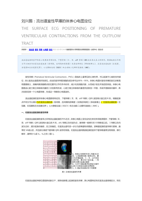
右室流出道间隔部起源的室早下壁导联通常没有顿挫,移行略早,通常在V3或V3、V4之间(图1)。
图1 右室流出道室性早搏 A 体表心电图室早在V1导联为QS型,V2导联移行晚于窦性QRS,提示为右侧起源,II、III、AVF呈直立的R型,提示位于流出道,I导联负向提示起源点偏前。
B CARTO激动顺序标测显示最早点位于右室流出道前交界部位。
C X-线证实起源点位于间隔侧。
右室流出道延伸部位是指肺动脉主干,即肺动脉瓣上起源的室性早搏,其心电图特征和右室流出道很难区分。
同右室流出道的早搏相比,肺动脉瓣上的早搏胸前导联移行更早,通常在V2/V3导联。
I导联R波更高或呈M型。
AVR和AVL导联QS波深浅与起源点位置前后高低不同相关(图2)。
图2 右室流出道肺动脉瓣上室性早搏A 体表心电图室早在V1导联rS型,V2导联移行晚于窦律QRS,提示为右侧起源。
II、III、AVF 呈直立的R型,提示位于流出道,I导联正向提示起源点偏后,AVR导联QS波较AVL导联深,提示起源点相对偏右。
B CARTO激动顺序标测显示最早点位于肺动脉瓣上并消融成功。
源自右室流出道室性早搏的心电图定位分析

o g a d f m r h etc l U W t c RV T n o i et a h f c v ehd t lc i h P r iai . ine o i r i t r gtvnr ua O  ̄O r t( O )ad t n sgt te e et e m to o oa z te V C o gnt n i r a v i e f i l e i o
0 5 。 I导联 呈 Q 或 rr提示 室早源 自R O .) 0 s s’ V T前部 , R或 r 起源 于后部 ,P<0 5 。 Ⅱ, , V 呈 R则 ( . ) 0 m A F导联 R波越 高 ( P<
0 5 , 导联 RS . )胸前 0 /移行越早 ( 0 5提示早搏距离 肺动脉瓣越近 。结论 】 R O 起 源的室早 中 Ⅱ,IA F p< . ) 0 【 在 VT 1,V 导联 R 1 波宽度
摘要:【 目的】 分析 右室流 出道 ( V T起 源室性早搏 的心 电图特征 及其与 消融 靶点 的关 系, R O 室性早搏有效 准确的 RO) 探讨 V T 心 电图定 位方法 。方法 】 【 回顾分析 20 —08 0 120 年在我 院行射频 消融 的 R O 室性 早搏 6 例 , VT 2 依据成 功消融靶 点 x 线定 位结 果, 分析其 与心电图各项 指标 的关系。结果 】 R O 早搏 中, 离壁与间隔部起源相 比 Ⅱ, A F 【 在 VT 游 m, V 导联 R 波宽伴有切迹 ( < P
室性早搏的心电图定位诊断

二尖瓣环起源
二尖瓣环起源的VPB其QRs波形特点类 似A型预激,呈右束支传导阻滞型,v1 导联以R波为主,胸导联移行早于V2导 联,V6导联呈Rs或RS型。 下壁导联的极性根据起源点的前后而不 同。部分游离壁起源的患者,在下壁导 联可以观察到顿挫。
.
.
谢谢!
.
室性早搏的心电图定位诊断
.
适应症
频发单型室性早搏和(或)非持续性室 速
Holter早搏次数非常多和(或)短阵室 速的次数较多
相关症状明显且多种药物治疗无效或希 望获得根治者
.
RVOT
无器质性心脏病患者发生的VPB,约80%起源 于右室,另外20%起源于左室。而在右室起源 的VPB中,绝大多数位于右室流出道。
.
.
.
二尖瓣环起源
二尖瓣环在解剖学上位于左心室的最后 方,此处起源的室速室早心肌除极方向 正对着胸前导联,故该组室速室早胸前 导联R波移行早(V2以前),V2--V6导 联以正向波为主的特点,多在V6 导联有 s或S波,三尖瓣起源室早偏离胸前导联所 以V6 导联无s或S波。二尖瓣环前侧壁和 侧壁起源的室性期前收缩在V6存在s波。
.
三尖瓣环起源
大致定位:首先,根据I导联及aVL导联均有 明显的正向波,可与右室流出道起源者相鉴别; 而III导联的极性则有助于判断起源点的前后; 对于游离壁和间隔部起源的区分, QRS波时 限>140ms、肢体导联存在切迹、V1联起始 波为正向对于前者的特异度均为100%,而胸 前导联转折区间≥V4的敏感度为100%,故当 具备QRS波时限>140ms、肢体导联存在切 迹、V1导联起始波为正向三个条件中的任何 一个,即可判断为游离壁起源,而当胸前导联 转折区间<V4即可排除游离壁起源。
右室流出道室早的心电图定位和消融PPT课件

临床病例分析
病例1--游离壁起源
I 导联:负向 S波:AVL>AVR
II/III/avF:切 迹 胸导联移行:V4
病例1--游离壁起源
病例2--RVOT与LVOT间起源
I 导联:正向 S波:AVR>AVL
胸导联移行:V2
病例2--RVOT与LVOT间起源
病例2--RVOT与LVOT间起源
RVOT解剖毗邻
RVOT解剖毗邻
右室流出道与左室流出道、 主动脉窦相毗邻,解剖位 置靠近,分别位于心脏的 右前与左后。 肺动脉窦处 于右心室流出道上方。
AP
LL
TIPs:RVOT室早消融失败 时,因这些位置的解剖毗 邻关系,应该考虑是否可 能为对侧(左室流出道、 主动脉窦)或者上方肺动 脉窦起源的可能。
右室流出道室早的心电图 定位导管操作技巧与消融
CONTENTS
右室解剖基础
室早定位原理 导管操作技巧 临床病例分析
右室解剖基础
RVOT解剖
Opened Right Ventricle Anterior View
肺动脉瓣 室上嵴
整个右心室的形态近似椎体, 解剖学正常存在的室上嵴将 其分成两部分:下面为固有 右心室,上方为漏斗部或称 肺动脉圆锥,其还被视为肺 动脉的起点。 右室流出道位于右室的左上 方,相当于右心室的心底部, 位置较高,该部位也称肺动 脉圆锥或漏斗部。 右室流出道起源于室上嵴的 游离缘,止于上方的肺动脉 瓣,外形近似于一个垂直整 个右室的短管,长约1.5厘米。 其内壁平滑而无肌小梁,其 向上延续为肺动脉出口。
大头双极电位提前QRS波起始25ms以上。 大头单极电图呈锐利的QS型,下降支常有切迹。 大头在该处起搏,起搏出与自身室早一致的图形。
- 1、下载文档前请自行甄别文档内容的完整性,平台不提供额外的编辑、内容补充、找答案等附加服务。
- 2、"仅部分预览"的文档,不可在线预览部分如存在完整性等问题,可反馈申请退款(可完整预览的文档不适用该条件!)。
- 3、如文档侵犯您的权益,请联系客服反馈,我们会尽快为您处理(人工客服工作时间:9:00-18:30)。
第19卷第1期2010年1月vol.19No.1Jan.2010武警医学院学报Acta Academiae Medicinae CPAF [收稿日期]2009-05-25;[修回日期]2009-10-23[作者简介]时向民(1970-),男,籍贯天津,医学博士,副主任医师,主要从事心脏电生理研究。
源自右室流出道室性早搏的心电图定位分析时向民1,王玉堂2,单兆亮2,杨庭树1(解放军总医院:1.南楼心内科;2.心内科;北京100853)摘要:【目的】分析右室流出道(RVOT )起源室性早搏的心电图特征及其与消融靶点的关系,探讨RVOT 室性早搏有效准确的心电图定位方法。
【方法】回顾分析2001-2008年在我院行射频消融的RVOT 室性早搏62例,依据成功消融靶点X 线定位结果,分析其与心电图各项指标的关系。
【结果】在RVOT 早搏中,游离壁与间隔部起源相比Ⅱ,Ⅲ,AVF 导联R 波宽伴有切迹(P <0.05)。
Ⅰ导联呈QS 或rsr’提示室早源自RVOT 前部,呈R 或rR 则起源于后部,(P <0.05)。
Ⅱ,Ⅲ,AVF 导联R 波越高(P <0.05),胸前导联R/S 移行越早(P <0.05)提示早搏距离肺动脉瓣越近。
【结论】在RVOT 起源的室早中Ⅱ,Ⅲ,AVF 导联R 波宽度判断早搏源于间隔或游离壁,Ⅰ导联QRS 波形态判断早搏源于RVOT 前部或后部,Ⅱ,Ⅲ,AVF 导联R 波高度结合胸前导联R/S 移行判断早搏接近肺动脉瓣或三尖瓣。
关键词:室性早搏;右室流出道;心电图;射频消融【文章编号】1008-5041(2010)01-0029-03【中图分类号】R540.4【文献标识码】AECG analysis of ventricular premature contraction originating from right ventricular outflow tractSHI Xiang-min,WANG Yu-tang,SHAN Zhao-Liang,YANG Ting-shu (Department of Cardiology of South Building,General Hospital of PLA,Beijing 100853,China)Abstract:【Objective 】To analyze the relationship between ECG and target ablation site of ventricular premature contraction (VPC)originated from right ventricular outflow tract (RVOT)and to investigate the effective method to localize the VPC origination.【Methods 】62patients(pts)hospitalized for radiofrequency ablation within 2001to 2008with VPC from RVOT were retrospectively analyzed,Correlative relation between successful ablation site based on X ray films and a variety of ECG parameters were studied.【Results 】In VPC from RVOT,wide and notched R wave of Ⅱ,Ⅲ,AVF indicated VPC originating from free wall (P <0.05),QS or rsr’type in lead Ⅰsuggested VPC originating from anterior wall,R or rR type indicated posterior wall origination (P <0.05).high R wave in Ⅱ,Ⅲ,AVF (P <0.05)with early R/S transition(P <0.05)revealed VPC adjacent to pulmonary valve.【Conclusions 】In VPC from RVOT the width of R wave in lead Ⅱ,Ⅲ,AVF was sensitive to localize VPC from free wall or septal.QRS configuration in lead Ⅰwas of use to distinguish VPC from anterior or posterior part within RVOT.The amplitude of R wave in lead Ⅱ,Ⅲ,AVF combined with R/S transition in precordial lead could predict VPC adjacent to pulmonary valve or tricuspid valve.key words:Ventricular premature contraction;Right ventricular outflow tract;ECG;Radiofrequency ablation右室流出道起源的室性早搏临床较为多见,患者多不伴器质性心脏病。
其发病机制可能与儿茶酚胺介导的晚期后除极以及局部微折返有关[1]。
该部位起源的频发室早或室速射频消融治疗虽然具有较高的成功率,但普遍存在寻找靶点时间较长,无效放电次数多,X 线曝光时间长的特点。
如能根据心电图特征预先判断早搏的起源位置,在标测时预见性的在流出道某一区域寻找靶点,对于提高手术效率,缩短手术时间至关重要。
本研究分析2001-2008年在我院行射频消融的62例右室流出道室早心电图特点,结合成功靶点X 线定位,总结了该部位早搏的心电图定位特点。
丐丐丐丐丐丐丐丐丐丐丐丐丐丐丐丐丐丐丐丐丐丐丐丐丐丐丐丐丐丐丐丐丐丐丐丐丐丐丐丐丐丐丐丐丐丐丐丐丐丐丐丐丐丐丐丐丐丐丐丐论著29第19卷第1期2010年1月vol.19No.1Jan.2010武警医学院学报Acta Academiae Medicinae CPAF 1资料和方法1.1一般资料2001-2008年我院住院行射频消融的右室流出道室性早搏患者62例,男28例,女34例,年龄范围14~64岁,平均年龄(39±25)岁。
患者室性早搏时胸前导联心电图均呈现左束支传导阻滞,额面心电轴右偏或者正常。
ⅡⅢAVF 导联QRS 直立。
有阵发性心悸病史1~10年,曾服用心律平,慢心律,乙胺碘呋酮,倍他乐克等药物治疗,效果欠佳,其中14例出现阵发性室速,室速图形与室早相同。
所有患者经超声心动图及胸部X 光片检查除外器质性心脏病。
术前停用抗心律失常药至少5个半衰期。
1.2方法1.2.1电生理检查:穿刺左侧股静脉送入6F 4极标测导管置于右心室心尖部。
53例患者术中出现频发室性早搏或室速,9例患者给予异丙肾上腺素1mg 加入生理盐水500ml 静脉点滴后诱发室早,且早搏形态与术前相同。
1.2.2消融方法:8F 加硬大头消融电极导管经股静脉送入RVOT。
X 线左前斜位60°(LAO )或右前斜位30°(RAO )投照下,将大头电极放在RVOT 的不同部位,LAO 确定电极位于间隔或游离壁,RAO 确定电极位置的高低和前后。
给予大头电极S1S1450~500ms 的刺激,选取刺激后心电图与自发室早完全相同的部位为靶点放电消融,能量为25~30W,电阻波动于100~130Ω,放电120~240s,如消融后早搏不再出现说明达到消融终点,如仍出现早搏则重新选择靶点消融。
1.2.3观测指标:对于RVOT 不同位置的室早或室速,观察Ⅰ导联形态及向量总和(R 或r 波与S 或s 波的总和),Ⅱ,Ⅲ,AVF 导联R 波幅度及高度,胸前导联R/S 波移行位置。
计数资料以( ±s )表示,采用t检验,P <0.05有显著性差异。
所用统计软件为Stata7.0。
2结果2.1射频消融结果62例患者在RVOT 消融成功,其中6例复发,再次射频消融后成功。
其中40例偏间隔部位,22例偏游离壁,27例位置偏前,35例位于中后部。
27例位置较高接近肺动脉瓣(上部),35例位于中下部。
2.2心电图分析Ⅱ,Ⅲ,AVF 导联R 波宽伴有切迹提示游离壁起源[(183.5±35.0)ms vs (140.2±30.6)ms;P <0.05],反之为间隔部起源。
Ⅰ导联呈QS 或rsr’提示室早源自RVOT 前部,呈R 或rR 则起源于后部[(-0.24±0.06)mv vs (0.15±0.08)mv;P <0.05],Ⅱ,Ⅲ,AVF 导联R 波高度越高[(1.54±0.52)mv vs (1.21±0.43)mv;P <0.05],胸前导联R/S 移行越早(V2-V3vs V4-V5;P <0.05)提示早搏距离肺动脉瓣越近。
见表1~3,图1~3(见封三)。
表1ROVT 前部与中后部起源的室早心电图对比部位(n )偏前部(27)偏中后部(35)Ⅰ导联形态QS 和rsr’R 或rR Ⅰ导联(R+S )值-0.03±0.25mv 0.20±0.28mv ①①:与ROVT 前部室早相比,P <0.05表2ROVT 上部与中下部起源的室早心电图对比部位(n )上部(27)中下部(35)Ⅱ,Ⅲ,AVF 导联R 波高度1.54±0.52mv 1.21±0.43mv ①胸导联R/S 波移行V2-V4V4-V6①①:与ROVT 上部室早相比,P <0.05表3ROVT 偏间隔与偏游离壁起源的室早心电图对比部位(n )偏间隔(40)偏游离壁(22)Ⅱ,Ⅲ,AVF 导联R 波宽度140.2±30.6ms 183.5±35.0ms ①①:与ROVT 偏间隔室早相比,P <0.053讨论流出道室性早搏在所有室性早搏中占有较高的比例,尤其多发于无器质性心脏病的患者,患者平时心电图正常。
其临床上具有运动及情绪激动易于诱发的特点,也有少部分室早易于在安静休息时发作[2]。
部分患者还可出现阵发性室速,以往认为此类室早预后良好,但近年研究发现频发流出道室早会损伤左室收缩功能,而射频消融去除室早后可使心功能恢复[3]。
