甲状腺穿刺细胞学液基制片流程
超声引导下甲状腺细针穿刺与液基薄层细胞学在甲状腺疾病诊断中的价值
超声引导下甲状腺细针穿刺与液基薄层细胞学在甲状腺疾病诊断中的价值【摘要】目的:分析超声引导下甲状腺细针穿刺与液基薄层细胞学在甲状腺疾病诊断中的适用价值。
方法:在我院收治的甲状腺结节患者中,择取2020年1月至2021年12月入院的84例作为实验对象,在超声引导下进行甲状腺细针穿刺,对穿刺取得的样本,分别实施组织病理学和液基薄层细胞学检查,就其检查结果进相比较。
结果:在对84例甲状腺结节患者进行液基薄层细胞学检查后,确定有良性61例、恶性22例,1例涂层不满意。
经过病理学诊断确定良性有62例,与细胞学检查结果一致(P>0.05);恶性有22例患者在细胞学检查后,予以手术切除,并及时对其病灶进行病理检查,确定其有21例为恶性,诊断准确率为95.45%。
结论:对甲状腺疾病予以超声引导下甲状腺细针穿刺与液基薄层细胞学检查,可对病变性质进行准确鉴别,创伤较小,可进行推广。
【关键词】超声;甲状腺细针穿刺;液基薄层细胞学;甲状腺疾病;诊断;价值甲状腺作为人体重要的代谢组织之一,其在存在异常后,很容易导致机体代谢紊乱。
甲状腺结节作为常见的一种甲状腺病变,其在近几年来其发病率呈上升趋势。
在以往,对于甲状腺结节多实施病理学检查来进行诊断,其准确率较高。
但病理学检查需要进行检查样本采集,其多是以手术的方式来进行,这就会对患者造成较大的损伤,且具有较强的事后性,无法对手术提供可靠的资料[1]。
在目前,提出以甲状腺细针穿刺细胞学检查来进行检查,其利用甲状腺细针穿刺取得细胞组织并实施液基薄层细胞学检查,以对疾病的性质进行辨别。
且在超声技术发展的状况下,临床以超声引导来进行细胞采集,可保证采集组织的确切性和快速性,效果更佳[2]。
在本次研究中,以84例甲状腺结节患者作为实验对象,就超声引导下甲状腺细针穿刺与液基薄层细胞学的诊断结果进行分析。
详细报道如下。
1一般资料与方法1.1一般资料在我院收治的甲状腺结节患者中,择取2020年1月至2021年12月入院的84例作为实验对象,男性46例、女性38例,年龄最大的67岁、最小的31岁,平均年龄(46.58±2.12)岁。
甲状腺结节细针穿刺病理诊断中液基细胞学技术的应用分析
甲状腺结节细针穿刺病理诊断中液基细胞学技术的应用分析摘要:目的:探究液基细胞学技术在甲状腺结节患者采取细针穿刺病理诊断中的临床应用。
方法:选取我院2020年3月-2021年3月期间内接诊的甲状腺结节患者50例,所有患者均采用细针穿刺病理诊断,把患者随机分为实验组和对照组,每组各25例,实验组患者采用液基细胞学技术进行制片(TCT),对照组患者则采用传统细胞学制片(CS),对比两组患者的细针穿刺病理诊断的良性符合率、恶性符合率和总准确率。
结果:实验组患者采取TCT制片其细针穿刺病理诊断的良性符合率(95.23%)、恶性符合率(100%)、准确率(96.00%),明显高于对照组患者采取CS制片的细针穿刺病理诊断良性符合率(70.00%)、恶性符合率(25.00%)、准确率(64.00%),数值差异明显具有统计学意义(P<0.05)。
结论:在甲状腺结节细针穿刺病理诊断中采用液基细胞学技术可有效提升病理诊断的准确性,为患者的治疗方案提供参考,具有重要的应用价值,值得在临床中推广并使用。
关键词:甲状腺结节;细针穿刺;病理诊断;液基细胞学技术传统细胞学制片容易受到红细胞白细胞的干扰,细胞重叠度较高从而影响诊断的准确率。
现有研究表明液基细胞学制片可以有效清除红细胞、白细胞等胶质蛋白或血细胞成分对于涂片的干扰,能够清晰显示细胞的单层排列状态、三位立体结构和细胞核结构,从而提高诊断准确率[1]。
本次研究以我院2020年3月到2021年3月接诊的50例采用细针穿刺病理诊断的甲状腺结节患者为研究对象,探究甲状腺结节细针穿刺病理诊断中液基细胞学技术的应用。
1资料与方法1.1一般资料选取我院2020年3月-2021年3月期间内接诊的甲状腺结节患者50例,所有患者均采用细针穿刺病理诊断,把患者随机分为实验组和对照组,每组各25例,所有患者均为女性。
实验组患者年龄在26-62岁之间,平均年龄为(44.38±2.07)岁。
甲状腺病变的液基细胞学诊断病理分析
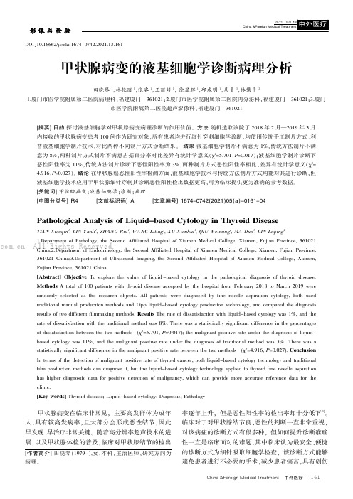
China &Foreign Medical Treatment中外医疗甲状腺病变在临床非常见,主要高发群体为成年人,具有较高发病率,且大部分会形成恶性结节,因此早发现、早治疗非常关键。
随着高分辨率超声技术的进展,以及甲状腺体检的普及,临床对甲状腺结节的检出率逐年上升,但是恶性阳性率的检出率却十分低下[1]。
临床对于对甲状腺结节良、恶性的判断一直非常重视,对该病症的诊断方式有很多种,但如何提升诊断准确性一直是临床面对的难题,其中临床认为最安全、便捷的诊断方式为细针吸取细胞学检查,该诊断方式能够避免患者进行不必要的手术,减少患者痛苦,具有创伤DOI:10.16662/ki.1674-0742.2021.13.161甲状腺病变的液基细胞学诊断病理分析田晓琴1,林艳丽1,张睿1,王丽婷1,徐显辉1,邱威明1,马多3,林鹭平21.厦门市医学院附属第二医院病理科,福建厦门361021;2.厦门市医学院附属第二医院内分泌科,福建厦门361021;3.厦门市医学院附属第二医院超声影像科,福建厦门361021[摘要]目的探讨液基细胞学对甲状腺病变病理诊断的作用价值。
方法随机选取该院于2018年2月—2019年3月内接收的甲状腺病变患者100例作为研究对象,所有患者均进行细针穿刺细胞学诊断,均使用传统手工制片方式、利普液基细胞学制片技术,对比两种不同制片方式诊断结果。
结果液基细胞学制片不满意为1%,传统方法制片不满意为8%,两种制片方式制片不满意占据百分率对比差异有统计学意义(χ2=5.701,P=0.017);液基细胞学制片诊断下恶性阳性率为11%,传统方法制片诊断下恶性阳性率为3%,两种制片方式恶性阳性率相比,差异有统计学意义(χ2=4.916,P=0.027)。
结论在甲状腺癌恶性阳性率检测方面,液基细胞学技术与传统方法制片方式均能对其进行诊断,但液基细胞学技术应用于甲状腺细针穿刺其诊断恶性阳性检出数据更高,可为临床提供更为准确的参考数据。
TLT制片技术在甲状腺FNAC标本制作中的应用价值探讨
TLT制片技术在甲状腺FNAC标本制作中的应用价值探讨【摘要】目的探讨TLT制片技术在甲状腺FNAC标本制作中的应用价值。
方法对120例甲状腺细针穿刺标本,分别采用液基薄层细胞制片技术和传统制片技术进行制作,并对标本质量进行对比分析。
结果采用液基薄层细胞制片技术所得标本中,诊断性细胞明显增多,细胞呈单层均匀分布,排列清晰,背景干净,具有诊断参考意义的成分得到部分保留。
结论液基薄层细胞制片技术能明显提高甲状腺FNAC标本的质量和诊断准确率,具有临床应用价值。
【关键词】液基薄层细胞制片技术;细针穿刺细胞学;甲状腺Abstract Objective To discuss the application value of thin layer technique (TLT) in specimen preparation of thyroid fine needle aspiration cytology (FNAC). Methods 120 specimen of thyroid FNAC were prepared with TLT and the traditional technique respectively, and a comparative analysis was made to the quality of the specimen.Results In the specimen with TLT, the diagnostic cells increased obviously, and the cells ranged neatly with a mean distribution of single layer, the background was clean and the parts of diagnostic reference werepartially preserved.Conclusions TLT can obviously raise the quality of thyroid FNAC specimen and the accuracy of diagnosis as well, it is of clinical application value.KEYWORDS TLT FNAC thyroid甲状腺肿块或结节是内分泌系统常见疾病,发病率为4%~7%;而甲状腺肿块中最大的问题是良恶性鉴别,尤其是不宜手术的患者,其良恶性的鉴别对临床治疗十分重要。
甲状腺结节细针穿刺病理诊断中液基细胞学技术的应用

甲状腺结节细针穿刺病理诊断中液基细胞学技术的应用发布时间:2021-12-19T02:10:14.728Z 来源:《航空军医》2021年6期作者:鞠学萍[导读] 同时以术后病理诊断为金标准,研究组检出准确率、良恶性诊断符合率均高于对照组(P<0.05)。
结论:甲状腺结节细针穿刺病理诊断中应用液基细胞学技术,可明显提高疾病诊断准确率,同时对结节良恶性有效鉴别,为后期治疗工作的开展提供有效参考。
鞠学萍大庆市第四医院 163712摘要:目的:评估液基细胞学技术应用于甲状腺结节细针穿刺病理诊断中的临床价值。
方法:遴选时段2019年10月-2020年10月内甲状腺结节患者80例,根据细胞学制片方法不同分2组,传统细胞学制片法40例(记对照组),应用液基细胞学技术40例(记研究组),对比分析两组诊断结果。
结果:对照组检出率为75.00%(30/40),研究组检出率为95.00%(38/40);同时以术后病理诊断为金标准,研究组检出准确率、良恶性诊断符合率均高于对照组(P<0.05)。
结论:甲状腺结节细针穿刺病理诊断中应用液基细胞学技术,可明显提高疾病诊断准确率,同时对结节良恶性有效鉴别,为后期治疗工作的开展提供有效参考。
关键词:甲状腺结节;细针穿刺病理诊断;液基细胞学技术;诊断价值Abstract: Objective: To evaluate the clinical value of liquid-based cytology in fine needle aspiration pathological diagnosis of thyroid nodules. Methods: 80 patients with thyroid nodules from October 2019 to October 2020 were selected. They were divided into two groups according to different cytological methods, including 40 cases of traditional cytology (recorded in the control group) and 40 cases of liquid-based cytology (recorded in the study group). The diagnostic results of the two groups were compared and analyzed. Results: the detection rate was 75.00% (30 / 40) in the control group and 95.00% (38 / 40) in the study group; At the same time, taking the postoperative pathological diagnosis as the gold standard, the detection accuracy and the coincidence rate of benign and malignant diagnosis in the study group were higher than those in the control group (P < 0.05). Conclusion: the application of liquid-based cytology in fine needle aspiration pathological diagnosis of thyroid nodules can significantly improve the accuracy of disease diagnosis, effectively distinguish benign and malignant nodules, and provide an effective reference for later treatment. Keywords: thyroid nodules; Fine needle puncture pathological diagnosis; Liquid based cytology; diagnostic value 甲状腺结节属于常见头颈部肿瘤及内分泌系统疾病的一种,有良性与恶性之分。
超声引导下甲状腺细针穿刺液基细胞
*器材应用与技术研究*超声引导下甲状腺细针穿刺液基细胞学与传统细胞涂片在甲状腺结节诊断中的意义陈良昆山市中医医院超声科,江苏昆山215300摘要目的评估并分析超声引导下甲状腺细针穿刺(US fine-needle aspiration,US-FNA)液基细胞学与传统细胞涂片对甲状腺结节的诊断价值。
方法选择2019年1月—2021年11月昆山市中医医院内接受US-FNA检查的90例甲状腺结节患者为研究对象,分别应用传统细胞涂片、液基细胞学技术,以手术病理学诊断结果为金标准,分析传统细胞涂片、液基细胞学单一检测及联合检测的诊断效能。
结果90例患者经手术病理诊断结果:恶性结节49例、良性结节41例;以手术病理诊断结果为金标准,传统细胞涂片检查:真阳性42例、假阳性2例、真阴性39例、假阴性7例;液基细胞学检查:真阳性40例、假阳性3例、真阴性38例、假阴性9例;联合检查:真阳性48例、假阳性1例、真阴性40例、假阴性1例;联合检查诊断灵敏度97.96%、准确度97.78%明显高于传统细胞涂片(85.71%、90.00%)和液基细胞学(81.63%、86.67%),差异有统计学意义(P<0.05);传统细胞涂片、液基细胞学单一诊断特异度(95.12%、92.68%)与联合检查特异度97.96%比较,差异无统计学意义(P>0.05)。
结论甲状腺结节US-FNA诊断中应用液基细胞学技术联合传统细胞涂片,可明显提高疾病诊断准确率,同时对结节良恶性有效鉴别,与病理组织学结果一致性高,可为后期治疗工作的开展提供有效参考。
关键词超声引导;甲状腺细针穿刺;液基细胞学;传统细胞涂片;甲状腺结节中图分类号R4文献标志码A doi10.11966/j.issn.2095-994X.2022.08.10.16Significance of Ultrasound-guided Thyroid Fine-needle Aspiration Liquid-based Cytology and Traditional Cell Smear in the Diagnosis of Thyroid NodulesCHEN LiangDepartment of Ultrasound,Kunshan Hospital of Traditional Chinese Medicine,Kunshan,Jiangsu Province,215300ChinaAbstract Objective To evaluate and analyze the diagnostic value of ultrasound-guided fine needle aspiration(US-FNA)liquid-based cytol⁃ogy and traditional cell smear for thyroid nodules.Methods A total of90patients with thyroid nodules who underwent US-FNA examination in Kunshan Hospital of Traditional Chinese Medicine from January2019to November2021were selected as the research objects.The tradi⁃tional cell smear and liquid-based cytology techniques were used to analyze the diagnostic efficacy of single detection and combined detec⁃tion of traditional cell smear and liquid-based cytology with the results of surgical pathology as the gold standard.Results90patients by sur⁃gical pathological diagnosis:49cases of malignant nodules,41cases of benign nodules;with the results of surgical pathological diagnosis as the gold standard,traditional cell smear examination:true positive42cases,false positive2cases,true negative39cases,false negative7 cases;liquid-based cytology:40cases of true positive,3cases of false positive,38cases of true negative,9cases of false negative;com⁃bined examination:48cases were true positive,1case was false positive,40cases were true negative,1case was false negative.The diagnos⁃tic sensitivity and accuracy of combined examination were97.96%and97.78%,which were significantly higher than those of traditional cell smear(85.71%,90.00%)and liquid-based cytology(81.63%,86.67%),the difference was statistically significant(P<0.05).There was no statisti⁃cally significant difference in the specificity of single diagnosis of traditional cell smear and liquid-based cytology(95.12%,92.68%)and the specificity of combined examination97.96%(P>0.05).Conclusion The application of liquid-based cytology technology combined with tradi⁃收稿日期:2022-08-04;修回日期:2022-08-24作者简介:陈良(1974-),男,本科,副主任医师,研究方向为腹部及浅表小器官超声诊断方面。
CS联合LBP制片技术在甲状腺细胞学诊断中的应用价值
有 患侧 甲状 腺组织 病理 学诊 断依 据 ; ④ 所有 组 织病
理学结 果需 明确 良 、 恶性 。
沉降式液基细胞学 ; 传统 细胞学 ; 甲状腺癌 ; 针 吸细
R 7 3 6 . 1 ; R 4 4 6 . 9
准 确 鉴别 甲状腺 良、 恶 性结节 , 可 避免 良性病 变患 者 不必 要 的严 重创 伤 检 查 和手 术 治 疗 , 是 目前 公 认 的 首选 诊 断方 法 和术 前 筛 选 检 查 【 。传 统 细 胞 学 手 工涂 片 ( c o v e n t i o n a l s m e a r , C S ) 常 受多 种 因素 影 响致
・
6 8 8・
安徽 医科 大s Me d i c i n a l i s A n h u i 2 0 1 3 J u n ; 4 8 ( 6 )
◇技 术与 方法 ◇
C S联合 L B P制 片技术在 甲状腺细胞学诊断中的应用价值
F N AC诊 断准 确率 、 敏感度 的方法 。
1 材 料与方 法
1 . 4 标 本 制作方 法
1 . 4 . 1 C S涂 片 将一 半 抽 取 物 迅 速 推 到玻 片 上 ,
徒 手用 针 头 均 匀 涂 抹 , 常规 涂 片 1~2张 , 立 即 予 9 5 % 乙醇湿 固定 , 苏 木精 一 伊红( HE ) 染色, 树 脂胶 封
后开风 , 李
摘要 分别采用沉降式 液基 细胞学制 片 ( L B P ) 技术 和传 统
俊 , 江洁美
4 8例 , 右叶者 7 2例 , 双 叶或 峡 部 者 8 4例 。病 例 入
甲状腺穿刺细胞学液基制片流程
甲状腺穿刺细胞学液基制片流程英文回答:The process of preparing liquid-based cytology (LBC) slides for thyroid fine-needle aspiration (FNA) involves several steps. First, the FNA procedure is performed by inserting a thin needle into the thyroid nodule to collect cells. The collected cells are then rinsed in a vial containing a liquid fixative, such as PreservCyt solution. This fixative helps to preserve the cellular structure and prevent degradation.Once the cells are in the fixative solution, the vial is gently agitated to ensure that the cells are evenly distributed. The fixative solution is then centrifuged to concentrate the cells at the bottom of the vial. The supernatant is carefully removed, leaving behind a pellet of cells.Next, the pellet of cells is resuspended in a smallamount of fixative solution. A drop of this cell suspension is then placed on a glass slide. The slide is tilted at a specific angle to allow the liquid to spread evenly across the surface. This helps to create a monolayer of cells on the slide, ensuring that individual cells can be easily identified and analyzed.After the liquid has dried on the slide, it is fixed by immersing it in a fixative solution, such as 95% ethanol. The fixative solution helps to preserve the cellular morphology and prevent any further degradation. The slide is then stained using a Romanowsky-type stain, such asDiff-Quik or Wright's stain. This stain helps to enhance the visibility of the cellular features and facilitates the identification of abnormal cells.Once the staining is complete, the slide is rinsed with water to remove any excess stain. It is then air-dried and coverslipped using a mounting medium. The coverslip helps to protect the slide and allows for easy examination under a microscope.中文回答:甲状腺穿刺细胞学液基制片的流程包括多个步骤。
液基细胞学操作规程
液基细胞学操作规程 Modified by JACK on the afternoon of December 26, 2020液基细胞学工作流程一.所需的耗材仪器的耗材均为一次性使用,每套耗材一般含:专用宫颈刷1个、细胞保存处理液1瓶、过滤膜袋1个(可选)、专用防脱载玻片1个、细胞制片器1个、一次性滴管1个二.采样的操作步骤A、准备1、要告诉病人进行检查的最佳时间(绝经前的妇女,月经中后期是最理想的)应避开经期。
2、要告诉病人检查前勿用阴道药膏,这样可以及防止因异物造成收集细胞的困难。
3、要告诉病人检查前48小时内禁止性交、盆浴和阴道检查。
B、采样1、将宫颈刷缓缓伸入。
2、使刷头导入宫颈管内,向前伸,紧贴宫颈口四周。
3、沿轴同向缓慢旋转十圈以上,切忌反向旋转。
C、漂洗1、将宫颈刷刷头推入细胞保存液瓶盖。
2、为使细胞充分漂洗到保存液中,要可适当振荡瓶子。
D、保存1、确保试样和检验申请单标记号码的一一对应性。
2、在处理液瓶子上标记相同号码后,将标本与检验申请单一同送往检验室。
三.注意事项1、取材部位应在宫颈鳞柱交界处。
2取材应尽可能避开经期,取材前24小时不上药,不冲洗,2、不过性生活。
分泌物较多时,要在采样前用棉签轻轻擦去不可用力擦。
3、应观察采样,使宫颈刷对所取部位有一定的压力,刷尖入颈管内,两边紧巾颈管外口。
4、采样过程中,宫颈出血明显时,应立即停止采样。
一般情况下尽量避免短期内(<3个月)重复取材,以免有假阴性结果。
5、器械应保持干燥清洁,不要用酒精、肥皂水及润滑剂等清洗。
6、申请单填写应尽量完整、字迹工整,尽可能提供相关的临床信息。
采样后应及时送往检验室。
四.传统取材样器与宫颈刷的对比传统取样器分为:1、圆形刷子,只能取子宫颈内的细胞2、棉棒,不能取出全部子宫颈细胞3、木质乔板,不能适合所有妇女宫颈结合部的开形状宫颈刷取样器:柔软鬃毛适合每种性状,避免可能的伤害,能够收集所有的细胞。
液基细胞制片工作站工作流程
液基细胞制片工作站是一种常用于细胞学检测的设备,其工作流程包括以下步骤:
1. 样本采集:通过采用刷子、刮片等方式,从患者的宫颈、尿道、口腔等部位采集细胞样本。
2. 样本处理:将采集到的样本放置在液基细胞制片管中,加入荧光染色剂和保护剂,振荡混合,使样本细胞均匀分布在液基细胞制片管中。
3. 液基细胞制片:将液基细胞制片管放入液基细胞制片工作站中,机器会自动进行制片。
液基细胞制片的过程包括先将细胞样本筛选出杂质和红细胞,然后将细胞在载玻片上进行喷涂、固定和染色等操作。
4. 细胞学检查:完成液基细胞制片后,载玻片会被送至细胞学实验室进行检查。
细胞学检查包括镜检和数字影像检查两种方式,镜检需要专业的细胞学医师通过显微镜进行观察,数字影像检查则通过数字显微镜或计算机影像处理系统进行诊断。
5. 报告解读:医师根据细胞学检查结果,结合患者的病史和临床表现,进行报告解读并提出诊疗意见。
在液基细胞制片工作站上,还可以通过连接医疗信息系统,将检测结果自动上传至患者的电子病历中,
方便医生进行远程诊断和管理。
总之,液基细胞制片工作站能够提高细胞学检测的质量和效率,对于协助医师进行早期诊断和治疗具有重要意义。
甲状腺细针穿刺术技术规范

甲状腺细针抽吸细胞学检查概述甲状腺细针抽吸活检现在已经确定为准确性很高的诊断方法,常规用于结节性甲状腺疾病诊断过程的第一步(1-4)。
流行病学研究显示结节性甲状腺疾病在临床很常见,北美洲成人的患病率为4% ~7%,每年新增发病率为0.1%,换句话说,美国每年大约出现275,000个新结节(5)。
最近一项对美国甲状腺协会临床成员的调查显示,他们中的绝大多数(96%)用细针抽吸细胞学检查来诊断甲状腺结节。
所以,我们估计仅美国每年进行的细针抽吸细胞学检查就有250,000 ~ 300,000例。
估计全世界每年进行细针抽吸细胞学检查者数以百万计。
因此,细针抽吸细胞学检查在甲状腺检查中的重要性怎么强调都不过分。
本章将介绍活检技术,细胞学诊断,并发症,细针抽吸结果,诊断缺陷,以及对甲状腺结节病人临床处理有用的其他信息。
定义Martin 和 Ellis于1930年首次报道用细针抽吸细胞学检查诊断甲状腺结节,当时他们使用的是18号针。
随后,有报道使用Silverman或Tru-Cut针行针刺切割活检组织学检查者。
但由于担心恶性肿瘤沿针剌穿剌道扩散、假阴性结果以及严重并发症等,这些技术均未被广泛接受。
然而,二十世纪六十年代北欧研究人员引入了甲状腺的细针抽吸活检,这一技术也逐渐被北美广泛接受,但是直到20世纪80年代才得以广泛应用(8,9)。
FNA活检多采用22-27号细针针头(通常为25号针头)。
顾名思义,这一活检技术就是通过抽吸获得甲状腺肿块的细胞或体液。
与经皮粗针穿刺活检不同,粗针活检是获取组织样本且需要组织固定,而抽吸活检所提供的是供细胞学检查的样本。
另一种细针非抽吸活检技术,可不用抽吸但仍可从甲状腺肿块得到供细胞学检查的标本。
尽管FNA技术看起来很简单,但是要学会并熟练的掌握活检技术则需要一定的时间和实践经验的积累。
有关谁最适合作FNA活检的争论仍在继续,但是很明显,如果已经掌握进行活检技巧的人进行活检的话就会得到最好的结果。
甲状腺穿刺技术
甲状腺细胞病理学细胞病理学又称临床细胞学或诊断细胞学,是以组织学为基础观察细胞结构和形态的一门科学,它包括脱落细胞学和针吸细胞学。
甲状腺的针吸细胞学检查始于1952年,由Soderstrom首次报道,20世纪70年代末在欧美被广泛推广。
我国开始于70年代。
目前认为甲状腺针吸细胞学检查有其局限性,诊断时思路要开阔。
甲状腺针吸细胞学的特点有如下几点:1、痛苦小,经济实用,病人易于接受。
2、操作简单,不需麻醉,极少有并发症。
3、诊断较准确、快速,是甲状腺结节的首选临床筛选方法。
4、操作越熟练,经验越丰富,诊断符合率越高。
5、每年穿刺500次以上可保持技术熟练程度。
6、标本不足者,不能诊断良性或阴性。
随着经验的积累,可更好地掌握标本足量的标本。
7、囊性病变吸出物细胞量较少,是造成假阴性诊断的主要原因。
8、对于不能诊断或B超有钙化点的病变,重复穿刺可提高诊断符合率。
9、针吸对非毒性结节性甲状腺肿、乳头状癌、髓样癌、未分化癌等许多甲状腺疾病的诊断准确率较高。
10、对于滤泡性病变,针吸是筛选手术病人的一种非常有效的方法。
11、细胞学诊断医师与临床医师经常相互交流有利于提高诊断符合率。
针吸技术与标本制备一、针吸器械细针是指针头外径不超过0.9mm的针,一般采用外径0.5-0.7mm的一次性无菌肌肉注射针头,即6-7号针头,相当于国外的22-25G针,注射器以10ml的为宜,要求严密,抽吸时不漏气,干蒸消毒,不能有水。
二、针吸操作1、负压抽吸穿刺法仔细检查甲状腺,选择穿刺点后,碘酒或酒精消毒皮肤,通常不需要麻醉。
左手固定肿物(肿物较大者用指腹按住,较小者用指尖或指甲按住),右手持注射器进针,当针尖进入肿块后回拉针栓,负压下针尖在肿物内前后迅速移动,当从针座后孔看到组织液时,释放负压,迅速拔针。
然后把吸出物推到玻璃片上。
目前国内外普遍采用此方法穿刺。
该方法吸出物多,处能直接涂片外,还可制作组织块,进行其他辅助检查。
免疫组化技术在甲状腺液基细胞学诊断为Ⅲ类-意义不明确细胞非典型病变中的应用价值
免疫组化技术在甲状腺液基细胞学诊断为m类-意义不明确细胞非典型病变中的应用价值张永生1王萍萍2,高萍1张立平、,李强4TTe applicatioe value of immunohisychemiseo V he diagnosis ot hyoid fluid based cy-toloon as class m atypical lesioas with uecleer significaaceZHANG Youdskepy1,WANG Pinuniny2,GAO Piny1,ZHANG Lipiny1,LI Qiany21Departmenh of Pathologn;2Departmeni cf Brenst nd Thyroia,CRzhao Central Hospital,bhangong Rizhao276300,China.【Abstract】Objective:To investigate the applicahon value of immunohisSchemistm in the diagnosis of thyroid fluid based cyslogy as class m atypical lesions with unclear significance.Methods:96cases of thyroid fine needle aspi-rahon liquid-based cyslogy were selected for diagnosis of class m atypical cell lesions with unclear significance.Allthe selected cases were diagnosed bp operahon.After the cell smear was comp/sd,the cell bloch was make with theremaining liquik based presevatSn solution.The posSperahve patPolovicgl specimen and cell bloch were sectionedand immunoXisSchemicai detection of CK16)Ga/ctin-3,HBME-1and TPO were carried out.According to the his-topatPolovicgl results,the selected cases were divided into thyroid papb/m gvxp and benign group.The expression offour Uinds of axtikobics in two groups in cell bloch immunoPisSchemicci staining and compamtive analysis.The consistency of cell bloch and hismpathoPgp in inimunobisSchemisWy was veri/ed.ResUts:In96cases of thyroid fineneedle aspimhon with liquik based cyslogy.13cases failed to make wax bloch due to the small amount of remainingcells,end78cases were divided into thyroid cancer group and benign group according to hismioxical diagnosis.Theexpression rate of CK19,GaPctin-3and HBME-1in thyroid cancer group was significantly higher than that in benign group(all P二2.400).The expression rate of TPO in benign group was significanhy higher than that in thyroidcancer group(P=2.002).The diRevnce was stahsticahy significant(P<2.25).The four inimunoPisSchemicaimarUers have high sensitivity,ecceracy and specificity in the diagnosis of thyroid cancer.The expression of CK19,Gc-lectin-3,HBME-1and TPO in thyroid cancer cell bloch was the same as that in posSperahve hismpathoPxp withno stahshcU diRevnce(P>0.45).Conclusion:The app/cahon value of cell bloch inimunobisSchemisWy in the diagnosis of thyroid fine needle aspimhon fluid based cyslogy as class m-atypical lesions with unclear significance isXrger.The coincidence rate between the results and the posSperahve hisSpathoPgp is high.It can improve the acce-racy of cyslogicai diagnosis.【Key woat t thyroid,puncture:liquik-based cyslogy,Bethesha reporting system,immunohisSchemistm,diagnosisand diRerenhai diagnosisMobern Oncology2021,29(11):1873-1372【摘要】目的:探讨利用免疫组化技术在甲状腺液基细胞学诊断为m类-意义不明确细胞非典型病变中的应用价值。
甲状腺肿瘤细针穿刺指南
甲状腺肿瘤细针穿刺指南【1】 (美国Papanicolaou细胞病理协会)甲状腺肿瘤细针穿刺的主要目的是鉴别需要外科治疗和不需要外科治疗的甲状腺肿块,以减少诊断性的甲状腺手术。
在有经验的人手里对一些甲状腺病变可以做明确的诊断如:胶性甲状腺肿,Hashimoto甲状腺炎,乳头状癌,高度滤泡性癌,髓样癌,未分化癌,大细胞淋巴瘤以及转移癌。
假阴性率要控制在2%以下,假阳性率要控制在3%以下。
要穿刺的甲状腺肿块类型:细针穿刺的主要指征是实体的(solitary)甲状腺肿块。
典型的多节结甲状腺病变很少是癌性肿块,有当某一个肿块快速增长成为主要的部分,或者结构,一致性发生变化的时候才有可能是癌变。
在这情况下其癌变的发生率和单个的实体肿块没有明显的差异。
值得注意的是Hashimoto甲状腺炎不但可以表现为结实的肿块类似肿瘤,而且还可以和甲状腺癌或者淋巴瘤同时存在。
维持熟练程度的最低要求:甲状腺细针穿刺可以在一般的医院或者医生诊所进行,不一定要在医学中心。
做细针穿刺的医生要有专业的培训.在短的时间内完成100例或以上穿刺是进行甲状腺细针穿刺培训的基本要求。
对阅片的细胞病理医生进行充分的培训也同等的重要。
建议在短的时间内进行较密集的培训,效果较好。
细胞病理医生在一年内完成30-40例甲状腺细针穿刺的阅片是维持熟练程度的基本要求。
细针穿刺的操作:甲状腺结节可以用25 Gauge针头(大约相当于国内6-7号针头)加或不加负压进行穿刺取样。
如果加压,可用5 毫升注射器,抽吸1-2 毫升负压,快速进出肿块。
针头拔出前应消除负压。
由于甲状腺组织血液丰富不加负压取样更简单易行,可以取得更干净,适当的样品。
囊性病变可以用大号的23号针头尽可能的抽干净液体,残留的肿块要用25 Gauge针头重复取样。
一般没有必要进行局部麻醉。
合适的取样是成功进行甲状腺细针穿刺诊断的前提。
穿刺后马上进行显微镜评估以决定取样是否合适,可以大大提高穿刺的成功率。
液基细胞学制片手册(新)Microsoft+Word+文档
液基细胞学薄层制片应用手册尊敬的客户:您好!感谢您使用我公司提供的康创液基细胞薄层制片技术(TCT),为更好方便您的制片工作,并达到最佳的制片效果,根据我公司的产品特点,特提供本产品的使用指导手册。
一前言康创液基细胞涂片系统是值得信赖的细胞液基涂片技术,该技术已在国内各大省市广泛应用。
提供了早发现、早诊断、早治疗的新方法,该方法具有简单年易行、普查成本较低,适用于大规模的宫颈癌普查的优点。
二液基细胞薄层制片的国际标准5000个上皮细胞两团以上宫颈内膜细胞细胞重叠少在40倍物镜视野下,每个视野可见11个上皮细胞三 实验室工艺流程1,机器制片流程 4、振荡器振荡消化5、消的标体液置离心机离心离心涂片机制片巴氏染色涂片及镜下照片、取材后,用镊子将刷头取下放入固定液中。
、将刷头置入宫颈口内旋转3-5周取材。
巴氏染色涂片及镜下照片2,手工涂片流程经离心后的标本,加入50%或75%乙醇混悬,用吸管吸取混悬液中间的标本进行手工涂片,涂片时,以中心点为圆圈逐渐向外画圈,不要重复,最大程度避免细胞重叠。
宫颈液基薄层制片流程4、振荡器振荡消化5、消化好的标体液置离心机离心3、宫颈标本管中加入SP 消化液制片经充分干燥后,分别做巴氏染色和DNA染色。
巴氏染色涂片及镜下照片2、取材后,用镊子将刷头取下放入固定液中。
1、将刷头置入宫颈口内旋转3-5周取材。
备好专门的标本管及宫颈刷巴氏染色涂片及镜下照片四 制片过程详细图解1 准备选择适合的筛查对象,准备好固定液和宫颈刷a 装有15ml 固定液的标本管b 厂家提供的宫颈刷2 取材: 标本采集是制片工作的开始,为了达到最好的制片效果,必须确定适合筛查的对象和遵守筛查原则,个体的选择非常重要,但按照以下采集原则可以避免可能出现的废片。
1、 窥阴器扩张阴道后,将宫颈刷头中央凸出部置入宫颈内,直到两翼刷毛和宫颈口两侧紧密接触。
其目的是取到细胞,从而避免病理科阅片时细胞量少而影响诊断的现象。
细胞穿刺制片流程
细胞穿刺制片流程下载温馨提示:该文档是我店铺精心编制而成,希望大家下载以后,能够帮助大家解决实际的问题。
文档下载后可定制随意修改,请根据实际需要进行相应的调整和使用,谢谢!并且,本店铺为大家提供各种各样类型的实用资料,如教育随笔、日记赏析、句子摘抄、古诗大全、经典美文、话题作文、工作总结、词语解析、文案摘录、其他资料等等,如想了解不同资料格式和写法,敬请关注!Download tips: This document is carefully compiled by the editor. I hope that after you download them, they can help yousolve practical problems. The document can be customized and modified after downloading, please adjust and use it according to actual needs, thank you!In addition, our shop provides you with various types of practical materials, such as educational essays, diary appreciation, sentence excerpts, ancient poems, classic articles, topic composition, work summary, word parsing, copy excerpts,other materials and so on, want to know different data formats and writing methods, please pay attention!细胞穿刺制片流程是一种常用于临床诊断和研究的技术,通过采集体液或组织细胞进行细胞学检查,可以帮助医生快速准确地诊断疾病。
- 1、下载文档前请自行甄别文档内容的完整性,平台不提供额外的编辑、内容补充、找答案等附加服务。
- 2、"仅部分预览"的文档,不可在线预览部分如存在完整性等问题,可反馈申请退款(可完整预览的文档不适用该条件!)。
- 3、如文档侵犯您的权益,请联系客服反馈,我们会尽快为您处理(人工客服工作时间:9:00-18:30)。
甲状腺穿刺细胞学液基制片流程甲状腺穿刺细胞学液基制片是一种常见的病理学检查方法,可用于评估甲状腺细胞的形态学特征和诊断甲状腺疾病。
以下是制片流程的详细描述:1.患者进入手术室,取患者的甲状腺穿刺液样本。
The patient enters the operating room, and a sample of thyroid aspiration fluid is taken from the patient.2.将甲状腺穿刺液样本倒入培养皿中。
Pour the thyroid aspiration fluid sample into a culture dish.3.加入液基固定液,使细胞固定在载玻片上。
Add liquid-based fixative to immobilize the cells on a glass slide.4.载玻片放置在离心机中,进行离心处理。
Place the glass slide in a centrifuge for centrifugation.5.注射已离心的细胞沉淀,形成细胞颗粒在载玻片上。
Inject the centrifuged cellular sediment, formingcellular particles on the glass slide.6.将载玻片在特定的温度和湿度条件下干燥。
Dry the glass slide under specific temperature and humidity conditions.7.用甲状腺穿刺细胞蘸液染色,增强形态学特征的观察。
Stain the thyroid aspiration cells with a dye to enhance observation of morphological characteristics.8.在显微镜下观察载玻片,注意细胞的形态学特征。
Observe the glass slide under a microscope, paying attention to the morphological characteristics of the cells.9.检查细胞的大小、形状、核仁及细胞浆等细胞学指标。
Examine cytological indicators such as cell size, shape, nucleus, and cytoplasm.10.鉴定细胞类型:正常甲状腺细胞、甲状腺癌细胞或其他病理细胞。
Identify cell types: normal thyroid cells, thyroid cancer cells, or other pathological cells.11.记录和分类细胞类型,进行细胞计数和分类统计。
Record and classify cell types, perform cell counting and classification statistics.12.根据细胞学结果,做出甲状腺疾病的诊断并提供治疗建议。
Make a diagnosis of thyroid disease and provide treatment recommendations based on cytological results.13.编制细胞学报告,详细描述细胞学特征和诊断结论。
Prepare a cytological report describing detailed cytological features and diagnostic conclusions.14.将制片完成的载玻片和细胞学报告送至临床医生进行解读。
Deliver the completed glass slide and cytological report to clinical doctors for interpretation.15.注意操作过程中的严格无菌原则,以避免细胞污染。
Pay attention to strict aseptic principles during the operation to avoid cell contamination.16.定期检查和维护实验设备,确保其正常运行和准确性。
Regularly inspect and maintain laboratory equipment to ensure its proper operation and accuracy.17.培养皿中的甲状腺穿刺液样本应适量,以确保细胞数量足够。
The thyroid aspiration fluid sample in the culture dish should be sufficient to ensure an adequate number of cells.18.液基固定液应正确配制,以保证细胞固定效果。
The liquid-based fixative should be properly prepared to ensure effective cell fixation.19.离心过程中的转速和时间应根据不同的细胞类型进行调整。
The centrifugation speed and time during the centrifugation process should be adjusted according to different cell types.20.注射细胞沉淀时要均匀注射,以获得均匀分布的细胞颗粒。
Inject the cellular sediment evenly to obtain a uniform distribution of cellular particles.21.载玻片的干燥时间不宜过长,以避免细胞形态学的改变。
The drying time of the glass slide should not be too long to avoid changes in cell morphology.22.选择合适的甲状腺穿刺细胞蘸液染色方法,以增强细胞特征的显现。
Choose an appropriate staining method for thyroid aspiration cells to enhance the visibility of cell features.23.显微镜的放大倍数和焦距应根据需要进行调整,以获得清晰的细胞图像。
Adjust the magnification and focal length of the microscope as needed to obtain clear cell images.24.观察细胞时要仔细、耐心,并遵循相关的细胞学标准。
Observe cells carefully and patiently, following relevant cytological standards.25.细胞计数时应避免重复计数和漏计,以保证统计结果的准确性。
Avoid duplicate or missed cell counting to ensure the accuracy of statistical results.26.细胞分类应参考国际上通用的细胞学分类标准和术语。
Cell classification should refer to internationally recognized cytological classification criteria and terminology.27.细胞计数和分类统计时要使用适当的计数器和统计方法。
Use appropriate counters and statistical methods for cell counting and classification statistics.28.诊断结果应基于多角度综合分析和专家判断,以提高准确性。
Diagnosis should be based on comprehensive analysis from multiple perspectives and expert judgement to improve accuracy.29.细胞学报告中的描述要准确、简洁,涵盖重要的细胞学特征。
The description in the cytological report should be accurate and concise, covering important cytological features.30.细胞学报告应及时送达临床医生,以帮助指导患者的诊疗过程。
The cytological report should be promptly delivered to clinical doctors to help guide the diagnosis and treatment process of patients.31.及时更新并遵守甲状腺穿刺细胞学液基制片的相关规范和指南。
Timely update and comply with relevant specifications and guidelines for thyroid aspiration cytology liquid-based preparation.32.员工在操作过程中应戴手套、口罩等个人防护用品,确保安全。
Staff should wear personal protective equipment such as gloves and masks during the operation to ensure safety.33.在实验室中的甲状腺穿刺细胞学液基制片区域,要保持干净整洁。
Keep the area for thyroid aspiration cytology liquid-based preparation in the laboratory clean and tidy.34.定期进行实验室的消毒和清洁工作,以防止交叉感染。
Regularly disinfect and clean the laboratory to prevent cross-infection.35.固定液、染色液等试剂应储存在适当的温度和湿度条件下,避免变质。
Fixatives, staining reagents, and other chemicals should be stored under appropriate temperature and humidity conditions to avoid deterioration.36.实验室人员要进行规范的操作培训,确保熟练使用相关仪器和设备。