股疝修补术ppt课件
合集下载
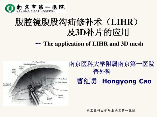
腹腔镜腹股沟疝修补术(LIHR)ppt课件

1
文档仅供参考,不能作为科学依据,请勿模仿;如有不当之处,请联系网站或本人删除。
LIHR的ห้องสมุดไป่ตู้用人群
具有腹膜前修补指证的人群
腹横筋膜薄弱患者(老年、直疝、复合疝等)
高腹内压患者
需要尽快恢复活动和工作的人群 有特殊需求和意愿的人群 复发疝和双侧疝患者
2
文档仅供参考,不能作为科学依据,请勿模仿;如有不当之处,请联系网站或本人删除。
22
TEP
腹膜前间隙的建立 文档仅供参考,不能作为科学依据,请勿模仿;如有不当之处,请联系网站或本人删除。
层次
在腹横筋膜浅层和腹膜之间, 而不是在腹横筋膜深、浅层之间!
腹横筋膜分前、后两层
➢前层(浅层):紧贴腹直肌和联合肌腱 深面,是真正意义上的腹横筋膜 ➢后层(深层):不规则增厚的纤维组织, 又称为腹膜前筋膜,术中要进行一定的 分离
补片必须覆盖整个肌耻骨孔,并有一 定的重叠!!
内侧:覆盖耻骨结节和腹直肌并超过中线!! 外侧:髂前上棘! 上缘:覆盖联合肌腱至少2cm!! 下缘外侧:精索“腹壁化”!! 下缘内侧:插入耻骨膀胱间隙(Retzius)!!
17
TAPP TAPP手术要点 文档仅供参考,不能作为科学依据,请勿模仿;如有不当之处,请联系网站或本人删除。 补片的固定
<20岁
青年患者
老年患者
单纯缝合
意愿 尽早活动 美观 复发疝 双侧疝
腹膜前修补
Lichtenstein 网栓充填术
LIHR
Open
首选 TEP
下腹手术史 复发疝 难复性疝 巨大阴囊疝
TAPP
反复多次复发
IPOM
3
3
腹外疝的修补层次 文档仅供参考,不能作为科学依据,请勿模仿;如有不当之处,请联系网站或本人删除。
文档仅供参考,不能作为科学依据,请勿模仿;如有不当之处,请联系网站或本人删除。
LIHR的ห้องสมุดไป่ตู้用人群
具有腹膜前修补指证的人群
腹横筋膜薄弱患者(老年、直疝、复合疝等)
高腹内压患者
需要尽快恢复活动和工作的人群 有特殊需求和意愿的人群 复发疝和双侧疝患者
2
文档仅供参考,不能作为科学依据,请勿模仿;如有不当之处,请联系网站或本人删除。
22
TEP
腹膜前间隙的建立 文档仅供参考,不能作为科学依据,请勿模仿;如有不当之处,请联系网站或本人删除。
层次
在腹横筋膜浅层和腹膜之间, 而不是在腹横筋膜深、浅层之间!
腹横筋膜分前、后两层
➢前层(浅层):紧贴腹直肌和联合肌腱 深面,是真正意义上的腹横筋膜 ➢后层(深层):不规则增厚的纤维组织, 又称为腹膜前筋膜,术中要进行一定的 分离
补片必须覆盖整个肌耻骨孔,并有一 定的重叠!!
内侧:覆盖耻骨结节和腹直肌并超过中线!! 外侧:髂前上棘! 上缘:覆盖联合肌腱至少2cm!! 下缘外侧:精索“腹壁化”!! 下缘内侧:插入耻骨膀胱间隙(Retzius)!!
17
TAPP TAPP手术要点 文档仅供参考,不能作为科学依据,请勿模仿;如有不当之处,请联系网站或本人删除。 补片的固定
<20岁
青年患者
老年患者
单纯缝合
意愿 尽早活动 美观 复发疝 双侧疝
腹膜前修补
Lichtenstein 网栓充填术
LIHR
Open
首选 TEP
下腹手术史 复发疝 难复性疝 巨大阴囊疝
TAPP
反复多次复发
IPOM
3
3
腹外疝的修补层次 文档仅供参考,不能作为科学依据,请勿模仿;如有不当之处,请联系网站或本人删除。
腹股沟疝之手术方式 ppt课件

ppt课件 4
LIHR的特点(优点)
• 真正的后入路手术,切口远离补片修复区 域,补片感染等并发症发生率几乎为0
• 治疗双侧疝无须增加切口,治疗复发疝可 避开原手术疤痕 • 对侧探查可发现隐匿疝并同时治疗 • 腹横筋膜后方操作,无须切开腹横筋膜 • 腹膜前间隙分离更方便,补片更容易展平
ppt课件
5
ppt课件 2
• 腹膜前修补术的原理是加强肌耻骨孔,一 劳永逸地修复一侧腹股沟区的所有薄弱区 域,理论上是最合理的术式。 • 但如果对于年轻斜疝病人,缝合或平片修 补即可达到目的,则腹膜前修补术可能属 于过度手术。 • 对于腹横筋膜松弛、腹内压增高的中老年 病人(如双侧疝、复合疝)来说,腹膜前 修补术应该是最合适的选择。
LIHR的适宜人群
• 复发疝和双侧疝
• 腹横筋膜薄弱的老年病人、直疝或复合疝, 存在腹内压增高因素的病人 • 需要尽快恢复体力活动、工作的病人 • 有特殊需求的病人
ppt课件
6
• 欧洲疝外科协会在2014年的《成人腹股沟 疝诊疗指南》中推荐Lichtenstein和TEP
ppt课件
7
• 经典腹股沟疝修补术(Bassini术,McVay术) • 常规腹股沟疝修补术(补片修补术) • 现在基本的认识是,腹腔镜腹股沟疝修补 术只能算是一种手术方法,而非微创手术
腹股沟疝之手术方式
手术方式的选择及评价
ppt课件
1
• Lichtenstein术(Malex平片),在腹横筋膜前 放置补片 • Rutkow术,Millikan术,UPP(网塞平片) • Stoppa于1969年开创的“巨大补片加强内脏囊 手术”(GPRVS) • Kugel于1994年设计一种具有记忆弹簧圈的补 片,通过后入路进行腹膜前修补 • MK术(双层补片) • Gilbert术(如PHS,UHS术) • Stoppa,Kugel术在腹横筋膜后植入补片
经腹股沟腹膜前腹股沟疝修补术课件

术中疝囊破裂的处理
疝囊破裂原因
疝囊破裂可能是由于手术操作不当、止 血不彻底或患者自身因素(如腹壁薄弱 )所致。
VS
处理方法
术中发现疝囊破裂,应立即采取措施防止 腹腔内容物进入阴囊。可以使用纱布或止 血材料进行压迫止血,同时注意保持手术 野清洁。如果疝囊破裂口较大,应使用补 片进行修补。
术中补片位置不正的处理
疝囊的游离
疝囊位于精索前内侧,游离疝囊周围组织,将疝囊与精索完 全分开。
若疝囊较小,可直接还纳至腹腔;若疝囊较大,可横断疝囊 后还纳。
补片放置与固定
将预置好的补片放置在精索深面 ,覆盖整个腹股沟管后壁,并延
伸至内环上方4cm。
用不可吸收线间断缝合补片下缘 与腹横筋膜、腹膜,以固定补片
。
用不可吸收线连续缝合补片上缘 与腹内斜肌、腹外斜肌腱膜,以
术后饮食及营养支持
术后饮食
术后6小时内应禁食水,6-12小时可少量饮 水,如无不适反应,可逐渐过渡到清淡流食 、半流食和普通食物。在进食过程中,应避 免辛辣、刺激性食物和饮料。
营养支持
在术后饮食中,应保证充足的蛋白质、维生 素和矿物质摄入,以促进身体恢复。同时, 对于营养不良或免疫功能低下的患者,可考
预防
严格无菌操作,术前积极控制基础疾病,如糖尿病、慢 性阻塞性肺疾病等,以降低感染风险。
处理
若发生感染,应积极抗感染治疗,如使用抗生素,必要 时需取出植入物。
慢性疼痛的预防与处理
预防
选择合适的补片材料,术中避免损伤神经和血管,术 后适当给予镇痛药物。
处理
若出现慢性疼痛,可采用非甾体类抗炎药、局部封闭 、物理治疗等方法缓解症状,严重者可考虑手术取出 植入物。
虑使用营养支持疗法来提高机体抵抗力。
腹腔镜腹股沟疝修(共13张PPT)

腹腔镜疝修补术图示(1)
腹腔镜疝修补手术注意事项
术后复发率低于其他疝修补术 。
结扎疝囊颈后切除疝囊 。
将补片送入腹膜前已游离的间隙内展平 ,钉合器固定。
分离时避免损伤生殖神经和股外侧皮神经 (精索血管外侧 )。
腹腔镜疝修补手术注意事项
腹腔镜疝修补发展的五个阶段
腹腔镜腹膜内补片修补手术要点
腹腔镜疝修补术的缺点
• 分离精索动、静脉和输精管。 • 结扎疝囊颈后切除疝囊 。 • 将补片送入腹膜前已游离的间隙内展平 ,
钉合器固定。
第九页,共13页。
腹腔镜疝修补手术注意事项
• 分离疝囊及周围腹膜应有足够大的范围,
补片能覆盖斜疝、直疝、股疝发生部位 , 从内侧加强整个腹股沟区 。
• 分离时避免损伤生殖神经和股外侧皮神经
(精索血管外侧 )。
• 止血可靠, 避免术后腹壁血肿和阴囊血肿发
生。
• 补片应在cooper氏韧带和腹横肌弓
状缘坚韧组织处钉合固定。
第十页,共13页。
腹腔镜疝修补术的优点
• 微创,痛苦小,恢复快 。 • 术后复发率低于其他疝修补术 。 • 对复发性疝,可避开原手术区 。 • 对双侧疝可同时修补, 不需另作切口 。
微创,痛苦小,恢复快 。
完整覆盖疝环,钉合器在缺损外2cm上钉夹固定。
对侧三孔法或经脐三孔法手术。
术中探查发现的隐性疝选择单纯内环口关闭术
对侧三孔法
经脐三孔法
完全腹膜外补片腹腔镜疝修补术
手术要点
术中探查发现的隐性疝选择单纯内环口关闭术
对侧三孔法
经脐三孔法
对侧三孔法 结扎疝囊颈后切除疝囊 。
经脐三孔法
• 全麻, 脚高头低位, 留置导尿管。 • 对侧三孔法或经脐三孔法手术。 • 分离精索动、静脉和输精管。 • 横断疝囊。 • 完整覆盖疝环,钉合器在缺损外2cm上钉
腹股沟疝无张力修补术PPT课件

36
Myopectineal Orifice
Prolene Hernia System
37
Myopectineal Orifice
• Prolene
Hernia System
38
39
40
2019/10/26
41
22
耻骨肌孔
•腹股沟外侧三角 •直疝三角 •股三角
腹壁下动脉
腹股沟斜疝
腹股沟韧带
腹股沟直疝 腹直肌外侧缘 耻骨
23
腹股沟外侧 三角 腹股沟韧带
腹横肌腱弓 腹壁下血管
24
直疝 三角 •直疝三角
•腹壁下血管 •腹股沟韧带
•腹直肌外侧缘
直疝三角=海氏三角 /腹壁下动脉的内侧
25
股三 角•股三角
•髂耻束 •髂腰肌 •上耻骨枝骨膜
2
历史回顾与演变
现代外科认为,这种传统的疝修补术是 将腹股沟区不同解剖结构组织和不同解剖层 次强行缝合在一起,不仅破坏了腹股沟管的 正常解剖结构,还达不到 真正的组织愈合,既不符 合解剖学基础,又不符合 外科手术原则。
3
历史回顾与演变
不管用何术式修补 ,均是 在腹股沟区有缺损的邻近组织上 修补。因此,在修补后局部组织 抗腹压的张力仍差,再加上随着 病人年龄的增大,腹股沟区各肌 肉腱膜、筋膜的胶原合成和转换 都存在着遗传性或后天性的退 变,均是导致术后 复发或新发 的解剖生理学基础。
17
腹股沟管解剖6
腹外斜肌+腱膜 :腹股沟管的前壁
18
腹膜前间隙 Bogros间隙:从脐下腹直肌后方向 外下分离。外侧为髂筋膜,前是腹 横筋膜,后为壁层腹膜。它是现代 疝外科后进路修补术式和腹腔镜修 补(TEP、TAPP)的必经之路,也 是腹膜前修补手术网片的放置空间。
Myopectineal Orifice
Prolene Hernia System
37
Myopectineal Orifice
• Prolene
Hernia System
38
39
40
2019/10/26
41
22
耻骨肌孔
•腹股沟外侧三角 •直疝三角 •股三角
腹壁下动脉
腹股沟斜疝
腹股沟韧带
腹股沟直疝 腹直肌外侧缘 耻骨
23
腹股沟外侧 三角 腹股沟韧带
腹横肌腱弓 腹壁下血管
24
直疝 三角 •直疝三角
•腹壁下血管 •腹股沟韧带
•腹直肌外侧缘
直疝三角=海氏三角 /腹壁下动脉的内侧
25
股三 角•股三角
•髂耻束 •髂腰肌 •上耻骨枝骨膜
2
历史回顾与演变
现代外科认为,这种传统的疝修补术是 将腹股沟区不同解剖结构组织和不同解剖层 次强行缝合在一起,不仅破坏了腹股沟管的 正常解剖结构,还达不到 真正的组织愈合,既不符 合解剖学基础,又不符合 外科手术原则。
3
历史回顾与演变
不管用何术式修补 ,均是 在腹股沟区有缺损的邻近组织上 修补。因此,在修补后局部组织 抗腹压的张力仍差,再加上随着 病人年龄的增大,腹股沟区各肌 肉腱膜、筋膜的胶原合成和转换 都存在着遗传性或后天性的退 变,均是导致术后 复发或新发 的解剖生理学基础。
17
腹股沟管解剖6
腹外斜肌+腱膜 :腹股沟管的前壁
18
腹膜前间隙 Bogros间隙:从脐下腹直肌后方向 外下分离。外侧为髂筋膜,前是腹 横筋膜,后为壁层腹膜。它是现代 疝外科后进路修补术式和腹腔镜修 补(TEP、TAPP)的必经之路,也 是腹膜前修补手术网片的放置空间。
左侧腹股沟疝修补术PPT课件

•
2.敷料:大包 手术衣
•
3.仪器:高频电刀
•Leabharlann 4.一次性物品:一次性电刀头,2/0、3/0、4/0丝线,手套,
大纱布10块,3包无菌棉球,10#、22#刀片,疝气修补片,8*20的角
针、圆针,6*14的圆针
手术步骤与术中配合
1.常规消毒,依次切开皮肤、皮下组织
清点手术器械,协助消毒铺单,递电刀止血
与洗手护士准确清点术中用物,仔细填写好手术记录单, 准确核对费用 术后协助医生固定切口敷料,填写好病人的信息 检查、整理病人,并协助病人转运 做好室内终末处理
护理问题
• 1.焦虑:与担心手术及预后有关 • 2.有坠床的危险:与病人年纪大,身体虚弱有关 • 3.电灼伤:与电刀使用不当有关
护理措施
• 焦虑
左侧腹股沟疝修补术
南京中医药大学
目录
患者信息及术前准备 手术步骤及配合 护理问题与措施
患者基本信息
• 顾卫群 科室:普外科
床号:601 住院号:354962 患者顾卫群,男,61岁,因“发现左侧腹股沟区可复性包块7个月”, 由门诊拟“狐疝”收住入院,刻下:神志清,精神可,包块有明显胀 痛,无头晕头痛,无恶心不适,平素饮食睡眠可,二便可,无既往史。
7.覆盖伤口
递伤口敷料
洗手护士:
洗手护士于术前15分钟洗手,穿手术衣,戴无菌手套, 铺置无菌手术台,与巡回护士共同清点纱布、手术器 械及缝针等,洗手护士清点巡回护士复述并记录 术中配合医生递取相应器械 术中、术后与巡回护士做好清点工作 术后清理术中器械
巡回护士:
协助病人自平车于手术床上,取仰卧位,注意保暖,输液 手臂至于托手架上,注意不要过度外展 协助麻醉师进行硬膜外麻醉 配合医生接电刀 调整无影灯于手术野,根据医生要求调整床位 做好配合工作
疝气修补手术护理课件

术前饮食与生活指导
饮食指导
告知患者术前应保持清淡易消化 的饮食,避免进食过多难以消化 的食物,以免术中发生呕吐。
生活指导
指导患者在术前保持良好的生活 习惯,如戒烟、戒酒、保持良好 的作息时间等,以降低术后并发 症的风险。
术前心理护理与沟通
心理护理
针对患者的焦虑、恐惧等情绪,进行 心理疏导和安慰,帮助患者树立信心 ,减轻心理压力。
处理
对于其他并发症,如肠粘连、肺部感染等,应根据具体情况进行处理,同时保持患者的舒适和安全。
06
疝气修补手术的未来展望
疝气修补手术的技术发展
微创手术
随着微创技术的不断发展,疝气修补手术将更加 精细、创伤机器人手术系统能够提高手术的精准度和稳定性 ,减少医生的操作难度和疲劳。
术后疼痛管理与控制
疼痛评估
对患者进行疼痛评估,了解疼痛程度 和性质,以便采取相应的疼痛控制措 施。
药物治疗
非药物治疗
采用物理治疗、按摩、放松等方法缓 解疼痛。
根据患者的疼痛程度,给予适当的止 痛药或镇痛药,缓解疼痛。
术后康复训练与指导
早期活动
在医生的指导下,逐步恢复患者 的活动能力,促进血液循环和伤
口愈合。
康复训练
根据患者的具体情况,制定个性 化的康复训练计划,包括肌肉锻
炼、关节活动等。
健康指导
向患者及其家属提供健康指导, 包括生活方式的调整、预防措施
等,以促进术后康复。
05
并发症预防与处理
术后感染的预防与处理
预防
严格遵守无菌操作原则,保持手术部位的清洁和干燥;遵医嘱使用抗生素。
处理
如果发生术后感染,应及时告知医生,并遵医嘱进行抗感染治疗,同时保持伤口 清洁和干燥。
腹腔镜下疝修补术ppt模板

饮食指导
术后根据患者恢复情况,逐步给予流 质、半流质至普食,保证营养均衡, 促进伤口愈合。
疼痛管理与并发症处理
疼痛评估
并发症预防与处理
定期评估患者的疼痛程度,采用数字 评分法或面部表情评分法等进行量化 评估。
密切观察患者有无出血、感染、肠梗阻 等并发症表现,一旦发现及时处理。同 时,采取预防措施降低并发症发生率。
近年来,随着腹腔镜技术 的普及和发展,腹腔镜疝 修补术已成为一种先进的 手术方式。
腹腔镜技术在疝修补术中的应用
手术适应症
腹腔镜疝修补术适用于各类腹 股沟疝、股疝等,尤其适用于
双侧疝和复发疝。
手术优势
腹腔镜疝修补术具有创伤小、 恢复快、疼痛轻、复发率低等 优点。
手术步骤
手术主要包括建立气腹、置入腹腔 镜和操作器械、分离疝囊、置入补 片、固定补片和缝合切口等步骤。
03 腹腔镜下疝修补术的手术 步骤
麻醉与体位选择
麻醉方式
通常采用全身麻醉,确保手术过程中患者无痛且肌肉松弛。
体位选择
患者取仰卧位,调整手术床使头低脚高,以利于术野暴露。
建立气腹及置入腹腔镜
气腹建立
通过Veress针或开放式技术建立气腹 ,维持腹内压在12-15mmHg。
腹腔镜置入
在脐部作一小切口,置入腹腔镜,观 察腹腔内情况。
术前评估
全面评估患者病情,制定个体化的手术方案。
术中监测
密切监测患者生命体征,及时发现并处理术中并 发症。
术后护理
加强术后护理,预防感染、出血等并发症的发生。
新技术、新材料的研发与应用前景
机器人辅助手术
机器人辅助腹腔镜手术具有更高的精准度和稳定性,有望成为未来 疝修补术的重要发展方向。
腹股沟疝无张力修补术护理查房ppt课件

增加蛋白质摄入
适量增加优质蛋白质的摄入,如瘦肉、鱼类、蛋类等,有助于组织 修复和增强身体免疫力。
多食蔬菜水果
增加蔬菜水果的摄入,以补充膳食纤维和维生素,促进肠道蠕动, 预防便秘,降低腹股沟疝复发的风险。
活动及锻炼指导
术后早期活动
在医生允许的情况下,尽早进行床上活动,如翻身、四肢活动等,有助于改善 血液循环,防止并发症的发生。
02 术前护理
心理护理
心理疏导
向患者详细解释手术过程、目的 及预期结果,消除其恐惧和焦虑
情绪。
增强信心
介绍成功案例,让患者了解手术的 高成功率,增强其对手术的信心。
家属沟通
与家属共同配合,给予患者充分的 关心和支持,减轻患者的心理压力 。
术前准备
01
02
03
04
身体检查
完善相关术前检查,如血常规 、尿常规、心电图等,以评估
活动指导
根据患者恢复情况,指导患者进行适当的床上活动和下肢运动, 预防深静脉血栓形成。
提升腹股沟疝无张力修补术护理质量的建议与措施
提高护理人员的专业水平
加强护理人员对腹股沟疝无张力修补术的专业知识培训,提高其 对术后护理的认识和技能水平。
强化患者教育
对患者进行详细的术前术后宣教,提高患者对手术的认知和自我护 理能力。
建立完善的随访制度
对患者进行定期随访,及时了解患者术后恢复情况,并提供针对性 的护理指导。
THANKS FOR WATCHING
感谢您的观看
腹股沟疝无张力修补术护理查房
汇报人:xxx 2023-11-20
目录
• 腹股沟疝无张力修补术概述 • 术前护理 • 术后护理 • 康复指导 • 护理查房讨论及总结
腹股沟斜疝修无张力补术ppt课件

4.辅助检查:血常规正常,出凝血时间正常,尿长规正常,
大生化正常,窦性心律。
5.诊断:中医诊断:狐疝、气虚下陷;西医诊断:左侧腹股
沟斜疝
拟于2015年12月20号下午在连续硬膜外麻醉下行左侧腹股
沟斜疝无张力修补术。
3
识
1.ห้องสมุดไป่ตู้剖
典型的腹外疝由疝环、疝 囊、疝内容物和疝外被盖 组成。疝环又称疝门,是 腹壁薄弱点或缺损所在, 亦是疝突出向体表的门户, 疝内容物是进入疝囊的腹 内脏器或组织,以小肠最 多见,大网膜次之,盲肠, 阑尾,乙状结肠,横结肠, 膀胱等均可作为疝内容物 进入疝囊,但较少,疝外 被盖是指疝囊以外的各层 组织。
发
症
腹股沟 区灼伤
手术区 腹肌无
力
血管 损伤
精索 损伤
16
八.护理目标
护理目标一:患者无负性情绪,能配合手术 ■多与患者交流,鼓励说出自己的想法; ■简单介绍手术室环境,手术治疗的目的及主要过程,可能 的不适等 ; ■提供与手术、麻醉、病人配合所需的相关知识。
护理目标二:病人对疾病及手术相关知识有一定的了解。 ■讲解疾病相关知识; ■加强宣教。使患者对手术方式及术前、术后的准备的知识 有一定的了解。
疝气
斜疝
直疝
5
二.疾病相关知识
3.病因 ■先天因素:胚胎早期,睾丸位于第2~3腰椎旁,以后逐渐 下降。睾丸逐渐下降带动内环处腹膜下移,形成腹膜鞘状突, 若鞘状突不闭或闭锁不全则成为先天性斜疝的疝囊。 ■后天因素:主要与腹股沟解剖缺损、腹壁肌或筋膜发育不 全有关。当腹内压增加时,内环处的腹膜自腹壁薄弱处向外 突出形成疝囊,腹内器官、组织随之进入疝囊。
9
四.术前准备
2.手术用物准备 阑尾包 大单、衣服 板线、组合缝合针、手套 电刀装置 补片
无张力腹股沟疝修补术课件

无张力腹股沟疝修补术课件
汇报人:小无名 2023-12-13
目录
• 引言 • 手术适应症与禁忌症 • 手术方法与步骤 • 并发症预防与处理 • 术后护理与康复指导 • 总结与展望
01
引言
疝气的定义与分类
疝气的定义
疝气是指人体组织或器官通过潜 在的腔隙或薄弱区域,由原来的 部位移位至另一部位的现象。
缝合切口
用适当的缝合方法缝合切口, 注意止血和预防感染。
切口选择
在腹股沟区选择合适的切口, 一般采用平行于腹股沟韧带的 斜切口。
修补疝囊
将疝囊回纳至腹腔,用适当的 材料修补疝囊。
术后处理
术后给予适当的抗生素和止痛 药,定期换药和随访。
04
并发症预防与处理
出血及血肿预防和处理
术前准备
完善相关检查,控制血压 、血糖等基础疾病,减少 术中出血风险。
无张力腹股沟疝修补术的意义
无张力腹股沟疝修补术不仅提高了手术效果,还降低了复发率,减轻了 患者痛苦,提高了患者生活质量。同时,该手术方法也适用于各种类型 的腹股沟疝,具有广泛的应用前景。
02
手术适应症与禁忌症
手术适应症
01
02
03
腹股沟疝
适用于各种类型的腹股沟 疝,包括直疝、斜疝和股 疝。
复发疝
神经损伤
术中注意保护神经,避免损伤。 如发生神经损伤,应立即采取相
应治疗措施。
复发
术后定期随访,及时发现并处理复 发情况。
其他并发症
如肠粘连、肠梗阻等,应针对不同 情况采取相应治疗措施。
05
术后护理与康复指导
术后护理要点
伤口观察
密切观察伤口有无渗血、渗液 及红肿等情况,如发现异常应
腹股沟斜疝修补术PPT课件

韧带中点上方约2cm处至耻骨结节,做与腹股沟韧 带平行的斜切口,儿童则于内环下方沿下腹部皮 肤横纹切开至耻骨结节上方)
SUCCESS
THANK YOU
2019/7/24
腹股沟斜疝修补术
用物准备 一般用物:
阑尾包、布类、手术衣、22#刀片、1.4.7#丝线 适量、小圆针、肥仔针、大角针、吸引管、电刀
困难
婴儿哭啼
妊娠、 腹水
腹股沟斜疝
手术方式
1. 传统方法:把缺损的边缘缝合在一起。
2.无张力疝修补术:用补片覆盖在缺损部位。
3.腹腔镜疝修补手术:通过腹腔镜器械把补片 放进腹腔或腹膜前间隙。
腹股沟斜疝修补术
麻醉方式: 硬膜外麻醉或腰硬联合
手术体位: 仰卧位
手术切口: 腹股沟切口(成人在腹股沟
腹股沟斜疝修补术
洗手护士配合的要点
1.熟悉手术步骤,准备好手术用物。 2.手术中严格遵守无菌操作,保持手术台面整 洁干燥。 3.认真敏捷,认真清点用物。
巡回护士配合的要点
1.认真核对病人做好解释工作。 2.调节室温,控制手术参观人员。
SUCCESS
THANK YOU
2019/7/24
特殊用物:
疝修补片
腹股沟斜疝修补术
手术步骤与手术配合
常规消毒、铺巾,贴手术薄膜
↓
→
碘酒酒精消毒术野皮肤,碘伏 消毒会阴部
沿腹股沟切口切开皮肤、皮下 组织及皮下筋膜
↓
切开腹外斜肌腱膜
↓
→ 22#刀切皮,干血垫拭血,小弯钳钳
扎,1#丝线结扎出血点或电凝止血
→ 更换22#刀片切开,递甲状腺
拉钩牵开暴露术野
→
小圆针4#丝线作荷包缝合疝囊颈, 直钳带4#丝线加固绑扎
SUCCESS
THANK YOU
2019/7/24
腹股沟斜疝修补术
用物准备 一般用物:
阑尾包、布类、手术衣、22#刀片、1.4.7#丝线 适量、小圆针、肥仔针、大角针、吸引管、电刀
困难
婴儿哭啼
妊娠、 腹水
腹股沟斜疝
手术方式
1. 传统方法:把缺损的边缘缝合在一起。
2.无张力疝修补术:用补片覆盖在缺损部位。
3.腹腔镜疝修补手术:通过腹腔镜器械把补片 放进腹腔或腹膜前间隙。
腹股沟斜疝修补术
麻醉方式: 硬膜外麻醉或腰硬联合
手术体位: 仰卧位
手术切口: 腹股沟切口(成人在腹股沟
腹股沟斜疝修补术
洗手护士配合的要点
1.熟悉手术步骤,准备好手术用物。 2.手术中严格遵守无菌操作,保持手术台面整 洁干燥。 3.认真敏捷,认真清点用物。
巡回护士配合的要点
1.认真核对病人做好解释工作。 2.调节室温,控制手术参观人员。
SUCCESS
THANK YOU
2019/7/24
特殊用物:
疝修补片
腹股沟斜疝修补术
手术步骤与手术配合
常规消毒、铺巾,贴手术薄膜
↓
→
碘酒酒精消毒术野皮肤,碘伏 消毒会阴部
沿腹股沟切口切开皮肤、皮下 组织及皮下筋膜
↓
切开腹外斜肌腱膜
↓
→ 22#刀切皮,干血垫拭血,小弯钳钳
扎,1#丝线结扎出血点或电凝止血
→ 更换22#刀片切开,递甲状腺
拉钩牵开暴露术野
→
小圆针4#丝线作荷包缝合疝囊颈, 直钳带4#丝线加固绑扎
无张力腹股沟疝修补术课件

2
手术操作步骤
手术准备
04
手术环境:确保手术室无
菌、温度适宜、设备齐全
03
手术器械:准备手术所
需的器械和耗材
02
麻醉方式:选择局部麻
醉或全身麻醉
01
术前检查:包括血常规、
尿常规、心电图等
手术过程
01
麻醉:局部麻醉或全 身麻醉
02
切口:在腹股沟区做 一长约3-5cm的切口
03
暴露疝囊:分离疝囊 与周围组织
2
术后恢复 时间缩短
3
术后复发 率降低
4
术后生活 质量提高
经验总结
STEP1
STEP2
STEP3
STEP4
手术技巧:熟 练掌握手术技 巧,提高手术 成功率
术前准备:充 分了解患者病 情,做好术前 准备
术后护理:加 强术后护理, 预防并发症
患者教育:加 强患者教育, 提高患者术后 康复效果
谢谢
02
预防出血:控制血压, 避免过度牵拉
03
预防神经损伤:仔细 操作,避免损伤神经
04
预防疝复发:正确缝 合,避免张力过大
术后康复指导
术后休息:术后 24小时内卧床 休息,避免剧烈 运动
01
04
康复锻炼:术后 2周开始进行适 当的康复锻炼, 如散步、慢跑等
饮食调理:多吃 高蛋白、高纤维 食物,避免刺激
无张力腹股沟疝修补术课件
演讲人
目录
01. 无张力腹股沟疝修补术概述 02. 手术操作步骤 03. 手术注意事项 04. 手术案例分析
无张力腹股沟疝
1
修补术概述
手术原理
01
无张力腹股沟疝修补术是一种微创手术,通过腹腔镜进行
腹腔镜腹股沟疝修补术PPT课件

南京医科大学附属南京第一医院
LIHR手术的优点 Advantages
5.术中可探查是否有隐匿疝,并得到及时的治疗。 Find and treat mutiple unexpected and concealed
hernia 6.治疗双侧疝、复合疝与复发疝具有一定的优势。 Ideally suitable for relapse hernia、bilateral
解剖结构的辨认 the working Anatomy
南京医科大学附属南京第一医院
SUCCESS
THANK YOU
2019/7/30
南京医科大学附属南京第一医院
解剖结构的辨认 Anatomy
腹壁下血管Epigastric Vessels
腹直肌Rectus Muscle
直疝区Direct Space 耻骨结节Pubic Tubercle
strangulated hernia 4.腹腔镜手术后严重粘连者。severe Post-
laparoscopic operation adhesion 5.复杂滑动疝。complicated sliding hernia 6.合并妊娠者。combined with pregnancy
南京医科大学附属南京第一医院
解剖结构的辨认 Anatomy
南京医科大学附属南京第一医院
解剖结构的辨认 Anatomy
南京医科大学附属南京第一医院
解剖结构的辨认 Anatomy
南京医科大学附属南京第一医院
解剖结构的辨认 Anatomy
南京医科大学附属南京第一医院
解剖结构的辨认 Anatomy
南京医科大学附属南京第一医院
南京医科大学附属南京第一医院
病人的体位 Position
腹股沟斜疝修补术PPT课件

休息
严格无菌操作, 保持敷料清洁、 干燥,避免大 小便污染, 尤其婴幼儿更 应加强护理, 必要时应
取平卧位3d, 膝部用小枕头 垫起使髋部 微屈,以缓和 缝合的张力,
密切观察伤口有 无渗血。腹股沟 斜疝术后切口放 置沙袋压迫12~ 24小时,并用 丁字带托起 阴囊。
注意保证 充足的
用抗生素预防 促进愈合。
When You Do Your Best, Failure Is Great, So Don'T Give Up, Stick To The End
13
感谢聆听
不足之处请大家批评指导
Please Criticize And Guide The Shortcomings
演讲人:XXXXXX 时 间:XX年XX月XX日
14
腹股沟斜疝修补手术的配合
1
病因:
慢性咳嗽 慢性便秘、排尿困难
妊娠、腹水 举重、从事重体力劳动
婴儿啼哭
腹内压增高
2
腹股沟斜疝
腹内脏器或组织经腹
股沟管突出即为腹股
沟斜疝,疝囊经过腹
壁下动脉外侧的腹股
沟管深环(内பைடு நூலகம்)突
出,向内、向下、向
前斜行经过腹股沟管,
再穿出腹股沟管浅环
(皮下环),并可进
入阴囊。
3
游离出髂腹下神经、
节、联合
髂腹股沟神经加以保护
腱
8
手术步骤:
以内环为参照,将补片在内环处剪成头尾两叶,两 叶绕过精索继续沿着腹股沟韧带、联合腱向上缝合
上缘补片的头、尾两叶交叉成鱼尾状并缝合 内环处以容纳一血管钳为宜(内环直径约4mm-5mm)
缝合腹外斜肌腱膜,逐层关腹 。
疝气修补手术课件

疝气修补手术课件
演讲人
目录
01
疝气修补手术概 述
02
疝气修补手术的 步骤
03
疝气修补手术的 并发症及处理
04
疝气修补手术的 注意事项
疝气修补手术概述
疝气的定义和分类
疝气:人体组织或 器官通过薄弱部位 进入另一部位形成
的肿块
分类:根据疝气的 位置和类型,可分 为腹股沟疝、股疝、
脐疝、切口疝等
腹股沟疝:最常见 的疝气类型,发生
在腹股沟区
股疝:发生在大腿 内侧的疝气
脐疝:发生在肚脐 周围的疝气
切口疝:发生在手 术切口部位的疝气
疝气修补手术的目的和意义
意义:减轻疼痛,提高 生活质量
保护内脏器官,避免疝 气导致的严重并发症
目的:修复疝气,恢复 腹壁的完整性
预防疝气复发,降低再 次手术的风险
疝气修补手术的适应症和禁忌症
✓ 适应症:疝气症状明显,
✓ 禁忌症:患有严重心肺功
影响日常生活,保守治
能不全、凝血功能障碍、
疗无效者
免疫功能低下等疾病者
12
34
✓ 相对禁忌症:年老体弱、
✓ 特殊情况:儿童疝气患者,
手术耐受性差、合并其
应根据病情和年龄选择合
他疾病者
适的手术方式
疝气修补手术的步 骤
术前准备
01
详细了解患者的病史、身体状况和手术史
02
完善各项检查,包括血液检查、心电图、胸部X线等
患者教育
术前准备:保持 良好的生活习惯, 避免吸烟、饮酒 等不良习惯
01
术后护理:注意 伤口护理,避免 感染,保持伤口
清洁干燥
02
04
康复锻炼:术后 进行适当的康复 锻炼,促进伤口 愈合和身体恢复
- 1、下载文档前请自行甄别文档内容的完整性,平台不提供额外的编辑、内容补充、找答案等附加服务。
- 2、"仅部分预览"的文档,不可在线预览部分如存在完整性等问题,可反馈申请退款(可完整预览的文档不适用该条件!)。
- 3、如文档侵犯您的权益,请联系客服反馈,我们会尽快为您处理(人工客服工作时间:9:00-18:30)。
7/7
It is important to tailor the closure carefully so the femoral vein is not compressed. Compression of the vein can lead to venous stasis and thrombosis.
6/7
6The dilated femoral ring can be narrowed by suturing inguinal ligament to pectineus fascia. In the alternative repair of a femoral hernia from above, the transition stitch of the McVay repair accomplishes the same thing. Some surgeons areo the canal as well.
4/7
4A femoral hernia passes through the femoral canal and reflects up toward the inguinal ligament. It is often difficult to differentiate a femoral from an inguinal hernia. If the majority of the bulge is below the inguinal ligament (a straight line between anterior superior spine and pubic tubercle) it is likely to be femoral..
3/7
3.A continuation of the transversalis fascia of the abdomen surrounds the femoral vessels forming the femoral sheath. The small space medial to the vein within the femoral sheath is the femoral canal through which lymphatics pass from the leg into the abdomen. The femoral nerve is outside the femoral sheath..
5/7
5The hernia is freed from surrounding tissue and reduced if no compromised bowel is present. Reduction is sometimes difficult. The femoral ring can be widened by incising the lacunar ligament medially or the inguinal ligament anteriorly. It is important to isolate only the fascial layer before cutting because of the presence of an aberrant obturator artery immediately above in up to 25% of individuals..
2/7
2. The femoral artery and vein occupy the space between the iliopsoas muscle and lacunar ligament. The femoral nerve emerges through the fibers of the iliopsoas muscle lateral to the vessels. Note the small space between the vein and the lacunar ligament..
股疝修补术
F1/7
1.The space between the inguinal ligament and the superior ramus of the pubis is filled laterally by the iliopsoas muscle. The pectineal (Cooper's) ligament is thickened periosteum over the pectineal ridge of the superior pubic ramus. The lowest fibers of the external oblique aponeurosis (inguinal ligament) reflect back onto the pectineal ligament forming the lacunar (Gimbernat's) ligament. The pectineus muscle originates from the superior ramus of the pubis below the pectineal ligament..
