金龟子绿僵菌附着胞分化及其与环腺苷酸cAMP的关联性研究_英文_
金龟子绿僵菌的藻胶颗粒剂型及其促进种群恢复作用

金龟子绿僵菌的藻胶颗粒剂型及其促进种群恢复作用农向群;王广君;张雄鹏;刘迅;张泽华【期刊名称】《中国生物防治学报》【年(卷),期】2016(032)006【摘要】为了保护绿僵菌的存活能力并促进其田间增殖,本试验研究了以海藻胶粒剂型化金龟子绿僵菌的成胶浓度、载体、促活组分、外膜等因素对绿僵菌的存活保护与增殖促进作用.结果确定了适合于金龟子绿僵菌与海藻酸钠、钙离子交联反应的最适条件为海藻酸钠2%、氯化钙0.2~0.3 mol/L;填充4%~10%凹凸棒土支撑颗粒内部胶网,有利于孢子存活和恢复生长;添加0.5%玉米粉或糊精作为促活剂,可使孢子增殖率提高60%;以1.5%海藻酸钠与3.5%聚乙烯醇联合形成的颗粒剂外膜对抵御外界微生物侵袭有一定作用.【总页数】5页(P762-766)【作者】农向群;王广君;张雄鹏;刘迅;张泽华【作者单位】中国农业科学院植物保护研究所/植物病虫害生物学国家重点实验室,北京100193;中国农业科学院植物保护研究所/植物病虫害生物学国家重点实验室,北京100193;乌兰察布市凉城县农牧业局,乌兰察布013750;中国农业科学院植物保护研究所/植物病虫害生物学国家重点实验室,北京100193;中国农业科学院植物保护研究所/植物病虫害生物学国家重点实验室,北京100193【正文语种】中文【中图分类】S476.12【相关文献】1.金龟子绿僵菌的藻胶颗粒剂型及其促进种群恢复作用 [J], 农向群;王广君;张雄鹏;刘迅;张泽华;2.金龟子绿僵菌对棉花枯萎病菌的拮抗作用研究 [J], 齐永霞;陈方新;李增智3.不同剂型草甘膦对金龟子绿僵菌MA4菌株生长发育的影响 [J], 杨腊英;刘磊;谢玉萍;黄俊生4.金龟子绿僵菌在草原羊草和克氏针茅根际的种群动态和根内宿存鉴定 [J], 蔡霓;张泽华;王峰;农向群;李红梅;王广君;黄训兵;崔伯阳;王露;涂雄兵5.swnR基因在金龟子绿僵菌合成苦马豆素中的作用 [J], 孙璐;宋润杰;路浩;王敬龙;莫重辉;赵宝玉因版权原因,仅展示原文概要,查看原文内容请购买。
cAMP-PKA信号通路与轴突再生

cAMP-PKA信号通路与轴突再生牛陵川;李长清【期刊名称】《国际神经病学神经外科学杂志》【年(卷),期】2007(34)3【摘要】成年哺乳动物中枢神经系统损伤后轴突不能有效再生是造成功能障碍的主要原因。
近年来的研究发现环腺苷酸(cAMP)及其类似物能够促进轴突有效再生,与以下机制有关:cAMP激活蛋白激酶A(PKA),能够拮抗RhoA对轴突再生的抑制作用,而RhoA信号途径是多种神经生长抑制因子抑制轴突再生的共同通路。
激活的PKA又激活转录因子-cAMP效应元件结合蛋白,使多胺合成增加,克服了髓鞘相关抑制因子对轴突再生的抑制作用,从而促进轴突再生。
还有实验证实cAMP-PKA 信号通路参与了神经营养因子促进神经再生的作用,也参与了对生长锥导向的调节。
【总页数】4页(P290-293)【关键词】环腺苷酸;蛋白激酶A;轴突再生;髓鞘相关抑制因子;神经营养因子【作者】牛陵川;李长清【作者单位】重庆医科大学附属第二医院神经内科【正文语种】中文【中图分类】R734.2【相关文献】1.mTOR信号通路对损伤诱导的轴突再生能力的影响 [J], 王秀丽;黄子威2.槲皮素通过抑制p38丝裂原活化蛋白激酶信号通路对大鼠脊髓损伤后胶质瘢痕形成及轴突再生的影响 [J], 李文俊;王业杨;李贵涛;孙鸿涛;周晓忠;罗俊男3.补肾益髓方及其拆方调控实验性自身免疫性脑脊髓炎小鼠轴突再生抑制信号通路相关分子的实验研究 [J], 王蕾; 安辰; 赵晖; 薛冰; 齐放; 李君玲; 金良韵; 张楠; 樊永平4.内源性信号通路在神经元轴突再生中的功能和机制研究 [J], 王燚锋; 王志萍5.受损脊髓神经轴突再生过程中Nogo-A/NgR及NGF/TrkA信号通路的交互作用[J], 杨林;邬瑶;周宾宾因版权原因,仅展示原文概要,查看原文内容请购买。
金龟子绿僵菌致病的分子机理研究进展
金龟子绿僵菌致病的分子机理研究进展刘颖;殷从松【摘要】金龟子绿僵菌(Metarhizium anisopliae)是一类重要的昆虫病原真菌,在害虫生物防治中起着重要作用.与化学农药相比,昆虫病原真菌开发的真菌杀虫剂作用时间持久、无环境污染、对非靶标生物安全,但存在致死时间长,杀虫效率低,防效不稳定等缺点,严重阻碍了真菌杀虫剂的广泛应用,为解决这一问题,从分子水平揭示昆虫病原真菌的致病机理尤为重要.从分子水平阐述了金龟子绿僵菌对寄主的识别与粘附、绿僵菌侵染寄主过程中附着胞的形成、绿僵菌对寄主体壁的穿透机制和绿僵菌进入寄主血淋巴后的适应机制.【期刊名称】《贵州农业科学》【年(卷),期】2010(038)010【总页数】5页(P96-100)【关键词】金龟子绿僵菌;致病分子机理;基因工程【作者】刘颖;殷从松【作者单位】重庆大学,生物工程学院,重庆,400000;重庆大学,生物工程学院,重庆,400000【正文语种】中文【中图分类】S476.12绿僵菌(Metarhizium anisopliae)是一种应用广泛的杀虫真菌,其系统分类地位争议较多,目前大家认可的是半知菌亚门(Deuteromycotina)丝孢纲(Hyphomycetes)绿僵菌属(Metarhizium)[1]。
金龟子绿僵菌侵染过程主要分为4个阶段:绿僵菌对寄主的识别与粘附,附着胞形成,分泌水解酶类穿透寄主表皮,适应寄主血淋巴环境并在多重机制下致死昆虫。
由于金龟子绿僵菌对寄主昆虫的致病力较强,田间防治效果好,对人畜和环境安全,人们对其寄予厚望。
多年来,国内外学者对绿僵菌在分子水平上进行了大量研究,取得了许多重大成果。
但另一方面,同细菌、病毒杀虫剂相似,金龟子绿僵菌也存在杀虫效果慢、受环境影响较大和效果不稳定等弱点,因而一定程度上限制了真菌杀虫剂的大规模应用。
因此,有必要弄清昆虫病原真菌侵染寄主过程中的分子机制,从而有目的地通过基因工程手段对生产菌株进行遗传改良,获得高效工程菌,加速真菌杀虫剂的产业化步伐。
金龟子绿僵菌的液固双相发酵研究

J o u r n a l o f He n a n Ag r i c u l t u r a l S c i e n c e s
金 龟 子 绿 僵 菌 的液 固双 相 发 酵 研 究
许 天 委 , 张世 清 , 黄 俊 生。
的优 点 , 先 生产 大 量 的菌 丝 或 芽 生孢 子 ,缩 短 了菌
子产 量和 菌丝生 长 量 , 试验 设计 3个重 复 , 以筛选 最
优 的液体 发酵 培养基 。
1 . 2 . 2 固体 培养基 的 筛选 将 菌 块 移接 到 最 优 的
s h a l l o w t r a y f e r me n t a t i o n , wh e n s o l i d c u l t u r e me d i u m F( r i c e 3 0 0 g , b r o k e n r i c e 6 0 0 g , H2 O 5 0 9 , 6 ,
Ab s t r a c t :M e t ar hi z i u m an i s o pl i ae i s a n i mpo r t a nt p e s t — p a t ho ge ni c f un gu s .Th i s e x pe r i me nt c a r -
KH 2 PO4 - K2 HPO4 b u f f e r 0 .0 5 , s u c r o s e 0 . 5 ) wi t h t h i c k n e s s o f 1 . 3 c m wa s e mp l o y e d , t h e
生孢子产 量和菌丝 生长量均 最高 , 分别达 到 4 . 3 ×1 O 。 个/ L、 5 . 9 9 g / L 。 固体 发 酵 时 , 浅 盘 发 酵 方 法 优
金龟子绿僵菌(Metarhizium anisopliae Ma83)几丁质酶的纯化及性质

金龟子绿僵菌(Metarhizium anisopliae Ma83)几丁质酶的纯化及性质蔡荟梅;刘斌;蔡敬民;杜先锋;樊美珍【期刊名称】《中国粮油学报》【年(卷),期】2010(025)004【摘要】由金龟子绿僵茵Ma83菌株产生的几丁质酶经硫酸铵盐盐析,Sephadex G-100柱层析,DEAE-纤维素柱层析分离纯化后,得到SDS-PAGE均一样品.酶的最适反应温度为50℃,半失活温度为65℃;酶的最适反应pH值为5.0,酶在pH4.0~7.0范围内较稳定.Ag+、Co2+、K+、Mg2+对Ma83几丁质酶有激活作用,而Hg2+、Zn2+、Pb2+对几丁质酶活力有抑制作用.经计算Ma83菌株几丁质酶对胶体几丁质的Km值为1.05 mg/mL.【总页数】5页(P56-60)【作者】蔡荟梅;刘斌;蔡敬民;杜先锋;樊美珍【作者单位】安徽农业大学茶与食品科技学院,合肥,230036;合肥学院化学与生物工程系,合肥,230022;合肥学院化学与生物工程系,合肥,230022;安徽农业大学茶与食品科技学院,合肥,230036;安徽农业大学虫生真菌研究所,合肥,230036【正文语种】中文【中图分类】Q814.1【相关文献】1.金龟子绿僵菌(Metarhizium anisopliae HN1)几丁质酶与谷胱甘肽S-转移酶GST在大肠杆菌中的高效融合表达 [J], 任文彬;张世清;黄俊生2.绿僵菌Ma83固态发酵几丁质酶和部分酶学性质的初步研究 [J], 蔡荟梅;刘斌;蔡敬民;吴克;樊美珍3.绿僵菌Ma83固态发酵几丁质酶和部分酶学性质的初步研究 [J],4.绿僵菌几丁质酶的分离纯化及性质 [J], 杨革;陈洪章;李佐虎5.金龟子绿僵菌(Metarhizium anisopliae HN1)几丁质酶基因的克隆及高效表达[J], 任文彬;张世清;黄俊生因版权原因,仅展示原文概要,查看原文内容请购买。
金龟子绿僵菌(Metarhizium_anisopliae_HN1)几丁质酶与谷胱甘肽S-转移酶GS
金龟子绿僵菌(M etar hiziu m anisopliae HN 1)几丁质酶与谷胱甘肽S -转移酶GST 在大肠杆菌中的高效融合表达任文彬1,2,张世清1,黄俊生1(1.中国热带农业科学院环境与植物保护研究所,海南儋州571101;2.仲恺农业工程学院,广东广州510225)摘要 使用RT -P CR 方法,从高毒力金龟子绿僵菌M etar h izium anisop liae H N 1中,克隆得到一个全长为1275bp 的几丁质酶基因,经B la st 分析此基因序列与M.anisop liae E 6的chi 1基因(AF 02749)同源率为96%。
将此基因克隆到pG EX -6p-1载体上,使之与载体上一个约26kD 大小的谷胱甘肽S -转移酶(G ST )相连,构建pG EX -ch i 融合表达载体,转化到大肠杆菌(E sche rich ia co li )B L 21中,经SD S -PAG E 结果分析显示:表达出的融合蛋白大小为68kD,此目的蛋白占表达总量的64.5%。
经破碎处理后可检测到几丁质酶活性。
关键词 金龟子绿僵菌;几丁质酶基因;融合蛋白;大肠杆菌中图分类号 S188 文献标识码 A 文章编号 0517-6611(2009)17-07900-03H ig h -e ffic ie n t F u s io n E xp re ss ion o f Ch it in a s e an d G STfrom M etar h iz i u m an isop liae HN 1in Escheich ia co li R EN W en -b in e t a l (E n v ironm en t an d P lan t P ro tection In s titu te ,C h i n e se A cade m y o f T rop ica l A g ricu ltu ra l S cien ces ,D an zh ou,H a in an 571101)A b s tra c t C h itin a se gen e s from M etarh iz ium an isop liae th a t is a n i m po r tan t en tom opa th o gen ic fu n gu s w e re co n s ide red on e o f th e k ey fac to rs to in va de th e ir h o sts .T o ta l RN A w a s e x trac ted from M etarh iz ium an isop liae H N 1s tra inan d ch itin a se gen e w a s am p li fied by R T -P CR.T h e w h o le len g tho f th is gen e w a s 1275bp ,an d th e nu cle o tide sequ en ce o f th e g en e w a s 96%si m ila r ity to th a t o f th e M.an isop liae E 6(A F 02749).T h enth e g en e w as su bclon e d in to prok an yo e x-p re ssion vec to r pG E X -6p -1an d con n ected w ith G S T.T h e clon e s w e re iden tif ied by en zym e d ig es tionan d sequ e n ced.T h is ex pre ssion p la sm id w a s tran s fo rm ed in -to E.co li s tra in B L 21an d e ffec tive fu s ion w a s e xp ressed.SD S -P AG Ean a ly sis in d ica tedth a t th e re com b in an t fu sio n pro te in w a s 68kD.T h e leve l o f exp re s-s ionfu sion p ro te in w a s a bou t 64.5%o f to ta l e xpre ssed pro te in s.T h e activ ity o f ch itin a se cou ld b e d e tec ted a fte r fragm en ta tiontrea tm en t .K e y w o rd s M e tarh iz iu m an isop liae ;C h iti n a se g en e ;F u s ion p ro te in ; E.co li基金项目 国家科技支撑计划(2007BAD 48B 00)。
不同来源金龟子绿僵菌菌株生物学特性

不同来源金龟子绿僵菌菌株生物学特性苏文晶【摘要】选择不同地理、寄主来源的15株金龟子绿僵菌(Metarhizium ankopliae)菌株,对各菌株菌落形态、生长速度、产孢量等生物学性状进行比较.结果表明:(1)根据菌丝质地和颜色、产孢能力、孢子颜色等将15株菌株分为6类,其中A、B、C类菌株分别有3、3、6株,D、E、F各有1株.(2)不同金龟子绿僵菌菌株的生长速度在初期(10 d以内)差异显著,MaQZ-01、MaJGZ-01和MaFZ-0,菌株10 d时菌落直径均超过6 cm,显著快于其他菌株;到生长后期(15 d),由于进入了产孢阶段,除Ma-20、MaJGSTR-01菌落直径较小外,其他菌株差异不显著.(3)不同菌株产孢量差异显著,MaQZ-01、MaZPTR-04和MaSHTR-05菌株产孢量显著高于其他菌株,分别达到0.147×10⁹、0.145×10⁹和0.137×10⁹个·cm-2.综合生长速度与产孢量两项指标,MaQZ-01菌株具备生长迅速、产孢量大等优良生产性能,可作为优良菌株进一步在害虫防治中加以利用.【期刊名称】《亚热带农业研究》【年(卷),期】2015(011)002【总页数】5页(P123-127)【关键词】金龟子绿僵菌;生物学特性;菌落形态;生长速度;产孢量【作者】苏文晶【作者单位】泉州市森林病虫防治检疫站,福建泉州362000【正文语种】中文【中图分类】S476.12Key words:Metarhiziumanisopliae; biological characteristics; colonial morphology; growth speed; sporulation绿僵菌[Metarhizium (Metsch.) Sorokin]是虫生真菌的重要类群之一,寄主范围广,能寄生8个目30个科约200种昆虫、螨类及线虫[1]。
金龟子绿僵菌发酵培养基响应面优化-精品文档资料

金龟子绿僵菌发酵培养基响应面优化金龟子绿僵菌(Metarhizium anisopliae)是一类重要的昆虫病原真菌,它作为生物防治制剂具有环境安全性好、昆虫不易产生抗性等特点。
此外,绿僵菌的寄主广泛性和广谱性使得其成为一种研究昆虫病原真菌侵染寄主过程的重要模式生物[1-2]。
目前,植物病害在全球范围内每年造成高达数千亿美元的损失,而其中又以昆虫病害最为严重。
金龟子绿僵菌具有寄生范围广、无残毒、后效期长等特点,已成为国内外杀菌、除虫的一线药物之一[3-4]。
然而,在金龟子绿僵菌发酵生产过程中,菌体生物量低,生产成本高,发酵周期长等问题限制了其推广应用[5-7];因此,对金龟子绿僵菌发酵培养基优化及条件探索具有重大意义。
本研究以金龟子绿僵菌NJYHWG 3820为试验菌株,应用响应面法系统考察了影响金龟子绿僵菌生物量的培养基成分,得到了最佳培养基配方,从而提高了生物量。
1 材料与方法1.1 菌种金龟子绿僵菌:Metarhizium anisopliae NJYHWG 3820,由笔者课题组保藏。
1.2 培养基斜面培养基:马铃薯200 g/L,蔗糖20 g/L,琼脂15~20 g/L,自然pH值。
种子培养基:蔗糖10 g/L,酵母膏10 g/L,KH2PO4 5 g/L,MgSO4 2 g/L,pH值6.5。
发酵培养基:蔗糖10 g/L,酵母膏10 g/L,KH2PO4 5 g/L,MgSO4 2 g/L,pH值6.5。
1.3 主要试剂和仪器蔗糖、葡萄糖、麦芽糖、乳糖、木糖、甘油、可溶性淀粉、蛋白胨、酵母膏、牛肉浸膏、甘氨酸、琼脂粉,由国药集团化学试剂XX公司提供;FeSO4?7H2O、MgSO4?7H2O、NaCl、K2HPO4?3H2O、CaCl2、NaNO3、CuSO4、CaCO3,由广东汕头市西陇化工厂提供;马铃薯,市售。
电热恒温培养箱,上海跃进医疗器械厂;灭菌锅,华粤行仪器XX公司;医用净化工作台,苏州净化设备厂;鼓风电热恒温干燥箱,上海试验仪器厂XX公司;台式高速离心机,德国艾本德股份公司;电子天平,瑞士梅特勒-托利多仪器XX公司;基础分析型纯水机,青岛富勒姆科技XX公司。
- 1、下载文档前请自行甄别文档内容的完整性,平台不提供额外的编辑、内容补充、找答案等附加服务。
- 2、"仅部分预览"的文档,不可在线预览部分如存在完整性等问题,可反馈申请退款(可完整预览的文档不适用该条件!)。
- 3、如文档侵犯您的权益,请联系客服反馈,我们会尽快为您处理(人工客服工作时间:9:00-18:30)。
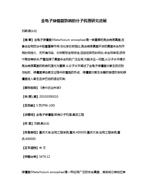
M ycosystema菌 物 学 报 15 September 2009, 28(5): 712-717jwxt@ISSN1672-6472 CN11-5180Q©2009 Institute of Microbiology, CAS, all rights reserved.Appressorial differentiation and its association with cAMP in the insect pathogenic fungus Metarhizium anisopliaeDUAN Zhi-Bing1 GAO Qiang1 LU Ding-Ding1 SHI Shao-Hua1BUTT Tariq M.2WANG Cheng-Shu1*1Institute of Plant Physiology and Ecology, Shanghai Institutes for Biological Sciences, Chinese Academy of Sciences, Shanghai 200032, China2School of Biological Sciences, University of Wales Swansea, Swansea SA2 8PP, UKAbstract: Host recognition and appressorium differentiation are the pivotal steps for insect pathogenic fungi to initiate infection process. In this study, appressorial differentiation was studied by comparing a mutant, which has been identified with the loss of genetic materials, with the wild-type strain of the insect pathogenic fungus Metarhizium anisopliae. The observations showed that both the mutant and wild-type strain could not only produce appressoria on the tips of newly germinated spores, but also form multiple infection structures from the terminals of branching mycelia on insect cuticle. In contrast to the wild-type, the frequency of appressorial formation was significantly reduced and in addition, no clear mucilaginous sheath was produced by the mutant appressoria. The study shows that the cuticle degrading enzyme subtilisin is not involved in appressorial differentiation, or indispensable in cuticle digestion. A significantly low frequency of appressorial differentiation by the mutant was coincided with its lower intracellular cAMP level in comparison to the wild-type. Addition of exogenous cAMP could significantly increase the frequency of appressorial differentiation by the mutant, indicating that cAMP signaling pathway is potentially involved in regulationof appressorial differentiation in M. anisopliae.Key words: insect pathogenic fungus, cuticle penetration, mucilaginous sheath, subtilisin金龟子绿僵菌附着胞分化及其与环腺苷酸cAMP的关联性研究段志兵1 高强1 吕丁丁1 石少华1 BUTT Tariq M.2 王成树1*1中国科学院上海生命科学研究院植物生理生态研究所 上海 2000322School of Biological Sciences, University of Wales Swansea, Swansea SA2 8PP, UK摘要:寄主识别与附着胞分化是虫生真菌启动侵染过程的首要步骤。
本文利用先前获得的金龟子绿僵菌基因缺失突变株与其野生型一起进行附着胞分化研究。
接种后不同时间下的观察表明,绿僵菌突变株或野生型的附着胞既可以在萌发不久的芽管顶端形成,也可以在伸长菌丝分支的顶端形成。
与野生型不同的是,突变株附着胞的分化频率显著下降,附着胞周围也缺Supported by The Knowledge Innovation Program of CAS (No. KSCX2-YW-G-037) and the Science and Technology Commission of Shanghai Municipality (No. 07PJ14101)*Corresponding author. E-mail: cswang@Received: 11-12-2008, accepted: 06-02-2009/jwxtcn乏粘液层的产生。
研究表明,绿僵菌的类枯草杆菌类体壁降解酶对于附着胞分化不产生影响,对体壁降解也非完全必需的。
与突变株附着胞分化频率显著降低相对应,其胞内环腺苷酸cAMP 水平显著下降,而添加外源cAMP 能够显著增加其附着胞分化频率,说明绿僵菌cAMP 信号途径对于调控附着胞分化起着重要的作用。
关键词:昆虫病原真菌,体壁穿透,粘液层,类枯草杆菌蛋白酶INTRODUCTIONDuring the process of fungal infection, the combination of enzymatic digestion and the mechanical force from the infection structure, appressorium, accounts for successful establishment of penetration through the host cuticle (Charnely & St Leger 1991; Clarkson & Charnely 1996). The ability of appressorium formation will greatly influence fungal pathogenicity and much attention has been paid to the mechanism exploitation of fungal infection structure differentiation (Wang & St. Leger 2005). For insect fungus M. anisopliae , adhesin mediates spore adhesion to insect cuticle is the first step (Wang & St. Leger 2007a). The studies on plant pathogenic fungi have shown that the signaling pathways, especially the cAMP signaling pathway has been evident to play important roles infungal morphogenesis and pathogenesis(Borges-Walmsley & Walmsley 2000; D’Souza & Heitman 2001). The reduction of intracellular cAMPlevel will greatly affect appressorial development andresult in the decrease of fungal pathogenicity as shown inplant pathogenic fungi, Colletotrichum lagenarium (Takano et al . 2001) and Magnaporthe grisea (Adachi &Hamer 1997).Until now, very little is known about cAMP effectsin insect pathogenic fungi, of which some have beendeveloped as promising biological control agents (Butt etal . 2001; Wang & St. Leger 2007b). A serendipitouslyacquired mutant of M . anisopliae has been characterizedwith the loss of genetic materials as well as the pathogenicity against mealworm, Tenebrio molitor (Wang et al . 2002, 2003). By using the mutant as amodel, we conducted experiments to study fungal appressorium differentiation and its associations with the cuticle degrading protease PR1 and intracellular cAMPlevel. Addition of exogenous cAMP was performed toexamine its influence on appressorium formation andspore germination of both the mutant and wild-typeisolates.1 MATERIALS AND METHODS1.1 Fungal culturesDuring a large scale of single spore isolation analysis of M. anisopliae strain V275, a pale instead of dark green color colony was unexpectedly acquired. Further analyses indicated mutations has occurred in the phenotypically altered colony with the loss of cuticle degrading proteins (Wang et al . 2002) and a conditionally dispensable chromosome (Wang et al . 2003). In this study, the mutant was further evaluated for appressorium differentiation. The wild-type and mutant cultures were maintained on potato dextrose agar (PDA, Difco) or in Sabouraud dextrose broth (SDB, Sigma) as described before (Wang et al . 2002, 2003).1.2 Scanning electron microscopy (SEM) studyThe front wings of the mealworm beetle were sterilized in 70% ethanol and dipped in the conidialsuspensions (105 conidia/mL) of the mutant and wild-type for 15s and then lined on the moisturized Whatman No. 1 filter paper in Petri dish and incubated at 25℃. Samples were taken out and fixed in 2%formaldehyde (aq. v/v) after 24, 36, 48, 60 and 72 hours post-inoculation. Appressorial development on the cuticle was studied using a Philips Scanning Electron Microscope (Philips Electron Optics). Briefly, the samples were placed in a specimen holder and treated through a series of increasing concentrations of ethanol for 10 minutes each: 20%, 50%, 70%, 90% and 100%, and then through ethanol:acetone (3:1) and anhydrous acetone to displace the water in specimens. The treatedspecimens were then mounted onto metal stubs and coated in gold dust for examination. 1.3 SDS-PAGE analysisProteins induced in 1% cockroach homogenate medium (Wang et al . 2002) were precipitated with 60% ammonium sulphate and dialyzed over night. To compare the differences of inductive protein profiles between thewild-type and mutant, equal amounts (10μg) of protein were analyzed by using 14% (w/v) and 19% sodium dodecyl sulfate-polyacrelamide gel electrophoresis (SDS-PAGE), respectively. The gel was documented after staining with Coomassie blue. Molecular weights of the proteins were determined by comparison with protein standards (Bio-Rad).1.4 cAMP assaysThe intracellular cAMP levels in the mutant and wild-type were assayed according to the method described by Filinger et al. (2002). Briefly, mycelia (0.1g), harvested from SDB after growth for four days at 25℃ and 120r/min in a rotator, were ground thoroughly under liquid nitrogen and suspended in 0.5mL of extraction buffer (50mmol/L Tris-HCl, pH 7.5). An aliquot of 0.1mL of this suspension was used for protein assay using a Bio-Rad protein assay kit. The rest was boiled for 5min and centrifuged at 13,000r/min for 5min. cAMP concentration in the supernatant was determined using a cAMP immunoassay kit (Sigma) according to the manufacturer’s instructions. Assays were conducted in triplicates from two independent cultures. The concentrations of cAMP were expressed in pmol/mg protein.1.5 Influence of cAMP on germination and appressorium formationFor the germination assay, the PDA plates (90mm in diameter) were amended by adding the stock solution of cAMP (Sigma) to a final concentration of 1 or 10mmol/L before inoculating with 0.1mL spore suspension (104 conidia/mL). The control plates were inoculated without adding cAMP. The appressorium formation assay was conducted according to the method described by St Leger et al. (1989a). Briefly, the aliquots of spore suspensions (104 conidia/mL) were added to a pre-sterilized growth medium (0.0125% yeast extract medium, YEM, pH 6.8) amended with final concentrations of 0, 1 and 10mmol/L cAMP and left for spore germination in polystyrene dishes. The conidia germinated with terminal swellings (morphologically similar to the appressoria formed on insect cuticles) were counted and the percentages were compared between the mutant and wild-type strain in different treatments. The student t-test was conducted to compare the differences between treatments.2 RESULTS2.1 Infection structure differentiationFor appressorial induction, both the wild-type and mutant conidia could produce appressorium-like structures with apical swellings shortly after spore germination on the hydrophobic surface of a polystyrene Petri dish (Fig. 1-A, B). SEM studies showed that spore germination behavior and appressorium formation on the cuticle were highly different between the mutant and wild-type conidia. The wild-type conidia could usually produce appressoria shortly after spore germination (Fig. 1-E) while the mutant conidia germinated but the germ tubes elongated without appressorial differentiation in a large proportion (data not shown). Most interestingly, by increasing the incubation time for up to 72 hours, multiple appressoria were found to be formed on the tips of branching mycelia by the wild-type (Fig. 1-D) but fewer appressoria were produced by the mutant (Fig. 1-F). There was a heavy mucilaginous sheath formed around each wild-type appressorium (Fig. 1-D) but no apparent mucilage could be found surrounding mutant appressorial cells (Fig. 1-C, F). However, the hydrolytic zone could be clearly observed beneath mutant appressorium (Fig. 1-C) and mycelium (Fig. 1-G), indicating that, even after the loss of subtilisin genes (Wang et al. 2002), the degrading enzymes secreted by the mutant could function effectively for cuticle digestion.2.2 Inductive protein profilesSDS-PAGE analysis indicated that the proteins produced by the wild-type and mutant in 1% cockroach homogenate inductive medium were different. Consistent with the previous study that the mutant has lost subtilisin PR1 genes (Wang et al. 2002), no PR1 (approximately 30kDa) was produced by the mutant (Fig. 2), suggesting that PR1 is not involved in the digestive activities which resulting in the formation of the hydrolytic zone observed above around the mutant appressorium (Fig. 1-C). Most of the other inductive proteins than 20kDa in molecular weight could not be well separated either on a 14% or 19% gel (Fig. 2).Fig. 1 Differences of appressorium formation between the mutant and wild-type strain of Metarhizium anisopliae V275. A: Appressorium of wild-type in YEM medium after induction for 12 hours; B: Appressorium of mutant in YEM medium 12 hours after incubation; C: A close-up look of the hydrolytic zone around a mutant appressorium 72 hours after incubation; D: Multiple appressoria formed on cuticle by the wild-type 72 hours after inoculation; E: Mutant mycelium showing the hydrolytic zone; F: Appressoria formed on insect cuticle by the mutant after inoculation for 72 hours; G: Wild-type appressorium formed on insect cuticle after inoculation for 36 hours; CO: Conidia; AP: Appressorium. Bar = 5μm.Fig. 2 SDS-PAGE profiles of total inductive proteins of the wild-type and mutant. A: Protein separation with a 14% acrylamide/biacrylamide gel; B: Protein separation with a 19% gel. M: Molecular marker; WT: Wild-type; MT: Mutant. The arrow shows that no PR1 protein produced by the mutant. 2.3 cAMP assay and its influence on appressorial differentiationA highly reduced cAMP level was detected in the mutant (32.32±2.57pmol/mg) in contrast to that in the wild-type strain (82.08±7.09pmol/mg) (P=0.001). The addition of exogenous cAMP had no considerable influence on spore germination for both the mutant and wild-type conidia on PDA plates (Fig. 3-A). However, consistent with SEM observations (Fig. 1-D, F), the mutant produced a significantly lower percentage of differentiated structures (13.07±2.52%) than did by the wild-type (54.36±3.11%) (P=0.002) (Fig. 3-B). The addition of exogenous cAMP could increase appressorium formations for both the wild-type and/jwxtcnmutant in a dose-dependent manner but more significantly for the mutant. Statistically, there was no significant increase for the wild-type examined at 1mmol/L cAMP amended medium (P=0.243) but a significant increase at 10mmol/L (P=0.049), while for the mutant both concentrations could highly increase appressorium structure formation when compared with the control (1mmol/L, P=0.029; 10mmol/L, P=0.020) (Fig. 3-B).Fig. 3 The influence of cAMP on spore germination and appressorium formation between the mutant and wild-type strain. A: Germination rates under different concentration of cAMP determined 12 hours post inoculation; B: Percentages of appressorium formation on the hydrophobic surface of polystyrene Petri dishes in 0.0125% YEM medium amended with indicated concentrations of cAMP 12 hours post inoculation.3 DISCUSSIONSAppressorial differentiation was usually described to occur shortly after fungal spore attachment and adhesion to a susceptible host surface (Clarkson & Charnely 1996; Dean 1997). Surprisingly, the observations of this study showed that appressoria could not only be formed on the tips of newly germinated germ tubes but also differentiated from the tips of branching mycelia to establish multiple penetration events. Despite the intricacy of fungal infection process, e.g. signals sensing, recognition, structure differentiation and penetration, “nutritional relationship” with the host has been reasonably explained as the only purpose for fungal infection structure differentiation (St Leger et al. 1989b). In this respect, the establishment of a single penetration by a germinated spore should be enough since it is an energy-cost event for infection structure differentiation. It remains open but highly intriguing what the environmental cues are and why the multiple appressoria can be formed from branching mycelia, especially from those close branching mycelia.In accordance with previous studies that the mutant isolate of M. anisopliae V275 lost genetic materials including subtilisin pr1 genes (Wang et al. 2002) and a dispensable chromosome (Wang et al. 2003), SDS-PAGE analysis in this study showed that the mutant produced no PR1 protein in inductive medium. The mutant, however, could still produce appressoria would indicate that the subtilisin protein PR1 is not involved in appressorial differentiation. Supportively, a previous study revealed that the target inhibition of PR1 did not prevent appressorial differentiation by M. anisopliae (St Leger et al. 1987). On the other hand, an array of proteins have been involved during the infection process of M. anisopliae (Freimoser et al. 2003; Wang et al. 2005) and different paralogous genes of pr1 from pr1A-pr1J have been identified from a single strain of M. anisopliae (Bagga et al. 2004). As demonstrated by SDS-PAGE analysis in this study, there was a large amount of proteins produced by the mutant cells in inductive medium, suggesting that these enzymes could either function complementarily during penetration or the mutant has developed an alternative strategy in pathogenesis.The frequency of appressorium differentiation coincided with intracellular cAMP level indicates that the cAMP signalling pathway plays a major role in appressorium formation in the insect pathogenic fungus. For M. anisopliae, two high-affinity of cAMP-binding proteins were detected during the early stage of conidial germination and appressorial differentiation (St Leger et al. 1990), indicating the involvement of cAMP in fungal early development. The experiments have demonstrated this is the case for plant pathogenic fungi (Takano et al. 2001; D’Souza & Heitman 2001; Lee et al. 2003). Theendogenous cAMP level was significantly low but detectable in the mutant indicating that, after the loss of genetic materials, its cAMP related genes might remain intact but with variations in upstream and or downstream gene regulations by comparison with the wild-type. Whatever the case, further studies are still required by employing the mutant as a powerful model to elucidate the pathogenic mechanisms of insect fungi. [REFERENCES]Adachi K, Hamer JE, 2002. Divergent cAMP Signaling pathways regulate growth and pathogenesis in the rice blast fungus Magnaporthe grisea.Plant Cell,10: 1361-1373Bagga S, Hu G, Screen SE, St Leger RJ, 2004. Reconstructing the diversification of subtilisins in the pathogenic fungus Metarhizium anisopliae. Gene, 324: 159-169Borges-Walmsley MI, Walmsley AR, 2000. cAMP signalling in pathogenic fungi: control of dimorphic switching and pathogenicity. Trends in Microbiology, 8: 33-141Butt TM, Jackson C, Magan N, 2001. Introduction - fungal biological control agents: progress, problems and potential. In: Butt TM, Jackson C, Magan N (eds.) Fungal biological control agents: progress, problems and potential. CAB International, Oxon. 1-8 Clarkson JM, Charnley AK, 1996. New insight into mechanisms of fungal pathogenesis in insects. Trends in Microbiology,4: 197-204 Dean RA, 1997.Signal pathways and appressorium morphogenesis.Annual Review of Phytopathology, 35: 211-234D’Souza CA, Heitman J, 2001. Conserved cAMP signaling cascades regulate fungal development and virulence. FEMS Microbiology Review, 25: 349-364Fillinger S, Chaveroche MK, Shimizu K, Keller N, d'Enfert C, 2002.cAMP and ras signalling independently control spore germination in the filamentous fungus Aspergillus nidulans. Molecular Microbiology, 44: 1001-1016Freimoser FM, Screen S, Bagga S, Hu G, St Leger RJ, 2003. Expressed sequence tag (EST) analysis of two subspecies of Metarhizium anisopliae reveals a plethora of secreted proteins with potential activity in insect hosts. Microbiology, 149: 239-247Lee N, D’Souza CA, Kronstad JW, 2003. Of smuts, blasts, mildews, and blights: cAMP signaling in phytopathogenic fungi. Annual Review of Phytopathology, 41: 399-427St Leger RJ, Charnley AK, Cooper RM, 1987. Characterization of cuticle-degrading proteases produced by the entomopathogenic fungus Metarhizium anisopliae. Archives of Biochemistry andBiophysics, 253: 221-232St Leger RJ, Butt TM, Goettel MS, Staples RC, Roberts DW, 1989a.Production in vitro of appressoria by the entomopathogenic fungusMetarhizium anisopliae. Experimental Mycology, 13: 274-288St Leger RJ, Butt TM, Goettel MS, Staples RC, Roberts DW, 1989b.Synthesis of proteins including a cuticle-degrading protease during differentiation of the entomopathogenic fungus Metarhizium anisopliae. Experimental Mycology, 13: 253-262St Leger RJ, Joshi L, Bidochka MJ, Roberts DW, 1995. Protein synthesis in Metahizium anisopliae growing on host cuticle.Mycological Research, 99: 1034-1040St Leger RJ, Laccetti LB, Staples RC, Roberts DW, 1990. Protein kinases in the entomopathogenic fungus Metarhizium anisopliae.Journal of General Microbiology, 136: 1401-1411Takano Y, Komeda K, Kojima K, Okuno T, 2001. Proper regulation of cyclic AMP-dependent protein kinase is required for growth, conidiation, and appressorium function in the anthracnose fungus Colletotrichum lagenarium. Molecular Plant-Microbe Interactions, 14: 1149-1157Wang CS, Skrobek A, Butt TM, 2003. Concurrence of losing a chromosome and the ability to produce destruxins in a mutant of Metarhizium anisopliae. FEMS Microbiology Letters, 226:373-378Wang CS, Typas MA, Butt TM, 2002. Detection and characterisation of pr1 virulent gene deficiencies in the insect pathogenic fungusMetarhizium anisopliae. FEMS Microbiology Letters, 213:251-255Wang CS, Hu G, St Leger RJ, 2005. Differential gene expression by Metarhizium anisopliae growing in root exudate and host (Manduca sexta) cuticle or hemolymph reveals mechanisms of physiological adaptation. Fungal Genetics and Biology, 42:704-718Wang CS, St Leger RJ, 2005. Developmental and transcriptional responses to host and nonhost cuticles by the specific locust pathogen Metarhizium anisopliae var. acridum. Eukaryotic Cell, 4: 937-947Wang CS, St Leger RJ, 2007a. A scorpion neurotoxin increases the potency of a fungal insecticide. Nature Biotechnology, 25:1455-1456Wang CS, St Leger RJ, 2007b. The MAD1 adhesin of Metarhizium anisopliae links adhesion with blastospore production and virulence to insects, and the MAD2 adhesin enables attachment to plants. Eukaryotic Cell, 6: 808-816/jwxtcn。