环氧化酶
环氧化物水解酶产生菌的筛选及其发酵条件的研究
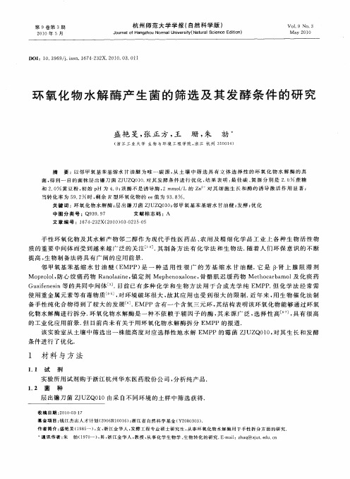
文 章 编 号 :l 7 — 3 X( 0 0 0 — 2 5 0 6 422 2 1 )30 1-5
手 性环 氧化 物及其 水解 产物 邻 二醇作 为现 代手 性 医药 品 、 用及 精 细化 学 品工业 上 各 种 生 物 活性 物 农 质的重 要 中间体 而受 到越 来越 广泛 的关 注l ]其 制备 方 法有 化 学 法 和 生物 法 . _ . 1 随着 人们 环 保 意识 的不 断
26 1 13 培 养 基 .
杭州 师范 大学学 报 ( 自然 科 学 版 )
21 0 0矩
1 3 1 筛选 培 养 基 每 10 0ml 养 基 中含 有 2 0g NH NO。 . 2 O . . 0 培 . ,1 5g NaHP ・ 1 H2 2 O,1 5 g .
提 高 , 物 制 备 法 将 具 有 广 阔 的应 用 前 景 . 生
邻 甲氧 基苯 基 缩 水 甘 油 醚 ( MP ) 一 种 适 用 性 很 广 的 芳 基 缩 水 甘 油 醚 . 是 J肾上 腺 阻滞 剂 E P 是 它 3 一 Mo r ll防心 绞 痛药 物 R n l ie 镇 定 剂 Me h n x 1n , 骼 肌 迟缓 药 物 Meh cr a l 化 痰 药 p oo , a oa n , z p e o ao e 骨 to ab mo 及 Gu i n s a e ei 的共 同 中间体_ . f n等 3 目前 已有 多 种化 学 和生 物方 法 用 于 合成 光 学 纯 E P 但 化学 法 经 常 需 ] MP . 使 用重 金属元 素 等有毒 物质 | ]对 环境破 坏 很大 , 4 , 故其 应 用 也受 到很 大 的 限制 . 近年 来 , 生 物催 化 法 制 用 备 手性 纯化合 物得 到 了较大 的发展 . MP ] E P含 有一 个 含氧 三元 环 , 结构 表 明该环 氧化 物 能够通 过环 氧 其 化 物水解 酶进 行拆 分. 氧化 物水 解酶 是一 种不 依 赖 于辅 因子 的酶 , 来 源 广 泛 , 环 其 选择 性 高 [ ] 具 有 很 高 6 , 。
环氧化酶的作用
环氧化酶的作用环氧化酶是一类重要的酶,在生物体内扮演着重要的角色。
它们能够催化环氧化反应,将某些化合物的双键转化为环氧化物。
环氧化反应是一种重要的有机化学反应,对于合成和代谢许多生物活性物质具有重要意义。
环氧化酶的催化机制是通过活性位点上的氧原子进行催化。
环氧化酶的活性位点通常由赖氨酸、组氨酸等氨基酸残基组成,它们能够与底物中的双键形成氢键和离子键,从而使底物的双键与氧原子接近,进而发生环氧化反应。
环氧化酶的活性位点还能够调节底物的立体化学特性,使得环氧化反应具有高度的立体选择性。
这种立体选择性使得环氧化酶在生物体内能够高效地催化合成特定立体异构体的环氧化物。
环氧化酶广泛存在于生物体内,包括动物、植物和微生物等。
它们在许多生物过程中发挥着重要的作用。
例如,环氧化酶在动物体内参与荷尔蒙的合成和代谢过程,调节生理功能的平衡。
同时,环氧化酶也参与了许多药物的代谢和解毒过程,对药物的效应和副作用具有重要影响。
在植物体内,环氧化酶参与了次生代谢物的合成,包括花色素、香气物质等。
此外,环氧化酶还在微生物的代谢途径中发挥着重要的作用,参与了许多生物催化反应。
环氧化酶的活性和稳定性受到多种因素的影响。
环境条件如温度、pH值等对其活性具有重要影响。
此外,环氧化酶的催化活性还受到底物的性质和结构的影响。
不同的环氧化酶对于不同结构的底物具有不同的催化效率和立体选择性。
因此,研究环氧化酶的催化机制和结构特征对于了解其功能和应用具有重要意义。
近年来,随着生物技术的发展,研究人员通过对环氧化酶的工程改造和基因重组等方法,成功地构建了多种具有特定催化性能的环氧化酶。
这些改造的环氧化酶在有机合成、废水处理、环境保护等方面具有重要应用价值。
例如,改造的环氧化酶可以高效催化有机合成反应,用于合成药物、农药和化学品等。
此外,环氧化酶还可以应用于废水处理,将废水中的有机物转化为无害物质,以减少对环境的污染。
环氧化酶作为一类重要的酶,在生物体内具有广泛的分布和重要的功能。
环氧化酶的作用
环氧化酶的作用环氧化酶是一类在生物体内广泛存在的酶,它在许多生物化学反应中起着重要的调节作用。
环氧化酶能够催化环氧化反应,将某些化合物中的不饱和双键氧化成环氧化合物。
这种反应在许多生物体内都是必需的,因为环氧化酶催化的环氧化反应能够产生一些重要的生理活性物质。
环氧化酶是一类金属酶,其催化活性依赖于其活性位点上的金属离子。
常见的环氧化酶活性位点上的金属离子包括铁离子、钴离子和锰离子等。
这些金属离子能够与底物分子中的不饱和双键形成配位键,使底物分子发生环氧化反应。
环氧化酶催化的环氧化反应具有高度的立体选择性,它能够选择性地将双键氧化成环氧化合物。
这种立体选择性是由环氧化酶的活性位点和底物分子的结构决定的。
环氧化酶的活性位点上存在着一些特定的氨基酸残基,它们能够与底物分子的特定部位发生氢键或范德华力作用,从而使底物分子以特定的立体构型进入活性位点,并与金属离子发生配位键,从而实现立体选择性催化。
环氧化酶催化的环氧化反应在生物体内起着重要的生理调节作用。
例如,环氧化酶能够催化一些内源性化合物的环氧化反应,从而产生一些具有生理活性的代谢产物。
这些代谢产物可以参与许多生理过程,如炎症反应、血管舒缩调节等。
另外,环氧化酶还能够催化一些外源性化合物的环氧化反应,从而使其具有更好的溶解性和生物活性。
这些外源性化合物包括一些药物和毒物,环氧化酶的催化作用能够影响它们的药效和毒性。
除了参与生理调节,环氧化酶还具有一定的应用价值。
由于环氧化酶具有高度的立体选择性和底物特异性,它可以用于合成一些具有特定立体构型和生物活性的化合物。
例如,环氧化酶可以催化一些合成药物的中间体的环氧化反应,从而提高合成药物的产率和选择性。
另外,环氧化酶还可以用于环境保护领域。
由于环氧化酶能够催化一些有机污染物的环氧化反应,将其转化为无毒或低毒的物质,因此可以用于处理工业废水和废气中的有机污染物。
环氧化酶是一类在生物体内广泛存在的酶,它能够催化环氧化反应,将某些化合物中的不饱和双键氧化成环氧化合物。
Cyclooxygenase-2 环氧化酶2
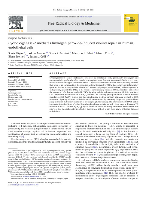
Original ContributionCyclooxygenase-2mediates hydrogen peroxide-induced wound repair in human endothelial cellsSonia Eligini a ,Izaskun Arenaz a ,1,Silvia S.Barbieri a ,Maurizio L.Faleri b ,Mauro Crisci a ,Elena Tremoli a ,c ,Susanna Colli a ,⁎a E.Grossi Paoletti Center,Department of Pharmacological Sciences,University of Milan,20133Milan,Italyb Divisione Anatomia Patologica,Ospedale Niguarda CàGranda,Milan,Italy cCentro Cardiologico Monzino IRCCS,University of Milan,20133Milan,Italya b s t r a c ta r t i c l e i n f o Article history:Received 29July 2008Revised 9February 2009Accepted 25February 2009Available online 6March 2009Keywords:Cyclooxygenase-2Endothelial wound Hydrogen peroxideReactive oxygen species sources Phosphatases p38MAPK Free radicalsCyclooxygenase-2(Cox-2)metabolites produced by endothelial cells,particularly prostacyclin and prostaglandin E 2,profoundly affect vascular tone,regional blood flow,and angiogenesis.We have previously shown that reactive oxygen species induce Cox-2expression in human endothelial cells (HUVEC),either on their own or as components of the signaling pathway triggered by TNF α,the prototypical in flammatory cytokine.Here we investigated the role of Cox-2induced by hydrogen peroxide (H 2O 2),either exogenous or endogenously generated by TNF α,in the repair of a mechanically wounded HUVEC monolayer and probed the sources of H 2O 2that are involved in TNF αsignaling and the pathways through which H 2O 2modulates Cox-2expression.Results indicate that H 2O 2-induced Cox-2activity participates in the repair of wounded monolayers.Both NADPH oxidase and the mitochondrial electron transport chain are involved in H 2O 2generation.Signaling triggered by H 2O 2for Cox-2induction acts by increasing the protein tyrosine kinase phosphorylation that follows inhibition of protein phosphatase activity.The activation of p38MAPK and its interaction in the inhibition of serine/threonine phosphatase activity are both critical steps in this event.We conclude that Cox-2induced by H 2O 2plays an important role in promoting endothelial wound repair after injury,so that the cardioprotective effect of Cox-2is due at least in part to its power of healing damaged endothelium.©2009Elsevier Inc.All rights reserved.Endothelial cells are pivotal in the regulation of vascular functions,including cell adhesion,in flammatory responses,regulation of permeability,and vasoactivity.Regeneration of the endothelial surface after vascular damage requires cell activation,migration,and proliferation,all events that are critical for neovascularization and wound repair [1].Reactive oxygen species (ROS)also play a central role in vascular physiology,and their effects on vascular function depend critically onthe amounts produced.The principal mediator of ROS-dependent signaling is hydrogen peroxide (H 2O 2),which is generated in response to receptor stimulation and is an ef ficient signal-transdu-cing molecule in endothelial cell migration [2].Its involvement as second messenger is based on two lines of evidence.First,H 2O 2accumulation follows the binding of growth factors and cytokines to their cognate receptors [3,4]and the downstream events are inhibited by scavenging enzymes or by antioxidants [5,6].Second,exposure of endothelial cells to H 2O 2induces the activation of signaling cascades [7,8].In particular,protein tyrosine and serine/threonine phosphatases are susceptible to H 2O 2-dependent inactiva-tion by oxidation or glutathionylation of a critical active-site-containing cysteine [9–11],with consequent phosphorylation-depen-dent activation of several signal transducers.Several sources of H 2O 2produced in response to receptor binding have been described in endothelial cells.The activation of nonin-flammatory NADPH oxidase family members,with subsequentspontaneous or enzymatic dismutation of superoxide anion (O 2U−)to H 2O 2,is the prime candidate for oxidant production in the plasma membrane microenvironment [12].H 2O 2can also be produced by mitochondria under physiological conditions and in response to receptor stimulation,as shown by the effects of inhibitors at speci ficFree Radical Biology &Medicine 46(2009)1428–1436Abbreviations:Cox-2,cyclooxygenase-2;Cox-1,cyclooxygenase-1;PGE 2,prosta-glandin E 2;HUVEC,human umbilical vein endothelial cells;ROS,reactive oxygen species;eNOS,NO synthase;TNF α,tumor necrosis factor-α;TTFA,thenoyltri fluor-oacetone;L-NAME,N ω-nitro-L -arginine methyl ester hydrochloride;AACOCF 3,arachi-donyl tri fluoromethyl ketone;SHP-2,protein tyrosine phosphatase containing Src homology domain 2;MTT,3-(4,5-dimethylthiazol-2-yl)-2,5-diphenyltetrazolium bro-mide;MAPK,mitogen-activated protein kinase;PCR,polymerase chain reaction;GAPDH,glyceraldehyde-3-phosphate dehydrogenase;MnTMPyP,Mn(III)tetrakis(1-methyl-4-pyridyl)porphyrin pentachloride;PP1,protein phosphatase 1;PP2A,protein phosphatase 2A;NADPH,nicotinamide adenine dinucleotide phosphate reduced form;VEGF,vascular endothelial growth factor;PTEN,phosphatase and tensin homolog.⁎Corresponding author.Fax:+390250318250.E-mail address:susanna.colli@unimi.it (S.Colli).1Current address:Unidad Mixta de Investigaciòn,University of Zaragoza,Spain.0891-5849/$–see front matter ©2009Elsevier Inc.All rights reserved.doi:10.1016/j.freeradbiomed.2009.02.026Contents lists available at ScienceDirectFree Radical Biology &Medicinej o u r n a l h o me p a g e :w w w.e l sev i e r.c o m /l oc a t e /fr e e ra d b i o me dmitochondrial sites in the electron transport chain upon oxygen sensing-related vascular signaling[13,14].In addition,other signifi-cant sources of endothelial ROS are cyclooxygenases(Cox's)and NO synthase(eNOS)[15,16,9].Arachidonic acid metabolites are important in the modulation of vascular homeostasis[17].The major rate-limiting enzymes involved in their synthesis are Cox's.Cox-1is constitutively expressed in most tissues and has general housekeeping functions,whereas Cox-2is responsible for high-level production of prostanoids in response to proinflammatory agents,tumor promoters,and growth factors[18]. Enzymatic Cox-2activity may serve as a compensatory mechanism to preserve the vasodilatory and antithrombotic properties of the vessel wall[19].Indeed,Cox-2metabolites produced by endothelial cells, particularly prostacyclin and prostaglandin E2(PGE2),have profound influences on vascular tone,regional bloodflow,vascular permeability and remodeling,angiogenesis,and wound repair[20,21].The balance between these autacoids is consequently critical in many pathophy-siological processes.We previously showed that ROS induce Cox-2expression in human endothelial cells,either on their own or as components of the inflammatory TNFαsignaling pathway[22].We therefore investi-gated whether Cox-2induced by H2O2affects the repair of a wounded endothelial monolayer and explored the signaling pathways whereby H2O2promotes Cox-2expression in endothelial cells.Materials and methodsMaterialsHuman recombinant tumor necrosis factor-α(TNFα)was from R and D Systems(Space Import–Export,Milan,Italy).PGE2,rote-none,thenoyltrifluoroacetone(TTFA),okadaic acid,calyculin A, H2O2,Nω-nitro-L-arginine methyl ester hydrochloride(L-NAME), sodium orthovanadate,sodium pyruvate,apocynin(4-hydroxy-3-methoxyacetophenon),and crystal violet were from Sigma–Aldrich S.r.l.(Milan,Italy).Stigmatellin and p-nitrophenyl phosphate were from Fluka(Sigma–Aldrich S.r.l.).SB203580,MnTMPyP,and NS-398were from Biomol(Tebu-Bio,Milan,Italy).Arachidonic acid sodium salt and the stable prostacyclin analog iloprost were from Cayman Chemical(Spi-Bio,Montigny Le Bretonneux,France). Arachidonyl trifluoromethyl ketone(AACOCF3)was from Alexis (Vinci-Biochem,Vinci,Firenze,Italy).NSC-87877,a selective inhibitor of the protein tyrosine phosphatase containing the Src homology domain2(SHP-2)was from Acros Organics(Nova Chimica,Milan,Italy).Antibodies against Cox-2(Ab29)and Cox-1 were gifts from Aida Habib(American University of Beirut, Lebanon)and from Cayman Chemical,respectively.Antibody against SHP-2and protein A/G–agarose were from Santa Cruz Biotechnology(Tebu-Bio,Milan,Italy).Antibody against phospho-tyrosine was from Upstate Biotechnology(Millipore,Milan,Italy). Antibodies against phosphorylated and total p38MAPK were from Biosource(Prodotti Gianni S.p.A,Milan,Italy).Peroxidase-con-jugated anti-mouse IgG antibody was from Jackson Immuno-Research Labs(Li StarFISH,Milan,Italy).Antibody directed against β-actin was from Sigma–Aldrich.Cell culture and treatmentHuman umbilical vein endothelial cells(HUVEC)were isolated from freshly acquired human umbilical veins,as described[22]. Informed consent was provided according to the Declaration of Helsinki.Cells were cultured in medium199(BioWhittaker Italia S.r.l., Bergamo,Italy)supplemented with10%heat-inactivated pooled human AB serum,1mM L-glutamine,50U/ml penicillin,50μg/ml streptomycin,0.1mg/ml neomycin,15μg/ml heparin,and50μg/ml crude extract of endothelial cell growth factor.Cells were used at the first passage.Heparin and endothelial cell growth supplement were omitted24h before stimulation.Incubations were carried out in M199medium supplemented with0.75%bovine serum albumin (fatty-acid-free and low in endotoxin;Sigma–Aldrich)and1%fetal calf serum.Inhibitors were added1h before stimulation.Cytotoxicity measurementCell viability was assayed with the use of either neutral red or MTT reduction assay and calculated as follows:relative viability= [(A e–A b)/(A c–A b)]×100,where A b is the background absorbance, A e is the experimental absorbance,and A c is the absorbance of controls.The various agents,at the concentrations tested,did not affect cell viability after either6or16h incubation.A slight reduction in viability,detected by MTT,was observed when HUVEC were incubated with stigmatellin and subsequently exposed to TNFα.Data (medians and ranges)were92.7%viable cells(76.5–100%),p=0.12, n=5,and84.5%viable cells(70–100%),p=0.25,n=4,vs cells incubated with TNFαfor6and16h,respectively.Prostanoid production assayCox-2activity was determined in HUVEC exposed to stimuli for6h. Cells were washed in Hanks'buffer,pH7.4,containing1mg/ml bovine serum albumin and incubated for30min with10μM arachidonic acid in the same buffer.Supernatants were collected and6-keto-PGF1αand PGE2levels were measured by enzyme immunoassay(Cayman Chemical).Western blotCells were harvested in lysis buffer,pH6.8,as described[22].Cell debris was removed by centrifugation(10,000g for5min)and protein concentration was determined by the micro-bicinchoninic acid assay. Equal amounts of lysates were subjected to SDS–polyacrylamide gel electrophoresis on7%gels and were transferred onto nitrocellulose membrane by a semidry transfer unit(Hoefer Scientific Instruments). The blots were then incubated with primary antibodies directed against Cox-1(1:500),Cox-2(1:10,000),and phosphorylated and total p38MAPK(1/1000).In some experiments,membranes were stripped and phosphotyrosine was detected with an antibody against phosphotyrosine(1:5000).Horseradish peroxidase-conjugated sec-ondary antibody was used at1:5000dilution.β-Actin was used as internal standard for control of protein load.Bands were detected using enhanced chemiluminescence(Amersham Pharmacia Biotech.).SHP-2immunoprecipitation,Western blotting,and activityCells were scraped in lysis buffer(50mM Tris–HCl,pH7.4,1% Nonidet P-40,0.25%sodium deoxycholate,150mM NaCl,1mM EDTA)supplemented with1mM PMSF,1μg/ml aprotinin,1μg/ml leupeptin,1μg/ml pepstatin,1mM Na3VO4,1mM NaF.Protein concentration was determined.Cell lysates(500μg)were incubated with the antibody to SHP-2(1.5μg)for2h at4°C and with protein A/G Plus–agarose beads for18h at4°C.Beads were washed(three times)in phosphatase buffer(62mM Hepes,pH7,6.25mM EDTA). Immune complexes were resuspended in electrophoresis sample buffer,boiled for3min,and immunoblotted with the anti-SHP-2 antiserum(1:500),as described above.SHP-2phosphatase activity was detected as reported[23].Briefly,immunocomplexes were washed twice in phosphatase buffer and resuspended in100μl of the same buffer containing the substrate p-nitrophenyl phosphate (10mM).Incubation was carried out for30min at37°C and stopped with NaOH(200mM).Substrate dephosphorylation was quantified spectrophotometrically at410nm.1429S.Eligini et al./Free Radical Biology&Medicine46(2009)1428–1436RNA isolation and reverse-transcription PCR(RT-PCR)Total RNA(1μg)was extracted from cells with TRIzol reagent, treated with DNase I(Invitrogen,Milan,Italy),and reverse transcribed(37°C for50min)by M-MLV reverse transcriptase (Invitrogen).The cDNA(1μl)was then subjected to22or30cycles of PCR for GAPDH and Cox-2,respectively(denaturation at94°C for 30s,annealing at55°C for30s,primer extension at72°C for60s), in a reaction mixture(50μl)containing 2.5U platinum Taq polymerase(Invitrogen)and200nM sense and antisense primers for Cox-2or GAPDH.The primers for Cox-2were as previously described[24].All reactions were performed in a Bio-Rad thermal cycler.PCR products were analyzed on2%agarose gel containing ethidium bromide(0.5μg/ml).GAPDH mRNA was used as a control for mRNA loading.Monolayer wound-healing migration assayThe wound-repair model employed confluent HUVEC monolayers maintained in M199supplemented with10%pooled human AB serum in35-mm dishes.The monolayer was wounded by dragging a1000-μl pipette tip along it[25,26],and HUVEC were cultured without (control)or with H2O2or TNFα.Just after wounding or after overnight incubation(16h),monolayers werefixed,stained with crystal violet, and photographed(50×magnification).Inhibitors were added1h before the stimuli.For the evaluation of wound closure undervariousFig.1.H2O2,either exogenously added or intracellularly generated by TNFα,induces closure of HUVEC monolayer wounds via a Cox-2-mediated mechanism.(A)Photomicrographs were taken just after wounding(prestimulation)or after16h incubation in medium alone(control)or in the presence of TNFα10ng/ml,H2O23μM,PGE210nM,or iloprost100nM. The inhibitor of Cox-2activity NS-398(5μM)was incubated with the cells for1h before the addition of TNFαor H2O2that lasted16h.Photomicrographs(50×original magnification)are representative of four individual experiments performed with HUVEC isolated from different cords.Boundaries of the wounds are indicated by lines.Insets represent enlarged regions of HUVEC monolayers.(B)For the evaluation of wound closure under various experimental conditions,the cells that migrated across the wound edge were counted in the regions of the premarked lines.Bar graph represents averaged data,expressed as cell number per threefields.Values are the means±SD for four independent experiments performed with HUVEC isolated from different cords.⁎p b0.05vs control.1430S.Eligini et al./Free Radical Biology&Medicine46(2009)1428–1436experimental conditions,the cells that migrated across the wound edge were counted in the regions of premarked lines(AxioVision4.7 software;Zeiss).Data are expressed as the average of cell number per threefields and the experiments were repeated at least three times using different umbilical cords.Statistical analysisResults are expressed as the mean±SD of the number of determinations indicated,performed with different umbilical cords. Means of two groups were compared using paired Student's t test. Grouped differences were compared with ANOVA(Dunn's and Dunnett's tests).Results from cytotoxicity experiments are expressed as medians and ranges.Medians were compared by Wilcoxon signed-rank test.A value of p b0.05was considered statistically different.ResultsH2O2and TNFαpromote the closure of a wound in a HUVEC monolayer via a Cox-2-mediated mechanismUsing a HUVEC wound repair assay that has been reported to be dependent on intracellular ROS production[25]as well as Cox-2 activity[26],we found that both TNFαand H2O2accelerate the closure of a wounded monolayer after mechanical disruption(Fig.1).NS-398, an inhibitor of Cox-2activity,opposed the entry of cells into denuded areas and wound closure,whereas addition of PGE2to the culture accelerated closure(Fig.1).A similar effect,albeit less pronounced, was observed after exposing monolayers to the prostacyclin analog iloprost(Fig.1).The data indicate that Cox-2activity is critical for H2O2-induced HUVEC monolayer repair.Woundedmonolayers Fig.2.H2O2,either exogenously added or intracellularly generated by TNFα,increases PGE2and6-keto-PGF1αlevels and induces Cox-2expression in HUVEC.(A)HUVEC wereleft unstimulated or were exposed to H2O2for6h.Medium was then replaced with Hanks'buffer containing10μM arachidonic acid and incubation was continued for30min. 6-Keto-PGF1αand PGE2levels were measured in the medium by enzyme immunoassays.The values shown are the means±SD of10independent experiments performed with HUVEC from different cords.⁎p b0.005vs untreated cells.Serum-starved HUVEC were incubated with H2O2or TNFα(B,C)for different times or(D)with increasing concentrations of H2O2for6h.Cox-1and Cox-2proteins were detected by Western analysis.β-Actin was used as a control for protein load.Blots are representative of four to six independent experiments performed with different HUVEC preparations.Densitometry is shown in the bar graphs.Signals were quantified relative toβ-actin.⁎p b0.05vs untreated cells.(E) Cox-2mRNA levels were measured by RT-PCR in HUVEC incubated with3μM H2O2or10ng/ml TNFαfor1h.GAPDH mRNA is shown as a control for RNA loading.Results are representative of six independent experiments.Densitometry is shown in the bar graph.Signals were quantified relative to GAPDH.⁎p b0.05vs untreated cells.1431S.Eligini et al./Free Radical Biology&Medicine46(2009)1428–1436exposed to H 2O 2or TNF αshowed morphologies similar to each other but different from the morphology of untreated cells (Fig.1A,insets).H 2O 2induces Cox-2in HUVECAs previously observed for TNF α[22],exogenous H 2O 2increased the levels of the Cox-2metabolites 6-keto-PGF 1αand PGE 2in the medium of HUVEC incubated with arachidonic acid (Fig.2A).Theincrease was attributable to augmented Cox-2protein levels induced by H 2O 2,either added as a bolus or endogenously generated by TNF α(Figs.2B and C ).Cox-2protein accumulation by H 2O 2was evident at 3h,persisted until 16h (Fig.2B),and was detectable at concentrations (b 10μM)that do not cause oxidative stress in cultured mammalian cells [9](Fig.2D).In contrast,Cox-1protein levels were unaffected (Figs.2B –D ).Cox-2mRNA levels,almost undetectable in unstimulated HUVEC,rose upon cell exposure to H 2O 2or TNF α(Fig.2E).Antioxidants prevent Cox-2induction by H 2O 2The redox status within the cell is critical for Cox-2induction by H 2O 2whether exogenous or endogenous.Alteration of the intracellular oxidant status by sodium pyruvate,which prevents H 2O 2-induced glutathione depletion in HUVEC [8],or by the membrane-permeative superoxide dismutase and catalase mimetic MnTMPyP [27]attenuated Cox-2induction by H 2O 2or TNF α(Fig.3).Different sources of H 2O 2mediate Cox-2induction by TNF αand regulate wound closure in HUVEC monolayersThe contribution of distinct sources of H 2O 2that coexist in endothelial cells was examined in HUVEC exposed to TNF α.First,the role of NADPH oxidase-derived H 2O 2was investigated by use of the NADPH oxidase inhibitor apocynin,which markedly prevented Cox-2induction (Fig.4A).Then the contribution of mitochondrial H 2O 2production was investigated in HUVEC incubated with inhibitors of the various complexes within the electron transport chain.Stigmatellin,which prevents electron transfer by complex III from ubiquinol to both iron –sulfur protein and heme b 566(Qo site)[28],reduced Cox-2levels (Fig.4B),which,conversely,were unaffectedbyFig.3.The intracellular redox status affects Cox-2expression in HUVEC.HUVEC were exposed to sodium pyruvate or to the superoxide dismutase mimetic MnTMPyP for 30min.TNF αor H 2O 2was then added and incubation was continued for 6h.Cox-2was detected by Western analysis.β-Actin was used as a control for protein load.Blots are representative of five or six independent experiments performed with different HUVEC cultures.Densitometry is shown in the bar graphs.Signals were quanti fied relative to β-actin.⁎p b 0.05vs TNF α-or H 2O 2-stimulatedcells.Fig.4.Different sources of ROS mediate Cox-2induction by TNF αand regulate wound closure in HUVEC monolayers.HUVEC were incubated for 1h (A)with apocynin or (B)with the inhibitors of mitochondrial electron transport complexes I,II,and III (rotenone,TTFA,and stigmatellin,respectively).TNF αwas then added and incubation was continued for 6h.Cox-2was detected by Western analysis.β-Actin was used as a control for protein load.Blots are representative of four or five independent experiments performed with different cell cultures.Densitometry is shown in the bar graphs.Signals were quanti fied relative to β-actin.⁎p b 0.05vs TNF α-treated HUVEC;#p b 0.05vs untreated cells.(C)Photomicrographs (50×original magni fication)of wounded HUVEC monolayers incubated with medium alone or with apocynin or stigmatellin for 1h and then exposed to TNF αfor 16h.Photomicrographs are representative of three or four individual experiments performed with HUVEC isolated from different cords.(D)Wound closure under the different experimental conditions was quanti fied by counting the cells that migrated across the wound edge in the regions of the premarked lines.Bar graph represents averaged data,expressed as cell number per three fields.Values are the means ±SD for three independent experiments performed with HUVEC isolated from different cords.⁎p b 0.05vs TNF α-treated HUVEC.1432S.Eligini et al./Free Radical Biology &Medicine 46(2009)1428–1436TTFA,an inhibitor of the electron transport from complex II to ubiquinone [14](Fig.4B).In contrast,rotenone,a complex I inhibitor [29],increased Cox-2levels in HUVEC whether the cells were resting or stimulated (Fig.4B).Finally,the respective contributions of H 2O 2generation by Cox's,lipoxygenases,and uncoupled eNOS were investigated in cells exposed to AACOCF 3(10–20μM)or L-NAME (1mM),which inhibit phospholipase A 2and eNOS activity,respectively.The results obtained exclude a role for H 2O 2from these sources in Cox-2induction (data not shown).The functional role of H 2O 2generated by either NADPH oxidase or mitochondrial complex III was de fined by a series of experiments performed in mechanically wounded HUVEC mono-layers.As shown in Fig.4C,both apocynin and stigmatellin retarded the closure of a wound promoted by TNF α.H 2O 2-mediated Cox-2induction:role of protein phosphatasesIn HUVEC,oxidants like H 2O 2produce biological effects by decreasing total cellular phosphatase activity,which facilitates a compensatory increase in cellular phosphotyrosine residues [30].As shown in Fig.5A,blocking protein tyrosine phosphatase activity by the general inhibitor orthovanadate resulted in Cox-2induction that went hand in hand with increased tyrosine phosphorylation (espe-cially within 97-and 116-kDa fractions).Additional experiments assessed the effect of H 2O 2or TNF αon the activity of cytosolic SHP-2,a protein tyrosine phosphatase that is critical for cell motility and is rapidly inactivated by oxidants [31,32].Both TNF αand H 2O 2signi ficantly reduced the activity of immunopuri fied SHP-2(Fig.5B),leaving protein levels unaltered (data not shown).However,the selective inhibition of SHP-2activity by NSC-87877(10–50μM)[33]did not affect Cox-2expression in HUVEC,either unstimulated or incubated with TNF αor H 2O 2(data not shown).Both calyculin A,a cell-permeative inhibitor of both serine/threonine phosphatases PP1and PP2A,and okadaic acid,a selective inhibitor of PP2A at 1nM [34],increased Cox-2levels in HUVEC,similar to H 2O 2or TNF α(Fig.5C).The increase in Cox-2levels paralleled the increase in phosphoty-rosine residues (particularly within the 66-and 116-kDa fractions).Moreover,the effect of calyculin A or okadaic acid was additive to that of H 2O 2or TNF α,in terms of both Cox-2up-regulation and protein tyrosine phosphorylation.Overall,serine/threonine phosphatase inhibition seems to be a candidate mechanism for Cox-2induction by H 2O 2in HUVEC.Involvement of p38MAP kinase in Cox-2induction by H 2O 2p38MAPK activation is among the signaling pathways involved in protein phosphatase inhibition [35].The role of p38MAPK activation as part of signal transduction driven by H 2O 2,either intracellularly generated or exogenously added,was therefore explored.Under our conditions,either H 2O 2or TNF αincreased the extent of p38MAPK phosphorylation,which peaked at 15min and returned to basal levels within 60min (Fig.6A).In addition,the p38MAPK inhibitor SB203580abrogated Cox-2induction by either H 2O 2or TNF αalmost completely (Fig.6B)and reduced the accumulation of Cox-2in HUVEC treated with calyculin A or okadaic acid (Fig.6C).Collectively,the data indicate that activation of p38MAPK and its interaction with serine/threonine phosphatase inhibition represent an essential step for Cox-2induction by H 2O 2in HUVEC.A diagram that illustrates the proposed signaling pathways through which Cox-2participates in H 2O 2-mediated endothelial wound repair is depicted in Fig.7.DiscussionOur results show that Cox-2up-regulation by H 2O 2increases cell migration to a wound made in a con fluent HUVEC monolayer.The H 2O 2can be either exogenously added or produced by the NADPH oxidase or mitochondrial chain in response to TNF α.The relevant pathways involve inhibition of serine/threonine phosphatases,acti-vation of p38MAPK,and theirinteraction.Fig.5.Inhibition of protein phosphatase activity results in Cox-2induction in HUVEC.(A)HUVEC were exposed to orthovanadate,a general protein phosphatase inhibitor,for 6h.Protein tyrosine phosphorylation and Cox-2expression were detected by Western analysis.β-Actin was used as a control for protein load.Blots are representative of three separate experiments performed on different cell cultures.(B)Effect of either H 2O 2or TNF αon the activity of the tyrosine phosphatases containing SHP-2.SHP-2activity was measured on immunopuri fied protein (see Materials and methods ).(C)HUVEC were exposed to TNF α,H 2O 2,okadaic acid (a speci fic inhibitor of PP2A at 1nM),or calyculin A (which inhibits both serine/threonine phosphatases PP1and PP2A)for 6h.Protein tyrosine phosphorylation and Cox-2expression were detected by Western analysis.β-Actin was used as a control for protein load.Blots are representative of three independent experiments performed with different cell cultures.The values shown for SHP-2activity are the means ±SD of seven independent experiments performed with HUVEC from different cords.⁎p b 0.05vs untreated cells.1433S.Eligini et al./Free Radical Biology &Medicine 46(2009)1428–1436The proangiogenic effects of Cox-derived products have been clearly established in malignancies [36].Their roles,particularly that of PGE 2,in endothelial cell migration and wound healing have recently been appreciated both in vitro and in vivo [37,38]in variousexperimental models of healing [39,40]and de novo vascularization [38].The ability of PGE 2to directly stimulate angiogenesis in HUVEC,separate from VEGF signaling,has been recently reported [41].ROS have long been known to play a role in many diseases and have also emerged as ef ficient signaling molecules.The principal mediator of ROS-dependent signaling is H 2O 2.Endothelial cell migration within the wound is dependent upon H 2O 2production,which contributes to cytoskeleton reorganization required for the migratory phenotype and for density-dependent inhibition of cell growth [25,42].Low concentrations of H 2O 2support the healing process in vivo [43],and endogenous H 2O 2exerts a cardioprotective effect during ischemia –reperfusion injury in an animal model of coronary microcirculation [44].Previous studies have recognized the important role of NADPH oxidase-derived ROS in HUVEC migration and proliferation [45,46],and this enzyme is a potential therapeutic target for the treatment of angiogenesis-dependent diseases [47].In line with these observations,our data indicate that inhibition of NADPH oxidase prevents Cox-2induction and retards the closure of wounded HUVEC monolayers promoted by TNF α.In addition,we show that H 2O 2derived from mitochondrial complex III takes part in wound healing via Cox-2induction,as documented by the reduced Cox-2levels detected upon complex III inhibition.This finding fits well with the recent observation of a crucial role of mitochondria-derived H 2O 2in the regulation of the angiogenic switch via PTEN oxidation in redox-engineered cell lines [48]and is in support of the crucial role of complex III in ROS generation observed in HUVEC [49].The inhibition of electron transport from complex I to ubiquinone by rotenone increased Cox-2levels in resting or stimulated HUVEC.As in othercellFig.7.Proposed role for Cox-2in H 2O 2-induced wound repair in human endothelialcells.Fig.6.Involvement of p38MAP kinase in Cox-2induction by H 2O 2either exogenously added or intracellularly generated by TNF α.(A)Time course of p38MAPK phosphorylation by TNF αor H 2O 2.Cell lysates were subjected to Western analysis for phosphorylated p38(phospho-p38)or total p38(p38MAPK).Densitometry (means ±SD,n =5)is shown in the bar graphs.Signals were quanti fied relative to total p38MAPK.⁎p b 0.05vs untreated HUVEC.(B,C)HUVEC were incubated with the p38MAPK inhibitor SB203580for 1h before the addition of (B)TNF αor H 2O 2or (C)calyculin A or okadaic acid.Incubation was continued for 6h.Cox-2was detected by Western analysis.β-Actin was used as a control for protein load.Blots are representative of four to six separate experiments performed with different cell cultures.Densitometry is shown in the bar graphs.Signals were quanti fied relative to β-actin.⁎p b 0.05vs TNF α-or H 2O 2-stimulated cells (B)or vs okadaic acid-or calyculin-treated HUVEC (C).1434S.Eligini et al./Free Radical Biology &Medicine 46(2009)1428–1436。
药物化学(青岛大学)智慧树知到答案章节测试2023年
第一章测试1.基本药物来源占比最高的是A:抗生素B:植物药C:半合成药物D:合成药物答案:D2.药物作用靶点与蛋白无关的是A:核酸B:酶C:离子通道D:受体答案:A3.药物的生物活性包括A:分子式B:化学结构C:治疗作用及毒副作用D:分子量答案:C4.下列哪个药物的合成标志着药物化学的形成A:贝诺酯B:吗啡C:对乙酰氨基酚D:阿司匹林答案:D5.与受体有关的药物分为A:还原剂B:激动剂C:氧化剂D:拮抗剂答案:BD6.药物化学的任务包括A:创制新药B:研究药物的作用机理C:提供使用药物的化学基础D:研究药物的生产工艺答案:ACD7.现代新药研究与开发模式应用于新药研究,传统新药研究与开发模式已经被弃用A:对B:错答案:B8.中国药品通用名称是药品的常用名,是最准确的命名A:对B:错答案:B9.现代新药研发要经过靶点的识别和确证,先导化合物的筛选、优化和确定,临床前研究和临床研究几个阶段A:错B:对答案:B10.CADN的中文意思是中国药品通用名称。
A:错B:对答案:B第二章测试1.下列药物哪个是褪黑素受体激动剂A:他美替胺B:扎来普隆C:氯巴占D:扑米酮答案:A2.下列药物哪个不是GABA受体激动剂A:非尔氨脂B:氨己烯酸C:普罗加比D:加巴喷丁答案:A3.下列药物哪个不是乙酰胆碱酯酶抑制剂A:他克林B:奥卡西平C:加兰他敏D:卡巴斯汀答案:B4.芬太尼属于的化学结构类型是A:苯吗喃类B:哌啶类C:吗啡喃类D:氨基酮类答案:B5.甲氧氯普胺是中枢及外周性多巴胺D2受体拮抗剂,具有A:止吐作用B:促动力作用C:抗精神病作D:催吐作用答案:ABC6.作用于阿片k受体的镇痛药是A:非那佐辛B:氟痛新C:阿尼利定D:喷他佐辛答案:ABD7.吗啡在体内代谢时,去甲基的一步发生在7位N 上。
A:错B:对答案:A8.美沙酮是具有开链结构的镇痛药。
A:对B:错答案:A9.巴比妥嘧啶环2位羰基上的氧原子用硫原子取代、作用时间更短。
环氧化酶的作用(一)

环氧化酶的作用(一)
环氧化酶的作用
简介
•环氧化酶是一类重要的酶类
•环氧化酶介导了多种化学反应
•研究环氧化酶的作用有助于解析生物体内的代谢过程
物理作用
•环氧化酶能够催化底物的氧化反应
•通过将底物的双键上的氧原子转移到其它位置,形成环氧化物生物过程
•环氧化酶参与多种生物过程
•在细胞中,环氧化酶常参与代谢物的转化和调节
•环氧化酶还能影响生物体内的信号传递
应用领域
•环氧化酶可应用于工业领域的催化反应中
•可以将环氧化酶用于研发新型药物
•还能用于分离和净化废水中的有机污染物
研究进展
•近年来,环氧化酶的研究取得了重要的突破
•通过结构生物学手段揭示了环氧化酶的催化机制
•进一步研究有助于设计和合成具有特定功能的环氧化酶
结论
•环氧化酶是一类重要的酶类,具有多种生物和工业应用价值•对环氧化酶的深入研究有助于揭示生物体内的代谢过程
•进一步研究和应用环氧化酶有助于推动生物技术和工业化进程的发展。
特异性环氧化酶

特异性环氧化酶[摘要]目的:探讨特异性环氧化酶(COX)2抑制剂rofecoxib 对MRL/lpr小鼠狼疮性肾炎的医治作用及其机制。
方式:将12只3月龄MRL/lpr自发狼疮小鼠随机均分为rofecoxib组和无药物干与的对照组,rofecoxib组予以rofecoxib 10mg・kg-1・d-1。
12周后别离测定各组小鼠尿蛋白排泄量、血肌酐水平;放射免疫法测定血清抗dsDNA抗体结合率、AngⅡ,尿TXB二、6KetF1α的转变;肾组织病理学检查及免疫组化测定肾组织COX二、TGFβ1蛋白的表达。
结果: 12周后,rofecoxib组尿蛋白排泄量、血肌酐水平降低(P<005);肾小球细胞外基质明显减少(P<005);免疫组化结果说明,rofecoxib 组肾组织内COX二、TGFβ1表达较对照组减少 (P<005)。
rofecoxib 组尿TXB2及血AngⅡ水平较对照组明显降低(P<),但两组血清抗dsDNA抗体结合率和尿6KetFla水平均无显著性不同(P>。
结论:COX2抑制剂对自发狼疮小鼠肾损害有良好的医治作用,能减少细胞外基质的聚积,降低蛋白尿,稳固肾功能,具有爱惜肾脏的作用。
[关键词]环氧化酶2抑制剂;狼疮性肾炎;转化生长因子β1;小鼠狼疮性肾炎(lupus nephritis, LN)是系统性红斑狼疮(systemic lupus erythematosus, SLE)最多见的并发症,肾脏损害的程度是阻碍SLE预后的重要因素。
SLE的发病与多种因素有关,最近几年有研究报导以为环氧化酶2(COX2)也是重要的炎症性介质,炎症部位的前列腺素(prostaglandin, PG)的合成增多是COX2过度表达的结果,PG也是重要的炎症性介质,增多的PG本身又激起更多的炎症细胞聚积,加重局部的炎症反映,形成恶性循环。
角鲨烯环氧化酶与癌症的研究进展
doi:10.3971/j.issn.1000-8578.2023.23.0407角鲨烯环氧化酶与癌症的研究进展郭良琦1,刘亚云1,2*,盛德乔1Research Progress of Squalene Monooxygenase and CancerGUO Liangqi 1, LIU Yayun 1,2*, SHENG Deqiao 1 (*: Contributed Equally as the First Author)1. Hubei Key Laboratory of Tumor Microenvironment and Immunotherapy, College of Basic Medical Science, China Three Gorges University, Yichang 443002, China; 2. National & Local Joint Engineering Research Center of High-throughput Drug Screening Technology, School of Life Sciences, Hubei University, Wuhan 430062, ChinaCorrespondingAuthor:SHENGDeqiao,E-mail:****************.cnAbstract: Squalene monooxygenase (SQLE) is the rate-limiting enzyme of cholesterol biosynthesis. It plays a crucial role in regulating cholesterol homeostasis. Increasing evidence shows that SQLE is closely related to the occurrence, development, metastasis, and poor prognosis of various cancers. SQLE can not only promote the proliferation of cancer cells and epithelial–mesenchymal transformation but also play an important role in maintaining the stemness of cancer stem cells and regulating cholesterol homeostasis. SQLE may be a potential molecular target for cancer therapy. In this review, the role of SQLE in regulating cholesterol homeostasis in vivo; its function in the occurrence, development, and metastasis of various cancers; and its molecular mechanism were summarized. Screening new SQLE inhibitors may provide new ideas for targeted cancer therapy.Key words: SQLE; Cancer; Cholesterol homeostasis; Inhibitors; Targeted therapyFunding: Natural Science Foundation of Hubei Science and Technology Department Youth Foundation Project, 2021 (No. 2021CFB065); National and Local Joint Engineering Research Center for High-Throughput Screening Technology, 2020 (No. M20201001); Health Research Foundation of Hubei Province, 2019 (No. WJ2019H532); Provincial Teaching and Research Project of Hubei Universities, 2022 (No. 2022246) Competing interests: The authors declare that they have no competing interests.摘 要:角鲨烯环氧化酶(SQLE )是胆固醇合成途径中的限速酶和关键控制点。
环氧化酶和环氧合酶
环氧化酶和环氧合酶
环氧化酶和环氧合酶是同一种酶的不同称呼,全称为环氧化酶(Cyclooxygenase,COX)。
环氧化酶是前列腺素(Prostaglandin,PG)生物合成过程中的关键酶,具有环氧化活性,可将花生四烯酸转化为前列腺素G2
(PGG2)。
它存在两种同工酶,即环氧化酶-1(COX-1)和环氧化酶-2(COX-2)。
COX-1 是一种结构型酶,在大多数正常组织中持续表达,参与维持正常的生理功能,如保护胃肠黏膜、调节血小板功能和肾功能等。
COX-2 是一种诱导型酶,在炎症刺激、细胞因子等因素的作用下表达增加,参与炎症反应、疼痛和发热等病理过程。
非甾体类抗炎药(Non-steroidal Anti-inflammatory Drugs,NSAIDs)如阿司匹林、布洛芬等,通过抑制环氧化酶的活性,减少前列腺素的合成,从而发挥抗炎、镇痛、解热等作用。
但NSAIDs 对环氧化酶的抑制作用是非选择性的,因此在抑制COX-2 的同时也会抑制COX-1,从而导致胃肠道不良反应等副作用。
近年来,选择性环氧化酶-2 抑制剂如塞来昔布等的研发,旨在减少对COX-1 的抑制,从而减少副作用,但其在临床应用中仍存在一些争议。
花生四烯酸 CYP 表氧化酶代谢产物 EET 对心肌病的保护作用
花生四烯酸 CYP 表氧化酶代谢产物 EET 对心肌病的保护作用周冰;何淑兰;谭武红【摘要】The incidence of cardiomyopathy has showed an increasing trend. Arachidonic acid and the epoxygenase product,epoxyeicosatrienoic acids (EETs),have important physiological and pathological roles,such as anti-myocardial ischemia and anti-inflammation effects.Besides,they can improve endothelial function,platelet adhesion and aggregation.EETs can be applied to a variety of ion channels of cardiovascular cells,and protect myocardial by regulating the expression of genes and proteins related to apoptosis.This review summarized the pathway of EETs and its protective effect and mechanism on myocardium.%心肌病发病率有逐年升高的趋势,花生四烯酸及其表氧化酶代谢产物环氧二十碳三烯酸(EETs)在心肌疾病中具有重要的生理病理作用,如抗心肌缺血、抗炎症反应、改善内皮功能、抗血小板黏附与聚集等作用,可作用于多种心血管细胞的离子通道,也可调节凋亡相关基因和蛋白的表达,从而起到心肌保护作用。
本文对 EETs 的产生途径以及对心肌的保护作用及机制加以阐述。
不饱和脂肪酸及其过氧化物通过酶和非酶途径降解成高级醇
不饱和脂肪酸及其过氧化物通过酶和非酶途径降解成高级醇1. 引言1.1 概述不饱和脂肪酸是一类重要的生物分子,它们在生物体内发挥着多种关键的生理功能。
通过氧化降解,不饱和脂肪酸可以转化为高级醇,这在代谢过程中具有重要意义。
而其过氧化物是一个中间产物,在不合适的环境条件下可能会引发细胞损伤和疾病。
因此,了解不饱和脂肪酸及其过氧化物的降解机制对于维持正常生理状态至关重要。
1.2 文章结构本文将分为五个部分来讨论不饱和脂肪酸及其过氧化物的降解机制。
首先,在“2. 不饱和脂肪酸降解途径”部分,我们将介绍不饱和脂肪酸通过酶和非酶途径进行降解的基本原理。
接下来,在“3. 不饱和脂肪酸过氧化物生成机制”部分,我们将探讨不饱和脂肪酸过氧化物形成的氧化反应过程以及影响其生成的因素。
然后,在“4. 不饱和脂肪酸降解为高级醇的机制研究进展”部分,我们将详细讨论酶催化和非酶途径下不饱和脂肪酸转化为高级醇的机制研究进展。
最后,在“5. 结论”部分,我们将总结本文的主要观点,并对未来相关研究进行展望。
1.3 目的本文旨在全面阐述不饱和脂肪酸及其过氧化物通过酶和非酶途径降解成高级醇的机制。
通过对这一领域最新研究进展的综述,我们希望揭示不饱和脂肪酸代谢过程中的重要生化反应,并为未来相关研究提供有益启示。
2. 不饱和脂肪酸降解途径2.1 酶途径不饱和脂肪酸是一类重要的生物活性分子,其降解过程主要依赖于一系列特定的酶催化反应。
这些酶催化反应可以按照不同的代谢路径进行分类。
首先是β氧化途径,该途径通过一系列酶的作用将不饱和脂肪酸转化为一分子较短的脂肪酰基辅酶A(acyl-CoA)。
其中关键的酶包括不同链长特异性的脂肪酰-CoA合成酶、环氧合酶以及羧基邻位迁移调节因子等。
接下来是卟啉环途径,该途径通过卟啉环含有五个双键结构,使得在约束条件下发生还原反应。
这个过程涉及到细菌中独特存在的戊二烯系统,以及由丙硫胺所催化的丙二烯恢复反应,最终产生从原始形态到C30-C50碳数范围内链长增加2个或4个碳原子链呼吸产物。
环氧化物水解酶的作用机制及应用研究

20 年第 , 卷第 2 06 3 7 期
文章编号 :0 6 4 8 20 ) 2 0 2 — 3 10 - 14( 0 6 0 — 0 9 0
《浙 江 化 工 》
・ 9・ 2
环氧化物水解酶的作用机制及应用研究 水
沈佳佳 ’张晓 军 2周 晓云 ( 、 江工 业大 学生物 与环 境 工程 学院 , 江 杭 州 300 ; , 1浙 浙 104 2 浙 江工业 大学 生物制 药研 究所 , 、 浙江 杭 州 3 00 ) 104
NC -环 ( 6 5 a 1 — 7a)
图 3 环 氧 化 物 水 解 酶 的 宏 观 结 构 图圈
帽
环氧化物水解酶属于 / p折叠型水解酶 , 其
微观结构 已经得到了充分的描述[ 是 由两个功能 4 1 , 性结构组成 : 核心结构和帽子结构。核心结构从 N 一
末端到 c 末端 , 由 8 p 折叠片组成的区域 , 一 是 个 一
从而产生较强的毒性和致癌性。
O
选择性的。
直到 19 和 1 9 年 , 91 9 3 两个独立的研究小组分 别发现 了微生物来源 的环氧 化物 水解酶能选择性
:
(1 d
^
地拆分环氧化物 , 得到的光学活性环氧化物能应用 于合成 。值得强调 的是 , 环氧化物水解酶的催化反 应要 比其他类型反应 ( 如酯类的生物催化反应 ) 要
其中,天冬氨酸残基亲核进攻环氧乙烷三元环中的
一
个碳原子 , 生成一个共价结合的酯 中间体 ; 在组氨
酸残基和羧酸残基的帮助下活化—个水分子 ,氢氧
使其水解成邻位二醇和酶。同 帽子结构 由五个螺旋组成 。图 3 描述了环氧化物水 根进攻将酯中间体 , Rn k等发现在 环氧化物水解酶的帽子结构中至 解酶的宏观结构[ 图中的 N 一环处于 N 催化末 时 , i 5 1 , C 一 端和帽子结构之 间 ,它的长度在 1-7 残基之 少存在—个酪氨酸残基 ,它为整个催化反应提供质 65 个 子, 并且和底物的结合程度有关 , 因此在环氧乙烷的 间, 帽子结构中有个帽子环 , 可插入 5 5 个残基。  ̄9
环氧化反应PPT课件

环氧化反应在有机化学、药物合成、 天然产物和材料科学等领域具有广泛 的应用,是许多生物活性分子和合成 中间体的关键合成步骤。
环氧化反应的类型
01
02
03
直接环氧化
使用氧化剂直接将烯烃转 化为环氧化物。
间接环氧化
通过中间体(如醇或酚) 进行氧化,然后与另一分 子醇或酚反应形成环氧化 物。
酶催化环氧化
04
环氧化反应的工业应用
石油化工领域的应用
环氧化反应在石油化工领域中有着广泛的应用,主要用于生产各种化学品和燃料 。例如,环氧乙烷是生产表面活性剂、乙二醇和其他聚合物的关键原料,而环氧 丙烷则是生产聚氨酯泡沫、丙烯醇和其他化学品的重要中间体。
此外,环氧化反应还用于生产润滑剂、抗磨剂和液压控制剂等特种化学品,以及 用于生产汽油和柴油等燃料。
由酶催化特定底物的氧化 ,生成环氧化物。
环氧化反应的机理
直接环氧化
双键受到亲电子剂(如过 氧酸)的攻击,形成环氧 化物和羧酸。
间接环氧化
底物首先被氧化为醇或酚 ,然后与另一分子醇或酚 反应形成环氧化物。
酶催化环氧化
酶作为催化剂,利用氧气 将底物氧化为环氧化物。
02
环氧化反应催化剂
催化剂的种类与特性
03
环氧化反应工艺流程
工艺流程图解
工艺流程图
应包括所有主要设备和装置的示 意图,以及物流和信息流的简单
描述。
主要设备
包括混合器、反应器、分离器和泵 等,应详细描述每个设备的用途、 结构和操作。
物流和信息流
应描述原料和产品的物流,以及温 度、压力和流速等参数的信息流。
工艺流程中的关键步骤与操作
关键步骤
新材料与催化剂
环氧化酶 (cox-i) 和细胞色素c还原酶
环氧化酶 (cox-i) 和细胞色素c还原酶下载提示:该文档是本店铺精心编制而成的,希望大家下载后,能够帮助大家解决实际问题。
文档下载后可定制修改,请根据实际需要进行调整和使用,谢谢!本店铺为大家提供各种类型的实用资料,如教育随笔、日记赏析、句子摘抄、古诗大全、经典美文、话题作文、工作总结、词语解析、文案摘录、其他资料等等,想了解不同资料格式和写法,敬请关注!Download tips: This document is carefully compiled by this editor. I hope that after you download it, it can help you solve practical problems. The document can be customized and modified after downloading, please adjust and use it according to actual needs, thank you! In addition, this shop provides you with various types of practical materials, such as educational essays, diary appreciation, sentence excerpts, ancient poems, classic articles, topic composition, work summary, word parsing, copy excerpts, other materials and so on, want to know different data formats and writing methods, please pay attention!环氧化酶 (COXI) 和细胞色素c还原酶:解析生物体内的氧气代谢在生物体内,氧气代谢是维持生命运行的关键过程之一。
环氧化物水解酶

微生物环氧化物水解酶引言环氧化物水解酶广泛分布于动物界(包括人类),在肝脏中,环氧化物水解酶主要分布在内质网网上,最近的研究表明,它也分布于肝细胞的核膜、胞浆中,而在环氧化物酶体、溶酶体和线粒体中缺失。
环氧化物水解酶以多种同工酶形式存在,酶的单体相对分子量为48k-54k之间,没有血红蛋白和黄素做辅基。
环氧化物水解酶也可催化内源性和外源性的环氧化物,其中对内源性环氧化物的速率远大于外源性的环氧化物。
因为环氧化物水解酶在致癌物的形成中扮演一定角色,所以被作为肝癌的早期标志二受到广泛关注!环氧化物(epoxide)在多种生物体的代谢过程中广泛存在,又在多种生物活性物质的合成中被广泛应用,它们是一类很有价值的手性有机合成砌块和中间体。
环氧化物具有环氧乙烷三元环,该环中各原子的轨道由于不能在正面充分重叠,而是以弯曲键连接,因而具有较强的张力,其碳氧键具有很强的亲电性,故其能与各种亲核试剂反应,通过选择性开环及官能团转换,就可以很方便地合成很多种光学活性物质,而且它们的反应活性高,其开环反应中通常具有极好的位置选择性和立体选择性。
环氧化物水解酶(epoxide hydrolase)又称环氧化物水合酶(epoxide hydratase)或环氧化物水化酶(epoxide hydrolase),能立体选择性地将水分子加到环氧底物上生成相应的手性1,2-二醇。
应用此酶,能够得到具有光学活性的剩余环氧化物和相应的二醇化合物。
该酶在生物体内的外源性化合物代谢中起着重要的作用。
因此,环氧化物水解酶在生物体内,尤其是微生物体内,是普遍存在的。
一、选育对细菌EH的研究最早是在1967年,Schroepfer等人在Pseudomonad(NRRL.2994)中发现了EH,并利用该酶成功催化水解了环氧油酸。
此后奥地利的K.Faber研究小组开辟了对微生物环氧化物的研究。
细菌环氧化物水解酶在自然界中普遍存在,分组成型和诱导型两大类。
药物的代谢反应-氧化反应1氧化反应

抗癌药物的氧化反应
总结词
抗癌药物在体内经过氧化反应后,通 常会失去药效或产生有害的代谢产物。
详细描述
抗癌药物如卡培他滨在体内经过氧化 后,会失去其抗癌活性,导致治疗效 果降低。同时,一些抗癌药物经过氧 化后会产生有害的代谢产物,可能对 正常组织造成损伤。
心血管药物的氧化反应
总结词
心血管药物经过氧化后,可能会产生具有毒性的代谢产物,影响药物的安全性和有效性。
03 药物氧化反应的酶促机制
微粒体氧化酶
1
微粒体是细胞内的一种细胞器,含有多种氧化酶, 其中最重要的是细胞色素P450酶。
2
微粒体氧化酶能够催化药物分子进行氧化反应, 使其结构发生变化,从而改变药物的性质和作用。
3
微粒体氧化酶的活性受到多种因素的影响,如药 物浓度、酶的种类和浓度、细胞内环境等。
了解药物代谢反应有助于预测药物在 体内的效果和安全性,为新药研发和 临床用药提供重要依据。
氧化反应在药物代谢中的角色
01
氧化反应是药物代谢的主要方式 之一,它能够使药物分子获得或 失去氧原子,从而改变药物的化 学性质和药效。
02
氧化反应通常由机体的氧化酶催 化,这些酶在肝脏、肠道等器官 中较为丰富,因此这些部位是药 物代谢的主要场所。
02 药物氧化反应的类型
脂肪族氧化
总结词
脂肪族氧化是药物代谢过程中常见的一种反应类型,主要涉及脂肪链的氧化。
详细描述
脂肪族氧化通常由脂肪族羟化酶催化,在药物分子中不饱和脂肪链的位置上引 入羟基,生成相应的醇类化合物。这些醇类化合物可能进一步氧化或与其它代 谢物结合,从而影响药物的活性和作用。
芳香族氧化
药物的电子云密度
药物的电子云密度决定了其氧化还原性质,电子云密度高的药物更 容易被氧化。
细胞色素p450酶环氧化反应
细胞色素p450酶环氧化反应细胞色素P450酶环氧化反应是一种重要的生物化学反应,它在生物体内发挥着关键的代谢作用。
本文将以人类的视角,结合相关知识,对细胞色素P450酶环氧化反应进行描述。
细胞色素P450酶是一类存在于生物体内的酶,它在细胞内负责氧化代谢一系列的化学物质。
其中,细胞色素P450酶环氧化反应是其重要的代谢途径之一。
它主要通过将底物的特定位置氧化,形成环氧化物,从而改变底物的结构和性质。
细胞色素P450酶环氧化反应的过程中,细胞色素P450酶首先与底物结合,形成底物-细胞色素P450酶复合物。
然后,细胞色素P450酶通过催化作用,将底物的特定位置上的氢原子替换为氧原子,形成环氧化物。
最后,环氧化物会进一步经过一系列代谢反应,最终转化为其他化合物。
细胞色素P450酶环氧化反应在生物体内具有重要的生理和毒理作用。
在生理方面,细胞色素P450酶环氧化反应参与多种代谢过程,如药物代谢、脂肪酸代谢和激素代谢等。
它可以使药物更易于排泄,或者转化为更活性的代谢产物,从而影响药物的疗效和毒性。
在毒理方面,细胞色素P450酶环氧化反应也可使一些有毒物质转化为无毒或低毒的代谢产物,起到解毒作用。
然而,有些化合物在经过细胞色素P450酶环氧化反应后,会转化为具有毒性的代谢产物,对生物体产生危害。
细胞色素P450酶环氧化反应的活性受多种因素影响,包括基因多态性、环境因素和其他药物的干扰等。
不同的细胞色素P450酶的亚型在人类中表达差异很大,这也是人体对药物反应差异的一个重要原因。
此外,环境因素如饮食、环境污染和吸烟等也可以影响细胞色素P450酶环氧化反应的活性。
总结起来,细胞色素P450酶环氧化反应是一种重要的生物化学反应,它在生物体内参与药物代谢和毒物解毒等多种生理和毒理过程。
通过对底物特定位置的氧化作用,细胞色素P450酶能够改变底物的结构和性质。
然而,细胞色素P450酶环氧化反应的活性受多种因素影响,这也是人体对药物反应差异的一个重要原因。
- 1、下载文档前请自行甄别文档内容的完整性,平台不提供额外的编辑、内容补充、找答案等附加服务。
- 2、"仅部分预览"的文档,不可在线预览部分如存在完整性等问题,可反馈申请退款(可完整预览的文档不适用该条件!)。
- 3、如文档侵犯您的权益,请联系客服反馈,我们会尽快为您处理(人工客服工作时间:9:00-18:30)。
环氧化酶
环氧化酶(Cyclooxygenase,COX)又称前列腺素内氧化酶还原酶,是一种双功能酶,具有环氧化酶和过氧化氢酶活性,是催化花生四烯酸转化为前列腺素的关键酶。
目前发现环氧化酶有两种COX-1和COX-2同工酶,前者为结构型,主要存在于血管、胃、肾等组织中,参与血管舒缩、血小板聚集、胃粘膜血流、胃黏液分泌及肾功能等的调节,其功能与保护胃肠黏膜、调节血小板聚集、调节外周血管的阻力和调节肾血流量分布有。
后者为诱导型,各种损伤性化学、物理和生物因子激活磷脂酶A2水解细胞膜磷脂,生成花生四烯酸,后者经COX-2催化加氧生成前列腺素。
COX-2的生物学性质
COX是花生四烯酸代谢过程中前列腺素(prostaglandins,PGs)合成的限速酶。
传统观念认为,COX 有两种结构亚型,即结构型COX-1和诱导型COX-2。
近期,COX的第三种同工酶——COX-3,在神经系统组织内被发现。
COX-1主要存在于正常的组织细胞中,催化产生维持正常生理功能的PGs。
COX-2是一种膜结合蛋白。
研究证实,在巨噬细胞、成纤维细胞、内皮细胞和单核细胞中COX-2均可被诱导表达。
生理状态下绝大部分组织细胞不表达COX-2;而在炎症、肿瘤等病理状态下受炎性刺激物、损伤、有丝分裂原和致癌物质等促炎介质诱导后,呈表达增高趋势,参与多种病理生理过程,具体是细胞膜磷脂通过磷脂酶A2途径被水解释放出花生四烯酸,在COX-2的催化下,合成前列腺素E2(PGE2),最后产生系列炎症介质,并通过瀑布式级联反应参与机体各生理、病理过程。
人类COX-2基因位于1号染色体q 25.2~q 25.3,长8.3 kb,含有10个外显子和9个内含子,编码604个氨基酸,含有17个氨基酸残基的信号肽。
COX-2与糖尿病
Pickup研究发现,炎症细胞因子、氧化应激等在2型糖尿病中具有重要作用,首次将2型糖尿病和亚临床炎症联系起来。
COX-2作为重要的炎症介质,参与糖尿病的发病机制以通过诱导合成PGs类衍生物来实现。
PGE2作为COX-2的产物之一,能抑制葡萄糖刺激胰岛素分泌,导致糖耐量减低。
COX-2抑制剂能增加β细胞胰岛素的合成,且呈剂量依赖形式。
同时,COX-2作为炎性反应的介质,能降低人体对胰岛素的敏感性。
相关动物实验显示,NOD小鼠和BALB/C小鼠进展为糖尿病前,COX-2仅仅表达于胰腺内分泌细胞,糖尿病时期主要表达于胰腺巨噬细胞和树突状细胞,并且COX-2和胰岛素在胰腺的不同细胞群中表达。
提示了COX-2病理表达会影响胰岛素分泌,并在胰岛β细胞的病理生理过程中发挥重要作用。
通过Heitmeier等的研究也证实,在1型糖尿病患者的胰岛细胞中,一些细胞因子如IL-1、IFN-γ、TNF-α通过诱导刺激COX-2表达,使PGE2合成增加,造成胰岛β细胞功能障碍和退化。
在高葡萄糖浓度的刺激下,1型和2型糖尿病患者的胰岛细胞、血管平滑肌细胞、内皮细胞及其离体培养的单核细胞中COX-2的表达均会增加。
表明高血糖的刺激或炎性因子诱导都能使COX-2异常表达,造成胰岛细胞损害。
COX-2与DN
DN作为糖尿病常见的微血管病变,又称肾小球硬化症。
COX-2主要在肾小球致密斑、皮质髓襻升支粗段、足细胞和肾髓质的间质细胞表达,与其他炎性介质共同参与肾小球和肾小管间质结构功能改变,在DN发生中起重要作用。
