HCMV IE1和pp65在人神经胶质瘤U251细胞中的时序表达
MicroRNA-4516、MicroRNA-198在视网膜母细胞瘤Y79细胞中的表达及其意义
欁欁欁欁欁欁欁欁欁欁欁欁欁欁欁欁欁欁欁欁欁欁欁欁欁欁欁欁欁欁欁欁欁欁欁欁欁欁欁欁欁欁欁欁欁欁欁欁欁欁欁欁欁欁欁欁欁欁欁欁欁欁欁欁欁欁欁欁欁欁欁欁欁欁欁欁欁欁氉氉氉氉引文格式:李萍,卢跃兵,孙爽,刘婷,孙先桃,余继锋.MicroRNA 4516、MicroRNA 198在视网膜母细胞瘤Y79细胞中的表达及其意义[J].眼科新进展,2022,42(3):200 204.doi:10.13389/j.cnki.rao.2022.0040【实验研究】MicroRNA 4516、MicroRNA 198在视网膜母细胞瘤Y79细胞中的表达及其意义△李 萍 卢跃兵 孙 爽 刘 婷 孙先桃 余继锋欁欁欁欁欁欁欁欁欁欁欁欁欁欁欁欁欁欁欁欁欁欁欁欁欁欁欁欁欁欁欁欁欁欁欁欁欁欁欁欁欁欁欁欁欁欁欁欁欁欁欁欁欁欁欁欁欁欁欁欁欁欁欁欁欁欁欁欁欁欁欁欁氉氉氉氉作者简介:李萍(ORCID:0000 0003 1666 754X),女,1978年7月出生,河南人,硕士,副主任医师。
研究方向:小儿眼科。
E mail:wmx1823711070l@163.com通信作者:余继锋(ORCID:0000 0003 2365 4560),男,1980年2月出生,河南人,博士,主任医师。
研究方向:小儿眼科。
E mail:jef fernyu@126.com收稿日期:2021 11 22修回日期:2022 01 15本文编辑:付中静△基金项目:河南省医学科技攻关联合共建项目(编号:LHGJ20190952);中国妇幼健康领域科研创新技术项目(编号:ZG FYBJXH KY2107)作者单位:450000 河南省郑州市,郑州大学附属儿童医院河南省儿童医院郑州儿童医院眼科(李萍,卢跃兵,孙爽,刘婷,孙先桃);100045 北京市,首都医科大学附属北京儿童医院眼科国家儿童医学中心(余继锋)【摘要】 目的 探究MicroRNA 4516(miR 4516)、MicroRNA 198(miR 198)在视网膜母细胞瘤(RB)Y79细胞中的表达及其临床意义。
HCMV IE1蛋白对小鼠Raw264.7细胞株TNF-α、IL-1β表达的影响

H MV. M eh d At rp 3 C tos f QE 0一I 1wa ie tdb n y s,I1fa me twa u co e no p D 3 1 e E sdg se ye z me E rg n ss b ln d it c NA . (一 ) ne — ,a u
o y o n s s cr to a oph g n c t kie e e in ofm cr a es,a o a uie t e ex erm enal si o u t ers u yig te t o enc m e a s o s t cq r h p i t ba s f rfrh t d n h pa h g i ch nim f
高尔基体概述

高尔基体概述高尔基体(Golgi apparatus)是由许多扁平的囊泡构成的以分泌为主要功能的细胞器。
又称高尔基器或高尔基复合体;在高等植物细胞中称分散高尔基体。
最早发现于1855年,1898年由意大利人卡米洛•高尔基(Camillo Golgi,1844-1926)在光学显微镜下研究银盐浸染的猫头鹰神经细胞内观察到了清晰的结构,因此定名为高尔基体。
因为这种细胞器的折射率与细胞质基质很相近,所以在活细胞中不易看到。
高尔基体从发现至今已有100多年的历史,其中一半以上的时间是进行关于高尔基体的形态甚至是它是否真实存在的争论。
细胞学家赋予它几十种不同的名称,也有很多人认为高尔基体是由于固定和染色而产生的人工假像。
直到20世纪50年代应用电子显微镜才清晰地看出它的亚显微结构。
它不仅存在于动植物细胞中,而且也存在于原生动物和真菌细胞内。
形态与组成高尔基体是由数个扁平囊泡堆在一起形成的高度有极性的细胞器。
常分布于内质网与细胞膜之间,呈弓形或半球形,凸出的一面对着内质网称为形成面(forming face)或顺面(cis face)。
凹进的一面对着质膜称为成熟面(mature face)或反面(trans face)。
顺面和反面都有一些或大或小的运输小泡,在具有极性的细胞中,高尔基体常大量分布于分泌端的细胞质中。
顺面和反面都有一些或大或小的运输小泡(图6-24),在具有极性的细胞中,高尔基体常大量分布于分泌端的细胞质中(图6-25)。
图6-24高尔基体各部分的名称图6-25培养的上皮细胞中高尔基体的分布(高尔基体为红色,核为绿色)引自/因其看上极像滑面内质网,因此有科学家认为它是由滑面内质网进化而来的。
扁平囊的直径为1μm,由单层膜构成,膜厚6~7nm,中间形成囊腔,周缘多呈泡状,4~8个扁平囊在一起,某些藻类可达一二十个,构成高尔基体的主体,称为高尔基堆(Golgi stack)。
高尔基体膜含有大约60%的蛋白和40%的脂类,具有一些和ER共同的蛋白成分。
人IL.
人IL作者:孙杰, 李伯安, 戚扬, 周继文, 朱雷, 迟淑萍, 程云【关键词】 ILCloning, expression and identification of human interleukin10gene[Abstract] AIM: To construct a prokaryotic expression vector containing human interleukin10 (IL10) gene, express and identify the protein. METHODS: Human IL10 gene from HepG2 was isolated andidentified by DNA sequencing, and then cloned into the expression vector PET28 a (+) to construct prokaryotic expression vector. The recombinant protein was expressed in BL21(DE3) and identified by Western blot and ELISA. RESULTS: The expressed protein was mainly located in the inclusion body. The relative molecular mass of the expressed product was identical to that of prediction. The expressed protein had binding activity with specific antibody. CONCLUSION: The human IL10 gene was successfully cloned and expressed in E.coli. The expressed product had antigenicity which provides foundation for preparation of monoclonal antibodies against IL10.[Keywords]IL10; cloning; prokaryotic expression[摘要] 目的: 构建含人IL10全基因序列的原核表达载体, 并进行表达及产物活性的鉴定。
【国家自然科学基金】_人巨细胞病毒(hcmv)_基金支持热词逐年推荐_【万方软件创新助手】_20140802
53 54 55 56 57 58 59 60 61 62 63
ul54 ul144 ul143 ul137 rna干扰 rna,小分子干扰 rna,信使 pp65蛋白 hbv cycline/cdk2激酶 5'-溴-2'-脱氧尿嘧啶核苷
1 1 1 1 1 1 1 1 1 1 1
53 54 55 56 57 58 59 60 61 62 63
科研热词 人巨细胞病毒 巨细胞病毒 预防 酵母双杂交 蛋白质相互作用 海马 治疗 核酶p 巨细胞病毒感染 多态性 基因沉默 凋亡 金银花 酵母双杂交系统 酵母双杂交实验 蛋白质pul23 肺炎 肝肿大 耐药 绿原酸 细胞学 细胞分化 糖蛋白m 神经胶质细胞瘤 神经生长因子 神经干细胞 短的egs 皮层蛋白pul23 病理学 疾病传播 疱疹病毒侵入介体 生物信息学 树突状细胞 抗病毒 感染 差异蛋白表达 巢蛋白 大鼠 外部引导序列(egs) 外部引导序列 基因 垂直 器官移植 即刻早期蛋l49基因 ul144基因
1 1 1 1 1 1 1 1 1 1 1 1 1 1 1 1 1 1 1
53 54 55 56 57 58
2011年 科研热词 推荐指数 人巨细胞病毒 8 巨细胞病毒 4 感染 3 巨细胞病毒感染/先天性 3 疾病 2 新生 2 婴儿 2 阻遏 1 金叶败毒颗粒 1 酵母双杂交系统 1 转录,遗传 1 蛋白质相互作用 1 脐血 1 肿瘤调节作用 1 肿瘤 1 肝脏移植 1 肝移植 1 聚合酶链反应 1 绒毛外细胞滋养细胞 1 细胞病变 1 糖蛋白类 1 相互作用 1 病毒血症 1 病毒感染 1 病毒包膜蛋白质类 1 病毒 1 生物信息学 1 甘露聚糖结合凝集素 1 排斥反应活动指数 1 抗病毒药物 1 慢性排斥反应 1 巨细胞病毒疫苗 1 巨细胞病毒感染/诊断 1 巨细胞病毒感染 1 实时定量p冬虫夏草水提物 1 免疫荧光 1 免疫印 1 人包皮成纤维细胞 1 vmia蛋白 1 ul23基因 1 ul133蛋白 1
人重组粒细胞集落刺激因子在大肠杆菌中的过表达说明书

IJMSVol 28, No.3, September 2003131Overexpression of Recombinant Human Granulocyte Colony-Stimulating Factor in E. coliAbstractBakground: Granulocyte colony-stimulating factor (G-CSF) is a cytokine that stimulates hematopoiesis and induces proliferation and differentiation of granulocyte progenitor cells as well as production of bone marrow neutrophilic granulocyte colonies. Nowadays, hu-man recombinant G-CSF(hr G-CSF) is used for the treatment of chemotherapy- and radiotherapy-induced neutropenia, and also in patients with bone marrow transplantation.Methods: A cDNA of human G-CSF (hG-CSF) was synthesized by PCR from recombinant cloning vector, with two altered nucleotides for increasing mRNA stability and overexpression, then inserted into a pET expression vector under the control of T7 promoter and cloned in E. coli strain BL21 (DE3).Results: After culture and induction of recombinant E. coli with IPTG, we achieved a high level expression of the hG-CSF, where it represented approximately 35% of the total protein as determined by SDS-PAGE and confirmed by western blotting with polyclonal and monoclonal hG-CSF antibodies.Conclusion: rhG-CSF was produced in a significantly high quantity with a yield of 35% of total protein as determined by SDS-PAGE. Since it is easily obtained by simple purification steps, it may be cost-effective, even on an industrial scale. Iran J Med Sci 2003; 28(3):131-134.Keywords • Granulocyte colony stimulating factor, recombinant • recombinant proteins • escherichia coli.Introductionranulocyte colony-stimulating factor (G-CSF) is a hemato-poietic growth factor which stimulates the proliferation and differentiation of neutrophil precursor cells as well as someof the functional properties of mature neutrophil granulocytes.1 It has been shown that G-CSF has a dramatic effect in the treatment of leukopenia, AIDS, MDS and bone marrow transplantation. It has also been reported that hrG-CSF plays an important role in modifying clinical infections secondary to chemotherapy .2 A single G-CSF gene per haploid genome exists on human chromosomeGOriginal ArticleArchive of SIDFallah M J, Akbari B, Saeedinia A.R, et al132 17 in region q21-q22. The gene consists of about 2500 nuclotides and is split by four introns.3 More than 80% of the G-CSF mRNA produced in human carcinoma cells including squamous carcinoma CHU-2 and bladder carcinoma 5637 cell lines en-code a protein of 204 amino acids (G-CSFb), while the remaining mRNA encode a protein of 207 amino acids (G-CSFa). These two different human G-CSF mRNAs are generated by alternative use of the 3' donor sequence of the intron 2 of the G-CSF gene. The N-terminal 30 amino. acids of G-CSFa and G-CSFb are the signal sequence for secretion of G-CSF. Mature G-CSFb, consists of 174 amino acids, has a molecular weight Of 18,671 and is at least 20 times more potent in colony stimulating activity than that consisting of 177 amino acids.1,4 Human G-CSF is O-glycosylated at Thr redisue (in the three amino acid deleted version) with a struc-ture of N —acetyl-neuraminic acid α(2-6)[galactose β(1-3)] N-acetylgalactosamine. The sugar moiety of the human G-CSF is not necessary for biological activity because human recombinant G-CSF pro-duced in E. coli is as active as the recombinant molecule produced in mouse cells.4,5 Several re-ports are available which point to the use of cell lines for synthesis of G-CSF cDNA.6, 7 Because the sequence bias of genes in nature and its correla-tion with tRNA are significantly different between procaryotes and eucaryotes, there is a limitation for the expression of human cDNA in E. coli system. Recently, however, the entire synthetic gene was used in order to increase the expression level of hrG-CSF.8 Also, the codon-anticodon interaction seems to be so sticky that it interferes with the translation of hG-CSF in E. coli, due to the abun-dance of GC rich codons in 5' end of hG-CSF cDNA.7 In this report, we used peripheral blood monocytes as RNA source for cDNA synthesis and its overexpression in E. coli , by altering the se-quence at the 5' end of the G-CSF-coding region and decreasing the G+C content without altering the predicted amino acids sequence.Materials and MethodsPlasmid, Bacterial strain and Reagents: pET23a was kindly provided by Biotechnology division of Pasteur Institute of Iran. E. coli Top10F' and BL21(DE3) were purchasted from Cinnagen (Iran). Restriction endonucleases, T4 DNA ligase and chemical reagent were purchased from Roche. Primers were synthesised by GENE SET OLIGUS (France).Fig 1: Structure of expression vector (pET) [pETG-CSF construct]Archive of SIDOverexpression of recombinant human granulocyte colony-stimulating factor in E. coli133DNA Recombinant Technology: Extraction of plas-mid, digestion, isolation, ligation, transformation, identification, PCR were performed as described elsewhere.9Construction of Expression Vector: For subcloning of cDNA without signal sequence, pBluescript II sk containing 650 bp fragment, previously con-structed, was used as template in PCR by the fol-lowing:CATATGACACCCCTAGGCCCTGCC as forward primer; and GAATTCATTAGGGCTGGGCAAGGT as reverse primer.PCR product was 540 bp hG-CSF cDNA without signal sequence. Then, 540 bp fragment inserted into pET23a expression vector under control of T7 promoter.Recombinant Human G-CSF Expression: Compe-tent E. coli BL21(DE3) cells were transformed with pET23a expression vector containing the hG-CSF cDNA. E. coli cells were grown in shaker flasks at 37ºc, in LB broth medium until the absorbance of 0.7 at 600 nm was reached. 10 μl IPTG (100 mM) was then added, to induce the production of hG-CSF. After 4 h, the cells were harvested by cen-trifugation at 3000 rpm for 5 min. SDS-PAGE was performed and for confirmation of rhG-CSF band in gel, western blotting with polyclonal and mono-clonal human G-CSF antibody were performed. In western blot, rabbit polyclonal antibody was used at1/1000 concentration and mouse monoclonal anti-body at 2.5 μg/ml.ResultsUsing PCR human G-CSF cDNA was obtained from previously constructed recombinant cloningvector of pBluescript SK-GCSF. 10The pBluescript containing 650 bp fragment was used as template in PCR with two primers into which Nde I and EcoR I sites were introduced . To increase mRNA stability and overexpression the forward primer was altered in two nucleotides. PCR product was 540 bp hG-CSF cDNA without signal sequence. The latter was inserted to pET23a expression vector under the control of T7 promoter (Fig 1). pETG-CSF recombinant vector transferred to E. coli BL21(DE3) strain and the transformant bacteria grown at 37˚C and induced by IPTG. The cell pellets were collected and lysed for SDS-PAGE (Fig 2). As shown in Figure 2. BL21(DE3) expressed a 18.6 KD molecular weight of G-CSF at a level of 35% of total cell protein as measured by densitometric scanning with photodoc and total lab software and Vilber lurmat Gel docu-mentation. The rhG-CSF was expressed as inclu-sion bodies. Western blotting with monoclonal and polyclonal hG-CSF antibodies confirmed the G-CSF band in gel (result not shown).DiscussionThe human G-CSF was formally first been applied to the leukopenia in US in 1991. At present, the hG-CSF is the most widely-used and clinically ef-fective haematopoietic growth factors. The ran-domized studies using rhG-CSF versus placebo after chemotherapy for cancers resulted in faster neutrophil recovery, less severe neutropenia, and infections reduced.2We constructed the procaryotic expression vec-tor pET23a containing human G-CSFb cDNA, and achieved high level expression of the hG-CSF in E. coli , which represented at least 35% of the total protein as determined by SDS-PAGE. The hG-CSF was expressed as inclusion bodies in E. coli . We used monocytes of peripheral Blood whereas Shu 2 and Nagata 3 used cell lines for RNA extrac-tion and cDNA synthesis . According to Delvin et al 7, a decrease in the G+C content of the 5' end of the coding region can increase the G-CSF expres-sion. Therefore, they altered 3-5 nucleotides in this region and reported 17% and 6.5% of the total pro-tein in the pL and trpP expression Systems whereas alteration of two nucleotides yielded 35% of the total protein of recombinant E. coli . Accord-ing to Kang et al.8, the limitation for the expression of human cDNA in E. coli system accounts for theFig 2: SDS-PAGE of E.coli BL-21 Recombinant strains. Lones, from left to right: 1. molecular weight marker (Kilodalton); 2. G-CSF (Filgrastim-Neupogen); 3. bacteria with plasmid 4 hours after induction; 4. bacteria with plasmid before induction; 5. bacteria without plasmid.Archive of SIDFallah M J, Akbari B, Saeedinia A.R, et al134 significant differences between sequence bias of genes in nature and their correlation with tRNA in procaryotes and eucaryotes. Therefore, they used the entire synthetic G-CSF genes and obtained 500-600 mg/lit rhG-CSF. We cultured recombinant E. coli in fermentor and produced 1.2 g/lit rhG-CSF. In conclusion, rhG-CSF was obtained in a signifi-cantly high quantity and the yield was 35% of total protein as determined by SDS-PAGE. Since it is easily obtained by simple purification steps, it may be cost- effective, even at an industrial scale.AcknowledgmentWe thank Vahid Sadeghi for the presentation of materials and regents and also Hossein Ali Sami for the editorial work.References1Nagata SH: Gene structure and function of granulocyte colony-stimulating factor. Bio es-says 1989; 10(4):113-7.2Shu zh, Qinong Y: Expression of cDNA for rhuG-CSF in E. coli and characterization of the Protein. Chin J Cancer Res 1998 ;(10):256-9. 3Nagata SH, Tsuchiya M, Asano S, et al: The chromosomal gene structure and two mRNAs for human granulocyte colony stimulating fac-tor. EMBO J 1986; 5(3):575-81.4Nagata SH, Fukunaga R: Granulocyte colony-stimulating factor and its receptor. Prog Growth Factor Res 1991; 3(2):131-41.5 Hoglund M: Glycosilated and non-glycosilatedrecombinant huG-CSF. What is the difference? Med Oncol 1998; 15:229-33.6 Souza LM, Boone TC, Gabrilove J, et al: Re-combinant human granulocyte colony stimulat-ing factor: effects on normal and leukemia myeloid cells. Science 1986; 232(4746):61-5.7 Devlin PE, Drummond RJ, Toy P, et al: Altera-tion of amino-terminal codons of huG-CSF in-creases expression levels and allows efficient processing by methionine aminopeptidase in E. coli. Gene 1988; 65(1):13-22.8 Kang SH, Park CI, Park JH, et al: high levelexpressionn and simple purification of recom-binant human granulocyte colony stimulating factor in E. coli. Biotechnol Lett 1995; 17(7): 687-92.9 Sambrook J, Russell DW: Molecular cloning: alaboratory manual. Cold Spring Harbor Labora-tory, 1989.10 Saeedinia A, Sadeghizadeh M, Maghsoudi N,et al: Construction and cloning of human granulocyte colony stimulating factor (hG-CSF) cDNA. Modarres J M S 2003; 5(1).Archive of SID。
CHO细胞表达系统研究进展
生物技术进展2016年㊀第6卷㊀第4期㊀239~243CurrentBiotechnology㊀ISSN2095 ̄2341进展评述Reviews㊀收稿日期:2016 ̄02 ̄22ꎻ接受日期:2016 ̄04 ̄04㊀作者简介:郑惠惠ꎬ技术员ꎬ主要从事真核重组抗原研发研究ꎮE ̄mail:shanjvqiuming@163.comꎮ∗通信作者:江洪ꎬ工程师ꎬ主要从事重组抗原研发研究ꎮE ̄mail:jiang@wondergen.comCHO细胞表达系统研究进展郑惠惠ꎬ㊀江㊀洪∗北京万达因生物医学技术有限责任公司ꎬ北京141017摘㊀要:CHO细胞表达系统是目前重组糖蛋白生产的首选系统ꎮ随着无血清悬浮培养技术㊁基因工程技术和大规模培养技术的应用和不断发展ꎬCHO细胞表达系统已经成为生物技术药物最重要的表达或生产系统ꎬ并被广泛应用于抗体㊁重组蛋白药物和疫苗等产品的研发和生产中ꎮ近年来ꎬ针对CHO细胞表达系统在某些重组蛋白的表达和大规模生产中存在的不足ꎬ研究者们通过利用基因工程技术手段ꎬ结合重组蛋白表达机制的研究成果ꎬ为优化和应用CHO细胞表达系统做出了不懈努力ꎮ从培养基的优化㊁高产重组CHO细胞株的构建㊁大规模培养三个方面综述了CHO细胞表达系统的最近研究进展ꎬ以期为CHO细胞表达系统的研究与应用提供参考ꎮ关键词:CHO细胞培养ꎻ细胞改造ꎻ重组抗原表达DOI:10.3969/j.issn.2095 ̄2341.2016.04.03ProgressofCHOExpressionSystemZHENGHui ̄huiꎬJIANGHong∗BeijingWondergenBio ̄medicineTechnologyCo.Ltd.ꎬBeijing141017ꎬChinaAbstract:CHOcellexpressionsystemisthepreferredsystemforrecombinantglycoproteinproduction.Withtheevolvingdevelopmentandapplicationsofserum ̄freesuspensionculturetechnologyꎬgeneticengineeringandthelarge ̄scaleculturetechnologiesꎬCHOcellexpressionsystemhasbecomethemostimportantexpressionorproductionsystemofbiotechnologyproducts.Thissystemiswidelyusedintheresearchandproductionofantibodiesꎬrecombinantproteinsandvaccines.Inrecentyearsꎬresearchershavemadegreateffortstoimprovetheexpressionandlarge ̄scaleproductionofrecombinantproteinsbyusinglatestbioengineeringtechnologyandthedevelopmentoftherecombinantproteinexpressionmechanism.ThisarticlebrieflyreviewedtherecentdevelopmentoftheCHOcellexpressionsysteminthreeaspects:theoptimizationoftheculturemediumꎬconstructionofengineeredCHOstrainsforhigh ̄levelproductionandlarge ̄scalecultureresearchꎬwhichwasexpectedtoprovidereferenceforresearchandapplicationofCHOcellexpressionsystem.Keywords:CHOcellcultureꎻcellengineeringꎻrecombinantantigenexpression㊀㊀CHO细胞是由Puck于1957年建成的中国仓鼠卵巢成纤维细胞系ꎮ发展至今ꎬCHO细胞已成为生物技术药物最重要的表达或生产系统ꎮ随着无血清悬浮培养技术㊁基因工程技术㊁生物反应器设计放大与强化技术㊁大规模高密度流加和连续灌注培养技术等的发展ꎬCHO细胞系统被广泛应用于抗体㊁基因重组蛋白质药物㊁病毒疫苗等生物技术产品的研究开发和工业化生产中ꎮCHO细胞是目前重组糖基蛋白生产的首选体系ꎮ因为它具有准确的转录后修饰功能ꎬ表达的蛋白在分子结构㊁理化特性和生物学功能方面更接近于天然蛋白分子ꎮ但CHO细胞在无血清培养基中会出现活力差㊁分泌外源蛋白能力弱等问题ꎮ所以建立稳定㊁高产的重组CHO细胞成为很多研究者的目标ꎮ近年来ꎬ研究者从细胞营养㊁代谢㊁凋亡㊁信号传导等角度ꎬ结合蛋白表达机制等研究成果ꎬ对这一目标的实现做出了很多努力ꎮ本文从培养基优化㊁高产重组CHO细胞株的构. All Rights Reserved.建㊁大规模培养三个方面综述了CHO细胞表达系统的最新研究进展ꎬ以期为CHO细胞表达系统的应用提供参考ꎮ1㊀培养基的优化研究发现ꎬ不同的细胞株甚至克隆对营养成分的需求都有差别ꎮ通过筛选比较不同培养基成分对重组抗原生产的影响ꎬ并开发适用于不同重组CHO细胞株的培养基ꎬ成为很多研究者提高CHO细胞表达系统产量的重要方式ꎮ为了维持细胞在无血清培养基中的正常生长ꎬ需要在基础培养基中添加很多其他因子ꎬ如激素㊁生长因子㊁蛋白水解物等ꎮ蛋白质水解物含有丰富的营养成分ꎬ可有效缩短细胞对无血清培养基的适应过程ꎮDavami等[1]通过组合比较不同来源的蛋白水解物对细胞密度及表达产量的影响ꎬ优化得到更适于DG44的培养基ꎮ酵母水解物作为一种成本较低的非动物源蛋白水解物ꎬ可以使细胞密度增加的同时ꎬ使重组表达抗体的表达量大幅提高[2]ꎮ大豆水解物等都可以被添加到基础培养基中[1ꎬ3ꎬ4]ꎮ由于蛋白水解物的构成复杂ꎬ且批间差异大ꎬ因此蛋白水解物的添加会影响细胞培养基批次间的稳定性ꎮ如果去除培养基中的蛋白质水解物ꎬ需要添加氨基酸或微量元素等ꎬ通过优化调整其比例ꎬ仍能支持高密度的CHO细胞培养[5]ꎮ刘兴茂等[6]采用Plackett ̄Burman实验对影响细胞生长的培养添加成分进行了考察ꎬ确定了腐胺㊁胰岛素及转铁蛋白对11G ̄S细胞的悬浮培养有明显的生长促进作用ꎮ设计的培养基可以使细胞最大生长密度达到4.12ˑ106cells/mLꎮXu等[7]采用Plackett ̄Burman设计与支持向量机(SVM)预测并实验确定了硫酸锌㊁转铁蛋白及BSA对CHO ̄K1细胞的生长有促进作用ꎮ另有研究表明ꎬ使用柠檬酸铁作为转铁蛋白的替代物ꎬ可以使细胞的密度达到7.0ˑ106cells/mLꎬ但是会降低转染效率[8]ꎮMiki等[9]研究发现ꎬ添加生长因子IGF ̄1和脂类信号分子溶血磷脂酸(LPA)也可以有效加速CHO细胞生长ꎮ优化培养基能有效提高重组CHO细胞的培养密度ꎮ高密度的CHO细胞培养是CHO细胞表达系统实现工业化生产应用的必要条件之一ꎮ与大肠杆菌和酵母表达系统相比ꎬCHO细胞有生长较慢㊁培养周期较长㊁产量较低等缺点ꎮ为了提高重组蛋白产量㊁扩大CHO细胞表达系统的生产应用范围ꎬ研究者们在优化培养基的实验基础上ꎬ构建高产的重组CHO细胞系ꎬ为大规模的重组蛋白生产提供基础ꎮ2㊀高产重组CHO细胞株的构建研究者们利用发展迅速的基因编辑技术对CHO细胞进行筛选和改造ꎬ得到高产的重组细胞株ꎮ研究者们通过过量表达或敲除某个基因ꎬ调整代谢途径㊁延缓细胞凋亡㊁增强转录表达效率ꎬ有效的增加了重组蛋白产量ꎮ通过结合全基因组测序和基因敲除技术的研究成果ꎬ研究者们为得到反应性更好的糖基化重组蛋白做出了不懈努力ꎮ2.1㊀调整代谢途径乳酸作为糖酵解产生的代谢产物会影响细胞生长ꎮZhou等[10]使用siRNA技术降低乳酸脱氢酶A(LDHa)和丙铜酸脱氢酶激酶(PDHKs)基因的表达ꎬ使乳酸的产生降低了90%ꎬ并增加了单抗的产量ꎮToussaint等[11]通过在rCHO中表达酵母丙酮酸羧化酶(PYC2)ꎬ改变了流加培养方式中葡萄糖的代谢速率ꎬ增长了细胞的对数生长期ꎬ从而增加了细胞密度及产量ꎮ2.2㊀延缓细胞凋亡为了延长细胞培养的时间从而增加产量ꎬ有研究者建立了能表达抗凋亡基因的CHO细胞系ꎮMajos等[12]通过在CHO中表达1个Asp29Asn突变的抑制凋亡基因ꎬ有效延缓了细胞凋亡ꎮ也有研究者通过敲除细胞中的促凋亡基因来延缓细胞凋亡ꎬ如Cost等[13]敲除了BCL2相关蛋白X(BAX)和BAK的基因ꎬ使单克隆抗体产量增加了5倍ꎮRitter等[14]发现8号染色体端粒区的缺失也可以使产物产量成倍增加ꎮ2.3㊀增强转录表达效率有研究者在细胞信号通路研究成果的基础上ꎬ通过表达转录及翻译过程中的相关蛋白ꎬ增强转录和表达效率ꎬ以增加目的重组蛋白的产量ꎮLeFourn等[15]通过在CHO中表达人信号受体蛋白SRP14ꎬ成功增加了分泌表达的重组蛋白的产042生物技术进展CurrentBiotechnology. All Rights Reserved.量ꎮPeng等[16]通过表达转录翻译相关蛋白SLY1㊁MUNC18C和XBP1ꎬ使IgG的产量提高了20倍ꎻRahimpour等[17]在CHO细胞中表达神经酰胺转移蛋白(CERT)的突变基因使t ̄PA的产量增加了35%ꎮ2.4㊀表达糖基化酶能产生糖基化的重组蛋白是CHO细胞表达系统重要的优势ꎬ研究者们通过建立能表达N ̄糖基化途径中不同酶类的细胞系以增加糖基化重组蛋白的反应性ꎮ如Goh[18]建立的一个含有N ̄乙酰氨基葡萄糖转移酶I基因的突变体CHO ̄gmt4细胞系ꎬ其表达的重组葡萄糖脑苷脂酶将不需要多糖重构可直接用于治疗戈谢病患者ꎮZhang等[19]通过CHO ̄gmt5细胞株表达的重组抗体ꎬ其Fc的N ̄多糖缺少岩藻糖和唾液酸能增强ADCC的作用ꎮ根据CHO ̄K1的基因组信息ꎬYang等[20]通过锌指核酸酶(ZFNs)基因敲除的方法ꎬ研究了19种包括作用于N ̄糖基链分支㊁半乳糖基㊁聚LacNAc延伸㊁唾液酸化加盖的N ̄糖基转移酶对N ̄糖基化作用的影响ꎬ为更准确的表达特定糖基化方式的重组蛋白提供了重要参考ꎮ重组CHO细胞表达重组蛋白能力的高低ꎬ不能简单的归结为某些关键基因的作用ꎮ为了得到高产的重组细胞株ꎬ需要研究者们综合考虑细胞的代谢情况㊁培养条件㊁蛋白表达效率和蛋白加工修饰能力等诸多因素ꎮ3㊀大规模培养研究基因工程技术㊁细胞融合技术及抗体类药物的迅速发展ꎬ推进了生物反应器培养技术在生物制药中的应用ꎮ由于CHO细胞能以悬浮培养的方式高密度培养ꎬ培养体积可达1000L以上ꎬ所以在大规模培养和重组蛋白的高产量生产中ꎬCHO细胞表达系统拥有广阔的发展前景ꎮ在大规模生产中ꎬ通常采用流加培养方式ꎬ通过添加营养物质来延长培养时间ꎬ增加细胞密度和目的产品的浓度ꎮ为了更大程度的提高重组蛋白的生产效率ꎬ研究者们需要根据不同细胞株的生长代谢特点ꎬ选择和优化起始培养基㊁补料培养基及补料策略ꎮ现代计算机技术㊁数学算法及理论的应用ꎬ也为研究者对细胞流加培养的优化提供了很大帮助ꎮ3.1㊀优化培养参数选择合适的培养基㊁优化细胞培养的参数(如温度㊁pH㊁溶氧㊁CO2浓度㊁渗透压等)对生产至关重要ꎮ同时ꎬ流加工艺参数(如流加培养基成分㊁流加时间等)均需根据不同的细胞株及反应器的特点来设计优化ꎮFan等[21]采用分批补料方式培养CHO细胞ꎬ实验显示培养基中的氨基酸和葡萄糖浓度对细胞的生长㊁IgG浓度和N ̄糖基化生成都很重要ꎮKim等[22]使用分批补料培养使IgG的产量达到2.3g/Lꎮ通过用小麦蛋白水解物(WGH)代替补料中的谷氨酰胺可以使t ̄PA的产量达到422mg/L[23]ꎮ3.2㊀应用新的培养技术微载体培养是一种动物细胞大规模培养技术ꎮ培养液中大量的微载体为细胞提供了极大的附着表面ꎬ从而可实现细胞的高密度培养ꎮ胡显文等[24]在搅拌式反应器中无血清培养分泌u ̄PA的DNA重组CHO细胞ꎬ通过部分更换Cytopore多孔微载体ꎬ解决了大规模细胞培养中细胞凋亡的问题ꎮ并使用周期变压刺激技术使u ̄PA的产量提高了10倍ꎬ且可以降低葡萄糖厌氧代谢产生乳酸的转化率ꎮVentini等[25]通过Cytodex微载体培养CHO ̄hTSH细胞的实验表明ꎬ培养基中微载体的数量及在rhTSH合成期开始时的细胞浓度是提高目的蛋白产量的重要参数ꎮ李智等[26]利用CHO细胞能在培养过程中自然结团的特性ꎬ采用超声沉降柱二合一灌流系统促进细胞结团和加强截留的特性ꎬ用无血清培养基连续灌流培养基因重组CHO细胞MK3 ̄A2株ꎬ分泌表达的rhTNK ̄tPA生产率平均为89mg/L dꎮ3.3㊀添加保护剂聚醚F68可以有效减少生物反应器中搅拌对细胞产生的机械损伤ꎮ针对F68对某些细胞株的生长及产量降低的情况ꎬ研究者发现0.05%或0.075%的500kDa的γPGA可以替代F68应用于CHODG44细胞的培养中[27]ꎮ在细胞培养工艺逐级放大的过程中ꎬ每一步都需要研究者们监控细胞在生长和表达方面的相关指标ꎮ生物反应器在线监控pH㊁溶氧等参数的功能㊁色谱和在线蛋白分解监测等技术为大规模培养的过程控制提供了帮助ꎮ142郑惠惠ꎬ等:CHO细胞表达系统研究进展. All Rights Reserved.4 展望CHO细胞是表达外源蛋白最多也是最成功的一类细胞ꎬ有其不可比拟的优点ꎬ同时也存在现行技术手段不能弥补的不足之处ꎮ结合生物信息学㊁细胞生物学㊁基因工程技术和生物反应器技术的研究成果ꎬ研究者们可以通过综合考虑细胞代谢特性㊁蛋白表达特性等影响因素ꎬ通过研发个性化培养条件及培养工艺ꎬ构建高表达载体ꎬ筛选稳定高产的重组细胞株ꎬ改造宿主细胞等角度继续优化CHO细胞表达系统ꎬ为产业化生产重组蛋白提供基础ꎮ用于产业化生产的重组CHO细胞ꎬ需要具备生长特性良好㊁能在无血清培养基中高密度培养㊁表达重组蛋白能力强㊁能正确的进行翻译后修饰等特点ꎮ糖基化是蛋白翻译后最重要的修饰之一ꎬ直接影响重组蛋白的空间结构㊁生物活性㊁稳定性㊁免疫原性和生物反应性等ꎮ对重组蛋白的糖基化研究一直是研发和生产真核重组蛋白的热点课题ꎮ随着基因编辑技术的发展ꎬ研究者们通过表达特定糖基化相关酶从而得到完整㊁准确的特定形式的糖链结构ꎬ为糖基化蛋白在免疫诊断㊁临床治疗等领域的持续发展奠定了基础ꎮ随着基因技术的不断发展ꎬ对细胞代谢㊁信号传导等方面研究的持续深入ꎬ构建能表达准确修饰的糖基化重组蛋白的高产重组CHO细胞株仍将成为研究热点ꎮ参㊀考㊀文㊀献[1]㊀DavamiFꎬEghbalpourFꎬNematollahiLꎬetal..EffectsofpeptonesupplementationindifferentculturemediaongrowthꎬmetabolicpathwayandproductivityofCHODG44cells:anewinsightintoaminoacidprofiles[J].Iran.Biomed.J.ꎬ2015ꎬ19(4):194-205.[2]㊀SungYHꎬLimSWꎬChungJYꎬetal..Yeasthydrolysateasalow ̄costadditivetoserum ̄freemediumfortheproductionofhumanthrombopoietininsuspensionculturesofChinesehamsterovarycells[J].Appl.Microbiol.Biotechnol.ꎬ2004ꎬ63(5):527-536.[3]㊀DavamiFꎬBaldiLꎬRajendraYꎬetal..PeptonesupplementationofculturemediumhasvariableeffectsontheproductivityofCHOcells[J].Int.J.Mol.CellMed.ꎬ2014ꎬ3(3):146-156.[4]㊀ChunBHꎬKimJHꎬLeeHJꎬetal..Usabilityofsize ̄excludedfractionsofsoyproteinhydrolysatesforgrowthandviabilityofChinesehamsterovarycellsinprotein ̄freesuspensionculture[J].Bioresour.Technol.ꎬ2007ꎬ98(5):1000-1005.[5]㊀张大鹤ꎬ易小萍ꎬ张元兴ꎬ等ꎬ适于重组CHO细胞培养的无血清培养基的制备[J].中国生物制品学杂志ꎬ2011(10):1152-1156.[6]㊀刘兴茂ꎬ刘红ꎬ叶玲玲ꎬ等ꎬCHO工程细胞无血清悬浮分批培养的生长代谢特征及动力学模型[J].生物工程学报ꎬ2010ꎬ(1):85-92.[7]㊀XuJꎬYanFRꎬLiZHꎬetal..Serum ̄freemediumoptimizationbasedontrialdesignandsupportvectorregression[J].Biomed.Res.Int.ꎬ2014ꎬdoi:10.1155/2014/269305. [8]㊀EberhardySRꎬRadzniakLꎬLiuZ.Iron(III)citrateinhibitspolyethylenimine ̄mediatedtransienttransfectionofChinesehamsterovarycellsinserum ̄freemedium[J].Cytotechnologyꎬ2009ꎬ60:1-9.[9]㊀MikiHꎬTakagiM.Designofserum ̄freemediumforsuspensioncultureofCHOcellsonthebasisofgeneralcommercialmedia[J].Cytotechnologyꎬ2015ꎬ67(4):689-697.[10]㊀ZhouMꎬCrawfordYꎬNgDꎬetal..DecreasinglactatelevelandincreasingantibodyproductioninChineseHamsterOvarycells(CHO)byreducingtheexpressionoflactatedehydrogenaseandpyruvatedehydrogenasekinases[J].J.Biotechnol.ꎬ2011ꎬ153(1-2):27-34.[11]㊀ToussaintCꎬHenryOꎬDurocherY.MetabolicengineeringofCHOcellstoalterlactatemetabolismduringfed ̄batchcultures[J].J.Biotechnol.ꎬ2015ꎬ217:122-131.[12]㊀MajorsBSꎬChiangGGꎬPedersonNEꎬetal..Directedevolutionofmammaliananti ̄apoptosisproteinsbysomatichypermutation[J].ProteinEng.Des.Sel.ꎬ2012ꎬ25(1):27-38.[13]㊀CostGJꎬFreyvertYꎬVafiadisAꎬetal..BAKandBAXdeletionusingzinc ̄fingernucleasesyieldsapoptosis ̄resistantCHOcells[J].Biotechnol.Bioeng.ꎬ2010ꎬ105(2):330-40. [14]㊀RitterAꎬVoedischBꎬWienbergJꎬetal..Deletionofatelomericregiononchromosome8correlateswithhigherproductivityandstabilityofCHOcelllines[J].Biotechnol.Bioeng.ꎬ2016ꎬ113(5):1084-1093.[15]㊀LeFournVꎬGirodPAꎬBucetaMꎬetal..CHOcellengineeringtopreventpolypeptideaggregationandimprovetherapeuticproteinsecretion[J].Metab.Eng.ꎬ2014ꎬ21:91-102.[16]㊀PengRWꎬFusseneggerM.MolecularengineeringofexocyticvesicletrafficenhancestheproductivityofChinesehamsterovarycells[J].Biotechnol.Bioeng.ꎬ2009ꎬ102(4):1170-1181.[17]㊀RahimpourAꎬVaziriBꎬMoazzamiRꎬetal..EngineeringthecellularproteinsecretorypathwayforenhancementofrecombinanttissueplasminogenactivatorexpressioninChinesehamsterovarycells:effectsofCERTandXBP1sgenes[J].J.Microbiol.Biotechnol.ꎬ2013ꎬ23(8):1116-1122. [18]㊀GohJSꎬLiuYꎬChanKFꎬetal..ProducingrecombinanttherapeuticglycoproteinswithenhancedsialylationusingCHO ̄gmt4glycosylationmutantcells[J].Bioengineeredꎬ2014ꎬ5242生物技术进展CurrentBiotechnology. All Rights Reserved.(4):269-273.[19]㊀ZhangPꎬHaryadiRꎬChanKFꎬetal..IdentificationoffunctionalelementsoftheGDP ̄fucosetransporterSLC35C1usinganovelChinesehamsterovarymutant[J].Glycobiologyꎬ2012ꎬ22(7):897-911.[20]㊀YangZꎬWangSꎬHalimAꎬetal..EngineeredCHOcellsforproductionofdiverseꎬhomogeneousglycoproteins[J].Nat.Biotechnol.ꎬ2015ꎬ33(8):842-844.[21]㊀FanYꎬJimenezDelValIꎬMullerCꎬetal..Aminoacidandglucosemetabolisminfed ̄batchCHOcellcultureaffectsantibodyproductionandglycosylation[J].Biotechnol.Bioeng.ꎬ2015ꎬ112(3):521-535.[22]㊀KimBJꎬZhaoTꎬYoungLꎬetal..Batchꎬfed ̄batchꎬandmicrocarriercultureswithCHOcelllinesinapressure ̄cycledrivenminiaturizedbioreactor[J].Biotechnol.Bioeng.ꎬ2012ꎬ109(1):137-145.[23]㊀KimdoYꎬChaudhryMAꎬKennardMLꎬetal..Fed ̄batchCHOcellt ̄PAproductionandfeedglutaminereplacementtoreduceammoniaproduction[J].Biotechnol.Prog.ꎬ2013ꎬ29(1):165-175.[24]㊀胡显文ꎬ肖成祖ꎬ高丽华ꎬ等.用多孔微载体大规模长期培养动物细胞的方法[J].生物技术通报ꎬ2001ꎬ(1):45-48. [25]㊀VentiniDCꎬDamianiRꎬSousaAPꎬetal..ImprovedbioprocesswithCHO ̄hTSHcellsonhighermicrocarrierconcentrationprovideshigheroverallbiomassandproductivityforrhTSH[J].Appl.Biochem.Biotechnol.ꎬ2011ꎬ164(4):401-409.[26]㊀李智ꎬ肖成祖ꎬ杨琴ꎬ等.CHO细胞无血清结团灌流培养:超声-沉降柱二合一灌流系统[J].中国生物工程杂志ꎬ2008ꎬ(4):53-58.[27]㊀ChunBHꎬLeeYKꎬChungN.Poly ̄gamma ̄glutamicacidenhancesthegrowthandviabilityofChinesehamsterovarycellsinserum ̄freemedium[J].Biotechnol.Lett.ꎬ2012ꎬ34(10):1807-1810.342郑惠惠ꎬ等:CHO细胞表达系统研究进展. All Rights Reserved.。
巨细胞病毒感染最新PPT课件

流行病学
• 国内孕妇CMV-IgG阳性率90-96.3%, CMV-IgM 3.1-13.4%,宫颈分泌物病毒分 离阳性率5.9-17.6%,母乳排出CMV为1327%。
19
流行病学
• 美国先天性感染CMV占新生儿的0.2-2.2% (平均1%)
• 象牙海岸国家先天性CMV感染的患病率 1.4%
干扰素反应
NK细胞活力
适应性免疫反应 CD8+,MHC限制性CTL CD4+反应
抗体激活 抗原变异 细胞因子,趋化因子反应 趋化因子受体(GPCRs) 趋化因子
UL8(pp65) TRS1 UL18 UL40
US2,US3,US11 US2 US6 TRL11 UL73,UL55,UL75
US28,US27 UL21.5
• 在绝大多数健康成人体内,HCMV表现为潜伏持续感染, 只有极少的病毒基因表达,在病毒裂解性复制被激活后, HCMV则通过多种策略逃避宿主的免疫监视,其中包括 阻止细胞调亡和干扰宿主免疫应答,同时HCMV病毒在 复制过程还会表达一些生长节制因子,调节HCMV在细 胞内的增殖,这些说明宿主与HCMV直接可能存在一种 彼此妥协的平衡关系,即HCMV并没有完全逃逸出宿主 的免疫监控体系,而宿主也很难将HCMV彻底清除。
8
病原学
CMV感染状态
• CMV侵入人体后表现为潜伏和裂解复制两 种状态。
机体免疫力正常
潜伏
免疫功能低下 裂解复制
9
病原学
裂解复制中的基因表达
根据时序分为:立即早期、早期、晚期
立即早期基因(细胞感染后4小时内表达):UL122/123和辅助因 子,编码IE72(IE1)和IE86(IE2)蛋白—立即早期抗原(immediate early antigen,IEA),启动DNA复制。 早期基因(细胞感染后4-24小时表达):编码非结构蛋白,参与病毒复 制的DNA聚合酶或修复酶的转录,及调节病毒免疫应答的蛋白等—早 期抗原。 晚期基因(细胞感染24小时以后):编码结构蛋白,在病毒的装配和成 熟中起作用—晚期抗原。
塞来昔布对人胶质瘤U251细胞增殖及NF-κB蛋白表达的影响

A s atObet e T vsgt teeet f e cxbo epofrtnhm nmagat lma di o c bt c : jci oi et a f c o l oi nt rleao u a l nn i s l ・ r v n i eh ce h i i i go a t m e n
中图分类号 : 7 94 R 3 .
文献标志码 : A
文章编号 :0 22 6 2 1 ) 1 0 60 10 - X( 00 3 - 2 -3 6 0
E fc fc lc xb o h rle a in a d NF—B e p e so fgima U2 eI f to ee o i n te po i rt n e f o K x rs in 0 l o 51 c l
ua c a i l rme h n s m.M e h d Hu n gima U2 1 c l n lg rtmi p a e wee dvd d i t e e o i r u n NF to s ma l o 5 el i o ai s h c h s r i i e no c l c xb g o p a d T —
Notch1、Notch3及Hes1在胃肠道间质瘤中的表达及临床意义

Notch1、Notch3及Hes1在胃肠道间质瘤中的表达及临床意义樊晓静;史志涛;孙昕;曾斌芳【摘要】Objective To investigate the expression of Notch1,Notch3 and Hes1 in gastrointestinal stromal tumors(GIST) and their clinical significance.Methods Quantitative real-time polymerase chain-reaction(Q-PCR) and Western blot were applied to detect the mRNA and the expression of Notch1,Notch3 and Hes1 in 135 matched GIST specimens and adjacent tissues.Meanwhile,the expression of Notch1,Notch3 and Hes1 was detected by immunohistochemistry,and the relationship between their expression and clinicopathological factors in GIST patients was analyzed.In addition,a total of 40 wild type mice(WT) and Notch1 knockout mice(KO) was divided into WT group,KO group,WT+ GIST group and KO+GIST group,and the expression of Notch1,Notch3 and Hes1 in each group was detected.Results Compared with adjacent tissues,the mRNA andthe expression of Notch1,Notch3 and Hes1 were up-regulated in GIST tissues(P<0.05).The positive rates of Notch1,Notch3 and Hes1 in the GIST specimens (59.26 %,65.19 % and 62.22 %) were higher than those in the adjacent tissues(17.780%,22.22 % and 17.78 %),and the difference was statistically significant(P<0.05).Statistical analysis showed that the expression of Notch1 was significantly correlated with the NIH grade of GIST(x2 =8.532,P=0.002);the expression of Notch3 was significantly related with tumor metastasis of GIST (x2 =7.532,P=0.003);the expression of Hes1was significantly associated with the tumor size of GIST(x2=6.781,P=0.012).The expression of Notch1,Notch3 and Hes1 was higher in WT+GIST group compared to the expression found in WT group(all P<0.05).There were no significant differences in the expression ofNotch1,Notch3 and Hes1 between WT+ GIST group and KO+GIST group.The expression of Notch1,Notch3 and Hes1 was lower in KO+ GIST group compared to the expression found in WT+GIST group(all P<0.05).Conclusion The expression of Notch1,Notch3 and Hes1 related to Notch signaling pathway is elevated in GIST tissues,and the activation of Notch signaling pathway may play an important role in the occurrence and progression of GIST.%目的探讨胃肠道间质瘤(GIST)患者肿瘤组织中Notch信号通路相关蛋白Notch1、Notch3及Hes1表达水平及临床意义.方法采用实时定量PCR (Q-PCR)和Western blot方法检测135例新鲜GIST标本及临近非肿瘤组织中Notch1、Notch3及Hes1 mRNA和蛋白表达情况.同时采用免疫组织化学方法检测Notch1、Notch3及Hes1蛋白表达情况,并分析各蛋白表达与GIST患者临床病理因素的关系.另将野生型小鼠(WT)和Notch1基因敲除小鼠(KO) 40只,分为WT组、KO组、WT+ GIST组和KO+GIST组,检测各组Notch1、Notch3及Hes1蛋白表达情况.结果与临近非肿瘤组织相比,GIST组织中Notch1、Notch3及Hes1 mRNA和蛋白表达均上调(P<0.05).GIST标本的Notch1、Notch3、Hes1阳性率(5926%、65.19%、62.22%)均高于临近非肿瘤组织(17.78%、22.22%、17.78%),差异有统计学意义(P<0.05).统计分析证实Notch1蛋白表达与GIST的NIH分级密切相关(x2=8.532,P=0.002);Notch3蛋白表达与GIST的肿瘤转移密切相关(x2=7.532,P=0.003);Hes1蛋白表达与GIST的肿瘤大小密切相关(x2=6.781,P=0.012).各组小鼠Notch1、Notch3及Hes1蛋白表达情况显示,与WT组相比,WT+ GIST组小鼠体内3种蛋白表达升高(P<0.05);与KO 组相比,KO+GIST组小鼠体内3种蛋白表达无明显变化(P>0.05);与WT+ GIST组相比,KO+ GIST组小鼠体内3种蛋白表达降低(P<0.05).结论 Notch信号通路相关蛋白Notch1、Notch3及Hes1在GIST患者组织中表达升高,Notch信号通路的激活可能在GIST发生、发展过程中起重要作用.【期刊名称】《重庆医学》【年(卷),期】2017(046)032【总页数】5页(P4500-4504)【关键词】胃肠道间质肿瘤;Notch1;Notch3;Hes1【作者】樊晓静;史志涛;孙昕;曾斌芳【作者单位】新疆医科大学附属自治区中医院肿瘤外科,乌鲁木齐830000;新疆医科大学附属自治区中医院肿瘤外科,乌鲁木齐830000;新疆医科大学附属自治区中医院肿瘤外科,乌鲁木齐830000;新疆医科大学附属中医医院肿瘤科,乌鲁木齐834000【正文语种】中文【中图分类】R735胃肠道间质瘤(gastrointestinal stromal tumors,GIST)是临床上较常见的胃肠道肿瘤疾病,其具有发病率高和危害性大的特点,严重影响患者的生活质量和身体健康[1]。
人巨细胞病毒蛋白pp65在胶质瘤中的表达及意义

人巨细胞病毒蛋白pp65在胶质瘤中的表达及意义李鹏起;费舟;李秉琴;闫志丰;陈晓燕;袁亮;董秋峰;杨鑫;李三中;甄海宁【摘要】Objective The expression and significance of pp65 protein of human cytomegalovirus (HCMV) in gliomas were investigated.Methods The expression of pp65 protein of HCMV was detected by immunohistochemistry in 175 cases of human brain ghoma and 10 cases of normal human brain tissue,as well as the relationship between its expression and clinicopathological features of gliomas was futher analyzed.Results Expression of pp65 protein in gliomas was significantly higher than that in normal human brain tissues (Z =-4.338,P=0.000).Immunostaining intensity of pp65 protein in malignant ghomas was significantly higher than that in benign gliomas (Z =-0.960,P=0.037),while the immunostaining intensity of pp65 protein was not significantly associated with the pathological grade (r =0.096,P=0.208),as well as with the gender (r =0.138,P =0.069),age (r =0.016,P =0.837) and tumor site (r =-0.147,P =0.52) of ghoma patients.Conclusion HCMV infection and its protein pp65 expression may be associated with the tumorigenesis of human brain gliomas,however its exact oncogenous mechanism needs further study.%目的探讨人巨细胞病毒(HCMV)蛋白pp65在胶质瘤中的表达水平及其意义.方法采用免疫组化方法检测HCMV蛋白pp65在175例人脑胶质瘤组织及10例正常人脑组织中的表达,分析其表达水平与胶质瘤临床病理学特征的关系.结果 pp65蛋白在胶质瘤组织中的表达水平明显高于正常人脑组织(Z=-4.338,P=0.000);pp65蛋白在高恶性度胶质瘤中的免疫染色强度明显强于低恶性度胶质瘤(Z=-0.960,P=0.037),而pp65蛋白免疫染色强度与胶质瘤的病理级别无明显的相关性(r =0.096,P=0.208),以及pp65蛋白免疫染色强度与胶质瘤患者的性别(r=0.138,P=0.069)、年龄(r=0.016,P=0.837)和肿瘤部位(r=-0.147,P=0.052)也均无明显相关性.结论 HCMV感染及其蛋白pp65表达可能与人脑胶质瘤的发生存在一定关系,但其确切的致瘤机制尚需进一步研究.【期刊名称】《中华神经外科疾病研究杂志》【年(卷),期】2017(016)003【总页数】5页(P202-206)【关键词】人巨细胞病毒;磷酸化糖蛋白65;胶质瘤;免疫组化【作者】李鹏起;费舟;李秉琴;闫志丰;陈晓燕;袁亮;董秋峰;杨鑫;李三中;甄海宁【作者单位】第四军医大学西京医院神经外科,陕西西安710032;第四军医大学西京医院神经外科,陕西西安710032;海东市乐都区人民医院妇产科,青海海东810700;第四军医大学西京医院神经外科,陕西西安710032;第四军医大学西京医院神经外科,陕西西安710032;第四军医大学西京医院神经外科,陕西西安710032;第四军医大学西京医院神经外科,陕西西安710032;第四军医大学西京医院神经外科,陕西西安710032;第四军医大学西京医院神经外科,陕西西安710032;第四军医大学西京医院神经外科,陕西西安710032【正文语种】中文【中图分类】R373;R739.41胶质瘤是中枢神经系统发病率最高的恶性肿瘤,其发病率约占颅内肿瘤的45%。
人巨细胞病毒gp52_pp65_pp150的融合表达及应用

论著 人巨细胞病毒gp52-pp65-pp150的融合表达及应用*张乐海**,马丽霞,王世富,姜忠强,彭振居,王素兰(山东大学齐鲁儿童医院,山东济南250022)摘要 目的 克隆表达人巨细胞病毒(human cy tomeg alov ir us,HCM V)特异性强的抗原决定簇基因,制备并纯化重组蛋白,并通过组装成捕获法EL ISA试剂盒对其抗原性进行评价。
方法 提取人巨细胞病毒A D169株的DN A,用PCR扩增H CM V的pp150(U L32)、g p52(U L44)、pp65(U L83)基因片段,将3个基因片段串联克隆至原核表达载体pG EX-5x-3进行融合表达和纯化,采用SDS-P AG E电泳法和免疫印迹法(W est ern blo t)对融合基因的克隆及重组蛋白的表达进行鉴定,并组装成捕获法EL ISA试剂盒对临床标本进行检测,评价试剂盒的各项性能指标。
结果 经序列测定各基因片段序列正确,成功构建了H CM V的高效表达融合基因的重组载体g p52-pp65-pp150,表达的融合蛋白经Western blot分析,分子量在50ku,具有良好的抗原性。
将该蛋白组装成捕获法EL ISA试剂盒,经232份血清标本的检测,与进口试剂盒相比,本试剂盒的灵敏度为93.91%;特异度为97.43%;粗一致性为96.1%;约登指数为0.901;批内变异系数为12.4%;稳定性良好,37 保存试剂与4 保存试剂进行对照试验,变异系数为13%。
结论 本实验通过基因工程技术的方法有效地获取纯度较高、特异性强的HCM V抗原(g p52-pp65-pp150重组蛋白),该融合蛋白构建的捕获法ELISA试剂与进口试剂盒比对,具有较高的灵敏度和特异性,经初步临床应用,具有进一步开发应用的价值,为不同临床感染阶段的H CM V的检测提供了技术基础。
关键词 人巨细胞病毒;g p52-pp65-pp150;免疫球蛋白M;融合基因;原核表达;免疫印迹法中图分类号 R373.11 文献标识码 A 文章编号 1673-5234(2011)01-0012-05[J our nal of Pathogen B iology.2011Jan;6(1):12-16.]The expression and use of the human cytomegalovirus gp52-pp65-pp150fusion geneZH ANG Le-hai,M A L-i x ia,WANG Sh-i fu,JIANG Zhong-qiang,PENG Zhen-ju,WANG Su-lan(T he labo rator y of Qilu Chil dr en s H osp ital of Shandong Univer sity,J inan250022,China)Abstract Objective T o clone and ex press antig en-determining g enes fro m human cyt omegalov irus(H CM V)w ith a high lev el o f specificit y,t o prepare and pur ify t he r eco mbinant prot ein,and to then ev aluate ant igenicit y using an EL ISA capture kit. Methods D NA was ex tracted fr om the H CM V str ain AD169,g ene frag ments o f pp150(U L32),gp52 (U L44),and pp65(U L83)w er e amplified with P CR,and then these thr ee g enetic fr agments wer e connected to the pr o-kar yot ic expressio n v ecto r pGEX-5x-3in a series for fusion,ex pr ession,and pur ificatio n.SDS-PA GE electro pho resis and Western blott ing w ere used to ev aluate clones with fusio n genes and ex pr essio n of recombinant pr otein.Clinical samples wer e tested with an EL ISA capture kit to evaluate the perfo rmance index o f the kit. Results Sequences of t he g enet ic frag ments all pro ved to be corr ect accor ding to sequencing.T he gp52-pp65-pp150recombinant vecto r was successfullyco nstr ucted w ith efficient expressio n of fusion genes of H CM V.Analysis of this ex pression by W estern blotting indicated that the mo lecular w eight of the fusion pr otein was50ku and that it had g ood ant igenicit y.T his pro tein w as subjected to assay w ith an ELI SA captur e kit.T esting of232ser um samples indicated that this kit had a sensitiv ity o f93.91%,spec-i ficity o f97.43%,crude co nsistency o f96.1%,Y ouden index o f0.901,and coefficient o f var iatio n o f12.4%.T he kit had satisfactor y stability compared to an impo rted kit.In a co ntr olled t est w ith the r eagent kept at37 and4 ,the co-efficient o f variation w as13%. C onclusion In this ex per iment,bio eng ineering effect ively y ielded HCM V antig en w ith better pur ity and specificity(gp52-pp65-pp150recombinant pro tein).Compared to impo rted r eagents,the EL ISA captur ekit consist ing of f usio n pro tein in a captur e assay had bet ter sensitiv ity and specificity.Pr eliminar y clinical use war rantsfur ther development and use of this kit.Furthermo re,it pr ovides a techno log ical basis fo r detection o f H CM V in different stag es of clinica l infection.Key words H uman cyto megalovirus(H CM V);g p52-pp65-pp150;immunog lobulin;fusion g ene;pr okary otic expres-sio n,wester n blo t人类巨细胞病毒(H CM V)感染非常普遍,多呈亚临床不显性感染和潜伏感染。
替莫唑胺耐药胶质瘤细胞U251的生物学特性的实验研究

替莫唑胺耐药胶质瘤细胞U251的生物学特性的实验研究刘来兴;陈磊;邓轶鑫;赵翠梅;王翔毅【摘要】目的观察耐替莫唑胺胶质瘤细胞的生物学特性.方法 U251 MG细胞的复苏、传代、细胞耐药分步诱导、MTT法检测U251 MG/TR对TMZ的耐药指数、CCK8法检测替莫唑胺对两组细胞增殖能力的影响、细胞集落法计数细胞集落.结果建立脑胶质瘤耐药细胞系U251/TR U251MG/TR比U251MG有更强的替莫唑胺耐药性,且对替莫唑胺的IC50是U251MG的11.14倍.结论本研究观察耐替莫唑胺胶质瘤细胞的生物学特性,为进一步研究替莫唑胺对胶质瘤的耐药机制建立基础,为临床替莫唑胺药物治疗抵抗提供新的研究思路.【期刊名称】《内蒙古医学杂志》【年(卷),期】2019(051)004【总页数】3页(P393-395)【关键词】胶质瘤细胞;替莫唑胺;耐药【作者】刘来兴;陈磊;邓轶鑫;赵翠梅;王翔毅【作者单位】内蒙古包钢医院神经外科,内蒙古包头 014010;内蒙古包钢医院神经外科,内蒙古包头 014010;内蒙古包钢医院神经外科,内蒙古包头 014010;内蒙古包钢医院神经外科,内蒙古包头 014010;内蒙古包钢医院神经外科,内蒙古包头014010【正文语种】中文【中图分类】R978.1脑胶质瘤又称脑神经胶质细胞瘤(GBM),是一种起源于神经上皮细胞的原发性脑肿瘤。
据统计,脑胶质瘤可占所有原发性中枢神经系统肿瘤的32%,占中枢神经系统恶性肿瘤的81%。
其5年病死率在全身肿瘤中仅次于胰腺癌和肺癌,位列第3位[1]。
此外,2012年中国肿瘤登记报告指出,中国脑及中枢神经系统恶性肿瘤死亡率为3.87/10万,位列十大高病死率肿瘤的第9位。
因此以恶性胶质瘤为代表的中枢神经系统恶性肿瘤已成为亟待解决的临床问题以及当今肿瘤研究的热点。
作为成人中最常见的恶性原发脑肿瘤以及最具侵袭性的肿瘤,GBM具有生长迅速、侵袭性强等特性[2]。
神经胶质瘤U251细胞中TRIM21和TRIM8之间相互作用研究

神经胶质瘤U251细胞中TRIM21和TRIM8之间相互作用研究王琳;刘雪梅;李慧;黄皑雪;赵越超;肖参;高波;邵宁生【期刊名称】《医学分子生物学杂志》【年(卷),期】2024(21)3【摘要】目的探讨TRIM21和TRIM8在神经胶质瘤U251细胞中的相互作用机制及其生物学效应。
方法在U251细胞中分别过表达和敲低TRIM21或TRIM8,通过蛋白质印迹和RT-PCR法检测两者的蛋白和mRNA表达水平。
利用免疫荧光实验分析TRIM21和TRIM8的亚细胞定位。
通过免疫共沉淀实验验证TRIM21和TRIM8之间的相互作用。
应用胞内泛素化实验检测TRIM21和TRIM8的泛素化修饰。
蛋白酶体抑制剂MG-132用于抑制蛋白酶体活性。
利用流式细胞术检测U251细胞凋亡。
结果在U251细胞中,TRIM21负向调控TRIM8蛋白水平,反之,TRIM8也可以负向调控TRIM21蛋白水平,并且都能抑制细胞凋亡。
机制研究显示,U251细胞中TRIM21和TRIM8之间存在相互结合,两者相互调控是通过催化泛素化介导的泛素-蛋白酶体依赖性降解途径实现的。
结论U251细胞中TRIM21与TRIM8可以通过泛素化修饰促进泛素-蛋白酶体依赖性的蛋白质降解相互负向调控,提示体内细胞正常生长分化需要TRIM21-TRIM8之间蛋白质水平的相对稳态,也即可能存在E3泛素连接酶蛋白稳态,稳态失衡可能与肿瘤的发生发展密切相关。
【总页数】9页(P187-195)【作者】王琳;刘雪梅;李慧;黄皑雪;赵越超;肖参;高波;邵宁生【作者单位】军事科学院军事医学研究院军事认知与脑科学研究所【正文语种】中文【中图分类】Q51;R753【相关文献】1.pcDNA3.1/Cx43真核表达质粒的构建及其在人神经胶质瘤U251细胞中的稳定过表达2.HCMV IE1和pp65在人神经胶质瘤U251细胞中的时序表达3.E2F3a 在U251胶质瘤细胞中的功能研究4.受体相互作用蛋白3对神经胶质瘤U251细胞增殖的影响5.AGPS在神经胶质瘤细胞中的相互作用蛋白及作用模式因版权原因,仅展示原文概要,查看原文内容请购买。
胃泌素释放肽及其受体对认知及情绪影响的研究进展
・综述・胃泌素释放肽及其受体对认知及情绪影响的研究进展相丹刘忠纯付林燕【关键词】胃泌素释放肽; 胃泌素释放肽受体; 认知功能;情绪doi:10.3969/j.issn.1009-6574.2016.04.030ResearchprogressintheeffectsofGRP/GRPRonthecognitionandemotionXIANGDan,LIUZhong-chun,FULin-yan.DepartmentofPsychiatry,RenminHospitalofWuhanUniversity,Wuhan430060,China【Keywords】Gastrin-releasingpeptide(GRP); Gastrin-releasingpeptidereceptor(GRPR);Cognitivefunction;Emotion胃泌素释放肽(Gastrin-releasingPeptide,GRP)是由27个氨基酸构成的哺乳类神经肽,是1971年由Anastasi首次从欧洲蛙类皮肤中提取出来的一种与蛙皮素同源的小调节肽,GRP与蛙皮素在结构上相似,拥有相同的7C-末端氨基酸序列,并且在多种哺乳动物细胞和组织中发挥相似的生物学作用。
GRP通过与GRP受体(GRPR)高亲和从而发挥生物学作用。
与其他类型哺乳类蛙皮素样肽(Bombesin-LikePeptides,BLP)受体一样,GRPR也属于G蛋白偶联受体家族,包含典型的7个跨膜结构域[1]。
GRPR由384个氨基酸构成,在人类、小鼠和大鼠分别定位于染色体Xp22.2-p22.13(人类),XF4(小鼠),Xq21(大鼠)。
在过去的研究中,越来越多的证据表明,GRP/GRPR系统广泛影响生理功能,包括痒感觉、疼痛感、记忆的形成和表达、应激反应、焦虑、恐惧、细胞增殖和免疫系统中的趋化作用等[2]。
1GRP/GRPR分布及信号传导通路GRP及其受体广泛地分布于人体胃肠道和中枢神经系统。
糖尿病患者外周血白细胞HCMV pp65基因表达状态研究

[ 关
键
词] 人 巨细胞病毒 ; 尿病 ; 糖 聚合酶链反应/ 转录一 聚合酶链反应 ; 逆 低基质磷蛋 白抗原 [ 文献标识码] A [ 文章编号] 17—93(O6O一O9 一O 61 6820 )4 28 5
LI e—in ,LI W e - u N W il g a U nh ,XU Ra - i g ,LI nxn NG iqa g。 1 Zh —i n ( .M igluC mm u i e lh S r ieC n n o o n t H a t e vc e — y
tt e ,Jin d n src ,Nig o3 5 4 ,C ia ;2 a g o gDitit n b 1 0 0 h n .Nig o 6hHo pi l n b t s t ,Nigb 1 0 0 a n o3 5 4 ,Ch n ia;3 .Shg iaU-
林 伟 良 刘文 虎 徐 冉行 凌 志强。 , , ,
(.宁波市江东 区明楼街道社 区卫生服务 中心 , 1 浙江 宁波
st fMe ia ce c s’a a 2 — 1 2 iyo dc l in e ,J p n 5 02 9 ) S
3 5 4 ;.宁波市第六 医院 , 10 0 2 浙江 宁波
nvri fMe i l cecs J p n5 0 29 ) ies yo dc i e , a a 2 - 1 2 t aS n
[ bt c] O j te oet lhpl r e hi r co P R n ee e r s iae C ( T C ) A s at b c v r - e i T a i y a a at n( C )adr r a c p s P R R —P R s bs o me s c n e i v stn rt
人巨细胞病毒IE1蛋白、WD重复蛋白5在高级别胶质瘤中的表达及意义
人巨细胞病毒IE1蛋白、WD重复蛋白5在高级别胶质瘤中的表达及意义作者:邱勇周军格陈俊瑜来源:《新医学》2020年第07期【摘要】目的探讨人巨细胞病毒IE1蛋白(HCMV-IE1)及WD重复蛋白5(WDR5)在高级别胶质瘤中的表达及意义。
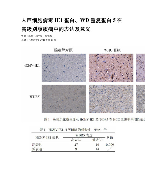
方法采用免疫组织化学(免疫组化)方法检测HCMV-IE1和WDR5在60份高级别胶质瘤组织及20份正常脑组织中的表达,分析HCMV感染与WDR5的相关性及两者对胶质瘤预后的影响。
结果 53 份(88.3%)高级别胶质瘤组织中的HCMV-IE1呈阳性表达,20份正常脑组织中均无HCMV-IE1表达(P < 0.001);HCMV-IE1表达程度低者总生存率优于表达程度高者(P = 0.037)。
49份(81.6%)高级别胶质瘤组织中WDR5表达阳性,11份(5.5%)正常脑组织中WDR5表达阳性(P = 0.009)。
在60份高级别胶质瘤组织中,HCMV-IE1与WDR5同时高表达有27例(45.0%),同时低表达有14例(23.3%),HCMV-IE1表达与WDR5表达呈正相关(r = 0.336,P = 0.009)。
结论 HCMV-IE1与高级别胶质瘤发生发展相关,是判断高级别胶质瘤预后的指标,其机制可能与HCMV-IE1和WDR5呈正相关有关联。
【关键词】人巨细胞病毒;高级别胶质瘤;WD重复蛋白5【Abstract】 Objective To investigate the expression and significance of human cytomegalovirus (HCMV)-IE1 protein and WDR5 protein in high grade glioma (HGG). Methods The expression of HCMV IE1 and WD repeat-containing protein (WDR5) proteins was detected by immunohistochemistry in 60 HGG tissues and 20 normal brain tissues. The correlation between HCMV infection and WDR5 protein and their influence on glioma prognoses were analyzed. Results HCMV-IE1 protein was positively expressed in 53 HGG tissues (88.3%) while not expressed in 20 normal brain tissues (P < 0.001). The overall survival rate of HGG patients with low expression level of HCMV-IE1 was better than those with high expression level of HCMV-IE1(P = 0.037). WDR5 protein expression was positive in 49 (81.6%) HGG tissues and in 11 (5.5%) normal brain tissues (P = 0.009). Among 60 HGG tissues, 27 cases (45.0%) had simultaneously high expression HCMV-IE1 protein and WDR5 protein, and 14 cases (23.3%)had simultaneously low expression. The expression of HCMV-IE1 protein was positively correlated with that of WDR5 protein (r = 0.336, P = 0.009). Conclusions HCMV-IE1 protein is associated with the initiation and development of HGG and is an indicator of the prognosis of HGG.The mechanism may be associated with the positive correlation between HCMV-IE1 and WDR5.【Key words】 Human cytomegalovirus;High grade glioma;WD repeat-containing protein膠质瘤是最常见的原发性神经上皮细胞恶性肿瘤,WHO依据肿瘤的恶性程度将胶质瘤分为Ⅰ~Ⅳ级,其中低级别胶质瘤(LGG)包含Ⅰ、Ⅱ级,高级别胶质瘤(HGG)包含Ⅲ、Ⅳ级[1]。
- 1、下载文档前请自行甄别文档内容的完整性,平台不提供额外的编辑、内容补充、找答案等附加服务。
- 2、"仅部分预览"的文档,不可在线预览部分如存在完整性等问题,可反馈申请退款(可完整预览的文档不适用该条件!)。
- 3、如文档侵犯您的权益,请联系客服反馈,我们会尽快为您处理(人工客服工作时间:9:00-18:30)。
进 行 免 疫 细胞 化 学 染 色 , 检测 I E 1及 p p 6 5的 表 达 水 平 。结 果 : 受染的 u 细 胞 出现 了 明 显 的 形 态 学 改 变, 并用 P C R 方 法检 测 到 了 HC MV核 酸 。免 疫 细 胞 化 学 染 色 表 明 , 蛋白I El在 感 染 后 3 0 mi n~2 h无 表 达, 4 h开 始 表 达 , l 4 h达 高峰 , 随后 下 降 ; 蛋白p p 6 5在 感 染 后 l h为 入 侵 型 p p 6 5. 4—9 6 h一 直 呈 低 水 平表 达 , 至 l 2 0 h表 达 急 剧 升 高 。 结 论 : u 细胞是 H C M V的 容许 细胞 , H C MV 在 其 中 的 表 达 具 有 时 序 性, 但是 其 时 序表 达较 在 人 成 纤 维 细 胞 中延 迟 。
[ 关键 词 ] 人 巨细 病 毒 ; 人 神 经胶 质 瘤 ; U 2 5 I 细胞 ; I E l ; p p 6 5; 时序 表 达 [ 中图分类号] R 3 7 3 [ 文献标识码 ] A [ 文章编号 ] l 6 7 2 — 7 3 4 7( 2 0 0 7 ) 0 4 — 0 5 5 l 一 0 6
A b s t r a c t : Ob j e c t i v e T o d e t e r m i n e w h e t h e r U 2 5 l c e l l s a r e p e r m i s s i v e f o r h u m a n c y t o me g a l o — v i r u s( H C MV) ,a n d t o i n v e s t i g a t e t h e c h a r a c t e i r s t i c s o f t e m p o r a l e x p r e s s i o n o f p r o t e i n s I E 1 a n d
p p65. M e t ho ds U2 5 1 c e l l s we r e i n f e c t e d wi t h HCMV , a n d t h e n t h e c e l l s we r e o b s e r v e d un d e r t h e t r a n s mi s s i o n e l e c t r o n i c mi c r o s c o p e,a n d t h e v i r a l n u c l e i c a c i d wa s d e t e c t e d b y PCR ,a n d t h e e x p r e s — s i o n l e v e l s o f I E 1 a n d pp 6 5 we r e a n a l y z e d b y i mmun o h i s t o c h e mi c a l a s s a y wi t h a n t i — I E 1 mo n o c l o n a l a n t i b o d y a n d a n t i — pp 6 5 mo n o c l o n a l a n t i b o d y a t v a io r u s t i me s p o s t i n f e c t i o n. Re s ul t s Mo r p h o l o g i c a l c h a n g e s o f t h e i n f e c t e d c e l l s a p p e a r e d un d e r t h e t r a n s mi s s i o n e l e c t r o n mi c r o s c o p e. T h e v i r a l n u c l e i c a c i d wa s d e t e c t e d s u c c e s s f u l l y b y P CR. Th e e x p r e s s i o n o f I E 1 wa s d e t e c t e d f i r s t l y a t 4 h p o s t i n f e c — t i o n,a n d r e a c h e d a p e a k wi t h i n 1 4 h,a n d t h e n d e c r e a s e d. Th e i nc o mi n g p p65 wa s de t e c t e d a t 1 h, t h e l o w e x p r e s s i o n l e v e l s o f p p 65 we r e d e t e c t e d f i r s t l y a t 4 h,a n d t h e y c o u l d r e ma i n r e l a t i v e l y c o n — s t a n t t h r o u g h 9 6 h, b u t t h e ma x i mu m e x p r e s s i o n o c c ur r e d a t 1 2 0 h. Co nc l u s i o n Hu ma n g l i o ma
( D e p a r t m e n t o f Mi c r o b i o l o g y,X i a n g y a S c h o o l f o Me d i c i n e ,C e n t r a l S o u t h U n i v e r s i t y,C h a n g s h a 4 1 0 0 7 8,C h i n a)
李建华 , 傅 鹰 , 陈利玉 , 戴 橄 , 罗敏 华 , 杨 滔 (中南 大 学 湘 雅 医学 院 微 生 物 学 系 , 长 沙 41 0 0 7 8)
[ 摘 要] 目的 : 研 究人 神 经胶 质 瘤 u 细胞 对 人 巨细 病 毒 ( h u m a n c y t o m e g a l o v i r u s ,HC MV) 的容许性 , 并探讨病毒蛋 白 I El及 p p 6 5的 时序 表 达特 点 。 方 法 : H C MV 感 染 u 蚓细胞 , 以透 射 电镜 观 察 u 细胞 的
T e mp o r a l e x p r e s s i o n o f HC MV I E1 a n d p p 6 5 i n h u ma n #o ma U2 s l c e l l s
L I J i a n — h u a,F U Yi n g, C HE N L i — y u,DAI Ga n, L UO Mi n— h u a,YAN G T a o
维普资讯
J c e S o u t ^ 西( Me d S c ) 2 O 0 7, 3 2 ( 4 )
5 5l
中南大学学报 ( 医学版 )
H C MV I E 1 和 p p 6 5在 人 神 经 胶 质 瘤 U 2 5 细 胞 中 的 时序 表 达
