20070918胃肠道组织学
胃肠道常见疾病介绍

胃肠道常见疾病介绍
1、总论 2、胃食管反流病 3、食管癌 4、胃炎 5、消化性溃疡 6、胃癌
胃肠道常见疾病介绍
胃炎
急性胃炎 慢性胃炎 特殊胃炎
Menetrier 病
应激(紧张、手术)、药物(阿司匹林 、化疗、铁剂、氯化钾、辣椒)、酒精 、胃镜下手术、反流、血循环障碍
病理
1、炎症:淋巴细胞、 浆细胞、中性粒细胞 2、化生:杯状细胞、 幽门腺细胞代替 3、萎缩:腺体破坏减 少 4、上皮内瘤变:不典 型增生
胃肠道常见疾病介绍
内镜下表现
胃炎
胃肠道常见疾病介绍
内镜下表现
胃炎
胃肠道常见疾病介绍
内镜下表现
胃炎
胃肠道常见疾病介绍
内镜下表现
胃炎
胃肠道常见疾病介绍
食胃管炎癌治治疗疗
胃肠道常见疾病介绍
食管癌
定义与表现
原发于食管上皮的恶性 肿瘤
早期: 胸骨后不适, 进食滞留 中晚期:进行性咽下困 难、食物反流、咽下痛 、恶病质
病因
1、亚硝胺、真菌毒素 2、慢性理化刺激及炎 症(粗糙、过烫食物、 烟丝、槟榔) 3、营养(缺少果蔬、 微量元素) 4、遗传医生 5、癌基因激活
病理
1、早期食管癌:充血 、糜烂、斑块、乳头型 2、中晚期食管癌:髓 质、蕈伞、溃疡、缩窄 、未定型 3、组织病理:90%鳞 癌、少数为腺癌
进展期胃癌:深度超过粘膜下层,侵入肌层为中期,侵入 浆膜层或之外为晚期胃癌。表现为恶心、呕吐、呕血、黑 便等
胃肠道常见疾病介绍
内镜表现
胃癌
胃肠道常见疾病介绍
内镜表现
胃癌
胃肠道常见疾病介绍
胃癌
胃肠道病理学知识点总结 考试复习资料
胃肠➢胃肠道正常组织:粘膜层:上皮层、固有层(Lamina propria)、黏膜肌层;粘膜下层;固有肌层(Muscularis propria);浆膜层(Serosa)➢食管炎1、发病因素:(1)胃内容物反流(反流性食管炎);(2)感染;(3)物理:胃管、放射;(4)化学:刺激物摄取、化疗药;(5)混合因素(Miscellaneous):尿毒症、GVHD;2、反流性食管炎临床表现:a) 吞咽困难;b) 疼痛胸骨后疼痛、心窝部烧灼感、返酸;c) 消化道出血,黑便3、食管炎并发症:溃疡、狭窄、B arrett’s食管;4、反流性食管炎病理变化:大体:食管粘膜充血发红,糜烂,溃疡,随病情呈进行性改变。
镜下:i. 鳞状上皮层内炎症细胞浸润(嗜酸性粒细胞,中性粒细胞,淋巴细胞)ii. 基底细胞增生,可占上皮总厚度的20%以上;iii. 固有层乳头延长,可延长至上皮层的上1/3;固有膜毛细血管充血。
➢Barrett食管1、*定义:食管远端粘膜的鳞状上皮被化生的胃肠腺上皮替代,柱状上皮之间可见杯状细胞;化生的柱状上皮可呈异性增生进而形成腺癌;多与长期胃食管反流有关。
2、*病理变化:大体:粘膜呈橘红色、天鹅绒样(velvet mucosa)不规则形病变;在灰白色正常食管粘膜的背景上呈补丁状分布;可继发有糜烂、溃疡、食管狭窄和裂孔疝。
(胃食管连接处可形成不规则的环形带,或呈舌状、岛状、斑片状等)镜下:由类似胃黏膜或小肠粘膜的上皮细胞和腺体构成。
Barrett粘膜的柱状上皮细胞兼有鳞状上皮和柱状上皮细胞的超微结构和细胞化学特征。
腺体排列紊乱,常有腺体的扩张、萎缩、纤维化和炎症细胞浸润。
局部粘膜肌层常增厚。
组织学上柱状上皮间由肠杯状细胞就可以确立诊断。
3、*临床意义:局部抗胃酸能力增强,但易引发食管溃疡、食管狭窄及食管腺癌。
4、并发症:食管溃疡、食管狭窄、食管腺癌;➢食管癌1、分布特点:鳞癌最常见(上1/3,20%;中1/3,50%;下1/3,30%);腺癌(下1/3);2、发病因素:(1)饮食:维生素、微量元素(trace metals)缺乏,食物真菌污染,亚硝酸盐摄入过多;(2)生活方式:烟酒;(3)食管动力紊乱或结构异常:食管炎,贲门失弛缓(achalasia),缺铁性吞咽困难综合征(Plummer-Vinson syndrome);(4)遗传易感:胼胝症(tylosis,手掌脚掌角化过度hyperkeratosis of palms and soles)、种族倾向(racial disposition);(5)腺癌与Barrett食管有关:Barrett 食管是食管腺癌的癌前状态。
胃肠道

胃肠道★充盈缺损(filling defect):胃肠道管壁形成向腔内的局限性隆起时,局部钡剂不能充盈,从而使由钡剂勾画出的消化管轮廓呈局限性凹陷表现。
主要见于一些肿瘤性病变,如癌、腺瘤、脂肪瘤等,和少数非肿瘤性病变,如息肉、异位胰腺等。
钡剂造影检查中,当粘膜面形成的凹陷或溃疡达到一定深度时可被钡剂充填,于切线位投照时形成突出于腔外的钡斑影像,称为龛影或壁龛,正位投照时表现为浓钡斑。
常见于炎症(如溃疡)、肿瘤(如溃疡性癌)等疾病。
消化管道局部发育不良、肌壁薄弱时,官腔内压增高导致的局限性管壁突出。
钡剂造影表现为消化道轮廓外的囊袋状突起,有黏膜伸入其内,有收缩和蠕动。
食管静脉曲张(下1/3多见)①管壁柔软;②与正常组织无明显分界;早期:食道下段粘膜皱襞轻度增粗、迂曲中期:食道中下段黏膜明显增粗、迂曲,呈串珠状、蚯蚓状充盈缺损;管壁边缘呈锯齿状或多发小圆型晚期:③食道全程黏膜明显增宽、迂曲,呈蚯蚓状、串珠状充盈缺损;④管壁边缘呈锯齿状,(管径扩大,)⑤蠕动减弱食管癌(40-70岁)①局部黏膜皱襞中断、破坏、消失;②管壁僵硬,蠕动消失;③狭窄段以上扩张;④病变位于管壁轮廓之内,与正常组织分界清。
浸润型:节段性管腔漏斗状或环状狭窄,病变段管壁僵硬,上段食管扩张溃疡型:切线位溃疡深入食管壁,形成不规则龛影,管壁偏心性狭窄髓质型:腔内不规则充盈缺损伴大小不等的龛影,黏膜破坏,管腔狭窄,与正常食管移行段呈斜坡状胃溃疡(20-50岁,小弯多见)——餐后痛直接征象:正位:圆形或椭圆形钡斑,边缘光滑整齐,密度均匀;周围黏膜纠集(钡斑+黏膜纠集)侧位:乳头状或半圆形龛影突出于胃轮廓之外;口部见一透明带(龛影+粘膜线/项圈征/狭颈征)间接征象:胃壁痉挛收缩,蠕动增强或减弱,瘢痕(变形或狭窄),分泌增加十二指肠溃疡(20-50岁,球部多见)——饥饿痛直接征象:正位:圆形或类圆形钡斑,边缘光滑整齐;周围黏膜纠集(钡斑+黏膜纠集)侧位:小锥形、乳头状或半圆形龛影,突出于轮廓之外;口部见一透明带(龛影+透明带)十二指肠球部变形(山字形、双叶形、三叶草形或花瓣形)间接征象:激惹征;幽门痉挛;张力和蠕动改变;分泌增加;固定压痛★胃癌(40-60岁,贲门、小弯、胃窦区)①胃腔内边缘不规则的充盈缺损——蕈伞型②胃腔狭窄,胃壁僵硬(革囊胃)——浸润型③恶性龛影:——溃疡型位于胃的轮廓之内,与正常胃壁分界清楚边缘不规则、裂隙征(两指压征之间指向口部的尖角)龛影周围:环堤征、指压征——半月征粘膜皱襞破坏消失癌病区蠕动消失黏膜线:胃溃疡龛影口部宽1-2mm的光滑整齐的透亮线,为黏膜水肿所致。
消化系统解剖生理

(三)小肠内消化
小肠是消化和吸收最重要的部位:
化学性消化: 胰液、胆汁、小肠液的参与 机械性消化: 小肠的运动
1. 胰液的分泌和作用
(1)碳酸氢盐:保护肠粘膜免受强酸侵蚀;为小肠 内消化酶发挥作用提供适宜PH值环境 (2)消化酶:胰淀粉酶、胰脂肪酶、胰蛋白酶和糜 蛋白酶
2. 胆汁的分泌和作用
成份:水、无机盐、胆盐、胆固醇、胆汁酸、胆色素 注意:胆汁能帮助消化,但不含消化酶 作用:对脂肪的消化吸收
胃肠道的解剖及生理
概述
消化系统组成: 消化管 消化腺
第1节 消化管
外膜
肌层 粘膜下层
粘 膜 层 上皮 固有层 粘膜肌层
一、消化管的一般微细结构(除口腔、咽)
(一)粘膜:消化管各段结构差异最大、功能最重
要的部分。
1.上皮 消化管的两端(口腔、咽、食管及肛门)为复 层扁平上皮;其余部分均为单层柱状上皮 2.固有层 为疏松结缔组织 3.粘膜肌层 为平滑肌
●
二、吸收
(一)吸收的部位:
1、口腔:吸收某些药物 2、食管:基本不吸收 3、胃:吸收酒精和少量的水分 4、小肠:食物的主要吸收场所 5、大肠:主要吸收水分和盐类
(二)主要营养物质的吸收:
糖:葡萄糖--主动转运入血(与钠离子协同转运)
蛋白质:氨基酸--主动转运入血
脂肪:+胆盐→乳糜微粒--入淋巴 胆固醇:游离胆固醇才能吸收入淋巴,胆盐和食物中
(二) 空肠和回肠
空肠
长度 位置 管径 管壁 颜色 血管 环状襞 近侧2/5 左上腹 较粗 较厚 较红 较多 密而高 淋巴滤泡 孤立
回肠
远侧3/5 右下腹 较细 较薄 较淡 较少 疏而低 集合
(三)小肠粘膜的微细结构特点:
2018胃肠道影像学诊断
角切迹
胃大弯
胃的正常影像解剖
形态: 四型:牛角型、钩形、长型、瀑布型 充盈相:轮廓光滑、密度均匀 粘膜相:小弯侧:粘膜皱襞及皱襞间沟光滑、规则、平行
胃底、胃体大弯侧:锯齿状---粘膜斜行或横行排列 胃窦:收缩时纵行为主;舒张时横行为主
功能: 蠕动:胃壁柔软,2~3个蠕动波 排空:2~4小时
胃正常影像解剖
粘膜皱襞呈放射状从四周向 病变区集中
消化道黏膜皱襞改变
十二指肠正常影像解剖
十二指肠球部充盈相
十二指肠球部粘膜相
十二指肠正常影像解剖
十二指肠正常影像解剖 影十
二 指 肠 低 张 造
小肠正常影像解剖
小 肠 气 钡 双 重 造 影
结肠(colon) 、直肠(rectum)正常影像解剖
肝曲
横结肠
脾曲
盲肠
阑尾
降结肠
乙状结肠 直肠
结肠正常影像解剖
3.食管、胃肠道的基本病变征象
钡餐造影检查: 钡剂单对比造影 气钡双重造影
空气造影 血管造影
CT: 胃肠道粘膜下及管腔外病变情况 管壁外侵犯、远处器官和淋巴结转移
MRI
各种检查方法的比较及选择
X线平片:
➢ 优势:价格便宜、简单易行 ➢ 劣势:消化道缺乏对比,征象较少 ➢ 应用:主要用于急腹症诊断,如胃肠
穿孔及肠梗阻等,而用于消化 道常规检查较少
牛角型胃
钩型胃
瀑布型胃
胃的分型示意图
髂嵴水平 长型胃
鱼钩型
牛角型 无力型ຫໍສະໝຸດ 瀑布型胃粘膜相
充盈相
DC相 CT
胃
粘
膜
放
粘膜皱襞
大
粘膜沟
像
胃正常影像解剖 胃底充盈相
消化系统疾病课件

72
.
胞浆疏松化
73
.
嗜酸性变
肝细胞气球样变及嗜酸性变
74
.
1.肝细胞的变质性变化
(2)肝细胞的凋亡及坏死
①凋亡: 表现为嗜酸性小体(acidophilic body)。 习惯上称嗜酸性坏死(acidophilic
necrosis)。
75
.
② 溶解坏死 (lytic necrosis)
严重的细胞水肿导致细胞崩解,最常见。 根据肝细胞坏死的程度及范围的不同,可分为:
66
.
各型肝炎病毒的特点
肝炎病毒型
HAV HBV
HCV HDV HEV HGV
67
病毒特点
传染途径
27nm,单链RNA 肠道(易暴发流行)
43nm,DNA 密切接触、输血、注射
30-60nm ,单链 RNA
缺陷性RNA 32-34nm,单链
RNA 单链RNA
同上 同上 肠道(易暴发流行) 输血、注射
镜下见:
① 出现特征性的桥接坏死和中 度碎片坏死
②小叶内可有纤维间隔形成,但小 叶结构大部分保存
98
.
碎片状坏死
99 (piecemeal necrosis)
.
桥接坏死
(bridging necrosis)
100
.
毛玻璃样肝细胞
101
.
临床病理联系及结局:
病程超过半年以上,肝功能持续 异常;可发展成肝硬化。
102
.
(3)重度慢性肝炎
肉眼观: 肝表面呈颗粒状,质地较硬。
镜下见:
① 出现重度碎片状坏死及大范围桥接坏死。
② 坏死区出现肝细胞不规则再生。
05-胃肠道

第一节胃的细胞病理学u胃癌是恶性肿瘤中最常见的一种。
胃癌侵至浆膜层术后五年生存率仅为8-17%,而早期胃癌的术后五年生存率达90%以上。
u早期诊断:最好方法为纤维胃镜。
(二)胃的组织学胃壁分四层:(1) 粘膜层(2) 粘膜下层(3) 肌层(4) 浆膜层(三)胃的细胞学Ø胃的正常上皮:均为柱状细胞。
①胃粘膜被覆上皮细胞②胃底腺细胞:主细胞、壁细胞、幽门腺细胞Ø胃外来的细胞:来自于口腔、鼻腔、十二指肠Ø非上皮性细胞:涂片见红细胞、中性粒细胞、淋巴细胞、浆细胞、嗜酸性粒细胞及组织细胞等。
Ø其他:食物残渣、细菌、寄生虫和原虫等Ø胃涂片背景:胞浆呈淡红色或淡黄色二、胃脱落细胞的采集与制片(一)电动洗胃机冲洗法(二)大量生理盐水洗胃法(三)蛋白水解酶洗涤法(四)网套气囊摩擦法(简称胃球法)(五)胃内窥镜简介-电动洗胃机冲洗法1.电动洗胃机构造由压缩机、控制台、储液瓶和胃管构成。
原理:利用空气压缩机为动力,操纵控制台可交替地向密闭的储液瓶中送正压或负压,以便将洗液通过胃管喷入胃内,或从胃内抽回瓶中。
2. 洗胃前的准备u饮食:少量碳水化合物,不宜进高蛋白、高脂肪和纤维质多食物u准备物品:12-14号胃管(十二指肠)1支生理盐水100毫升醋酸缓冲液(pH5.6)300-400毫升注射器(50或100毫升)1支烧杯(50-100毫升)各1个万能试纸适量载玻片4枚离心机(3000/分钟以上)1台3. 冲洗方法:u病人空腹吞服胃管后,将洗胃用水全部注入胃内。
此时病人可变动体位帮助冲洗。
如此反复减压冲洗数次,最后抽出的冲洗液即第一液,进行外观检察记录后弃掉。
变换贮液中洗胃用水为PH5.6醋酸缓冲液300-400ml。
用上述同样方法反复加压冲洗3-4次,病人同时变换体位,以利于充分冲洗,最后将全部洗胃液(第二液)抽出,放冰盘或冰箱中静置后用于检查。
4. 离心、涂片、固定、染色u将最后之冲洗液放入4个50-100ml的尖底离心管内3000转/分的速度离心5min,弃去上清液,用乳头吸管或刮匙选择性吸取残渣涂片,如有血液或食物残渣时,可取其上层残渣涂片。
【医学PPT课件】消化系统组织学

一、唾液腺的微细结构
1、浆液性腺泡:
一 腺泡 2、粘液性腺泡:
般
3、混合性腺泡:
结
构
1、闰管:
导管 2、纹状管:
3、小叶间导管、总导管:
31
浆液性腺泡
混合性腺泡
闰管
纹状管
粘液性腺泡
二、胰的微细结构
1、腺泡
1、外分泌部
2、导管
胰
1、A细胞
2、内分泌部 2、B细胞
(胰岛) 3、D细胞
4、PP细胞
2、肝门管区
41
肝小叶 中央静脉
肝板
❖ 肝细胞排列的放射状 的板状结构称为肝板。
❖ 在切片上,肝板断面 呈索状,称肝索。
43
肝细胞
1 肝细胞体积大,有 血窦面 细胞连接面 胆小管面
2、肝细胞核圆大居中,浅染,核仁清楚,核和核 仁可为多个或多倍体核。Mi, RER, SER, Gi, Ly, 过 氧化物酶体等细胞器丰富。
❖ 2.胃主细胞 A.主要分布在胃底腺颈部 B.大量粗面内质网聚集于顶部胞质 C.分泌的胃蛋白酶原被盐酸激活 D.分泌内因子
❖ 3、.潘氏细胞分布于 A.幽门腺
B.小肠腺 C.十二指肠腺 D.大肠腺
❖ 4、.关于小肠绒毛,哪项错误 A.由粘膜和粘膜下层共同突起形成 B.有丰富的毛细血管网 C.含中央乳糜管 D.上皮细胞在其顶端脱落
❖ 粘膜下层:无特殊。
❖ 肌层:内斜、中环、外纵三层 平滑肌,环行肌增厚形成贲门 括约肌和幽门括约肌。
❖ 外膜:浆膜。
11
胃底腺
分布:胃体和胃底部; 结构:分支管状腺,可分 颈、
体与底部;开口于胃小凹。 组成:主细胞、壁细胞、颈粘液
细胞及内分泌细胞。
肠道组织学描述-概述说明以及解释

肠道组织学描述-概述说明以及解释1.引言1.1 概述肠道组织学是研究肠道组织的结构、组成、功能以及与疾病关系的学科。
肠道是人体消化系统中最长的一段,起到吸收营养物质、水分和排除废物的重要作用。
肠道组织学的研究可以帮助我们更好地了解肠道的生理和病理过程,为肠道相关疾病的诊断和治疗提供科学依据。
肠道组织学主要关注肠道的微观结构和细胞组成。
肠道由黏膜、黏膜下层、肌层和浆膜组成,其中黏膜是最关键的部分。
黏膜中包含许多微细的结构,如肠绒毛、肠腺和免疫相关的淋巴组织。
这些结构对于吸收和消化食物至关重要。
肠道组织学的研究方法主要包括组织染色、免疫组织化学、电镜等技术。
通过对肠道组织进行切片并染色,可以观察到细胞和组织的形态特征,并了解其功能和异常改变。
免疫组织化学技术可以用于检测和定位特定的蛋白质和细胞因子,揭示其在肠道中的作用机制。
电镜则能够提供更高分辨率的图像,详细观察细胞的超微结构。
对肠道组织学的研究具有重要的临床意义。
许多肠道相关疾病,如炎症性肠病、肠道感染和肠道肿瘤,都与肠道组织的结构和功能异常有关。
通过对肠道组织进行病理分析,可以帮助医生进行准确的诊断,并指导治疗方案的选择。
此外,了解肠道组织的结构和功能对于新药的研发和治疗策略的优化也具有重要意义。
未来,肠道组织学的研究将继续深入。
随着技术的不断进步,我们可以望见更多关于肠道组织结构和功能的新发现。
同时,肠道组织学也将与其他学科相互融合,如生物学、微生物学和免疫学等,共同揭示肠道健康与疾病发生发展的机制。
相信未来的肠道组织学研究将为人们提供更深入的认识和更有效的治疗手段。
文章结构部分的内容应该包括本文所使用的章节结构和每个章节的主要内容简介。
在本文中,文章结构部分可以这样描述:文章结构如下:1. 引言1.1 概述1.2 文章结构1.3 目的2. 正文2.1 肠道组织学基本概念2.2 肠道组织学的研究方法3. 结论3.1 对肠道组织学的重要性的总结3.2 对未来肠道组织学研究的展望在引言部分,我们将提供一个概述,用于引入读者对肠道组织学的主题的基本了解。
胃肠道解剖ppt

• 二、大肠
大肠(large intestine)起始段在右髂窝 处与回肠相接,末端终于肛门,长约1.5m, 分为盲肠、结肠、直肠和肛管4部分。 盲肠和结肠在外形上有3个特征:
结肠带(colic bands)是肠壁的纵行肌 聚集而成的带状结构,共3条,起于兰尾根 部,沿肠管的表面纵行排列,止于乙状结 肠末端 ;
瓣边缘共同围成锯齿状环行线,环绕肠管;齿状线以下的上皮为
复层扁平上皮。齿状线上方由内脏神经分布,下方由躯体
神经分布。齿状线也是直肠动脉供应、静脉和淋巴回流的
分界线。在齿状线下方,由于肛门内括约肌紧缩,而形成 一宽约lcm略微凸起的环形带,称肛梳(anal pecten)。肛 梳下缘有一不甚明显的环形线,称白线(white line)。
• 1.上部(superior part) 又称球部,于第1腰椎的右侧起自 幽门,行向右后方,至肝门下方,胆囊颈附近,急转向下 续为降部,转折处称十二指肠上曲 (superior duodenal flexure)。上部靠近幽门约2.5cm的一 段肠管,肠壁较薄,粘膜多较平滑,称十二指肠壶腹又称 十二指肠球(duodenal bulb)。 2.降部(descending part) 十二指肠上曲沿第1~3腰椎 体的右侧下降,至第3腰椎水平,急转向左连接水平部, 转折处称十二指肠下曲(inferior duodenal flexure)。降部 的粘膜形成许多环形襞,在其后内侧壁上,有一纵行的粘 膜皱襞,称十二指肠纵襞(longitudianl fold of duodenum)。 纵襞的下端有一隆起,称十二指肠大乳头 (major duodenal papilla),是胆总管和胰管共同开口之处。 在大乳头上方1~2cm处有时可见有十二指肠小乳头,是副 胰管的开口部位。
消化道形态学和组织学
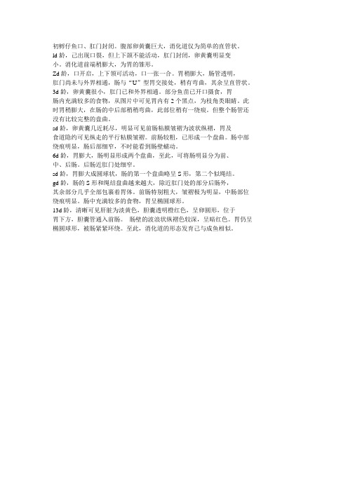
初孵仔鱼口、肛门封闭。
腹部卵黄囊巨大,消化道仅为简单的直管状。
ld龄,己出现口裂,但上下颌不能活动,肛门封闭,卵黄囊明显变
小。
消化道前端稍膨大,为胃的雏形。
Zd龄,口开启,上下领可活动,口一张一合。
胃稍膨大,肠管透明,
肛门尚未与外界相通,肠与“U”型胃交接处,稍有弯曲,其余呈直管状。
3d龄,卵黄囊很小,肛门已和外界相通。
部分鱼苗已开口摄食,胃
肠内充满较多的食物,从图片中可见胃内有2个黑点,为枝角类眼睛。
此时胃稍膨大,在肠的中后部稍稍弯曲,此部位稍有一绕痕。
但整个肠管还没有比较完整的盘曲。
sd龄,卵黄囊几近耗尽,明显可见前肠粘膜皱褶为波状纵褶,胃及
食道隐约可见纵走的平行粘膜皱褶。
前肠较粗,已形成一个盘曲。
肠中部绕痕明显,肠后部细窄,不时能看到肠壁蠕动。
6d龄,胃膨大,肠明显形成两个盘曲,至此,可将肠明显分为前、
中、后肠。
后肠近肛门处细窄。
sd龄,胃膨大成圆球状,肠的第一个盘曲略呈S形,第二个似绳结。
gd龄,肠的S形和绳结盘曲越来越大,除近肛门处的部分后肠外,
其余部分几乎全部包裹着胃体。
前肠特别粗大,皱褶极为明显,中肠部位绕痕明显。
肠中充满较多的食物,胃呈椭圆球形。
13d龄,清晰可见肝脏为淡黄色,胆囊透明橙红色,呈卵圆形,位于
胃下方,胆囊管通入前肠。
_肠壁的波浪状纵褶色较深,呈暗红色。
胃仍呈椭圆球形,被肠紧紧环绕。
至此,消化道的形态发育己与成鱼相似。
消化系统病理

食道溃疡
正常食道
杯状细胞
肠上皮化生
食道粘膜
复层鳞状上皮 主要功能:运送食物 常见病理改变
上皮内炎细胞浸润:中性、嗜酸、淋巴 上皮破坏脱落:糜烂、溃疡 上皮增生 肠上皮化生
Lower esophageal sphincter
胃
粘液细胞 壁细胞
主细胞
胃体
胃窦
胃炎
幽门螺杆菌
肠上皮化生正常胃窦粘膜层(mucosa):是消化管各段结构差异最 大、功能最重要的部分
上皮和腺体
– 复层扁平上皮:口、咽、食管、肛门 – 单层柱状上皮:胃、肠、阑尾
固有层:结缔组织、血管淋巴组织 粘膜肌层:薄层平滑肌
Squamous Columnar
Cuboidal Goblet
食道
嗜酸性粒细胞
淋巴细胞
食道炎症
消化系统正常及常见 病变形态简介
北京大学医学部病理学系 石雪迎 2014.9.25
Tel: 82805488 E-mail: shixueying@
消化系统的组成
消化道
食道 胃 小肠:十二指肠、空 肠、回肠 大肠:结肠、直肠
消化腺
肝脏 胰腺 涎腺
消化管壁的一般结构
粘膜层 粘膜下层 肌层 外膜(浆膜)
小叶中央型坏死 点、灶状坏死
碎片状坏死 ----界板炎
桥接坏死 亚大片坏死 大片坏死
坏死和凋亡
根据病变范围和分布
小叶中央型坏死 点、灶状坏死
碎片状坏死 ----界板炎
桥接坏死 亚大片坏死 大片坏死
炎细胞渗出
坏死引起炎细胞聚集 细胞毒性T淋巴细胞攻击病毒感染细胞 汇管区炎细胞浸润 Kupffer细胞吞噬凋亡细胞 肉芽肿性炎
胃肠道系统病理学二

五、肿瘤
(一)良性上皮性肿瘤
1 、十二指肠腺瘤( Brunner 腺腺 瘤) ,以组织学正常的十二指肠的 结节性增生为特征,伴有导管和散 在的间质成分。 2 、腺瘤: 可单发或多发,有蒂或 无蒂,包括管状腺瘤,绒毛腺性息 肉或绒毛状腺瘤。易恶变。 3 、错构瘤性息肉: 大体与一般的 腺瘤性息肉无不同,镜下腺体由平 滑肌纤维束支撑,病变中心平滑肌 纤维束较粗,而边缘则较细,腺体 和平滑肌纤维紧密交错排列,酷似 浸润形态,易误诊癌。易恶变。
1 、吸收障碍综合征可以是多器质性异常道德结果。小肠活检对乳糜 泻的评估很有帮助,特征是小肠粘膜完全扁平。大体见十二指肠皱襞 减少或消失,这是内窥镜检查时的一个重要诊断线索。镜下:绒毛萎 缩或消失,但整个粘膜厚度基本正常,伴粘膜固有层内含免疫球蛋白 的浆细胞数目增加,表层上皮内大的脂滴沉积。 2 、乳糜泻发病机制尚不清楚。可并发肠恶性肿瘤,如发现病人对去 除谷蛋白的反应发生逆转、发热、体重下降、腹痛、杵状指、肠溃疡 或血清IGA水平持续升高,要考虑淋巴瘤。 并发慢性非特异性溃疡性十二指肠空肠回肠炎,本病特点是在营养吸 收障碍综合征的基础上发生腹痛和发热,这种情况可能多半是恶性淋 巴瘤的初期阶段。 并发癌少见。
(二)恶性肿瘤
1 、腺癌: 小肠腺癌比大肠腺癌少 40-60 倍,可发生在小肠任何部位。 肿瘤常为中分化腺癌,产生粘液, 大多数患者就诊时肿瘤已扩散到肠 壁内并可能已经转移到局部淋巴结。 预后主要决定于有无局部淋巴结受 累。有淋巴结转移者88%死于肿瘤, 无淋巴结转移者仅有45%死于肿瘤。 2、其他类型癌 包括小细胞癌、腺鳞癌、间变性 (肉瘤样)癌。
三、溃疡
1 、十二指肠消化性溃疡是一种十 分常见的疾病,发病机制与侵袭性 因素、防御性因素的相互作用有关, 完好的胃底粘膜是十二指肠消化性 溃疡形成的先决条件。
胃肠专业知识
疾病分类病因食道炎食物,抗生素,胃部炎症,贲门松弛等反流性食道炎胆汁反流性胃炎糜烂性胃炎(疣状胃炎)胃溃疡Hp感染,滥用抗生素浅表性胃炎没有得到及时治愈饮酒胃下垂多见体型偏瘦的人群,悬韧带松弛导致胃部下沉浅表性胃炎初期胃部炎性病变萎缩性胃炎各类胃炎未治愈引起,长期服用抑酸药物导致。
幽门螺旋杆菌传染,自身免疫力低下胃息肉肠息肉十二指肠溃疡由于胃部功能受损导致胃部逆向蠕动或幽门口关闭不全导致(反流性疾病属功能型病变,先功能受损后引发炎症黏膜上皮受损,饮食不洁,乱用抗生素药物同上各类炎症刺激引起的炎性肿瘤结肠炎细菌感染,长期作息时间紊乱溃疡性结肠炎结肠炎合并长期感染炎症刺激,免疫低下直肠炎免疫障碍检查症状词我院特色治疗治疗项目关于病种包装特点GS靶向治疗体系糜烂/溃疡性胃炎:此类疾病主要是由于胃内粘膜层受损导致。
引导方向在于突出胃粘膜合成细胞(三叶因子)促进三叶因子的合成,使胃部自愈并非药物治愈,这样治愈后防复发的效果显著。
一般治疗3-7天一疗程需2-4疗程,费用在每疗程几百元到几千元不等。
反流性胃炎/食道炎:由于这种疾病是逆向蠕动,所以采用此类治疗办法效果显著,也是其他医院没有的治疗办法。
正常胃部蠕动的方向是向肠道方向。
但是病发后就是向上蠕动了。
所以采用仪器刺激胃部向下蠕动。
一般一到两次效果很结肠炎:这类患者一般长期服药效果不明显。
大肠水疗主要包装为患处给药。
通过灌输药物来对肠道形成一层保护层。
防止粪便等有害菌对炎症面的二次刺激。
各类肠炎:原理可同上,通过水疗的方式刺激肠道内部有益菌增长。
缓解便秘或者是腹泻的症状。
调整菌群后患处给药消炎即可治愈。
胃/肠动力治疗仪大肠水疗萎缩性胃炎:这类疾病是由于胃部细胞萎缩,没有分泌胃酸的功能引起。
重点放在靶向治疗可以针对性的激活萎缩的细胞,促使细胞腺体有分泌其他慢性胃炎:靶向定位治疗利用中药贴片,在体外针对患处进行导入。
(利用大型生物磁场经络导入技术)控制幽门螺旋杆菌繁殖摧毁其生长的环境。
胃肠道解剖护理课件
THANK YOU
疼痛管理
评估患者的疼痛程度,采用适当的止痛方法,如药物治疗、物理治疗 等,缓解患者疼痛。
康复锻炼
鼓励患者进行适当的康复锻炼,如散步、太极等,促进肠道蠕动和功 能恢复。
心理支持
关注患者的心理状态,提供心理疏导和支持,帮助患者保持积极心态, 增强康复信心。
提高生活质量的方法与技巧
01
02
03
04
健康生活方式
症,保障患者健康。
患者及家属的自我管理与教育
疾病知识教育
向患者及家属普及胃肠道疾病 的相关知识,提高其对疾病的
认知和理解。
自我监测与记录
指导患者及家属进行自我监测 和记录,及时发现异常情况并 就医。
药物与治疗管理
向患者及家属说明药物的作用、 使用方法及注意事项,指导正 确的治疗方法。
心理调适与应对
向患者及家属提供心理支持,鼓励他 们积极面对疾病,增强治疗信心。
定期检查与筛查
定期进行胃肠道检查
通过胃镜、肠镜等检查手段,及早发 现胃肠道病变,提高治愈率。
筛查高危人群
对有家族史、不良生活习惯等高危人 群进行定期筛查,针对性地进行预防 和干预。
健康饮食与生活习惯
均衡饮食
保持食物的多样性,适量摄入高 纤维食物,减少油腻、刺激性食 物的摄入。
药物治疗与护理
01
02
03
药物治疗
遵医嘱按时服药,确保药 物剂量和用药方式的正确 性,避免漏服或错服。
药物观察
密切观察患者用药后的反 应,如出现不良反应或过 敏症状,应及时就医。
药物指导
向患者及家属说明药物的 用法、用量、注意事项及 副作用,提高患者的自我 管理意识。
消化道病理

• ( 2 )贲门粘膜特点:小凹与胃体粘膜相似, 腺体呈分枝管泡状腺上皮主要为粘液细胞,夹 少许壁细胞,粘膜肌不整齐。 • (3)胃窦粘膜特点:小凹较深占1/2—1/3,腺 管分枝弯曲,间质较多,有少数淋巴细胞,粘 膜肌整齐。 • ( 4 )移行部粘膜特点:小凹介于胃体与胃窦 之间,腺体为体窦混合 • (5)十二指肠粘膜特点:表面有许多绒毛, 主要有吸收细胞和杯状细胞,固有膜内有肠腺, 由分泌细胞,杯状细胞和神经内分泌细胞组成, 粘膜肌层不整齐。
• ( 1 )慢性浅表性胃炎:这是胃粘膜最常见的 一种病变,国内报道检出率高达 50%,其病理 特征为不同程度的炎症细胞浸润,以淋巴及浆 细胞为主,表面或小凹上皮可增生或化生。 • 按炎症细胞浸润的深度可分为轻、中、重。凡 炎症细胞浸润范围不超过粘膜浅层的1/3者称轻 度;浸润达粘膜深度2/3者称中度;浸润范围超 过2/3,且已累及粘膜全层者称重度。但从日常 所见胃粘膜的切片来看,在慢性重度浅表性胃 炎中,也往往见到固有腺体的萎缩性变化,因 此要区分较重的慢性浅表性胃炎和较轻的慢性 萎缩性胃炎,有时是很困难的,特别在胃窦部 粘膜的切片中。
• 根据资料来看,发现结肠型不完全性肠化生和 肠型胃癌有密切关系。在肠上皮化生中,化生 的上皮细胞,可以数量增多,排列紊乱,细胞 和核出现异型,形成不典型增生。肠化可分为 三级:轻度,肠化细胞取代胃粘膜固有细胞1/3 之内;中度,肠化细胞取代胃粘膜固有细胞 1/3—2/3之内;重度,肠化细胞取代胃粘膜固 有细胞2/3以上。
上消化道常见的病理
福建省立医院病理科 江淑萍 福建省立医院内科 郑铭
胃粘膜病理
• 一.胃粘膜解剖和组织学特点 1.解剖特点: 胃是消化道最膨大的部分。胃可分为前壁和后 壁,前、后壁相连的上缘为胃小弯,下缘为胃 大弯。胃小弯上端有食管的进口称为贲门,下 端有移行于十二指肠的出口称为幽门。一般将 胃分为四部分:贲门、胃底、胃体、胃窦部。 贲门:位于贲门口下3cm范围,胃底:自贲门 向左上方突出的部分,胃体:上界胃底,下界 胃角切迹,胃窦部:幽门口上7cm范围。
- 1、下载文档前请自行甄别文档内容的完整性,平台不提供额外的编辑、内容补充、找答案等附加服务。
- 2、"仅部分预览"的文档,不可在线预览部分如存在完整性等问题,可反馈申请退款(可完整预览的文档不适用该条件!)。
- 3、如文档侵犯您的权益,请联系客服反馈,我们会尽快为您处理(人工客服工作时间:9:00-18:30)。
submucosa muscularis: smooth muscle
inner oblique, middle circular, outer longitudinal
serosa: mesothelium
STOMACH: Divisions
Fundus
Cardia Corpus
Pylorus
Pylorus region
Secrete mucus and enzyme lysozyme G cells secret gastrin, D cells secret somatostatin
SMALL INTESTINE
consist of duodenum, jejunum and ileum the main site of food digestion, metabolite absorption, and endocretion
STOMACH: Cell types
Mucous neck cells present in cluster or as single cells between parietal cells in the necks of gastric glands. parietal cell/oxyntic cell Chief/Peptic cell
•Intestinal villi: out growths of the mucosa
(epitheliam plus lamina propria) into the lumen
•Plicae circulares: the semilunar or spiral
projection of mucosa and submucosa Function: increasing the area of contact between the intestinal surface and food.
Cardiac region
Gland is often coiled and has a large lumen
Secrete mucus and lysozyme
Junction of esophagus and cardia
Glands have long pits and short coiling
Characteristics of different small intestine:
(from duodenum to ileum)
Decreased plicae circulares Increased goblet cells in mucous Increased lymphoid tissue in lamina propria and submucous
Enteroendocrine cell: secret hormone
Undifferentiated cell: gastric stem cells
goblet cell
absorption cell paneth cell
goblet cell absorption cell paneth cell
mucosa
epithelia lining: (stratified squamous or simple culumnar)
lamina propria: LCT(vessel, nerve, gland, LT)
muscularis mucosa: thin smooth macular
submucosa
Gastric stem cell few in number, these cells can differentiate into mucous neck, parietal, chief and enteroendocrin cells
Enteroendocrine cell small, pale, few; granules are located basally for release into the lamina propria
submucosal nerve plexus
gland(only in esophagus and duodenum)
muscularis: inner circular, outer longitudinal
(skeletal or smooth muscular)
adventitia:thin loose connective tissue
Tunica submucosa with glands
ESOPHAGUS
Junction of esophagus and cardia
STOMACH
mucosa: epithelium, lamina propria, mucularis mucosae
gastric pit, gastric gland
plicae circulares
duodenum
jejunum
intestinal villi
Central lacteal
Mucous membrane
•Microcilli: cylindrical protrusion of the apical
cytoplasm and the cell membraneduodenumFra bibliotekileum
LARGE INTESTINE
• Consists of rectum, colon and appendix • Long intestine glands, no villi • Great abundance of goblet and absorptive cells,
Intestinal villus
absorption cell goblet cell
striated border (microvilli)
Intestine gland
Absorption cell: tall columnar cells with striated
border(microvilli)
large intestine
junction of rectum and anus
APPENDIX
• an evagination of cecum with small, narrow and irregular lumen
• abundance lymphoid follicles in its wall
constitute
solitary lymphoid nodule aggregated lymphoid nodule
M cell(microfold cell)
M cell
location: overlaying the lymphoid follicles of Peyer’s patches morphology: numerous membrane invaginations forming pit at their apical and lateral surfaces and intraepithelial lymphocytes are contained in it function:endocytose antigens and transport them to lymphoid cells, so that initiate immune responses
Histology shows corpus & fundus to be the same = “fundic stomach”
surface mucous cell
gastric pit--surface epithelia extent into lamina propria
Cardiac gland lamina propria Fundic gland
}
Tunica submucosa
Tunica muscularis
skeletal in upper 1/3rd mixed in middle 1/3rd smooth in lower 1/3rd
Fibrosa
Epithelium (non keratinized stratified squamous)
LM: small than parietal cell, basophilic cytoplasm
EM: have the characteristics of a protein synthesizing Function: secreting pepsinogen which activated in acidic environment into pepsin.
(fibrosa or serosa)
TONGUE
Lower surface:smooth
mucous
Dorsal surface:irregular
Filiform papillae Fungiform papillae Circumvallate papillae
Taste bud
muscularis:mass of striated skeletal muscular
Digestion & Nutrition
教学目标及基本要求 1. 掌握消化管的基本结构。 2. 了解口腔粘膜的结构。 3. 了解舌的结构和牙的结构。 4. 掌握消化管各段的结构特点及功能。 5. 了解消化管的免疫功能。 6. 了解消化管内分泌功能。
General structure of digestive tract
Filiform papillae
Fungiform papillae
Foliate papillae