DWI在脑部的应用
DWI在脑部的应用

要点二
详细描述
脑卒中发生后,由于缺血缺氧导致脑组织水肿和坏死,水 分子扩散受限。DWI能够通过扩散系数值定量分析脑组织 损伤程度,从而预测患者神经功能恢复情况。研究表明, 高扩散系数值与不良预后相关,而低扩散系数值可能预示 着较好的神经功能恢复。
脑白质病变预后评估
总结词
DWI可用于评估脑白质病变的进展和预后, 为临床治疗提供依据。
供参考依据。
脑卒中治疗方案选择与优化
治疗方案选择
预防复发
DWI技术可以用于评估脑卒中患者的 病情严重程度,帮助医生选择合适的 治疗方案,如溶栓治疗、机械取栓等。
对于脑卒中患者,DWI技术可以用于 长期监测,及时发现复发的迹象,为 预防复发提供依据。
治疗效果评估
DWI可以监测脑卒中治疗后的效果, 通过观察病灶大小和扩散程度的变化, 评估治疗效果。
详细描述
脑白质病变是指脑白质区域的髓鞘脱失或轴 突损伤,常见于多发性硬化、脑白质营养不 良等神经系统疾病。DWI能够通过扩散系数 值定量分析白质病变的严重程度,从而评估 疾病的进展和预后。研究表明,扩散系数值 的变化与疾病活动性相关,可用于监测治疗 效果和调整治疗方案。
05 DWI在脑部病变治疗中的 应用
详细描述
脑白质病变是神经系统常见疾病,DWI通过观察水分子扩散运动的变化,能够反 映脑白质病变的病理生理特征。不同类型的脑白质病变在DWI上表现出不同的扩 散受限程度和扩散系数值,有助于临床医生进行准确的鉴别诊断。
04 DWI在脑部病变预后评估 中的应用
脑部肿瘤预后评估
总结词
DWI有助于评估脑部肿瘤的恶性程度和 预后,为治疗方案的选择提供依据。
脑白质病变检测
总结词
DWI有助于发现脑白质病变,为早期诊断和治疗提供依据。
DWI在颅内病变的应用 弥散加权成像
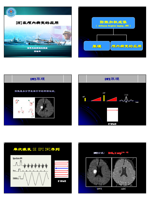
K-SPACE
DWI
ADC
表观弥散系数(ADC值)
反映水分子活动的自由度。单位mm2/s 如果需要作量化分析,则应测量ADC值。
-
=
B=1000
B=0
ADC
T2 shine through effect
DWI
弥散
T2
当受检组织的T2 值明显增高,在 DWI上有明显的T2 值图像对比存在
ADC=1.06×10⁻³
DWI在急性脑梗塞中的应用
早期诊断急性脑梗塞 鉴别新旧梗塞灶 评价缺血半暗带
一早期诊断急性脑梗塞
二鉴别新旧梗塞灶
ADC
EADC
CO中毒
三评价缺血半暗带
缺血缺氧性脑病
脱髓鞘病变
中枢神经系统淋巴瘤
恶性脑膜瘤
表皮样囊肿
可用于手术后复查
脑脓肿
鉴
别
脑
脓
肿
与
转
移
瘤
0.60×10⁻³mm2/s
脑脓肿 转移瘤
线粒体脑肌病
膜结构的阻挡1
A
B
膜结构的阻挡2
肿瘤细胞 水分子
恶性肿瘤
大分子蛋白物质的吸附作用
大大分分子子
水分子 水分子
脑脓肿、表皮样囊肿
EPI-DWI的伪影
膜结构的阻挡3
肿瘤细胞
恶性肿瘤
微血管内流动血液的影响
血管
炎细胞
水分子 炎性病变时,炎细胞对水分子的限制被血管内流动水分子的高弥散所抵消
磁敏感伪影
N/2鬼影
DWI在颅内病变的应用
海军总医院磁共振室 黄敏华
弥散加权成像
( Diffusion Weighted Imaging , DWI )
DWI在脑梗死中的应用
DWI 在脑梗死中的应用摘要:DWI在诊断脑梗死中发挥着重要的作用。
脑梗死时DWI信号改变是基于脑梗死后,脑细胞肿胀,导致细胞毒性水肿,弥散成像呈现为异常高信号影,称为弥散受限,提示新发性脑梗塞。
对超急性期和急性期脑梗死患者的确诊,其灵敏度明显高于头颅CT、T1WI和T2WI,最早在发病的2个小时便可以发现异常高信号——脑梗死病灶。
目前,基于这种超急性期和急性期脑梗死的诊断的灵敏度,DWI已经属于临床上不可缺少的手段,为临床的颅脑治疗和脑保护治疗提供了非常大的影像学依据。
关键词:DWI 脑梗死超级性期急性期脑梗死(Cerebral infarction,CI),又称为缺血性脑卒中(Cerebal ischemic stroke,CIS),是一种由于脑部血液循环不通畅,导致相应供血区域脑组织缺血缺氧而发生坏死或软化的疾病。
若大脑供血动脉中出现粥样硬化和(或)血栓栓塞致动脉形成狭窄和闭塞,其结果是急性局灶性脑功能不全[1]。
其患病率呈现逐年上升趋势,并逐渐年轻化。
根据流行病学统计显示,我国脑血管疾病的死亡率位居于世界第一[2],高于心脏系统疾病与恶性肿瘤,其中脑梗死死亡率占50%~60%,急性期病死率达到5%~15%,幸存者致残率高达50%,这严重影响了患者生活质量。
因此,早发现、早诊断、早治疗的“三早”原则对于提高疾病预后,改善病人生活质量显得尤为重要。
如何在脑缺血后和不可逆损伤发生前作出准确诊断是一个重要的研究课题[3]。
就脑梗死检查方法来讲,常规MRI在鉴别脑梗死是否为出血性方面具有重要意义。
若排除颅内出血,根据病灶所出现的位置确定其中风的亚型。
我们还可利用该预测中风的后果。
在CT和MRI上,脑梗死成像表现出典型地特征,在CT图像上,梗死区表现为与供血动脉相对应的低密度区;在MRI图像上,梗死区表现为长T1长T2,即在T1WI呈现低信号、T2WI呈现高信号,同时在FLAIR压脂图像上也呈现高信号[13]。
dwi医学名词解释

dwi医学名词解释
Dwi是医学上的缩写,代表"Diffusion Weighted Imaging",
即扩散加权成像。
在医学影像学中,DWI是一种利用水分子在组织
中的随机运动来生成图像的成像技术。
它通过测量水分子在组织中
的自由扩散,可以提供关于组织微结构和功能的信息。
DWI通常用
于检测和诊断中风、脑部肿瘤和其他神经系统疾病。
在临床实践中,DWI常常与MRI(磁共振成像)结合使用,可以提供高对比度和高分
辨率的图像,有助于医生进行准确诊断和治疗规划。
从技术角度来看,DWI利用了磁共振成像中的梯度脉冲序列,
通过测量水分子在梯度磁场中的运动来生成图像。
由于不同类型的
组织对水分子的扩散有不同的特征,DWI可以显示出组织的微观结
构和病变情况,对于早期发现病变和评估治疗效果具有重要意义。
此外,DWI还可以结合其他成像技术,如ADC(Apparent Diffusion Coefficient,表观扩散系数)成像,来提供更全面的信息。
ADC成像可以衡量组织中水分子扩散的速度和方向,从而进一
步帮助医生进行疾病诊断和评估。
总的来说,DWI作为一种重要的医学成像技术,对于神经系统
疾病的诊断和治疗起着至关重要的作用,它的应用不断拓展和深化,为临床医学带来了许多益处。
DWI的原理及临床应用
DWI的原理及临床应用1. DWI简介扩散加权成像(Diffusion-Weighted Imaging,简称DWI)是一种基于核磁共振成像(Magnetic Resonance Imaging,简称MRI)的技术,它通过测量水分子在组织中的自由扩散,提供了关于组织微结构的信息。
DWI在医学影像学领域具有广泛的临床应用。
2. DWI的原理DWI是基于水分子的自由扩散现象来获取影像信息的。
水分子在组织中的自由扩散受到许多因素影响,例如细胞膜的完整性、细胞分布密度以及细胞内外溶液的分子浓度等。
DWI使用一种特殊的梯度来限制水分子的自由扩散,从而使得在某个方向上的水分子质量浓度的变化能够更容易地被检测到。
通过对不同方向上的梯度进行扫描和测量,可以获得组织中水分子自由扩散的信息。
3. DWI的临床应用DWI在临床应用中具有广泛的用途,以下是一些常见的应用。
3.1 脑卒中和脑损伤评估DWI可以用来评估脑卒中和脑损伤患者的病情。
脑卒中后,受损的脑组织中的水分子的自由扩散会受到限制,导致DWI图像上的信号改变。
通过对DWI图像的分析,可以帮助医生判断脑卒中患者的病情严重程度以及影响范围。
3.2 肿瘤检测和分析DWI可以用于肿瘤的检测和分析。
肿瘤组织中的细胞密度常常较高,导致水分子的自由扩散受到限制。
因此,DWI可以准确地检测出肿瘤的存在,并提供关于肿瘤的信息,例如肿瘤的大小、位置和形态。
3.3 炎症和感染的评估DWI也可以用于炎症和感染的评估。
炎症和感染通常导致组织细胞密度的增加,从而限制水分子的自由扩散。
通过对DWI图像进行分析,可以检测出炎症和感染的存在,并提供有关病情的额外信息。
3.4 白质疾病的诊断DWI是评估白质疾病的一种重要工具。
白质疾病是指影响脑的白质部分的一类疾病,例如白质卒中和多发性硬化症。
通过检测和分析DWI图像,可以帮助医生判断白质疾病的类型和程度。
3.5 弥漫性疾病的检测DWI还可以用于检测一些弥漫性疾病,如弥漫性肝病和弥漫性肾病。
dwi基本原理及其在中枢神经系统中的应用

dwi基本原理及其在中枢神经系统中的应用
DWI(Diffusion weighted imaging)是一种MRI(Magnetic Resonance Imaging)技术,能够测量组织内水分子的自由扩散程度。
DWI原理基于布朗运动理论,即水分子在组织中不停地随机运动。
DWI采用梯度强度以及梯度方向不同来衡量水分子扩散方向和速度,这些信息被整合在一起形成图像,即DWI 图像。
DWI在中枢神经系统中的应用广泛,因为DWI可以反映大脑中白质和灰质的微结构和组织完整性。
白质病变、水肿和缺血性损伤等神经系统疾病都可以通过DWI检测到。
DWI对于急性缺血性脑卒中的早期诊断和治疗提供了重要的支持,因为发生脑卒中后,组织坏死开始导致扩散系数降低,DWI可以显示出白质区域的异常高信号或强度减低。
DWI还可以用于定位肿瘤和神经网络功能区域的准确识别,可以帮助医生提供更好的手术规划和处理。
dwi名词解释

dwi名词解释
DWI是磁共振检查中的一种特殊扫描序列,中文名称为弥散加权成像。
它利用正常组织和病理组织之间水扩散程度和方向的差别来成像,因此,DWI 可以用于区分正常组织和病变组织。
在临床应用中,DWI主要用于诊断急性脑梗死,其敏感性为94%,特异性为100%。
此外,DWI还可以用于鉴别蛛网膜囊肿与表皮样囊肿、硬膜下积脓与积液、脓肿与肿瘤坏死等。
在颅内其他病变如肿瘤、感染、外伤和脱髓鞘等的诊断、鉴别诊断和评价中,DWI也能提供有价值的信息。
以上内容仅供参考,建议咨询专业医生获取更准确的信息。
磁共振DWI在急性脑梗死中的应用
磁共振DWI在急性脑梗死中的应用摘要目的分析磁共振弥散加权成像(DWI)在急性脑梗死中的应用价值。
方法120例急性脑梗死患者作为研究对象,所有患者均经CT扫描、常规核磁共振成像(MRI)扫描及DWI扫描,回顾性分析患者的临床资料,就其检测结果展开分析。
结果经临床追踪随访及影像复查确诊,120例患者中,48例为超急性期脑梗死、60例为急性期脑梗死、12例为亚急性期脑梗死。
120例患者中共检出病灶144个,其中新发病灶105个,合并慢性期梗死病灶39个。
经CT扫描检出率为65.83%,MRI检出率为73.33%,DWI检出率99.17%,DWI 检出率显著高于CT扫描和MRI检查,差异有统计学意义(P<0.05)。
结论针对急性脑梗死患者可行磁共振DWI检查,能够准确的检出患者病灶情况,诊断患者疾病情况,值得临床推广。
关键词磁共振;弥散加权成像;急性脑梗死;检出率脑梗死包括脑血栓形成腔隙性梗死和脑栓塞等,是由于脑组织局部供血动脉血流突然减少导致该血管脑组织缺血、缺氧造成脑组织坏死软化,临床症状分为主观症状、脑神经症状及躯体症状。
CT和MRI检查是目前临床上用于诊断和判断脑梗死的主要手段,但针对急性期脑梗死同时合并有其他疾病的患者检测还存在一定的局限性。
因此,磁共振DWI就成为了人们关注的焦点[1]。
本次研究就本院收治的120例急性脑梗死患者作为研究对象,分别行CT扫描、MRI检查及DWI检查,就其诊断情况展开分析,现报告如下。
1 資料与方法1. 1 一般资料选取本院2015年5月~2016年2月收治的120例急性脑梗死患者作为研究对象,所有患者均经急诊、神经内科门诊检查,并符合血管性认知障碍诊断标准。
120例患者中男64例,女56例,年龄56~75岁,平均年龄(65.5±6.5)岁。
其中经临床追踪随访及影像复查确诊,120例患者中,48例为超急性期脑梗死、60例为急性期脑梗死、12例为亚急性期脑梗死。
DWI技术在脑部疾病诊断中的应用
DWI技术在脑部疾病诊断中的应用
刘怀祥;田宗皎
【期刊名称】《中国社区医师:综合版》
【年(卷),期】2012(28)15
【摘要】目的:探讨弥散加权磁共振成像(DWI)技术在脑部疾病的诊断中的应用价值.方法:采用DWI技术检查患者70例,进行回顾性分析,通过影像学资料对比探讨DWI在脑部疾病的诊断中的应用价值.结果:在对脑梗死的诊断中,DWI和FLAIR序列图像阳性检出率高,尤其是针对超急性期脑梗死的阳性检出率,DWI序列优势十分明显;针对胶质瘤水肿的诊断,DWI序列图像不如其他3种序列图像;对于脑出血的诊断,CT仍然是诊断脑出血的"金标准",DWI序列检查作为一种补充诊断.结论:DWI技术在脑病的诊断和鉴别方面,有着非常重要的临床价值.
【总页数】1页(P290-290)
【作者】刘怀祥;田宗皎
【作者单位】河南永城市永煤集团总医院,476600
【正文语种】中文
【中图分类】R743.33
【相关文献】
1.DWI技术在脑部疾病诊断中的应用 [J], 刘怀祥;田宗皎
2.脑部弥散张量成像数据处理技术在疾病诊断研究中的应用进展 [J], 张建梅;戴培山;李玲;盛韩伟;吴静
3.1H-MRS联合DWI在脑部星形细胞瘤诊断中的应用 [J], 王彦朋;张云萍;曹志勇
4.T2WI-FLAIR增强序列在脑部疾病诊断中的应用进展 [J], 苏梦瑶;周智鹏
5.快速FLAIR技术在脑部疾病诊断中的应用 [J], 王秀河;黄耀熊;黄力;刘斯润
因版权原因,仅展示原文概要,查看原文内容请购买。
最新DWI在脑部疾病诊断中的应用教学内容

扩散加权成像
(Diffusion Weighted Imaging,DWI)
目前在人体内进行水分子扩散测量与成 像的唯一影像学方法 DWI的出现是MRI发展的一个里程碑 在宏观成像中反映活体组织中水分子的 微观扩散运动
T2透过效应
表现为一些长T2信号的病灶在DWI图像上也表 现为高信号。
F/55Y,房颤患者,左侧脑室旁急性期梗塞
DWI鉴别新旧梗塞
M/61Y, 右侧肢体麻木无力9天 既往有脑梗塞病史
脑出血
MR信号强度随血红蛋白氧和状态改变 超急性期脑出血在常规T2WI表现为混杂高信号,
与脑梗塞难以鉴别 DWI上可见病灶边缘低信号灶,为特征性改变 高b值DWI对非血肿形式出血显示不佳
有研究报道血栓的DWI表现可提示预后 脑内静脉性梗塞为血管源性水肿,ADC值轻度
降低或升高
F/38Y,头疼15天
左侧横窦内血栓 形成
F/21Y, 右侧肢体 无力2天
脑静脉的闭塞产生局部脑水肿和静脉型梗死
肿瘤
胶质瘤 转移瘤 脑膜瘤 海绵状血管瘤 淋巴瘤 血管母细胞瘤 表皮样囊肿 蛛网膜囊肿
Ⅱ
级 星 形 细 胞 瘤
ADC值1.516×10⁻³mm2/s,DWI低信号, DWI未明确显示瘤周水肿
Ⅲ
级 星 形 细 胞 瘤
ADC值1.408×10⁻³mm2/s,DWI高信号, DWI模糊显示瘤周水肿
肝 癌 脑 转 移 瘤
ADC值1.105×10⁻³mm2/s,DWI略高信号,坏死区低信号, DWI未明确显示瘤周水肿
脓腔内脓液粘稠,水分子弥散明显受限 DWI帮助与肿瘤囊变坏死鉴别
脑脓肿
M/42Y
脑脓肿
磁共振dwi的原理及应用

磁共振DWI的原理及应用1. 介绍磁共振扩散加权成像(Diffusion-Weighted Imaging,DWI)是一种用于检测组织水分子运动状态的成像技术。
通过测量水分子在生物组织内的随机热运动,可以提供有关组织微结构及功能的信息。
本文将介绍磁共振DWI的原理及其在临床应用中的重要性。
2. 原理磁共振DWI的原理基于分子热运动对水分子的偏移造成的相位差异。
在常规磁共振成像中,脉冲序列通过对磁化强度和相位信息进行编码来生成图像。
而对于DWI,通过应用梯度场,在磁化感应的基础上加入梯度方向对水分子进行编码。
这样可以探测水分子在组织中的扩散运动。
3. 应用3.1 体内器官的病理检测•DWI可以用于检测与炎症相关的组织病理变化,如脑梗死、炎性肠病等。
通过检测组织的扩散系数,可以提供与病变强度和范围相关的信息。
•在肿瘤学中,DWI被广泛应用于检测肿瘤的早期诊断和治疗反应。
高度病态的组织通常会导致DWI成像中高信号区域的出现。
3.2 脑部疾病诊断•DWI广泛应用于脑部疾病的诊断,如脳梗死、脳炎等。
脑组织中的扩散系数变化可以提供关于缺血和细胞水肿的信息。
•在癫痫诊断中,DWI可以检测到癫痫灶附近的水肿,帮助确定病灶的位置和范围。
3.3 肝脏疾病诊断•DWI在肝脏疾病中的应用日益重要。
例如,肝癌和肝血供不良通常导致肝组织的扩散系数下降,可以通过DWI成像来检测和定量评估这些疾病。
3.4 心脏疾病的评估•DWI可用于评估心肌梗死区域的程度和扩散变化。
心肌梗死区域通常导致水分子的扩散减慢,可以通过DWI成像来定量评估。
3.5 肾脏疾病的评估•DWI可以用于评估肾脏疾病,如肾癌、肾血供不足和肾梗死等。
通过测量肾组织的扩散系数,可以提供关于肾功能和病理变化的定量信息。
4. 结论磁共振DWI作为一种非侵入性的成像技术,可以提供关于组织微结构和功能的有用信息。
其在医学诊断和临床应用中的重要性不断增加。
通过对DWI成像的分析和评估,可以帮助医生对疾病进行早期诊断、评估治疗反应以及指导治疗方案的制定。
DWI基本原理及其在脑部疾病中的应用【2024版】

SDW为DWI信号强度;r是质子密度;D是水分子的扩散系数
• DWI的信号强度主要取决于组织内水分子的扩散系数(D) 和T2以及扩散敏感梯度因子b值。
DWI图像的影响因素——b值
•
b值为0时,DWI图像近似T2加权图像。
•
较小的b值得到的图像信噪比较高,但对水分子扩散运动的检
测不明高,而且组织信号的衰减受其它运动的影响比较大,如组织血
流灌注造成的水分子运动等,这些运动模式相对水分子的扩散运动来
说要明显的多。
DWI图像的影响因素——b值
•
在b值较低时,由于受血流灌注等因素的影响,所测得的ADC
值偏高,而且b值越小,测得的ADC值越偏高。
运动伪影
磁敏感伪影
涡漩电流伪影
运动伪影 (癫痫发作)
成像序列:SE-EPI扩散加权成像
基于SE的EPI扩散成像是临床最常用、最 实用的扩散成像技术。 EPI技术是可缩短EPI的回波链并大大缩 短TE,从而可大大减轻磁敏感伪影。
成像序列:螺旋桨弥散加权成像(Propeller DWI)
多发性硬化
DWI高信号 ADC稍高信号
DWI图像的影响因素
• 在水分子扩散自由的区域,检测到的DWI信 号应为低信号,同时测量得到高的ADC值。 • 但在临床实践中,我们看到的DWI图像实际 上会受到组织T1、T2,甚至毛细血管灌注等多种 因素的影响,导致DWI图像并不表现为理想的情 况。
DWI图像的影响因素
ADC=ln(S2/S1)/(b1-b2)
• S1、S2 不同b值时的DWI信号强度
• exp
简述弥散加权成像技术的临床应用

简述弥散加权成像技术的临床应用
弥散加权成像(DWI)是一种基于磁共振成像(MRI)的技术,用于检测组织内水分子的扩散情况。
它在临床上有广泛的应用,包括但不限于以下几个方面:
1. 急性脑卒中的诊断:DWI 对急性脑卒中,尤其是急性脑梗死的诊断具有很高的敏感性和特异性。
在急性脑梗死发生后的数分钟到数小时内,DWI 上可出现高信号,而在常规 MRI 上可能没有明显的异常。
2. 肿瘤的诊断和鉴别诊断:DWI 可以帮助区分良性和恶性肿瘤,以及肿瘤的分级。
恶性肿瘤通常具有较高的细胞密度和较低的水分子扩
散,因此在 DWI 上呈现高信号。
3. 脓肿和炎症的诊断:脓肿和炎症组织由于细胞外水分增加,水分子扩散受限,在 DWI 上也表现为高信号。
4. 外伤性脑损伤的诊断:DWI 可以检测出脑挫裂伤、弥漫性轴索损伤等外伤性脑损伤引起的水分子扩散受限。
5. 神经系统变性疾病的诊断:某些神经系统变性疾病,如多发性硬化、肌萎缩侧索硬化等,可导致水分子扩散异常,DWI 有助于发现这些异常。
6. 腹部疾病的诊断:DWI 在肝脏、脾脏、胰腺等腹部器官的疾病诊断中也有一定的应用价值,可以帮助区分实性肿瘤和囊性肿瘤、脓肿等。
总之,DWI 作为一种无创性的影像学检查技术,在许多疾病的诊断、治疗监测和预后评估中都具有重要的临床应用价值。
头颅磁共振dwi高信号的解读

头颅磁共振dwi高信号的解读头颅磁共振扫描(DWI)是一种非侵入性的影像学检查,用于评估大脑和颅内结构。
DWI常用于检测颅内异常,如脑卒中、脑梗塞等。
头颅磁共振DWI显示出高信号区,可能意味着以下几种情况。
首先,高信号可以表示急性缺血。
脑组织中的神经元常常需要大量的能量来维持正常的功能,当血供中断或受限时,细胞会进入缺血状态,这可能导致细胞死亡和功能障碍。
头颅磁共振DWI可以显示出此类缺血区域,显示为高信号。
这种高信号区域通常伴随着相邻脑组织的低信号区域,称为脑卒中。
其次,高信号也可以表示脑异常增生。
某些情况下,脑组织的异常增殖可能导致头颅磁共振DWI的高信号区域。
这可能是由于肿瘤、炎症或其他脑内病变引起的。
这些高信号区域可能有不同的形状、大小和分布,具体取决于异常增生的性质和位置。
此外,高信号还可以代表其他病理变化。
在头颅磁共振DWI中,高信号可以出现在多发性硬化症、脑炎、脑血管病变等疾病中。
这些疾病通常具有不同的病理机制和临床表现,但它们在头颅磁共振DWI中都可能显示为高信号区域。
需要指出的是,头颅磁共振DWI的高信号区域并不总是代表病理变化。
有时,高信号可能是正常变异或扫描过程中的伪影。
因此,在解读头颅磁共振DWI中的高信号区域时,需要综合考虑患者的临床症状、其他影像学表现和实验室检查结果,以更准确地评估患者的病情。
总之,头颅磁共振DWI的高信号区域可能代表多种病理变化,包括急性缺血、脑异常增生和其他病理变化。
这些高信号区域的特征和分布可以提供有关患者病情和疾病类型的重要信息。
然而,在解读时需要综合其他临床资料,以获得更准确的诊断。
DWI基本原理及其在脑部疾病中的应用

当前存在问题和挑战剖析
图像分辨率与信噪比
当前DWI技术仍面临图像分辨率和信 噪比的挑战,尤其是在低场强MRI系
统中。
扫描时间与运动伪影
较长的扫描时间和头部运动可能导致 图像伪影,影响DWI图像的准确性和
可靠性。
标准化与可重复性
DWI技术的标准化和可重复性仍需进 一步提高,以便在不同中心和不同设
。
癫痫
02
DWI可用于检测癫痫患者脑内的异常放电区域,为手术治疗提
供定位依据。
帕金森病
03
DWI可用于评估帕金森病患者黑质-纹状体通路的受损情况,为
疾病诊断和治疗提供重要信息。
04
DWI技术进展与新兴应用
高分辨率DWI技术发展现状
高场强MRI技术
利用更高场强的MRI扫描仪,提 高DWI的空间分辨率和信噪比, 实现更精细的脑部结构成像。
DWI能够反映组织微观结构的改变, 特别是在脑部疾病中,如脑梗死、脑 肿瘤等,能够提供重要的诊断信息。
DWI信号产生与检测
DWI信号的产生依赖于水分子的扩散运动。在核磁共振成像 中,通过对组织施加特定的扩散敏感梯度,使得水分子的扩 散运动对信号产生影响。
检测DWI信号需要使用特定的脉冲序列和参数设置,以获取 扩散加权图像。常用的脉冲序列包括自旋回波序列和梯度回 波序列等。
扩散敏感梯度设置
扩散敏感梯度是DWI中的关键参数之一,用于测量水分子的扩散运动。通过设置 不同的扩散敏感梯度强度和持续时间,可以获取不同扩散加权程度的图像。
扩散敏感梯度的设置需要考虑到组织的特性和病变的特点,以达到最佳的成像效 果。
水分子扩散特性描述
DWI在急性缺血性脑卒中的临床

DWI检查结果与检查时间有关,发病后一定时间内才能检 测到梗死灶。
对新旧病灶区分困难
DWI难以区分新旧病灶,可能会影响对预后的准确判断。
对小型病灶敏感性较低
对于较小的梗死灶,DWI的检测效果可能不佳,导致漏诊 。
05
结论与展望
DWI在急性缺血性脑卒中临床应用的意义
早期诊断
DWI技术能够早期发现急性缺血性脑卒中,为患者提 供及时的治疗。
预后评估
DWI可以评估脑卒中病灶的大小和位置,预测患者的 预后情况。
治疗方案选择
根据DWI结果,医生可以制定更个性化的治疗方案, 提高治疗效果。
DWIБайду номын сангаас急性缺血性脑卒中临床应用的展望
技术改进
随着医学技术的不断发展,DWI成像质量将得到进一步提升,为临 床提供更准确的诊断信息。
联合其他影像技术
未来,DWI可以与其他影像技术如MRI、CT等结合使用,为急性缺 血性脑卒中提供更全面的诊断信息。
DWI在急性缺血性脑卒中治疗中的局限性
1
DWI对出血的敏感性较低,对于出血性脑卒中诊 断存在局限性。
2
DWI对某些部位如后循环缺血的诊断效果不佳。
3
DWI对早期脑梗死的诊断存在假阳性,需要结合 其他影像学检查进行确诊。
04
DWI在急性缺血性脑卒中预后评估中的应 用
急性缺血性脑卒中预后评估方法
01
临床普及
随着人们对DWI认识的深入,其在急性缺血性脑卒中的临床应用将得 到更广泛的普及和应用。
THANKS
感谢观看
DWI能够准确检测脑组织缺血程 度和梗死范围,为预后评估提供 重要依据。
预测功能恢复
dwi序列的原理及应用

DWI序列的原理及应用1. DWI序列简介DWI(Diffusion-Weighted Imaging)序列是一种采用磁共振成像(MRI)技术检测分子扩散的方法。
它利用水分子的扩散运动提供有关生物组织微观结构和组织区域功能活动的信息。
DWI序列可以通过测量水分子在组织中扩散的程度来定量评估组织的微观结构和水分子的流动状态。
2. DWI序列的原理DWI序列的原理是利用梯度磁场脉冲对水分子进行标记,通过测量该标记水分子在空间中的移动情况进行成像。
在DWI序列中,采用了一组梯度脉冲,将水分子沿不同方向推动,然后通过成像技术测量水分子的扩散运动。
根据不同的梯度方向,可以获取一系列的DWI图像。
3. DWI序列的应用DWI序列在医学影像学中有着广泛的应用。
以下是一些常见的应用领域:3.1 脑部成像DWI序列可用于评估脑部组织的健康状况。
通过测量水分子在脑组织中的扩散情况,可以检测到脑缺血、脑梗塞等疾病。
此外,DWI序列还可以用于评估肿瘤的侵袭性、脑肿瘤的诊断和治疗等。
3.2 肝脏成像DWI序列可以用于评估肝脏组织的健康状态。
由于肝脏组织中存在着各种病理变化,如肝癌、肝纤维化等,通过测量水分子在肝脏组织中的扩散情况,可以提供有关这些病理变化的信息。
利用DWI序列还可以评估肝脏移植术后的功能状态。
3.3 前列腺成像DWI序列在前列腺成像中也有重要的应用。
前列腺癌是男性常见的恶性肿瘤之一,采用DWI序列可以提供有关前列腺癌的定量信息,辅助医生进行诊断和治疗。
3.4 乳腺成像DWI序列在乳腺成像中的应用越来越受到重视。
乳腺癌是女性最常见的恶性肿瘤之一,利用DWI序列可以提供乳腺肿瘤的定量信息,有助于早期发现和诊断。
3.5 过程监控DWI序列广泛应用于过程监控领域。
例如,在肿瘤治疗过程中,可以通过DWI序列评估治疗效果;在脑卒中患者的治疗过程中,可以评估患者的神经恢复情况。
4. DWI序列的优势和局限性4.1 优势•DWI序列对于检测组织的微观结构和功能状态具有高度敏感性,并且成像速度快。
- 1、下载文档前请自行甄别文档内容的完整性,平台不提供额外的编辑、内容补充、找答案等附加服务。
- 2、"仅部分预览"的文档,不可在线预览部分如存在完整性等问题,可反馈申请退款(可完整预览的文档不适用该条件!)。
- 3、如文档侵犯您的权益,请联系客服反馈,我们会尽快为您处理(人工客服工作时间:9:00-18:30)。
Company Logo
contents
Infection Trauma Hemorrhage DWI Vasculitis and vasculopathies Leukodystrophies
Neoplastic lesions
Encephalopathies
Epilepsy
/moban
Company Logo
Introduction
Cytotoxic edema – characterized by abnormal cellular uptake of water and myelin edema – characterized by intramyelinic accumulation of vacuolated or free water – have high signal intensity on the diffusion trace, with decreased ADC as a result of isotropically restricted water diffusion. On the other hand,vasogenic edema, caused by increased permeability of the blood–brain barrier, and interstitial edema, caused by subependymal water diffusion in acute hydrocephalus have intermediate signal on the DW image with increased ADC DWI区分水肿的性质: 细胞毒性水肿和和髓鞘性水肿——由于同向性弥散受限,弥散相呈 高信号,ADC值下降; 血管源性水肿和间质性水肿——弥散不受限,弥散相呈中等信号, ADC值升高。
/moban
Company Logo
Introduction
Recently DWI has been applied to various other cerebral diseases. 现在DWI被应用在多种脑部疾病 In this paper,I demonstrate different DWI patterns of non-infarct lesions of the brain which are hyperintense in the diffusion trace image, such as infectious, neoplastic and demyelinating diseases, encephalopathies – including hypoxic–ischemic, hypertensive, eclamptic, toxic, metabolic and mitochondrial encephalopathies – leukodystrophies, vasculitis and vasculopathies, hemorrhage and trauma 本课件讲述不同弥散图像高信号的脑部非梗塞性疾病, 例如炎症、肿瘤、脱髓鞘疾病、脑病变包括缺血缺氧性、 高血压、子癫、中毒、代谢性和线粒体脑病-脑白质病 变,血管炎和血管病变、出血及外伤
Diffusion weighted MR imaging in non-infarct lesions of the brain DWI在非梗塞性脑病变中的应用
林亚南 2012-7-11
Introduction
Diffusion weighted imaging (DWI) is based on the sensitivity of MR to microscopic mobility of water molecules within tissues. DWI基于MR对组织内水分子微观运动的敏感性 DWI consists of a DW image and an apparent diffusion coefficient (ADC) map. DW image, together with qualitative and quantitative assessment of the ADC map has been widely used in the diagnosis of acute cerebral infarction, owing to the reliable distinction of cytotoxic and vasogenic edema. 由于DWI联合定性及定量的ADC图可以鉴别细胞毒性和血管源性 水肿,已广泛地应用在急性脑梗塞的诊断 Edema is a non-specific reaction of brain parenchyme to diffenernt factors ,which can be differentiated by DWI. DWI可鉴别不同因素导致的脑实质的非特异性反应--水肿
/moban
Company Logo
Introduction
Although DWI has been tried for different organ systems, it has been found its primary use in the central nervous system. The most widely used clinical application is in the detection of hyperacute infarcts and the differentiation of acute or subacute infarction from chronic infarction 尽管DWI已经应用在不同系统的器官,但是主 要应用在中枢神经系统。 临床最广泛的应用是诊断超急性脑梗塞和鉴别 急性或亚急性脑梗塞与陈旧脑梗塞
/moban
Company Lsimplex encephalitis: 30 years old, male. (a)shows a hyperintense lesion in the left temporal lobe. hipocampal and parahypocampal gyri, (b) the diffusion trace shows high signal with increased ADC (c). Single voxel proton MR spectroscopy shows decreased N-acetyl aspartate with a prominent lactate peak(d).
/moban
Company Logo
男性,65y
Fig.1.Cerebral absecess:65 years old ,male.A right temporal lobe absecess with a thick rim enhancing capsule,surrounded by massive edema.On the DWI,the lesion has high signal with partially deceased ADC.
/moban
Company Logo
单纯疱疹病毒性脑炎(Herpes simplex encephalitis) DW image shows high signal in the lesions with usually decreased ADC values representing cytotoxic edema and rarely higher ADC values representing vasogenic edema. 病变DWI表现为高信号,ADC值降低代表细胞 毒性水肿,少数高ADC值代表血管源性水肿 Areas of cytotoxic edema correspond to a worse outcome compared to areas of vasogenic edema 细胞毒性水肿与血管源性水肿相比,结局较差
/moban
Company Logo
克雅氏病 ( Creutzfeld-Jakob disease ) Creutzfeld-Jakob disease is one of several spongiform encephalopathies. Characteristic findings are rapidly progressive dementia, myoclonus and periodic sharpwave complexes on electroencephalography. 克雅病(皮质-纹状体-脊髓变性 ) 是几种海绵状脑病 之一。典型临床表现为:快速进展的痴呆、肌阵挛,脑 电图表现为周期性的锐波。 MR imaging is helpful in differentiating the two forms of the disease. 磁共振可以帮助鉴别克雅氏病的两种类型。
/moban
Company Logo
单纯疱疹病毒性脑炎(Herpes simplex encephalitis)
Herpes simplex encephalitis is one of the most common viral infections. T2-hyperintense lesions with typical temporal and frontal localization with petechial hemorrhage are characteristic findings. 单纯疱疹病毒性脑炎是最常见的病毒感染性疾病之一, T2高信号和典型的颞叶、额叶点状出血是其特征表现。 Conventional MR imaging and clinical findings might be non-specific, necessitating the proof of evidence of viral DNA by polymerase chain reaction in the cerebrospinal fluid. 常规MRI和临床表现可能是非特异性的,需要脑脊液中 病毒DNA的PCR试验来验证
