儿童的牙髓治疗
变异干髓术在乳牙牙病治疗中应用
变异干髓术在乳牙牙病治疗中的应用【摘要】目的探讨变异干髓术在儿童牙病治疗中的应用。
方法采用变异干髓术治疗200例患儿的乳牙牙髓根尖周病。
结果治疗急性牙髓坏炎成功率达95.7%,慢性牙髓炎91.8%,治疗急性根尖周炎成功率达91.4%,慢性根尖炎达82%。
结论采用变异干髓术治疗儿童乳牙牙髓根尖周病简单方便,切实可行。
【关键词】变异干髓术;乳磨牙;牙髓炎;根尖周炎文章编号:1004-7484(2013)-02-0684-01牙髓炎及根尖周炎是儿童乳牙的常见病。
由于儿童的特殊生理特点,理解能力差,配合程度差,所以治疗应力求简单有效,以达到消除感染和炎症的目的,扩大乳牙保留范围,尽力将换牙保存到替牙时期[1]。
我们选用变异干髓术治疗门诊一些不配和、无法完成根管治疗的患儿的乳磨牙牙髓炎及根尖周炎,取得了不错的临床效果。
具体报告如下:1 资料与方法1.1 一般资料共收治患者200人,年龄3-7岁。
患牙共265颗,其中上颌乳磨牙97颗,下颌乳磨牙168颗。
根尖炎患者85颗,其中急性根尖炎35颗,慢性根尖炎50颗;牙髓炎180颗,其中急性牙髓炎94颗,慢性牙髓炎86颗。
1.2 方法常规开髓,对疼痛剧烈的患儿可在局麻下开髓,或开髓后用蘸有表麻膏的小棉球放置髓室内做表面麻醉。
然后用挖匙除去冠髓,并尽量去除根管内的部分牙髓,3%双氧水和生理盐水交替反复冲洗髓腔,擦干窝洞后,隔离唾液,在髓腔内封入fc和碘仿糊剂,丁香油氧化锌糊剂封固。
1周后如无症状,取出暂封药物,将干髓剂覆盖在根髓口,压入根管内,垫底后永久充填。
1.3 疗效评价成功:治疗后无临床症状,乳牙功能良好,不影响恒牙胚的发育,乳牙根正常吸收。
失败:治疗后,出现根尖周脓肿,x片显示牙囊骨壁和恒牙胚受损,影响恒牙胚的发育,或乳牙牙根吸收过早。
2 结果(见表1)3.1 乳牙的健康对颌骨和牙弓的正常发育,恒牙的正常萌出和良好排列有着密切的关系。
乳牙牙髓病和根尖周病的治疗目的就是去除感染和慢性炎症,消除疼痛,恢复牙齿牙齿功能,保持乳牙列的完整,维持咀嚼功能,以便儿童健康成长。
儿童口腔医学—牙髓病和根尖周病讲义

儿童口腔医学—牙髓病和根尖周病讲义乳牙乳牙牙髓病和根尖周病的诊断方法疼痛疼痛是诊断牙髓病的重要症状之一,它包括激发痛和自发痛。
年龄较大的儿童、青少年临床可采用冰块测试,但对幼小儿童不宜采用。
幼小儿童询问患儿是否在玩耍、看书或睡觉时牙痛,以资鉴别。
乳牙牙髓病和根尖周病的疼痛表现悬殊较大,通常有疼痛历史的表明牙髓已有炎症或已经坏死肿胀肿胀是根尖周炎的一个主要特征。
乳牙牙髓组织疏松,血运丰富。
乳牙慢性牙槽脓肿往往由龈沟排脓,年轻恒牙也偶有龈沟排脓情况。
慢性根尖周脓肿或牙槽脓肿往往在患牙附近留有瘘管孔。
口外肿胀主要表现是颌面部蜂窝织炎。
单根乳牙引起肿胀或出现瘘管时,牙髓多完全坏死,单根年轻恒牙则可能残留部分活髓;多根乳牙和年轻恒牙可能出现某一或双根管牙髓已经坏死,而其他根管内仍可能为活髓或残留活髓。
叩痛和松动当乳牙牙髓炎、牙髓坏死的炎症感染影响到根尖周组织或牙周组织时,患牙可出现松动和叩痛。
牙髓活力测验乳牙和年轻恒牙的解剖结构,儿童神经发育,感知及言语能力的限制使得儿童不宜做温度和电活力测验。
X线检查X线检查是一项很重要的检查方法,对牙髓病和根尖周病的诊断和疗效的判断有重要意义。
在乳牙的X 线片中应注意观察:龋病的深度及与髓腔的关系。
髓腔内有无钙变和牙体内吸收。
根尖周围组织病变的状况和程度。
乳牙牙根是否出现生理性或病理性吸收。
恒牙胚发育状况包括恒牙胚发育程度、位置、牙胚外包绕的牙囊骨壁是否完整。
X线检查还可以显示治疗后根尖周组织愈合情况或牙髓治疗是否成功。
乳牙牙髓病乳牙牙髓病临床表现特点乳牙牙髓病包括牙髓炎症、牙髓坏死和牙髓变性。
乳牙牙髓病多由深龋感染引起,为龋病的并发症。
除龋病感染外,牙齿外伤也可引起。
乳牙牙髓病临床症状不明显,以慢性炎症为主,急性炎症往往是慢性炎症急性发作引起。
乳牙牙髓病治疗技术乳牙牙髓病治疗技术FC(戊二醛)断髓术:手术前准备常规治疗器械,甲醛甲酚或戊二醛制剂。
术前X线片了解根尖周组织及牙根吸收情况,牙根吸收1/2时不宜做活髓切断术。
乳牙牙髓病的治疗教材教学课件

选择合适根管器械和药物
根据乳牙根管特点选择合适的器械和 药物,避免过度预备和刺激。
注意根管充填时机和方法
根管充填应在炎症得到控制后进行, 选择合适的充填材料和方法,确保根 管严密封闭。
根管治疗后疼痛处理方法
轻度疼痛
可给予口服镇痛药物,如布洛芬 等,缓解疼痛症状。
中度疼痛
在口服镇痛药物的基础上,可加用 抗生素控制感染,如阿莫西林等。
如血小板源性生长因子、转化生长因子等,具有促进细胞增殖、分 化和组织修复的作用。
组织工程
利用生物材料、细胞和生长因子等构建人工牙髓组织,为乳牙牙髓病 治疗提供新的途径。
THANKS FOR WATCHING
感谢您的观看
激光治疗
激光治疗具有消炎、镇痛、促进组织 修复等作用,可作为乳牙牙髓病的辅 助治疗手段之一。
03 乳牙牙髓病并发症预防与 处理
根管治疗中并发症预防策略
熟练掌握根管解剖结构
熟悉乳牙根管系统的特点,避免器械 超出根尖孔或损伤恒牙胚。
严格无菌操作
根管治疗过程中应严格遵守无菌原则, 防止根管内感染扩散。
分类
根据病程和临床表现,乳牙牙髓 病可分为急性牙髓炎、慢性牙髓 炎、牙髓坏死等类型。
发病原因及危险因素
发病原因
乳牙牙髓病的主要发病原因是细菌感 染,通常是由于龋齿、牙外伤等导致 细菌侵入牙髓组织引起感染。
危险因素
口腔卫生不良、饮食习惯差、免疫力 低下等是乳牙牙髓病的危险因素。
临床表现与诊断依据
临床表现
闭合和根尖周组织愈合。
拔牙
对于无法保留的患牙,应及时拔 除,避免炎症扩散影响恒牙胚发 育。拔牙后应根据情况进行间隙
保持或正畸治疗。
04 儿童口腔保健与乳牙牙髓 病关系
生物陶瓷iRoot_BP_Plus在儿童牙外伤导致的牙露髓治疗中的应用效果

- 126 -用[J].中外医学研究,2022,20(23):30-33.[12]李克锋,陈静刚,杨景帆,等.踝关节骨折切开复位内固定术中修复断裂下胫腓前韧带的临床意义[J].中国骨与关节损伤杂志,2022,37(6):642-643.[13]王新标,翁荔芳,阮原芳,等.锚钉修复三角韧带联合切开复位内固定术治疗踝关节骨折合并三角韧带损伤患者的效果[J].中外医学研究,2023,21(18):1-5.[14]周金华,张文玺,刘国旗,等.韧带修复与拉力钉固定下胫腓联合损伤的比较[J].中国矫形外科杂志,2022,30(18):1716-1719.[15]林需枰,刘庆军,丁真奇,等.下胫腓螺钉固定联合下胫腓韧带修复治疗踝关节骨折合并下胫腓联合损伤的疗效[J].中华创伤杂志,2022,38(5):424-429.(收稿日期:2023-10-18) (本文编辑:程旭然)①漳州卫生职业学院附属口腔医院 福建 漳州 363000生物陶瓷iRoot BP Plus在儿童牙外伤导致的牙露髓治疗中的应用效果方雅君①【摘要】 目的:分析生物陶瓷iRoot BP Plus 在儿童牙外伤导致的牙露髓治疗中的应用效果。
方法:回顾性分析2020年12月—2022年12月于漳州卫生职业学院附属口腔医院就诊的70例牙外伤导致的牙露髓患儿的资料。
均应用活髓切断术治疗,根据术中应用盖髓剂的不同分对照组(应用氢氧化钙)、研究组(应用生物陶瓷iRoot BP Plus),各35例。
比较两组手术相关指标、牙齿功能及牙齿美观度、牙本质桥形成率及牙齿变色发生率。
结果:研究组手术时间、肿胀持续时间及疼痛持续时间均短于对照组,差异有统计学意义(P <0.05)。
术后6个月时,研究组固定功能、舒适功能、美观度评分高于对照组,差异有统计学意义(P <0.05);但两组语言功能及咀嚼功能评分比较,差异无统计学意义(P >0.05)。
术后3个月、6个月,研究组牙本质桥形成率高于对照组,牙齿变色发生率低于对照组,差异有统计学意义(P <0.05)。
Vitepex糊剂根管充填与空管疗法治疗乳牙牙髓炎的临床疗效比较
Vitepex糊剂根管充填与空管疗法治疗乳牙牙髓炎的临床疗效比较作者:刘文莉庞雪晶韩秀丽来源:《中国实用医药》2015年第03期【摘要】目的比较Vitepex糊剂根管充填与空管疗法治疗乳牙牙髓炎的临床疗效。
方法180例乳牙牙髓炎患儿随机分为根管充填组和空管疗法组,分别采用根管充填治疗和空管疗法治疗,并比较疗效。
结果根管充填组的总有效率为92.09%,空管疗法组的总有效率为93.33%,两组差异无统计学意义(P>0.05)。
结论根管填充和空管疗法治疗乳牙牙髓炎都能获得满意的疗效, Vitapex糊剂作为根管充填材料性能优越,应用范围广,操作便捷,是一种较理想的根充材料。
空管药物疗法操作简便,更容易被患儿接受,更适合于对儿童乳牙牙髓炎的临床治疗,值得应用推广。
【关键词】 Vitepex糊剂;根管充填;空管疗法;乳牙牙髓炎DOI:10.14163/ki.11-5547/r.2015.03.090目前,儿童龋病仍是影响儿童身体健康的重要疾病之一。
乳牙牙髓炎是儿童口腔科的多发病[1]。
根管治疗术是治疗该病最有效的方法之一,同时根管充填材料的不断进步使根管治疗术的成功率越来越高, Vitapex 糊剂是目前一种常用的根充糊剂,其疗效已得到充分肯定[2]。
本院2009年6月~2011年6月采用Vitepex 糊剂根管充填与空管疗法治疗乳牙牙髓炎,对两种疗法的临床疗效进行比较,现报告如下。
1 资料与方法1. 1 一般资料 180例患儿均为本院收治的乳牙牙髓炎患儿,共计259颗患牙;男82例,女98例;年龄3~16岁,平均年龄9.7岁;按照随机原则,设根管充填组90例139颗患牙和空管疗法组90例120颗患牙,两组患儿性别、年龄、病因及牙位分布比较经检验差异无统计学意义(P>0.05),具有可比性;所有患儿身体健康,治疗前拍摄牙片,无牙周或根尖周病变,无系统性疾病,患牙就诊前未经过治疗,依从性好。
1. 2 治疗方法 Vitapex根管充填材料由日本森田株式会社生产,根管长度测量仪为德国VDW GmbH公司生产,手用不锈钢K锉。
多次法根管治疗与一次性根管治疗儿童牙体牙髓病的临床疗效分析

DOI:10.16662/ki.1674-0742.2023.21.062多次法根管治疗与一次性根管治疗儿童牙体牙髓病的临床疗效分析姬莉1,周龙2,陈俊俊31.长春市口腔医院儿童口腔科,吉林长春130000;2.长春市口腔医院修复科,吉林长春130000;3.吉林大学第二医院药学部,吉林长春130000[摘要]目的研究多次法根管治疗和一次性根管治疗儿童牙体牙髓病的临床价值。
方法便利选取2021年2月—2022年1月长春市口腔医院收治的112例牙髓病患儿作为研究对象,采用随机分组法分为两组,每组56例。
分为一次性根管治疗的研究组和多次法根管治疗的参照组。
比较两组效果。
结果研究组总有效率为98.21%,高于参照组的78.57%,差异有统计学意义(χ2=10.529,P=0.001)。
研究组视觉模拟评分量表低于参照组,差异有统计学意义(P<0.05)。
研究组不良反应发生率低于参照组,差异有统计学意义(P<0.05)。
结论对于牙髓病患儿来说,多次法和一次性根管治疗都可改善患儿的症状,但后者的疗效更显著,可有效缓解患儿的疼痛。
[关键词]多次法根管治疗;一次性根管治疗;儿童;牙体牙髓病;疗效分析[中图分类号]R4 [文献标识码]A [文章编号]1674-0742(2023)07(c)-0062-04Clinical Analysis of Multiple Root Canal Treatment and One-time Root Canal Treatment in Children with EndodonticsJI Li1, ZHOU Long2, CHEN Junjun31.Department of Pediatric Stomatology, Changchun Stomatological Hospital, Changchun, Jilin Province, 130000 China;2.Department of Prosthetics, Changchun Stomatological Hospital, Changchun, Jilin Province, 130000 China;3.Depart⁃ment of Pharmacy, Jilin University Second Hospital, Changchun, Jilin Province, 130000 China[Abstract] Objective To study the clinical value of multiple root canal treatment and one-time root canal treatment in children with endodontics. Methods A total of 112 children with endodontic diseases admitted to Changchun Stomato⁃logical Hospital from February 2021 to January 2022 were conveniently selected as the study objects and divided into two groups by random grouping method, with 56 cases in each group. They were divided into the study group of one-time root canal therapy and the reference group of multiple root canal therapy. Compared the effects of the two groups.Results The total effective rate of the study group was 98.21%, higher than that of the reference group (78.57%), and the difference was statistically significant (χ2=10.529, P=0.001). The visual analog score scale of the study group was lower than that of the reference group, and the difference was statistically significant (P<0.05). The incidence of ad⁃verse reactions in the study group was significantly lower than that in the reference group, the difference was statisti⁃cally significant (P<0.05). Conclusion For children with endodontic disease, both multiple root canal treatment and one-time root canal treatment can improve the symptoms of children, but the latter is more effective and effective in relieving pain in patients.[Key words] Multiple root canal treatment; One-time root canal treatment; Children; Endodontics; Curative effect analysis[基金项目]吉林省卫生健康科技能力提升项目(2021LC025)。
乳牙深龋的间接牙髓治疗

乳牙深龋的间接牙髓治疗吴偲;刘映伶;邹静;周学东;郑黎薇【摘要】乳牙是人类的第一副牙齿,其正常萌出建并行使生理功能对儿童的身心发育具有重要意义.乳牙龋病是儿童慢性疾病之首,是儿童口腔医学临床最常见的疾病之一.根据世界卫生组织调查数据显示,世界范围内60%~90%的学龄儿童患有龋病.乳牙龋病在我国具有患龋率高,就诊率低下的特点,如不及时治疗,可导致牙体组织缺损、生理间隙丢失、牙髓和根尖周病变及颌面间隙感染,严重者可致乳牙早失并伴发牙列畸形及后续恒牙萌出障碍等不良结果,影响儿童口腔健康及身心发育.因此,对深龋乳牙采取积极有效的治疗措施对保存必要乳牙及其牙髓活力,恢复正常生理功能,维持牙列完整性,诱导后续恒牙正常萌出建具有重要意义.本文从目前深龋乳牙间接牙髓治疗的研究认识现状出发,通过文献资料收集整理,对间接牙髓治疗、间接盖髓术、暂时性保髓充填、部分去龋法、分步去龋法和非创伤性修复治疗等相关概念进行了对比分析,阐明了乳牙间接牙髓治疗的技术内涵和治疗意义,对乳牙深龋的临床治疗路径完善提供了理论依据.【期刊名称】《华西口腔医学杂志》【年(卷),期】2018(036)004【总页数】6页(P435-440)【关键词】乳牙深龋;间接牙髓治疗;间接盖髓术;暂时性保髓充填;非创伤性修复治疗【作者】吴偲;刘映伶;邹静;周学东;郑黎薇【作者单位】口腔疾病研究国家重点实验室国家口腔疾病临床医学研究中心四川大学华西口腔医院儿童口腔科,成都610041;口腔疾病研究国家重点实验室国家口腔疾病临床医学研究中心四川大学华西口腔医院儿童口腔科,成都610041;口腔疾病研究国家重点实验室国家口腔疾病临床医学研究中心四川大学华西口腔医院儿童口腔科,成都610041;口腔疾病研究国家重点实验室国家口腔疾病临床医学研究中心四川大学华西口腔医院牙体牙髓病科,成都610041;口腔疾病研究国家重点实验室国家口腔疾病临床医学研究中心四川大学华西口腔医院儿童口腔科,成都610041【正文语种】中文【中图分类】R781.05正常生理条件下,个体牙列发育要经历无牙列期、乳牙列期、混合牙列期和恒牙列期四个阶段。
奥硝唑合剂联合一次性根管填充治疗儿童牙体牙髓病的效果观察

奥硝唑合剂联合一次性根管填充治疗儿童牙体牙髓病的效果观察发布时间:2022-01-05T08:56:32.657Z 来源:《医师在线》2021年10月19期作者:徐晴琳[导读]徐晴琳(成都是青羊厚济医院有限责任公司;四川成都610000)【摘要】目的:探究对于儿童牙体牙髓病患儿,选择奥硝唑合剂联合一次性根管填充治疗的效果。
方法:随机抽选在我院收治的儿童牙体牙髓病患儿(n=60),分别予以一次性根管填充治疗,以及奥硝唑合剂联合一次性根管填充治疗,前者纳为对照组,后者纳为观察组,每组30例,收治时间为2020.1-2021.1。
将2组的治疗效果、并发症、咬合力以及咀嚼功能情况进行对照。
结果:在治疗总有效率上,观察组(96.67%)较高于对照组(86.67%)(P<0.05);在并发症总发生率上,观察组(3.33%)较低于对照组(16.67%)(P<0.05);治疗前,两组患儿的咬合力以及咀嚼功能没有明显差异(P>0.05),治疗后与对照组相比,观察组患儿的咬合力以及咀嚼功能明显提升(P<0.05)。
结论:对于儿童牙体牙髓病患儿,在一次性根管填充治疗基础上,联合奥硝唑合剂有助于提升治疗效果,并减少不良反应,预后效果较为明显。
【关键词】儿童;牙体牙髓病;奥硝唑合剂;一次性根管填充治疗急慢性牙髓炎和龋齿是最常见的牙髓疾病,由于儿童口腔尚处于发育阶段,对外界因素的抵抗力不强,由于细菌刺激,牙髓疾病的发病率逐渐增加。
它不仅影响牙齿的美观,而且并发症也会影响口腔健康[1-2]。
一次性根管充填治疗具有方便快捷的优点,但消毒不彻底易影响疗效。
奥硝唑属于第三代硝基咪唑衍生物,其主要作用是抑制口腔厌氧菌,因此具有很好的杀菌作用。
为了探究对于儿童牙体牙髓病患儿,选择奥硝唑合剂联合一次性根管填充治疗的效果,本研究随机抽选在我院收治的儿童牙体牙髓病患儿(n=60),现报告如下。
1.资料与方法1.1一般资料随机抽选在我院收治的儿童牙体牙髓病患儿(n=60),分别予以一次性根管填充治疗,以及奥硝唑合剂联合一次性根管填充治疗,前者纳为对照组,后者纳为观察组,每组30例,收治时间为2020.1-2021.1。
儿童牙髓炎治疗方法
儿童牙髓炎治疗方法
儿童牙髓炎是指牙髓组织发生炎症,引起牙髓腔内疼痛和感染。
以下是几种常见的儿童牙髓炎治疗方法:
1.根管治疗:这是治疗牙髓炎最常见的方法。
医生会使用特殊的仪器和工具清除受感染的牙髓组织,并清洗和消毒髓腔,然后填充和封闭根管。
2.药物治疗:医生可能会开出药物来缓解疼痛和消炎。
例如,消炎药、口服抗生素等可以帮助控制感染和减轻症状。
3.牙齿拔除:在一些严重的病例中,如果根管治疗无法解决问题,医生可能会建议拔除受感染的牙齿。
这适用于那些不能进行根管治疗的情况,例如牙齿根部骨质破坏或者牙齿无法恢复功能。
4.根尖手术:在某些情况下,根管治疗后仍然存在问题,如根管感染无法解除或出现根尖囊肿等。
医生可能会选择进行根尖手术,即手术切除牙根尖部的病变组织。
需要注意的是,儿童牙髓炎的治疗应该由专业的口腔医生来进行,并且家长和孩子应积极配合治疗过程中的注意事项和口腔卫生保护。
及时治疗可以避免牙齿感染扩散和其他并发症的发生。
教案 儿童牙髓病和根尖病

教案:儿童牙髓病和根尖病第一章:儿童牙髓病和根尖病的概述教学目标:1. 了解儿童牙髓病和根尖病的定义和特点。
2. 掌握儿童牙髓病和根尖病的发生原因和临床表现。
3. 理解儿童牙髓病和根尖病的诊断和治疗方法。
教学内容:1. 儿童牙髓病和根尖病的定义和特点解释儿童牙髓病和根尖病的概念介绍儿童牙髓病和根尖病的发生率和发展趋势2. 儿童牙髓病和根尖病的发生原因讲解儿童牙髓病和根尖病的病因和发病机制分析儿童牙髓病和根尖病的危险因素3. 儿童牙髓病和根尖病的临床表现描述儿童牙髓病和根尖病的典型临床症状介绍儿童牙髓病和根尖病的临床检查方法第二章:儿童牙髓病和根尖病的诊断教学目标:1. 掌握儿童牙髓病和根尖病的诊断方法。
2. 学会评估儿童牙髓病和根尖病的严重程度。
3. 理解儿童牙髓病和根尖病的鉴别诊断。
教学内容:1. 儿童牙髓病和根尖病的诊断方法介绍儿童牙髓病和根尖病的常用诊断方法讲解儿童牙髓病和根尖病的诊断流程2. 评估儿童牙髓病和根尖病的严重程度介绍评估儿童牙髓病和根尖病严重程度的指标和方法讲解儿童牙髓病和根尖病的分级和分期3. 鉴别诊断儿童牙髓病和根尖病介绍鉴别诊断儿童牙髓病和根尖病的相关疾病讲解鉴别诊断的步骤和注意事项第三章:儿童牙髓病和根尖病的治疗方法教学目标:1. 掌握儿童牙髓病和根尖病的治疗方法。
2. 学会制定儿童牙髓病和根尖病的治疗计划。
3. 理解儿童牙髓病和根尖病的术后注意事项。
教学内容:1. 儿童牙髓病和根尖病的治疗方法介绍儿童牙髓病和根尖病的保守治疗和手术治疗讲解各种治疗方法的适应症和禁忌症2. 制定儿童牙髓病和根尖病的治疗计划介绍制定治疗计划的依据和原则讲解治疗计划的实施步骤和注意事项3. 儿童牙髓病和根尖病的术后注意事项介绍术后注意事项的重要性讲解术后护理和复查的方法和时间第四章:儿童牙髓病和根尖病的预防措施教学目标:1. 了解儿童牙髓病和根尖病的预防措施。
2. 学会实施儿童牙髓病和根尖病的预防策略。
复方阿替卡因在儿童牙体牙髓无痛治疗中的效果探讨
•296•云南医药2020年第41卷第3期•基层医师园地・复方阿替卡因在儿童牙体牙髓无痛治疗中的效果探讨邹美玲(河南省职工医院口腔科,河南郑州450000)关键词:复方阿替卡因;牙体牙髓;无痛治疗;利多卡因中图分类号:R781.31文献标志码:B文章编号:1006-4141(2020)03-0296-02儿童牙体牙髓病为一种儿科常见口腔疾病,若治疗不及时,可能会影响患儿的生存质量。
婴幼儿时期为牙齿的重要发育阶段,受口腔不良习惯、牙外伤、頻齿等导致的乳牙替换异常等,导致婴幼儿时期牙科疾病高发,若治疗不及时,不仅会影响牙齿健康,还会影响儿童的健康成长发育,甚至并发全身疾病,因此临床应提高儿童牙科疾病治疗的重视叫临床治疗过程中,考虑到儿童自身耐受性差,不耐受疼痛,临床需实施无痛麻醉治疗。
利多卡因为临床常用的口腔疾病治疗药物,复方阿替卡因为一种口腔科专用麻醉药物,关于这两种麻醉药物的具体应用效果,临床缺乏相关方面的研究。
本次研究中,临床提出给予儿童牙体牙髓麻醉中分别应用利多卡因、复方阿替卡因注射治疗,对比不同麻醉用药治疗效果,现报告如下。
资料与方法一、一般资料自医院从2017年9月-2019年2月收治的儿童牙体牙髓患儿中抽选100例进行研究,患儿无严重的精神疾病、脏器功能障碍、本次研究麻醉用药禁忌症。
随机分为2组,对照组50例,男30例,女20例,年龄为5~12岁,平均年龄为(9.0±2.2)岁,观察组50例,男25例,女25例,年龄为4~13岁,平均年龄为(8.9±2.0)岁。
2组统计资料经统计学分析,组间并无显著差异(P>0.05),可进行研究。
二、方法2组患儿入院后,先接受常规检査。
对照组麻醉用利多卡因浸润麻醉,具体注射方法:前牙注射药物剂量2mL,后牙注射药物剂量3mL。
观察组注射用复方阿替卡因,实施浸润麻醉,根据患儿体重确定具体用药剂量,注射剂量为患儿体重(kg)xl.33倍,注射剂量单位为毫克。
华北理工儿童口腔医学课件05儿童牙髓病与根尖周病

• 由于乳磨牙根分叉的结构特点牙髓
感染易通过该处扩散,因此乳磨牙 尖周炎症多发生于根分叉的根周组 织内,表明绝大多数尖周病变与乳 磨牙的髓底解剖结构有关。
• 乳牙与恒牙胚关系 密切,乳牙
外伤或牙髓、尖周组织感染都有 可能导致恒牙胚的损害。如出现 矿化、发育不全或形成异常等现 象。
二、 检查和诊断方法 (一) 检查方法:
儿童牙髓病与根尖周病
定义
• 牙髓和根尖周病 是发生在牙髓组织和根尖周
膜及牙槽骨的疾病的总称,牙髓和根尖周病与龋
病一道被称为牙体牙髓病
硬组织特点
乳牙的牙髓腔形态和牙齿外形一致,与恒牙相比有如下特点:
1. 乳牙硬组织薄,钙化度低,尤其在牙颈 部,牙本质小管粗大,渗透性强。故牙 髓易受外界细菌侵犯,临床上闭锁性牙
常规检查:问诊、望诊、扪诊、 探诊、叩诊、冷热诊
特殊检查:牙髓活力测验、x线片等
牙髓病、尖周病 检查、诊断的仪器 这是目前牙髓学的薄弱环节,缺乏反映客观
检查情况的仪器,绝大多数都是根据患者的反应
电活力试验器:主要还是判断牙髓是否有活 力,据报道准确率约85%。
第一节 诊断方法
一、收集病史
疼痛:是牙髓炎、根尖周炎的突出症状和重要临
根尖炎时可为活髓。
5.根和根管的数目。
乳牙根和根管
乳 前 牙:单根—单根管 上乳磨牙:三根—三根管
下第一乳磨牙:二根
2根管型 3根管型* 4根管型
下第二乳磨牙:二根
3根管型 4根管型*
• 乳磨牙髓室底薄、侧副根管较多,与牙
周膜相通,往往牙髓尚未坏死,炎症则 已扩散到根分叉处的牙周组织而形成脓 肿,一旦发生脓肿,瘘管也多位于根分 叉的牙龈处。
炎,慢性根尖周炎或牙槽脓肿往往在患牙附近留有瘘 道孔。
一次性乳牙根管治疗术治疗乳牙牙髓病及根尖周病疗效观察
一次性乳牙根管治疗术治疗乳牙牙髓病及根尖周病疗效观察李俊玲;刘伟;纪彩娜【摘要】Objective To study the short-term efficacy and pain of primary root canal therapy of deciduous teeth caries and periapical diseases, so as to provide effective treatment for root canal therapy of dental deciduous teeth. Methods 145 patients in our hospital from April 2015 to April 2016 were studied. according to the random number method, the experimental group (n.= 75 cases) and control group (n = 70 cases) were divided into two groups. Results The experimental group ( 50.67%, 93.33%) and control group Pain ratios ( 64.29% ), the treatment success rate ( 77.14% ) was significantly different, and the incidence of adverse reaction in experimental group(5.71%_was significantly lower than the control group ( 22.86% ), the difference was statistically significant (P<0.05 ). conclusion The use of disposable dental root canal therapy in the treatment of deciduous teeth caries and periapical periodontitis can effectively improve the clinical therapeutic effect and improve the incidence of short-term pain in patients, it is worthy of clinical application.%目的:探讨一次性乳牙根管治疗术治疗乳牙牙髓病及根尖周病的近期疗效,为牙科乳牙根管治疗提供行之有效的治疗方案.方法:收集2015年4月-2016年4月在笔者科室接受治疗的145例患者,按照随机数字表法分为实验组与对照组,对照组:75例,行常规根管治疗;实验组:70例,采用一次性根管治疗,对比两组患者术后近期效果及疼痛发生率.结果:两组疼痛率比较,实验组(49.33%)与对照组(64.29%)存在显著性差异;两组治疗成功率(93.33%vs 77.14%)比较存在显著性差异,且实验组(5.71%)不良反应发生率明显低于对照组(22.86%),差异具有统计学意义(P<0.05).结论:采用一次性乳牙根管治疗术治疗乳牙牙髓病及根尖周病患者,能有效提升临床治疗效果,改善患者近期内疼痛发生率,值得在临床上推广使用.【期刊名称】《中国美容医学》【年(卷),期】2017(026)005【总页数】3页(P60-62)【关键词】乳牙牙髓病及根尖周病;一次性乳牙根管治疗术;近期效果;疼痛率【作者】李俊玲;刘伟;纪彩娜【作者单位】青岛市中心医院口腔科山东青岛 266042;青岛市中心医院口腔科山东青岛 266042;青岛市肿瘤医院口腔科山东青岛 266042【正文语种】中文【中图分类】R788+.3乳牙牙髓病及根尖周病是儿童最常见的乳牙疾病,也是导致乳牙早失的关键,乳牙早失会影响牙列完整、乳恒牙正常功能发挥情况。
牙髓切断术与牙髓摘除术在无症状深龋露髓乳牙治疗中的效果分析

牙髓切断术与牙髓摘除术在无症状深龋露髓乳牙治疗中的效果分析摘要:目的:分析牙髓切断术与牙髓摘除术在无症状深龋露髓乳牙治疗中的应用效果。
方法:筛选本院于2020.01-2021.11收治100例无症状深龋露髓乳牙患儿作为研究对象,依据治疗方法不同将纳入患儿分为两组,单组例数50,组别设置对比组(牙髓摘除术)、研究组(牙髓切断术),比较两组临床有效率、不良反应发生率。
结果:研究组临床总有效率(92.00%)显著较对比组(76.00%)更高(P<0.05)。
研究组持续性疼痛、牙周肿胀、瘘管形成、牙尖周和根分叉病变以及根管内外吸收等不良反应总发生率(8.00%)明显较对比组(26.00%)更低(P<0.05)。
结论:牙髓切断术与牙髓摘除术在无症状深龋露髓乳牙治疗中均可取得一定效果,与牙髓摘除术比较,牙髓切断术疗效、安全性更为理想,值得临床借鉴与推广,但是,在实际治疗期间应结合患儿具体情况,予以全面、综合分析后选择最佳牙髓治疗方法。
关键词:深龋;乳牙;牙髓摘除术;牙髓切断术;临床效果龋损是口腔常见、多发疾病,指在口腔内多种因素复合作用所致的牙体硬组织损害性疾病,且呈进行性进展状态[1]。
深龋通常是龋损进展至牙本质深层时的病损,一旦发生,患儿对冷热、酸甜等刺激可产生强烈疼痛反应,当刺激去除,症状即可消失[2]。
但是,由于小儿其神经功能等正处于发育阶段,加之受乳牙解剖生理特点,部分情况下,即使发生深龋,患儿自觉症状也不是非常明显[3]。
同时,相关研究指出,在龋损乳牙治疗过程中,约有40%左右患儿患牙会出现露髓现象,若未能及时进行有效治疗,可对患儿健康发育与成长造成不良影响,因此,探寻高效、安全治疗方案十分重要[4]。
本次研究筛选本院于2020.01-2021.11收治100例无症状深龋露髓乳牙患儿,观察评估牙髓切断术与牙髓摘除术治疗效果,旨在为临床治疗无症状深龋露髓乳牙提供参考与指导。
1资料与方法1.1一般资料筛选本院于2020.01-2021.11收治100例无症状深龋露髓乳牙患儿作为研究对象,依据治疗方法不同将纳入患儿分为两组,单组例数50,组别设置对比组:男26例、女24例,年龄最低3岁、最高9岁,均值(5.59±1.32)岁;研究组:男27例、女23例,年龄最低3岁、最高8岁,均值(5.46±1.28)岁;两组一般资料比较(P>0.05),存在可比性。
第七章 儿童口腔常见疾病护理技术(2)(1)
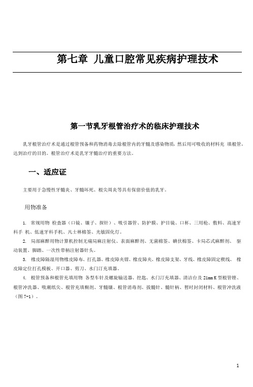
第七章儿童口腔常见疾病护理技术第一节乳牙根管治疗术的临床护理技术乳牙根管治疗术是通过根管预备和药物消毒去除根管内的牙髓及感染物质,然后用可吸收的材料充填根管,达到治疗的目的。
根管治疗术是乳牙牙髓治疗的重要方法。
一、适应证主要用于急慢性牙髓炎、牙髓坏死、根尖周炎等具有保留价值的乳牙。
用物准备1.常规用物检査器(口镜、镶子、探针)、吸引器管、防护膜、护目镜、口杯、三用枪、敷料、高速牙科手机、低速牙科手机、凡士林棉签、光敏固化灯。
2.局部麻醉用物计算机控制无痛局麻注射仪、表面麻醉剂、无菌棉签、碘伏棉签、卡局芯式麻醉剂、驱动装置、脚踏、一次性带柄注射器针头。
3.橡皮障隔湿用物橡皮障布、打孔器、橡皮障夹钳、橡皮障夹、橡皮障支架、牙线、橡皮障固定楔线、橡皮障定位打孔模板、开口器、剪刀、水门汀充填器。
4.根管预备和根管充填用物各型车针及螺旋输送器、挖匙、水门汀充填器、清洁台及21mm K型根管铿、根管冲洗器、吸潮纸尖、根管充填糊剂、牙髓镶、根管消毒剂、拔髓针、髓针柄、暂时封闭材料、根管冲洗液(图7-1)。
图7-1根管预备和根管充填用物①各型车针及螺旋输送器;②挖匙;③水门汀充填器;④清洁台及21mm K型根管挫;⑤根管冲洗器;⑥吸潮纸尖;⑦根管充填糊剂;⑧牙髓镶;⑨根管消毒剂;⑩拔髓针;⑪髓针柄; ⑫暂时封闭材料;⑬根管冲洗液5.垫底及充填用物水门汀充填器、树脂雕刻刀、树脂压光器、酸蚀剂、粘接剂、小棉棒、光固化复合树脂、咬合纸、调拌刀、调拌板、成形片、金钢砂车针、玻璃离子水门汀粉和液(图7-2)o图7-2垫底及充填用物①水门汀充填器;②树脂雕刻刀;③树脂压光器;④酸蚀剂;⑤粘接剂;⑥小棉棒;⑦光固化复合树脂;⑧咬合纸;⑨调拌刀;⑩调拌板;⑪成形片;⑫金刚砂车针;⑬玻璃离子水门汀粉和液三、乳牙根管治疗术医护配合流程(表7-1)医生操作流程护士配合流程1.治疗前准备(1)做好患儿的心理护理引导患儿坐于综合治疗椅上,向患儿及家长讲解治疗的主要过程,减轻患(2)局部麻醉儿的焦虑情绪用凡士林棉签润滑口角,防止口镜牵拉造成患儿痛苦(3)橡皮障隔湿安装局部麻醉药物到注射仪上,递碘伏棉签予医生,注射时双手协助固定患儿头部,避免突然摆动造成误伤按照常规安装橡皮障系统2.去净腐质,制备洞形,揭净髓室顶安装牙科手机及车针,去腐时协助吸唾,强力吸引器管置于患牙旁,保持3.探查根管数目,拔髓术野清晰,随时调整灯光,用挖匙去除腐质时及时用棉球清除上面残留物质,保持器械清洁(图7-3)递探针予医生- 表7-1乳牙根管治疗术医护配合流程6 .7 .续表医生操作流程护士配合流程根管消毒封药,放置暂时封闭材料取适量根管消毒剂递予医生,安装螺旋输送器。
儿童牙髓治疗

Pulpotomy in Primary Teeth 适应症: 乳牙的各种早期牙髓炎,感染仅限于冠髓。Coronal Pulpitis
具体临床情况: a.无自发痛史,临床检查无松动、叩痛、牙龈无红肿 和瘘管
Pulpotomy in Primary Teeth
禁忌症: 牙髓感染不仅限于冠髓Coronal Pulp,已侵犯根髓Radicular Pulp,形成牙髓弥漫性炎症,
乳牙的牙髓状态判断
Pulpitis的特点
早期症状不明显,疼痛史的有无不能作为乳牙牙髓炎的 绝对诊断标准。 一旦出现自发痛,可说明牙髓有广泛的炎症,甚至牙髓坏死;无自发痛史不能说明牙髓无炎症存在。 乳牙牙髓炎多为慢性过程,出现急性症状时,常为慢性炎症急性发作。 X线片上应无病变
Periapical Periodontitis的特点
戊 二 醛冠髓切断术 Pulpotomy using Glutaraldelyde
乳牙的牙髓治疗
Glutaraldelyde的特点:
1.
对组织的作用是有自限性的,其分子不渗透出根尖孔,而FC中的甲醛分子可透过根尖孔;
2.
对组织蛋白的凝固性坏死是不可逆的,且立即固定,而FC所需时间长,且是可逆的过程;
乳牙根管治疗术 Root Canal Treatment
适应症: 各型急慢性牙髓炎、根尖炎和牙髓坏死
乳牙根管治疗术 Root Canal Treatment
禁忌症: 1. 根吸收在三分之一以上; 2. 根尖周广泛的病变,或病变波及恒牙胚; 3. 髓底较大的穿孔;牙源性囊肿和滤泡囊肿的存在; 4. 根管弯曲不通。
髓可为活髓。即:乳牙根尖炎时可为活髓。
5.
根和根管的数目。
乳牙根和根管
液氮冷冻疗法在儿童牙髓治疗中的临床应用价值分析

液氮冷冻疗法在儿童牙髓治疗中的临床应用价值分析摘要:目的分析在儿童牙髓治疗中应用液氮冷冻疗法的应用价值。
方法抽选76例于2021年8月至2022年5月来我院接受未穿隧的龋源性急性牙髓炎治疗的儿童资料,利用奇偶法随机均匀分成参照组与研究组,前者接受常规麻醉,后者接受液氮冷冻疗法,汇总并计算镇痛效果,评估患儿焦虑抑郁情绪。
结果观察组患儿镇痛有效率高达94.73%显著高于参照组的78.94%,差异符合统计学原理(P<0.05);治疗前,两组患儿SAS评分与SDS评分无差异(P>0.05);治疗后,观察组患儿SAS评分与SDS评分显著低于参照组(P<0.05)。
结论在儿童牙髓治疗中应用液氮冷冻疗法有助于提高镇痛效果,消除患儿内心恐惧,为后续诊疗指明了方向。
关键词:液氮冷冻疗法;儿童牙髓治疗;应用价值;分析急性牙髓炎是口腔科诊疗中常见病症,具有疼痛剧烈等特点,若不及时治疗容易影响患儿正常生活。
儿童属于临床诊疗特殊群体,具有灵活好动、恐惧心理强等特点,在常规治疗下产生的疼痛会加剧患儿不适,配合度更低,如何提高患儿配合度成为牙科诊疗关注焦点【1】。
目前,临床在急性牙髓炎治疗时,常给予麻醉治疗,但效果偏差,为解决该问题,更多医院引进液氮冷冻疗法来提高患儿依从性,本文将围绕76例患儿展开研究,现报道如下。
1 资料与方法1.1一般资料抽选76例于2021年8月至2022年5月来我院接受未穿隧的龋源性急性牙髓炎治疗的儿童资料,利用奇偶法随机均匀分成参照组与研究组,每组各38例,参照组患儿男女比例为20:18,年龄在5~10岁之间,平均年龄(7.25±0.82)岁;研究组患儿男女比例为19:19,年龄在6~9岁之间,平均年龄(7.33±0.90)岁,两组患儿拥有相似的临床资料(P>0.05),具有可比性。
医护人员已阐述研究意义,家属同意并签署知情同意书;伦理委员会已批准此次研究。
纳入标准:依从性良好;临床资料完整;符合乳牙急性牙髓炎诊断标准;年龄低于14岁。
- 1、下载文档前请自行甄别文档内容的完整性,平台不提供额外的编辑、内容补充、找答案等附加服务。
- 2、"仅部分预览"的文档,不可在线预览部分如存在完整性等问题,可反馈申请退款(可完整预览的文档不适用该条件!)。
- 3、如文档侵犯您的权益,请联系客服反馈,我们会尽快为您处理(人工客服工作时间:9:00-18:30)。
乳牙的牙髓治疗乳牙的解剖生理特点一、乳牙硬组织的特点二、乳牙牙髓组织的特点。
三、乳牙牙根及根周组织的特点四、乳牙牙根的生理性吸收五、乳牙与恒牙胚的关系Goal of the pulp therapy in the primary and mixed dentitions1.successful treatment of the pulpally involved primary tooth, allowing the tooth to remain in a non-pathologic state.2.maintenance of arch length and tooth space;3.restoration of comfort with the ability to chew;4.prevention of speech abnormalities and abnormal habits.检查和诊断一、疼痛史(history of pain) 二、露髓和出血(pulpal exposure and hemorrhage)三、肿胀和瘘管( abscess and fistula) 四、叩痛和松动 (percussion test and mobility)五、牙髓敏感测试( vitality tests) 六、X线检查 (radiographic interpretation)乳牙牙髓治疗方法保存全部活髓的方法:盖髓术保存部分牙髓的方法:牙髓切断术保存患牙的方法:牙髓摘除术(根管治疗术)Indications for primary pulp therapyFurther to the signs and symptoms, additional indications for pulp therapy are:▪cooperative child and carers.▪avoidance of the psychological trauma of extraction.▪absence of the permanent successor tooth.▪to maintain an intact arch in the primary dentitions.一、盖髓术(pulp capping)定义:是一种用药物覆盖于近髓的牙本质上或露髓的牙髓创面上,使牙髓病变得以恢复并保存全部生活牙髓的治疗方法。
间接盖髓术(indirect pulp capping): 直接盖髓术(direct pulp capping)间接盖髓术:适应证:深龋近髓或外伤牙冠折断近髓无明显牙髓炎症状的患牙,或症状轻微的轻度牙髓充血患牙。
治疗步骤:去龋、制洞盖髓充填修复注意事项:1、治疗前正确判断牙髓状况。
2、遵循去龋制洞原则,操作轻巧,尽量避免对牙髓的刺激,特别是在接近洞底时。
3、如对牙髓状况判断不清,可先行盖髓观察,待无症状后再行永久性充填。
4、定期复查。
5、失败的处理:意外穿髓,继发龋,牙髓炎。
直接盖髓术:适应证:制洞时的意外露髓,露髓孔小于1mm的患牙,外伤冠折新鲜露髓的患牙。
治疗步骤:隔湿消毒盖髓充填修复注意事项:1、选择好适应证是成功的关键。
2、加强无菌操作,避免附加感染。
3、操作中要保护好穿髓点避免损伤牙髓创面。
4、注意止血。
5、注意清除牙本质碎屑。
6、术后定期复查。
7、失败及处理:牙髓炎,牙髓坏死,牙髓钙变或内吸收。
对在乳牙进行盖髓术有不同看法,但一般认为乳牙直接盖髓术成功率较低。
direct pulp capping is not successful in the primary dentition.It may be indicated in permanent teeth.影响盖髓术疗效的因素:1、治疗前的临床诊断及适应证的选择2、治疗中的无菌操作及避免损伤3、良好的盖髓剂和密闭性能良好的修复材料二、牙髓切断术(Pulpotomy)定义:在局部麻醉下切断和去除有炎症或受创伤的冠部牙髓,保留根部生活牙髓的治疗方法。
Pulp amputation or pulpotomy involves the removal of the coronal portion of the pulp, leaving residual pulp tissue in the root canals.适应证:深龋、部分冠髓牙髓炎;前牙外伤性冠折牙髓外露;制洞时意外穿髓且穿髓点较大Clinical criteria for coronal pulpitis1. no history of spontaneous toothache.2. no tenderness to percussion, no excessive tooth mobility, and no abscess or fistula.3. no evidence of periapical or furcal patholoty in radiographic examination.4. pulp without serous or purulent drainage, and ability to control hemorrhage following a coronal pulp amputation during operation.Formocresol pulpotomy FC牙髓切断术(1930年)▪Glutaraldehyde pulpotomy戊二醛牙髓切断术▪The calcium hydroxide pulpotomy 氢氧化钙牙髓切断术 (80年代)▪Pulpotomy capping with MTA(2001年)▪[Mineral trioxide aggregation (MTA)]▪Ferric sulphate vital pulpotomy 硫酸铁活髓切断术氢氧化钙牙髓切断术(vital pulpotomy) The calcium hyduoxide pulptomy:▪治疗步骤:(1)麻醉:local anaesthesia (2)制备洞形:caries removed and endodontic access cavity (3)切断冠髓:excavation of coronal pulp (4)止血haemorrhage control(5)盖髓copping with materials (6)充填restored with materialsFC牙髓切断术(半失活牙髓切断术):▪定义:在局麻下切断冠髓后,用甲醛甲酚(FC)或戊二醛处理牙髓创面并覆盖其糊剂,利用甲醛甲酚或戊二醛的作用,使与其接触的牙髓组织固定、防腐。
▪适应证:深龋、部分冠髓牙髓炎▪术前准备:FC糊剂▪治疗步骤:(1)麻醉(2)制备洞形(3)切断冠髓(4)FC糊剂覆盖断髓面(5)充填▪关于FC的毒性:导致根内吸收和根外吸收;下方牙胚形成障碍(人类病例研究报道);诱导基因突变和致癌的潜能(灵长类,细胞培养和大鼠的研究报道);胚胎毒性和致畸作用(鸡胚研究报道);在牙周韧带、骨、牙本质、肝、肺、胃、脑都可以检测的FC(狗的研究);2004年6月国际癌症协会(International Agency for Research on Cancer, IARC)发出了甲醛甲酚蒸汽是对于人类具有致癌性的警告。
▪FC的替代品:氢氧化钙制剂(calcium hydroxide agents)骨形成蛋白(Bone-morphogenic protein矿物三氧化物聚合体(MTA)电刀(Electrosurgery):利用电流对组织蛋白的凝固作用,消除牙髓感染同时使牙髓失活激光(Lasers):在激光处理的牙髓断面处,形成凝固坏死层,隔绝外界对牙髓的刺激硫酸铁(Ferric sulphate):铁与硫酸根离子与血液蛋白凝集,在断裂的血管末端形成机械屏障▪硫酸铁活髓切断术:治疗步骤:(1)麻醉;(2)制备洞形;(3)切断冠髓;(4)用棉球或合适的器械将15%的硫酸铁溶液置于牙髓断面15s;(5)用氧化锌丁香油粘固粉直接覆盖在牙髓断面上(6)充填MTA 、FC、Calcium hydroxide 临床成功率success rateMTA-97% FC-83%(70%~98%) Ca(OH)-60%(82%)▪MTA的不足之处: expensive cannot be kept once opened▪The evidence of success in pulpotomy therapyClinical criteria Radiographic criteria▪No subjective symptoms * No pathologic internal or external resorption▪No tenderness to percussion *No deterioration of bone in periapical and bifurcation area▪No excessive tooth mobility *Developing of permanent tooth embryo▪No abscess or fistula *Evidence of dental bridge of calcium hydroxide was used as thecapping material▪牙髓切断术的注意事项:1、严格掌握适应证 2、严格无菌操作 3、避免损伤牙髓4、术后复查和疗效评定5、失败及处理:牙髓炎,牙髓坏死和根尖周炎,根髓钙化和内吸收三、牙髓摘除术(Pulpectomy/root canal therapy)▪定义:在局部麻醉下或牙髓失活后,将全部牙髓摘除,预备根管,去除感染物质对根尖周组织的不良刺激, 用可吸收的充填材料充填根管,达到促进根尖周病愈合并保留患牙的目的。
This technique is to gain access to the root canals, remove as much dead and infected material as possible and fill the canals with a suitable material to maintain the primary tooth in a non-infected state.▪适应证:牙髓炎症涉及根髓,不宜行牙髓切断术之患牙。
外伤,▪术前准备:常规治疗器械根管充填材料 X线片Contraindication for pulpectomy in primary teeth1.a nonrestorable tooth2.radiographically visible internal resorption in the roots3.teeth with mechanical or carious perforation of the floor of the pulp chamber4.excessive pathologic resorption, excessive tooth mobility.5.excessive pathologic loss of bone support with loss of the normal periodontal attachment, or involving permanent tooth bud6.The presence of a dentigerous or follicular cyst▪治疗步骤:(一次、二次)(1)麻醉(2)制备洞形(3)摘除牙髓,预备根管(4)充填根管(5)牙体修复▪急性根尖周炎的应急处理:髓腔引流切开脓肿引流抗菌药物全身治疗1.常规去龋,开髓,揭全髓室顶:位置大小方向无痛安全2.根管预备:去根管内残余坏死牙髓,小号锉去根管内壁感染牙本质,不强调“根管成形”切勿将坏死组织推出根尖孔3.冲洗(配合吸唾)4.根管消毒5.去封药,冲洗管腔6.干燥根管,注射法糊剂充填7.常规窝洞充填后不锈钢冠恢复牙体形态:常用充填材料:复合体,玻璃离子粘固粉后牙不锈钢冠前牙树脂冠套▪注意事项:(1)根管预备时勿将根管器械超出根尖孔(2)根管充填材料仅可采用可吸收的、不影响乳恒牙替换的糊剂(3)术前摄X线片,了解病变和根吸收情况(4)不宜对瘘道进行深搔刮术,避免伤及恒牙胚(5)在牙根尚未形成或根吸收1/3以上的情况下,根管消毒应选用药性温和的药物如樟脑酚(camphorated parachlorophenol CP)、碘仿(iodoform)和氢氧化钙药尖(calcium hydroxide gutta point)等。
