abcam抗体中文使用说明大全
β-Actin抗体-β-Actin内参抗体
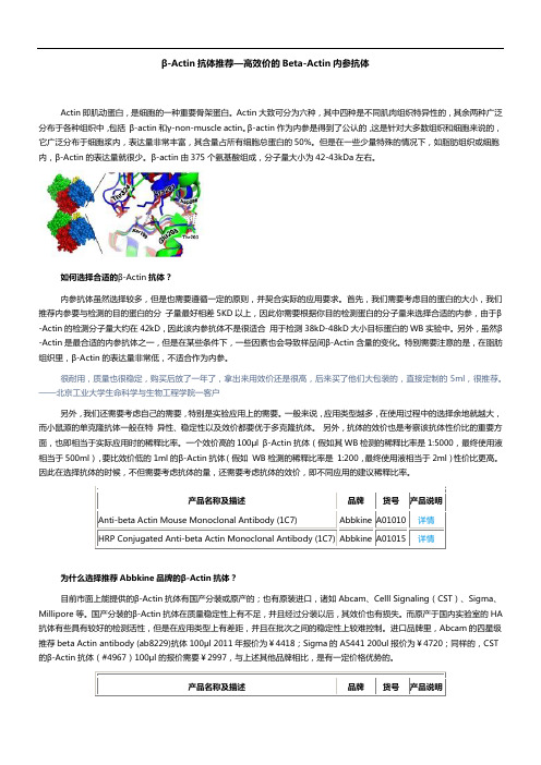
β-Actin抗体推荐—高效价的Beta-Actin内参抗体Actin即肌动蛋白,是细胞的一种重要骨架蛋白。
Actin大致可分为六种,其中四种是不同肌肉组织特异性的,其余两种广泛分布于各种组织中,包括β-actin和γ-non-muscle actin。
β-actin作为内参是得到了公认的,这是针对大多数组织和细胞来说的,它广泛分布于细胞浆内,表达量非常丰富,其含量占所有细胞总蛋白的50%。
但是在一些少量特殊的情况下,如脂肪组织或细胞内,β-Actin的表达量就很少。
β-actin由375个氨基酸组成,分子量大小为42-43kDa左右。
如何选择合适的β-Actin抗体?内参抗体虽然选择较多,但是也需要遵循一定的原则,并契合实际的应用要求。
首先,我们需要考虑目的蛋白的大小,我们推荐内参要与检测的目的蛋白的分子量最好相差5KD以上,因此你需要根据你目的检测蛋白的分子量来选择合适的内参,由于β-Actin的检测分子量大约在42kD,因此该内参抗体不是很适合用于检测38kD-48kD大小目标蛋白的WB实验中。
另外,虽然β-Actin是最合适的内参抗体之一,但是在某些条件下,一些因素也会导致样品间β-Actin含量的变化。
特别需要注意的是,在脂肪组织里,β-Actin的表达量非常低,不适合作为内参。
很耐用,质量也很稳定,购买后放了一年了,拿出来用效价还是很高,后来买了他们大包装的,直接定制的5ml,很推荐。
——北京工业大学生命科学与生物工程学院一客户另外,我们还需要考虑自己的需要,特别是实验应用上的需要。
一般来说,应用类型越多,在使用过程中的选择余地就越大,而小鼠源的单克隆抗体一般在特异性、稳定性以及效价都要优于多克隆抗体。
另外,抗体的效价也是考察该抗体性价比的重要方面,也即相当于实际应用时的稀释比率。
一个效价高的100μl β-Actin抗体(假如其WB检测的稀释比率是1:5000,最终使用液相当于500ml),要比效价低的1ml的β-Actin抗体(假如WB检测的稀释比率是1:200,最终使用液相当于2ml)性价比更高。
(完整版)★抗肿瘤药物临床应用指导原则全文

抗肿瘤药物临床应用指导原则(征求意见稿)目录第一章抗肿瘤药物临床应用的基本原则 (4)一、权衡利弊,最大获益 (4)二、目的明确,治疗有序 (4)三、医患沟通,知情同意 (4)四、治疗适度,规范合理 (5)五、熟知病情,因人而异 (5)六、不良反应,谨慎处理 (5)七、临床试验,积极鼓励 (6)第二章抗肿瘤药物临床应用的管理 (7)一、抗肿瘤药物的管理 (7)(一)分级管理 (7)(二)使用管理 (8)(三)配置管理 (9)(四)人员资质管理 (9)二、落实与督查 (10)第三章各类抗肿瘤药物的适应证和注意事项 (11)一、细胞毒类药物 (11)(一)作用于DNA化学结构的药物 (11)(二)影响核酸合成的药物 (15)(三)作用于核酸转录的药物 (19)(四)作用于DNA复制的拓扑异构酶抑制剂 (20)(五)主要作用于有丝分裂M期干扰微管蛋白合成的药物 (21)(六)其他细胞毒药物 (23)二、激素类药物 (24)(一)芳香化酶抑制剂 (24)(二)雌激素和抗雌激素 (27)(三)雄激素与抗雄激素 (29)(四)孕激素 (31)(五)RH-LH激动剂/拮抗剂 (32)三、肿瘤分子靶向和生物治疗 (33)(一)生物反应调节剂 (33)(二)单克隆抗体 (36)(三)细胞分化诱导剂 (40)(四)细胞凋亡诱导剂 (41)(五)新生血管生成抑制剂 (42)(六)表皮生长因子受体抑制剂 (43)(七)基因治疗 (43)(八)多靶点小分子抑制剂 (44)四、肿瘤治疗辅助药物 (45)(一)造血生长因子 (45)(二)止吐药 (51)(三)镇痛药 (53)(四)抑制破骨细胞药 (55)(五)神经精神用药 (57)第四章各类肿瘤的治疗原则 (61)一、头颈部恶性肿瘤 (61)(一)鼻咽癌 (62)(二)鼻腔和鼻旁窦恶性肿瘤 (62)(三)喉癌 (63)(四)甲状腺癌 (66)二、胸部肿瘤 (67)(一)非小细胞肺癌 (67)(二)小细胞肺癌 (71)(三)胸腺肿瘤 (73)(四)恶性胸膜间皮瘤 (74)三、消化系统肿瘤 (75)(一)食管癌 (75)(二)贲门癌 (77)(三)胃癌 (77)(四)结直肠癌 (79)(五)胆管癌、胆囊癌 (82)(六)胰腺癌 (83)(七)肝癌 (85)四、乳腺癌 (86)(一)复发转移乳腺癌药物治疗 (87)(二)可手术乳腺癌术后抗肿瘤药物治疗 (89)五、泌尿系统、男生殖系统肿瘤 (91)(一)肾上腺肿瘤 (91)(二)肾脏肿瘤 (93)(三)尿路上皮癌 (94)(四)前列腺癌 (97)(五)阴茎肿瘤 (100)(六)睾丸肿瘤 (101)六、妇科肿瘤 (103)(一)宫颈癌 (103)(二)卵巢癌 (105)(三)子宫内膜癌 (108)(四)外阴癌 (110)(五)阴道癌 (112)(六)妊娠滋养细胞肿瘤 (113)七、血液淋巴系统肿瘤 (116)(一)白血病 (116)(二)恶性淋巴瘤 (120)(三)多发性骨髓瘤 (140)八、颅脑肿瘤 (142)(一)胶质瘤 (142)(二)髓母细胞瘤 (145)(三)脑转移瘤 (147)(四)中枢神经系统淋巴瘤 (149)(五)颅内生殖细胞瘤 (151)(六)颈静脉球瘤 (153)(七)垂体腺瘤 (155)九、原发恶性骨与软组织肿瘤 (159)(一)骨肉瘤 (159)(二)尤文氏肉瘤 (164)(三)软组织肉瘤 (166)《抗肿瘤药物临床应用指导原则》编审专家名单 (167)第一章抗肿瘤药物临床应用的基本原则正确合理地应用抗肿瘤药物是提高肿瘤患者生存率和生活质量,降低死亡率、复发率和药物不良反应发生率的重要手段,是肿瘤综合治疗的重要组成部分。
原位杂交蛋白酶abc型的原理_解释说明以及概述

原位杂交蛋白酶abc型的原理解释说明以及概述1. 引言1.1 概述原位杂交蛋白酶ABC型(ATP-binding cassette transporter)是一种广泛存在于生物体细胞内的重要转运系统。
该转运系统通过利用化学能量(ATP)驱动,进行底物的跨膜运输,并在维持细胞内稳态和应对环境变化中扮演着重要角色。
本文将详细介绍原位杂交蛋白酶ABC型的工作原理、其在生物学中的应用以及展望其未来的发展方向。
1.2 文章结构本文共分为5个部分,具体内容包括引言、原位杂交蛋白酶ABC型的原理、解释说明原位杂交蛋白酶ABC型的工作原理、概述其在生物学中的应用以及结论。
下面将逐一介绍每个部分内容。
1.3 目的本文旨在深入了解并全面阐述原位杂交蛋白酶ABC型的工作原理,探讨其在生化研究和疾病诊断治疗等领域中的应用价值,并对该领域未来发展方向做出展望。
通过本文对原位杂交蛋白酶ABC型的全面解析,旨在促进相关学科领域的研究进展,并为实际应用提供科学依据。
以上是“1. 引言”部分的详细内容,对于文章大纲中其他部分的详细内容可以参考指定的目录。
2. 原位杂交蛋白酶abc型的原理2.1 ABC转运系统介绍ABC转运系统是一种广泛存在于生物界的重要蛋白质家族,包括ABC输入通道、ABC输出通道和ABC导致蛋白(ATP结合盒)三个主要部分。
其中,ABC输入通道由多个转录子编码的蛋白质组成,主要功能是将底物从胞外传输到细胞内。
ABC输出通道也由多个蛋白质组成,其作用是将底物从细胞内排出。
ABC导致蛋白是整个系统的“引擎”,通过ATP酶活性提供能量,并驱动底物的转运。
2.2 ABC转运系统的组成与功能ABC转运系统由多个不同类型的蛋白质组成。
每种类型的蛋白质在系统中扮演着特定的角色。
例如,输入通道中主要包括底物结合蛋白和跨膜导管蛋白;输出通道则涵盖了底物排出途径和有助于形成稳定复合体的辅助因子。
这些不同组分之间通过相互作用形成复杂而高效的工作系统。
抗体选择指南,抗体保存指南,抗体使用说明书样本

抗体选择指南抗体保存指南抗体说明书样本一抗体选择指南:检测任何目的靶蛋白都有不止一种抗体可供选择,为缩小抗体的选择范围选中合适的抗体,需要考虑如下几种因素:1. 分析或应用的类型2. 样本蛋白的结构性质3. 样本的种属4. 抗体宿主的种类5. 抗体的标记和检测1分析试验的应用类型一般抗体说明书都列出该抗体经试验验证过适用于何种分析类型,如:可以应用于WB IHC ICC ELASA分析等,如果抗体说明书没有提及的应用类型,并不意味着该抗体不适用于此种分析应用类型,而仅是说明尚未经过此种分析试验验证,如果抗体不适用某些分析试验,则会在抗体说明书上标注出来不适于某分析试验。
2 样本蛋白的结构性质了解样本蛋白的结构性质有助于选择最合适的抗体,至少两方面因素需要考虑(1)..待测样本蛋白的结构域:抗体是由各种不同免疫原免疫宿主而制备得来,其中的免疫原包括:全长蛋白、蛋白片断、多肽、全有机体(如:细菌)或细胞,抗体说明书一般都有免疫原的描述,如果打算检测的是蛋白片断或一种特殊的同型物或蛋白全长的某一区域,则必须选择用含此片段域的免疫原制备出的抗体。
如果打算用FACS流式检测活细胞的表面蛋白,则需要选择含该表面蛋白的胞外域来免疫制备的抗体。
(2)样本的提取或处理过程:某些抗体要求样本经过某些特殊处理,例如:许多抗体只识别还原和变性的、表位已暴露不受二级四级结构阻碍的蛋白样本,另一方面,某些抗体仅识别天然折叠状态的蛋白。
当选择免疫组化的抗体时,应注意某些抗体只识别未固定的冷冻的组织,而另一些抗体则适用于无需抗原修复解交联步聚的甲醛固定石蜡包埋的组织,这些都会在抗体说明书上应用部分标示出来3 样本的物种应选择物种相同或有交叉反应的抗体,抗体可能与不同物种的同种靶蛋白有交叉反应,因其氨基酸序列同源性较高,如果样本的种类未列入抗体说明书上的交叉反应种属表中,并不意味着该抗体不适用于检测该物种的蛋白,而只是表示该物种尚未用此抗体检测验证过,应通过序列比对的方法来预测交叉反应,可应用Expasy 和 NCBI BLAST来进行不同物种蛋白同源性比对。
Abcam抗体产品保存指南

Abcam产品保存指南抗体保存得当与否,直接决定了抗体的活性和使用效果!如果抗体保存得当,大部分抗体活性都可以维持数月甚至数年。
本指南以Abcam抗体保存方法为主,同样也可以应用到其他绝大部分公司的抗体产品!请谨记!无论何时请按照说明书推荐的保存条件正确保存抗体!1. 收到抗体后请务必在12000rpm离心1-5分钟后再打开管盖进行分装和保存(如果抗体体积小于50ul,请延长离心时间至5分钟,以保证全部抗体均离心下来)!Abcam的抗体使用非常特殊的储存管,只有在12000rpm离心1-5分钟才能将附着在管壁或盖内的抗体全部离心下来。
即使是在10000rpm离心也会导致离心不完全,导致出现抗体的量不足的现象!2. 对于Abcam大部分抗体,比较合适的保存方式是分装后保存在 -20℃ or-80℃。
o对绝大多数抗体来说,保存在-20℃是完全足够了。
没有任何证据显示保存在-80℃会有更多的好处!o分装可以最大程度的降低反复冻融对抗体活性的损害,同时也降低了由于多次从同一管中吸取抗体造成的污染可能性。
o分装的量以一次实验用完为好,最少不能少于10ul每份。
因为分装体积越小,抗体的浓度越可能会受到蒸发以及管壁吸附的影响o复融后的分装抗体如果一次用不完,将剩余母液保存在4℃,避免再冻起来!o抗体工作液应该当天配制当天用完,在4℃尽量不要超过1天。
o绝对避免将抗体保存在自动除霜冰箱中。
尽量将抗体保存在冰箱里层,而不是门上3. 大部分抗体收到后4℃短暂保存1-2周对抗体活性是没有影响的。
如果抗体很快(1-2周)会使用,推荐在4℃保存,这是为了避免反复冻融对抗体活性的损害。
如果要长期保存则最好是在-20℃ or -80℃。
最关键的一点是要根据说明书推荐的方式来正确的保存抗体!但是,腹水形式的产品请在收到后立即冻起来!因为该类产品含有大量的蛋白酶,长期在4oC保存会导致抗体的降解!4. 大部分抗体的运输条件:一般的运输过程需要1-2周的时间,所以是在4℃的条件下来完成的。
His抗体实验大全
His抗体推荐—Abbkine全系列His标签抗体His标签是由6个组氨酸(His-His-His-His-His-His)组成的短肽,专门设计用于重组蛋白质的吸附纯化。
由于分子量较小,并且较容易分离和纯化,His-tag 融合标签与其他标签相比有很多明显优势, 是目前用于纯化的融合标签中使用最为广泛的一种。
His 抗体可以用于检测和His标签融合表达蛋白的表达、细胞内定位,以及纯化、定性或定量检测His融合表达蛋白等。
然而,目前商品化His-tag的单克隆抗体(mAb)种类不多, 且价格也十分昂贵。
如何选择合适的His标签抗体?我们在选择His抗体时,主要根据自己的需要,特别是实验应用上的需要。
其中需要考虑的因素包括了,His抗体的应用检测类型、His抗体的克隆性及制备来源等。
比如该His抗体是否能用于Western Bloting的检测?是否可以用于免疫荧光IF实验?我是否需要特异性更好的单克隆His标签抗体?我是否需要考虑下是选择小鼠来源的还是兔来源的His抗体?之前有使用SSS(一家著名品牌的His标签抗体,名称隐去)的His标签抗体,一开始还好,过了半年后,就发现产品质量不稳定,做IF实验不行了,后来试用了下Abbkine的,IF结果很漂亮,顺带还买了他们的DyLight荧光二抗——广州南方医科大学一客户一般来说,应用类型越多,在使用过程中的选择余地就越大,而小鼠源的单克隆抗体一般在特异性、稳定性以及效价都要优于多克隆抗体。
另外,抗体的效价也是考察该抗体性价比的重要方面,也即相当于实际应用时的稀释比率。
一个效价高的100μl His 抗体(假如其WB检测的稀释比率是1:5,000,最终使用液相当于500ml),要比效价低的1ml的His抗体(假如WB检测的稀释比率是1:200,最终使用液相当于2ml)性价比更高。
因此在选择抗体的时候,不但需要考虑抗体的量,还需要考虑抗体的效价,即不同应用的建议稀释比率。
Protein A G MagBeads 技术手册说明书

Protein A/G MagBeads Cat. No. L00277Technical Manual No. TM0249 Version 08212013 Index1.Product Description2. Instruction For Use3.Troubleshooting4. General Information1.Product Description1.1Intended UseGenScript Protein A/G MagBeads are ideal for small‐scale antibody purification and immunoprecipitation (IP) of proteins, protein complexes or other antigens.1.2PrincipleThe sample containing antibody is added to the Protein A/G MagBeads. The antibody will bind to beads during a short incubation. Then the beads‐bound antibody may be eluted from the beads by using a magnetic separation rack, or used for immunoprecipitation (IP). A cross‐linking procedure may be needed before IP to prevent co‐elution of the primary antibody. Magnetic separation eliminates the changes of micro tubes, minimizes the loss of sample and removes excessive steps of traditional centrifugation method.1.3Description of MaterialMaterial SuppliedGenScript Protein A/G MagBeads are super paramagnetic beads of average 40 μm in diameter, covalently coated with recombinant Protein A/G. The beads are supplied as 25% slurry in phosphate buffered saline (PBS), pH 7.4, containing 20% ethanol. The Protein A/G MagBeads have a binding capacity of more than 10 mg Goat IgG per 1 ml settled beads (e.g. 4 ml 25% slurry).Protein A/G is a genetically engineered protein (MW≈43 kDa) that combines the IgG binding sites of both Protein A and Protein G. 6×His‐tag was attached to its N‐terminal to facilitate the purification. The secreted Protein A/G contains four Fc‐binding domains from Protein A and two from Protein G, making it a more universal tool to bind and purify immunoglobulins.Cat. No. L00277 Size: 2 ml.Additional Material RequiredMixing/Rotation DeviceMagnetic Separation RackTest tubes and pipettesBuffers and solutions (see below)Additional Buffers NeededBinding/Wash Buffer: 20 mM Na2HPO4, 0.15 M NaCl, pH 7.0Elution Buffer: 0.1 M glycine, pH 2‐3Neutralization Buffer: 1 M Tris, pH 8.51×SDS Sample Buffer: 62.5 mM Tris‐HCl (pH 6.8 at 25°C), 2% w/v SDS, 10% glycerol, 50 mM DTT,0.01% w/v bromophenol blue2.Instruction For UseThe protocol uses 100 μl Protein A/G MagBeads, this may be scaled up or down accordingly.2.1Preparation of the MagBeadspletely resuspend the beads by shaking or vortexing the vial.2.Transfer 100 μl beads into a clean tube.3.Place the tube on a magnetic separation rack to collect the beads. Remove and discard the supernatant.4.Add 1 ml Binding/Wash Buffer to the tube and invert the tube several times to mix. Use the magnetic separationrack to collect the beads and discard the supernatant. Repeat this step twice.2.2Separation of Target IgG1.Resuspend the beads in 100 μl Binding/Wash Buffer.2.Add the sample containing target IgG to the tube and gently invert the tube to mix.3.Incubate the tube at room temperature with mixing (on a shaker or rotator) for 30 – 60 minutes.e the magnetic separation rack to collect the beads and discard the supernatant. If necessary, keep thesupernatant for analysis.5.Add 1 ml Binding/Wash Buffer to the tube and mix well, use the magnetic separation rack to collect the beads anddiscard the supernatant. Repeat the wash step three more times.6.Proceed to elution of isolated IgG (Section 2.3).2.3Elution of Isolated IgG1.Add 100 μl Elution Buffer to the tube and mix well. Incubate for five minutes at room temperature with occasionalmixing.e the magnetic separation rack to collect the beads and transfer the supernatant that contains the eluted IgG intoa clean tube.3.Repeat Step 1 and 2 twice.4.Add 10 μl of Neutralization Buffer to each 100 μl eluate to neutralize the pH. If needed, perform a buffer exchangeby dialysis or desalting.2.4ImmunoprecipitationBound IgG will be co‐eluted along with the target when using elution methods. If the presence of IgG does not disturb desired detection system, go directly to section 2.4.2 below. For applications where co‐elution of the IgG is not desired, the primary IgG can be cross‐linked to the Protein A/G MagBeads as described in section 2.4.1 below.2.4.1Cross‐linking IgG to the Beads1.Add 1 ml 0.2 M triethanolamine, pH 8.2 to the Protein A/G MagBeads with immobilised IgG. Wash twice using themagnetic separation rack with 0.2 M triethanolamine, pH 8.2 as the washing buffer.2.Resuspend the beads in 1 ml of 20 mM dimetyl pimelimidate dihydrochloride (DMP) in 0.2 M triethanolamine, pH 8.2(5.4 mg DMP/ml buffer). This cross‐linking solution must be prepared freshly.3.Incubate the beads with rotational mixing for 30 minutes at room temperature. Use the magnetic separation rack tocollect the beads and discard the supernatant.4.Resuspend the beads in 1 ml of 50 mM Tris, pH 7.5 to stop the reaction and incubate for 15 minutes at roomtemperature with rotational mixing.e the magnetic separation rack to collect the beads and discard the supernatant.6.Wash the cross‐linked beads three times with 1 ml PBS, pH7.4.2.4.2Binding Antigen to the IgG Cross‐linked Beads1.Add sample containing target antigen to the beads. For a 100 kD protein, use a volume containing approximate 25 µgtarget antigen/ml beads to assure an excess of antigen. If dilution of antigen is necessary, PBS or 0.1 M phosphate buffer (pH 7‐8) can be used as dilution buffer.2.Incubate with tilting and rotation for one hour at room temperature.3.Place the tube on the magnetic separation rack for 2 minutes to collect the IgG‐coated Beads‐target complex. Forviscous samples, double the time on the rack. Discard the supernatant.4.Wash the beads 3 times using 1 ml PBS.2.4.3Elution of Target ProteinA.Denaturing elution1.Place the tube from section2.4.2 on the magnetic separation rack to collect the beads and discard the supernatant.2.Add 100 µl 1XSDS Sample Buffer to the tube and mix well.3.Heat the tube at 100°C for five minutes.e the magnetic separation rack to collect the beads and transfer the supernatant containing desired sample into anew tube.5.Analyze the sample by SDS‐PAGE followed by Western blot analysis.B.Non‐denaturing elution1.Place the tube from section2.4.2 on the magnetic separation rack to collect the beads and discard the supernatant.2.Add 100 µl Elution Buffer to the tube and mix well. Incubate for five minutes at room temperature with occasionalmixing.e the magnetic separation rack to collect the beads and transfer the supernatant into a new tube.4.Repeat Step 2 and 3 twice.5.Add 10 μl Neutralization Buffer to each 100 μl of eluate to neutralize the pH.3.TroubleshootingReview the information below to troubleshoot your experiments using the GenScript Protein A/G MagBeads. Problem Possible Cause SolutionThe beads are difficult toimmobilize using the magneticseparation rack.Too many beads are used.Decrease the volume of MagBeadssuspension.A considerable amount of samplehas been added, but very fewspecific antibody of interest isdetected.The antibody of interest is at verylow concentration.Use a serum‐free medium for cellsupernatant samples.Affinity‐purify the antibody using itsspecific antigen coupled to anaffinity supporting material.The antibody of interest is purified,but it is degraded (as determined byloss of function in downstreamassay).The antibody is sensitive to low‐pHelution buffer.The downstream application issensitive to the neutralized elutionbuffer.Try another elution reagent, such as3.5 M MgCl2, 10 mM phosphate, pH7.2.Desalt or dialyze the eluted sampleinto a suitable buffer.No antibody is detected in anyeluate.The antibody in the sample cannotbind to Protein A/G.Try GenScript Protein A MagBeadsor Protein G MagBeads.4.General Information4.1Storage and StabilityThis product is stable until the expiration date stated on the COA, when stored unopened at 2–8°C. Do not freeze the product. Keep the MagBeads in liquid suspension during storage and all handling steps. Drying will cause loss of binding capacity and result in reduced performance. Resuspend the beads well before use. Be careful to avoid bacterial/fungal contamination.4.2Technical SupportPlease contact GenScript for further technical information (see contact details). Certificate of Analysis/Compliance is available upon request. The latest revision of the package insert/instructions for use is available on .4.3Warning and LimitationsThis product is for research use only. Not intended for any animal or human therapeutic or diagnostic use unless otherwise stated. This product contains 20 % EtOH as a preservative. Flammable liquid and vapor. Flash point 38°C. R‐10 flammable. Material Safety Data Sheet (MSDS) is available at .4.4Related MagBeads ProductsCat. No. Product NameL00273 Protein A MagBeadsL00274 Protein G MagBeadsL00295 Ni‐Charged MagBeadsL00327 Glutathione MagBeadsL00275 Mouse Anti‐His mAb MagBeadsL00336 Mouse Anti‐GST mAb MagBeadsGenScript USA Inc.860 Centennial Ave.,Piscataway, NJ 08854Toll‐Free: 1‐877‐436‐7274Tel: 1‐732‐885‐9188, Fax: 1‐732‐210‐0262Email: *********************Web: 。
Bactobac表达系统中文版说明书

Bac-to-Bac杆状病毒表达系统试剂盒内容物:Introduction:Overview:Bac-to-Bac杆状病毒表达系统提供快速有效的方式产生重组杆状病毒。
此方式基于让已经转入杆状病的质粒(杆粒)的位点特意转座子的表达框的质粒在Ecoli中扩增。
Bac-to-Bac杆状病毒表达系统要紧包括:*pFastBac捐献质粒的选择,它要能够产生包括目的位点的表达结构,那个目的基因的产生被杆状病毒特意位点启动子操纵。
*一个Ecoli宿主,DH10Bac,包括杆状病毒质粒(杆粒)和辅助质粒,在转染pFastBac 表达结构后能够产生重组杆粒。
*一个操纵表达的质粒,包括Gus和/或CAT基因,以便在感染细胞后产生重组杆状病毒,表达β-葡萄糖酸酐酶和/或氯霉素乙酰转移酶。
Bac-to-Bac表达系统的优势:利用那个系统产生重组杆状病毒较传统的同源重组有以下优势:*与利用同源重组产生重组杆状病毒所需的4-6周相较,分辨纯化重组病毒少于两周*减少了从斑点挑选重组病毒DNA所包括亲缘和非重组病毒的概率*能够快速同时进行大量重组,适合表达功能性研究的蛋白选择pFastBac菌体(Vector):大量的pFastBac菌体都适于进行Bac-to-Bac表达系统。
选择关于你的需要最适合的菌体。
指南用途:指南提供了一个关于Bac-to-Bac表达系统的概述,并对以下提供指导:1、克隆目的基因到pFastBac TM供体质粒的选择2、转化pFastBac TM 结构到最高效的DH10Bac TM产生重组质粒3、转染重组质粒DNA到昆虫细胞产生重组杆状病毒4、扩增滴定(Amplify and titer)杆状病毒株,利用病毒株感染昆虫细胞表达目的重组蛋白重要的:Bac-to-Bac杆状病毒表达系统是用来帮忙你产生重组杆状病毒,在昆虫细胞中进行高水平表达目的基因的系统。
虽然他能够帮忙你很容易的产生杆状病毒表达你的重组蛋白,可是利用这系统更偏向于有杆状病毒生物学和昆虫表达背景的利用者。
c-Myc标签Co-IP试剂盒中文说明
23620描述ProFound™ c-Myc Tag IP/Co-IP Kit 包含了足够25个与c-Myc –标签蛋白IP或Co-IP的成分,包括10个c-Myc –标签阳性对照的实验试剂盒成分:Immobilized Anti-c-Myc, 62.5 g固定的珠状琼脂加上125g 抗体,以含25%的溶液和0.1%叠氮钠的PBS 的形式提供(总体积为250ul)。
BupH™ Tris Buffered Saline Pack,一个包装,用500 ml超纯水稀释后,溶液成分为25 mM Tris, 0.15 M NaCl, pH 7.2Elution Buffer, 50 ml, pH 2.8Lane Marker Non-Reducing Sample Buffer (5X), 5 ml, 包含0.3 M Tris•HCl, pH 6.8, 5% SDS, 50% 甘油, 电泳指示剂。
Handee™ Spin Columns Accessory Pack, 含提前放置好的玻璃原料,顶盖和底盖的27Collection Tubes and Caps Accessory Pack, 100 有刻度的2 ml 管子和塞盖c-Myc-tagged Positive Control, 500ul,1 mg/ml E. coli 提取的包含c-Myc-标签GST(古胱氨酸S-转移酶)储藏: 收到后,将c-Myc-标签阳性对照放在-80度冰箱,(在-20度可放6个月),将其他所用到的成分放在4度,c-Myc-标签阳性对照用干冰运输。
注意:ProFound™ c-Myc Tag IP/Co-IP Kit是ProFound™ c-Myc Tag IP/Co-IP Application Set (23622) 和c-Myc-tagged Positive Control (23633)的联合体。
介绍ProFound™ c-Myc Tag IP/Co-IP Kit与用蛋白A/G琼脂糖的IP方法相比,传统的提供了一个简单快速的方法来研究c-Myc-标签蛋白。
Elekta呼吸门控治疗的ABC系统使用指南说明书
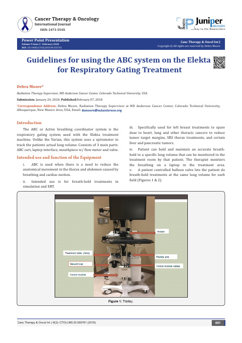
Power Point PresentationVolume 9 Issue 3 - February 2018DOI:Canc Therapy & Oncol Int JCopyright © All rights are reserved by Debra MooreGuidelines for using the ABC system on the Elektafor Respiratory Gating TreatmentDebra Moore*Radiation Therapy Supervisor, MD Anderson Cancer Center, Colorado Technical University, USA Submission: January 24, 2018; Published:February 07, 2018*Correspondence Address: Debra Moore, Radiation Therapy Supervisor at MD Anderson Cancer Center, Colorado Technical University, Albuquerque, New Mexico Area, USA, Email:Cancer Therapy & OncologyInternational JournalISSN: 2473-554XIntroductionThe ABC or Active breathing coordinator system is the respiratory gating system used with the Elekta treatment machine. Unlike the Varian, this system uses a spirometer to track the patients actual lung volume. Consists of 3 main parts: ABC cart, laptop interface, mouthpiece w/ flow meter and valve.Intended use and function of the Equipmenti. ABC is used when there is a need to reduce the anatomical movement in the thorax and abdomen caused by breathing and cardiac motion.simulation and XRT.iii. Specifically used for left breast treatments to spare dose to heart, lung and other thoracic cancers to reduce tumor target margins, SRS thorax treatments, and certain liver and pancreatic tumors.iv. Patient can hold and maintain an accurate breath- hold to a specific lung volume that can be monitored in the treatment room by that patient. The therapist monitors the breathing on a laptop in the treatment area. v. A patient controlled balloon valve lets the patient do breath-hold treatments at the same lung volume for each field (Figures 1 & 2).Figure 1: Trolley.Patient control switch: Patient presses the button during treatment and only releases it to stop the breathold. When the button is released the balloon valve is deflated. If the patient presses the button twice is one second it will send a distress signal that will show upPatient control switchreleases it to stop the breathold. When the button is released the balloon valve is deflated. If the patient presses the button twice is one second it will send a distress signal that will show up on the laptop screen (Figure 3).Patient Respiratory System (Figure 4)a. Sensor pneumatics cable assembly: measures the amount of air the patient breathes that is then sent to the ABC control moduleb. Balloon valve: inflates to prevent air flow through the spirometer when a breathold starts. It deflates when the breathold is completed or interrupted.c. Balloon valve tube: how the ABC control module controls the balloon valve.d. Mechanical connector: attaches to the flexible arme. Filter adapter: mouthpiece and filter kit attaches tothis (Figures 5 & 6).Patient respiratory system.Filter adapter.Figure 6 : Measures the quantity of air a patient breathes through the ABC system. ABC Response Control Box (Figure 7)Figure 7 : ABC Response control Box.i. The response control box enables/disables the response gating control system.ii. Green lights on USB connect, Relay module, Gating on/ off, Gating enabled/fault will allow for the patient loaded into ABC system to be treated. Any red lights and system will not allow treating.iii. Gating enable button: enables/disables gating of the accelerator when pressed continuously for 3 seconds. Also used to change from a fault mode to gating enabled mode.Introduction Recommended cleaning for the ABC is as follows:Clean surfaces using a mild detergent solution then dry with a clean, lint free clothFigure 8 : Laptop Connection to ABC cart using the networkcable.Figure 9 : Connection of the cart to a power outlet and turn thepower to the cart ON.Figure 10 : Turn the Active Breathing Coordinator box ON(green light should be present.)a. Morning QAi. Connect laptop to ABC cart using the network cable. Cable runs from laptop to cart network port located next to ii. Ensure that all cords are firmly attached (Figures 8-10). iii. Turn the Active Breathing Coordinator box ON (green light should be present).iv. Turn on laptop (Figure 11).Figure 11 : Turn on laptop.v. Login to ABC computer (Password: abc lowercase) (Figure 12).Figure 12 : Login to ABC computer (Password: abc lowercase.) vi. Double click on ABC icon.vii. ABC System Check should all be green (connected.)viii. At this time, the laptop should have connected to the mouse and monitor on the cart.ix. ABC will prompt user to load an existing patient. Select the morning QA patient.x. A QA patient should be created by the physicist for daily QA.xi. Once the QA patient is loaded, put in the current date under session ID.Connect the spirometer to the automated morning QA device (Figure 13).Figure 13 : Connect the spirometer to the automated morning the morning QA device (Figure 14).xiv. Press the play button to initiate test.xv.The QA device will run tests and a prompt will come up and tell you if the test has passed or failed (Figure 15). xvi. Record if passed or failed on the daily QA sheetLCD menu to find the manual option (Figure 16).xviii. This setting will give you 3 inhale and exhale cycles for testing.xix. Press the play button each time for an inhale and exhale.xx. This will be done 6 times for 3 inhales and 3 exhales. xxi. Record inhale and exhale volume values on the daily QA sheet.CT Simulation using ABC (Figure 17)a. Turn on computer.b. Take ABC cart into Tx room and connect network cable from the laptop into cart.c. Plug in cart unit to nearest outlet.d. Turn on control module power switch.e. Remove table clamp from cart and attach to superior/Figure 17 : CT Simulation using abc.Figure 18 : Deploy laptop and turn on.f. Deploy laptop and turn on (Figure 18).g. Log into ABC.h. Select Create New.Populate Patient Data (Figure 19)Figure 19 : Populate Patient Data.i. Patient and session Information ii. Session ID is CT Simiii. Gating Option – Manualiv. Select Breath holdv. 30 seconds for full scanvi. Treshold defaults at 1.2Lvii. Inhalationviii. Save.Modification to set up can be made by selecting Patient Settings and clicking and entering desired levels (Figures 20). Physics will provide instructions to the patient explaining the steps to the ABC procedure (Figure 21). Position mouth piece, nose clamp and patient control switch. Ask patient to take in a big breath and take note as to how many liters are inspired. Repeat 3 times and adjust threshold to approximately 80% of maximum volume. Activate System by pressing space bar once. Have patient depress green button and take in a big breath. System will cut up breath upon reaching the preselected volume. Have them let go of the button and breath normally. After patient balloon device and lock in place.Figure 21 : Physics will provide instructions to the patientexplaining the steps to the ABC procedure.Treatment using ABC (Figure 22).Remove Laptop from cart and place on counter. Connect computer to Elekta Response Box and network cable. Turn on 23). Turn on computer. Take ABC cart into Tx room and connect network cable into cart (Figure 24). Plug in cart unit to nearest outlet. Turn on control module power switch. Remove table clamp from cart and attach to superior/top of table. Insert flexible arm into table clamp. Attach patient respiratory device to flexible arm along with mouth piece (Figure 25). Login to ABC computer (Password: abc lowercase). Double click on ABC icon. ABC System Check should all be green (connected) (Figure 26). Trouble shooting (if needed). Load existing patient. Click on ABC patient data (Upper left corner). Select correct patient from existing list. Highlight patient for treatment. Select – open patient (Figure 27). ABC patient setting Box will appear (Figure 28). Under session record today’s date. Click DONE (lower left). Load Patient information (plan) on linac. Highlight field and add pre-port film (must image daily).Figure 23 : Turn on Response Box on (hold down until green light appears).Figure 24 : Take ABC cart into Tx room and connect network cable into cart.Figure 25 : Attach patient respiratory device to flexible arm along with mouth piece.Figure 26 : ABC System Check should all be green (connected).Figure 27 : Highlight patient for treatment.Figure 28 : ABC patient setting Box will appear. Select port needed. Instruct patient to hold control switch (Figures 29-31). 2nd therapist should be providing patient with breathing instructions over intercom. Patient instructed to release green button as needed with breathing crisis, (release of button will necessitate 2nd therapist to hit space bar to reactivate ABC control) Hit space bar to activate breath control with linac. ABC control is represented by green bar. Instruct patient to gently take in a deep breath. ABC control will prevent patient from normal breathing once dose delivery threshold is reached. Treatment delivery achieved within countdown window (Figure 32).Figure 29 : Instruct patient to hold control switch.Figure 30 : Red Line Indicates control Switch is not depressed.Figure 31 : Blue line indicates control switch is depressed.Figure 32 : Treatment delivery achieved within countdown window. Trouble Shooting (Figure 33)Figure 33 : Trouble shooting.Most common problem when launching ABC software is that no USB connections are identified.and network connections.iii. These are the ABC cart connections.the culprit as it works loose with gravity (Figure 35).Figure 35 : Micro USB cable connected on the bottom left is often the culprit as it works loose with gravity.module is located (Figure 36).Figure 36 : Power switch for the control module is located. vi. Green light on top indicates power to the module and control unit are on (Figure 37).Figure 37 : Green light on top indicates power to the module and control unit are on.vii. Red light on top along with audible alarm indicatesFigure 38 : Red light on top along with audible alarm indicates power switch to the module and control unit are off..viii. Power switch for the ABC control cart is located on theFigure 39 : Power switch for the ABC control cart is located on the side of the unit along with the network connector.Figure 40 : Check that the network cable is plugged securely into the correct jack.into the correct jack (Figure 40).Figure 41 : If all connections are securely plugged try to re-launch the USB server from the control laptop.Figure 42 : All green lights indicate ABC is in the correct state for treatment.Figure 43 : Make sure patient is holding the patient control button down. This will change the respiratory indicator line blue.If all connections are securely plugged try to re-launch the USB server from the control laptop (Figure 41). All green lights indicate ABC is in the correct state for treatment (Figure 42). While delivering treatment you find that the space barCancer Therapy & Oncology International Journalwill not activate the system. Make sure patient is holding the patient control button down (Figure 43). This will change the respiratory indicator line blue. If line is blue and space bar still does not work then press pause then record to re-activate system (Figure 44). This is the correct activated treatment state (Figure 45). If beam will not deliver check that the response box is active (Figure 46). Press and hold power switch till light turns green. If light is red, press and hold till light turns off then turn it back on. This is what the response box should look like when ready to deliver beam. If beam will not deliver check that the response box is active. Press and hold power switch till light turns green.Figure 44 : If line is blue and space bar still does not work thenpress pause then record to re-activate system.Figure 45 : This is the correct activated treatment state.Figure 46 : If beam will not deliver check that the response box is active. Press and hold power switch till light turns green.AcknowledgementTherapy Staff – Supervisor – Debra Moore, Roshan Cherian Brandon Held, Leslie Robson Virginia Hernandez Bernal, Ameenah Roybal Janis Roybal Garcia, Dosimetry – Paul Chavez, Erik Frija and Rocio Bergamo, Nursing – Elfrida Bauer, Celina Trujillo and Jayme Runyan, Dr. Ramesh Gopal – Radiation Oncologist, Dr. Amit Garg – Radiation Oncologist, Dr. Kara Bucci – Radiation Oncologist, Special Thanks to Physics – Elder Calderon, Mark Garcia and Mark Marshall.This work is licensed under Creative Commons Attribution 4.0 License DOI: 10.19080/CTOIJ.2018.09.555761。
免疫细胞使用说明书

免疫细胞使用说明书(V1.0)【适用范围】该产品说明书适用于人和动物PBMC、SMC、CBMC、BMMNCs等单个核细胞和人、小鼠CD3+T、CD4+T、CD8+T、CD19+B、单核、NK细胞等亚型免疫细胞的复苏、保存、使用等过程。
【产品描述】单个核细胞是白细胞中只有一个细胞核的细胞,相对密度在1.076~1.090。
根据其组织来源不同,又可分为外周血单个核细胞(Peripheral Blood Mononuclear Cell,PBMC)、脐带血单个核细胞(Cord Blood Mononuclear Cell,CBMC)、脾脏单个核细胞(Spleen Mononuclear Cell)、骨髓单个核细胞(Bone Marrow Mononuclear Cells,BMMNCs)等。
IPHASE单个核细胞均是由新鲜的组织或器官,利用细胞比重的差异,通过密度梯度离心的方法获得。
免疫细胞(Immune cell),由多种不同类型的细胞组成,包括淋巴细胞、单核细胞、巨噬细胞、树突状细胞、自然杀伤细胞等。
免疫细胞作为免疫系统的重要成员,具有吞噬异物并产生抗体、机体伤病的损伤治愈、抗御病原体入侵等能力,在执行免疫应答和免疫功能方面担负重任。
IPHASE免疫亚型细胞均是通过免疫磁性细胞阴选分选的方法,从新鲜制备的单个核细胞悬液中分选而来。
【包装规格】5×10⁶cells/管1×107cells/管2×107cells/管5×107cells/管【应用方向】可作为大分子药物临床前药代动力学和药效学研究、药物毒性筛选、免疫学研究、疫苗开发以及移植免疫等多个领域研究的原材料。
【合规保障】合规是研究的开始。
按照国家要求,凡涉及到人和猴相关的细胞产品均需办理合规审批流程,只有审批通过,且取得证明文件后才可买卖细胞。
IPHASE/汇智和源分离免疫细胞的组织或器官均由合规渠道获得,来源清晰,免除客户后顾之忧。
pAAV-MCS腺病毒载体说明(图)

pAAV-‐MCS编号 载体名称北京华越洋VECT10024 pAAV-‐MCSpAAV-‐MCS载体基本信息:载体名称: pAAVMCS, pAAV-‐MCS, p AAV M CS质粒类型: 腺病毒相关载体表达水平: 高启动子: CMV克隆方法: 多克隆位点,限制性内切酶载体大小: 4650 b p5' 测序引物及序列: CMV-‐F: 5'-‐CGCAAATGGGCGGTAGGCGTG-‐3'3' 测序引物及序列: hGH-‐PA-‐R: C CAGCTTGGTTCCCAATAGA载体标签: -‐-‐载体抗性: 氨苄备注: -‐-‐稳定性: /组成型: -‐-‐病毒/非病毒: 腺病毒pAAV-‐MCS载体质粒图谱和多克隆位点信息pAAV-‐MCS载体序列:ORIGIN1 C CTGCAGGCA G CTGCGCGCT C GCTCGCTCA C TGAGGCCGC C CGGGCAAAG C CCGGGCGTC 61 G GGCGACCTT T GGTCGCCCG G CCTCAGTGA G CGAGCGAGC G CGCAGAGAG G GAGTGGCCA 121 A CTCCATCAC T AGGGGTTCC T GCGGCCGCA C GCGTGGAGC T AGTTATTAA T AGTAATCAA 181 T TACGGGGTC A TTAGTTCAT A GCCCATATA T GGAGTTCCG C GTTACATAA C TTACGGTAA 241 A TGGCCCGCC T GGCTGACCG C CCAACGACC C CCGCCCATT G ACGTCAATA A TGACGTATG 301 T TCCCATAGT A ACGTCAATA G GGACTTTCC A TTGACGTCA A TGGGTGGAG T ATTTACGGT 361 A AACTGCCCA C TTGGCAGTA C ATCAAGTGT A TCATATGCC A AGTACGCCC C CTATTGACG 421 T CAATGACGG T AAATGGCCC G CCTGGCATT A TGCCCAGTA C ATGACCTTA T GGGACTTTC 481 C TACTTGGCA G TACATCTAC G TATTAGTCA T CGCTATTAC C ATGGTGATG C GGTTTTGGC 541 A GTACATCAA T GGGCGTGGA T AGCGGTTTG A CTCACGGGG A TTTCCAAGT C TCCACCCCA 601 T TGACGTCAA T GGGAGTTTG T TTTGCACCA A AATCAACGG G ACTTTCCAA A ATGTCGTAA 661 C AACTCCGCC C CATTGACGC A AATGGGCGG T AGGCGTGTA C GGTGGGAGG T CTATATAAG 721 C AGAGCTCGT T TAGTGAACC G TCAGATCGC C TGGAGACGC C ATCCACGCT G TTTTGACCT 781 C CATAGAAGA C ACCGGGACC G ATCCAGCCT C CGCGGATTC G AATCCCGGC C GGGAACGGT 841 G CATTGGAAC G CGGATTCCC C GTGCCAAGA G TGACGTAAG T ACCGCCTAT A GAGTCTATA 901 G GCCCACAAA A AATGCTTTC T TCTTTTAAT A TACTTTTTT G TTTATCTTA T TTCTAATAC961 T TTCCCTAAT C TCTTTCTTT C AGGGCAATA A TGATACAAT G TATCATGCC T CTTTGCACC1021 A TTCTAAAGA A TAACAGTGA T AATTTCTGG G TTAAGGCAA T AGCAATATT T CTGCATATA 1081 A ATATTTCTG C ATATAAATT G TAACTGATG T AAGAGGTTT C ATATTGCTA A TAGCAGCTA 1141 C AATCCAGCT A CCATTCTGC T TTTATTTTA T GGTTGGGAT A AGGCTGGAT T ATTCTGAGT1201 C CAAGCTAGG C CCTTTTGCT A ATCATGTTC A TACCTCTTA T CTTCCTCCC A CAGCTCCTG1261 G GCAACGTGC T GGTCTGTGT G CTGGCCCAT C ACTTTGGCA A AGAATTGGG A TTCGAACAT 1321 C GATTGAATT C CCCGGGGAT C CTCTAGAGT C GACCTGCAG A AGCTTGCCT C GAGCAGCGC 1381 T GCTCGAGAG A TCTACGGGT G GCATCCCTG T GACCCCTCC C CAGTGCCTC T CCTGGCCCT 1441 G GAAGTTGCC A CTCCAGTGC C CACCAGCCT T GTCCTAATA A AATTAAGTT G CATCATTTT1501 G TCTGACTAG G TGTCCTTCT A TAATATTAT G GGGTGGAGG G GGGTGGTAT G GAGCAAGGG 1561 G CAAGTTGGG A AGACAACCT G TAGGGCCTG C GGGGTCTAT T GGGAACCAA G CTGGAGTGC 1621 A GTGGCACAA T CTTGGCTCA C TGCAATCTC C GCCTCCTGG G TTCAAGCGA T TCTCCTGCC1681 T CAGCCTCCC G AGTTGTTGG G ATTCCAGGC A TGCATGACC A GGCTCAGCT A ATTTTTGTT 1741 T TTTTGGTAG A GACGGGGTT T CACCATATT G GCCAGGCTG G TCTCCAACT C CTAATCTCA1801 G GTGATCTAC C CACCTTGGC C TCCCAAATT G CTGGGATTA C AGGCGTGAA C CACTGCTCC 1861 C TTCCCTGTC C TTCTGATTT T GTAGGTAAC C ACGTGCGGA C CGAGCGGCC G CAGGAACCC 1921 C TAGTGATGG A GTTGGCCAC T CCCTCTCTG C GCGCTCGCT C GCTCACTGA G GCCGGGCGA 1981 C CAAAGGTCG C CCGACGCCC G GGCTTTGCC C GGGCGGCCT C AGTGAGCGA G CGAGCGCGC 2041 A GCTGCCTGC A GGGGCGCCT G ATGCGGTAT T TTCTCCTTA C GCATCTGTG C GGTATTTCA 2101 C ACCGCATAC G TCAAAGCAA C CATAGTACG C GCCCTGTAG C GGCGCATTA A GCGCGGCGG 2161 G TGTGGTGGT T ACGCGCAGC G TGACCGCTA C ACTTGCCAG C GCCCTAGCG C CCGCTCCTT 2221 T CGCTTTCTT C CCTTCCTTT C TCGCCACGT T CGCCGGCTT T CCCCGTCAA G CTCTAAATC2281 G GGGGCTCCC T TTAGGGTTC C GATTTAGTG C TTTACGGCA C CTCGACCCC A AAAAACTTG 2341 A TTTGGGTGA T GGTTCACGT A GTGGGCCAT C GCCCTGATA G ACGGTTTTT C GCCCTTTGA 2401 C GTTGGAGTC C ACGTTCTTT A ATAGTGGAC T CTTGTTCCA A ACTGGAACA A CACTCAACC 2461 C TATCTCGGG C TATTCTTTT G ATTTATAAG G GATTTTGCC G ATTTCGGCC T ATTGGTTAA2521 A AAATGAGCT G ATTTAACAA A AATTTAACG C GAATTTTAA C AAAATATTA A CGTTTACAA2581 T TTTATGGTG C ACTCTCAGT A CAATCTGCT C TGATGCCGC A TAGTTAAGC C AGCCCCGAC2641 A CCCGCCAAC A CCCGCTGAC G CGCCCTGAC G GGCTTGTCT G CTCCCGGCA T CCGCTTACA2701 G ACAAGCTGT G ACCGTCTCC G GGAGCTGCA T GTGTCAGAG G TTTTCACCG T CATCACCGA 2761 A ACGCGCGAG A CGAAAGGGC C TCGTGATAC G CCTATTTTT A TAGGTTAAT G TCATGATAA 2821 T AATGGTTTC T TAGACGTCA G GTGGCACTT T TCGGGGAAA T GTGCGCGGA A CCCCTATTT 2881 G TTTATTTTT C TAAATACAT T CAAATATGT A TCCGCTCAT G AGACAATAA C CCTGATAAA2941 T GCTTCAATA A TATTGAAAA A GGAAGAGTA T GAGTATTCA A CATTTCCGT G TCGCCCTTA3001 T TCCCTTTTT T GCGGCATTT T GCCTTCCTG T TTTTGCTCA C CCAGAAACG C TGGTGAAAG3061 T AAAAGATGC T GAAGATCAG T TGGGTGCAC G AGTGGGTTA C ATCGAACTG G ATCTCAACA 3121 G CGGTAAGAT C CTTGAGAGT T TTCGCCCCG A AGAACGTTT T CCAATGATG A GCACTTTTA3181 A AGTTCTGCT A TGTGGCGCG G TATTATCCC G TATTGACGC C GGGCAAGAG C AACTCGGTC 3241 G CCGCATACA C TATTCTCAG A ATGACTTGG T TGAGTACTC A CCAGTCACA G AAAAGCATC3301 T TACGGATGG C ATGACAGTA A GAGAATTAT G CAGTGCTGC C ATAACCATG A GTGATAACA 3361 C TGCGGCCAA C TTACTTCTG A CAACGATCG G AGGACCGAA G GAGCTAACC G CTTTTTTGC 3421 A CAACATGGG G GATCATGTA A CTCGCCTTG A TCGTTGGGA A CCGGAGCTG A ATGAAGCCA 3481 T ACCAAACGA C GAGCGTGAC A CCACGATGC C TGTAGCAAT G GCAACAACG T TGCGCAAAC 3541 T ATTAACTGG C GAACTACTT A CTCTAGCTT C CCGGCAACA A TTAATAGAC T GGATGGAGG3601 C GGATAAAGT T GCAGGACCA C TTCTGCGCT C GGCCCTTCC G GCTGGCTGG T TTATTGCTG3661 A TAAATCTGG A GCCGGTGAG C GTGGGTCTC G CGGTATCAT T GCAGCACTG G GGCCAGATG 3721 G TAAGCCCTC C CGTATCGTA G TTATCTACA C GACGGGGAG T CAGGCAACT A TGGATGAAC 3781 G AAATAGACA G ATCGCTGAG A TAGGTGCCT C ACTGATTAA G CATTGGTAA C TGTCAGACC 3841 A AGTTTACTC A TATATACTT T AGATTGATT T AAAACTTCA T TTTTAATTT A AAAGGATCT3901 A GGTGAAGAT C CTTTTTGAT A ATCTCATGA C CAAAATCCC T TAACGTGAG T TTTCGTTCC3961 A CTGAGCGTC A GACCCCGTA G AAAAGATCA A AGGATCTTC T TGAGATCCT T TTTTTCTGC4021 G CGTAATCTG C TGCTTGCAA A CAAAAAAAC C ACCGCTACC A GCGGTGGTT T GTTTGCCGG 4081 A TCAAGAGCT A CCAACTCTT T TTCCGAAGG T AACTGGCTT C AGCAGAGCG C AGATACCAA 4141 A TACTGTCCT T CTAGTGTAG C CGTAGTTAG G CCACCACTT C AAGAACTCT G TAGCACCGC4201 C TACATACCT C GCTCTGCTA A TCCTGTTAC C AGTGGCTGC T GCCAGTGGC G ATAAGTCGT4261 G TCTTACCGG G TTGGACTCA A GACGATAGT T ACCGGATAA G GCGCAGCGG T CGGGCTGAA 4321 C GGGGGGTTC G TGCACACAG C CCAGCTTGG A GCGAACGAC C TACACCGAA C TGAGATACC 4381 T ACAGCGTGA G CTATGAGAA A GCGCCACGC T TCCCGAAGG G AGAAAGGCG G ACAGGTATC 4441 C GGTAAGCGG C AGGGTCGGA A CAGGAGAGC G CACGAGGGA G CTTCCAGGG G GAAACGCCT 4501 G GTATCTTTA T AGTCCTGTC G GGTTTCGCC A CCTCTGACT T GAGCGTCGA T TTTTGTGAT4561 G CTCGTCAGG G GGGCGGAGC C TATGGAAAA A CGCCAGCAA C GCGGCCTTT T TACGGTTCC 4621 T GGCCTTTTG C TGGCCTTTT G CTCACATGT//pAAV-‐MCS其他腺病毒表达载体:pNTAP-‐Shuttle-‐B pAAV-‐CAG-‐RFPpAAV-‐CaMKIIa-‐EGFP pCTAP-‐Shuttle-‐ApAAV-‐CMV-‐Rluc pNTAP-‐Shuttle-‐ApAAV-‐CAG-‐GFP pBHGloxdelE13cre pCTAP-‐Shuttle-‐C pDC511pDC515 pDC312pAdTrack-‐CMV pAAV-‐IRES-‐hrGFP pShuttle-‐IRES-‐hrGFP-‐1 pShuttle-‐CMV-‐EGFP-‐C pacAd5 9.2-‐100 pacAd5 C MV-‐GFP pAAV-‐Syn-‐Rluc pCMV-‐MCS pShuttle-‐IRES-‐hrGFP-‐2 pShuttle-‐CMV pAAV-‐Syn-‐Rluc pAAV-‐hSyn-‐RFP pAAV-‐CMV-‐iRFP pAAV-‐UbCpDC415 pAAV-‐CAG-‐Fluc pAAV-‐LacZ pNTAP-‐Shuttle-‐B pDC316 pDC516pAAV-‐CA pDC416pNTAP-‐Shuttle-‐C pDC311pDC512 pAAV-‐hrGFPpDC411 pAAV-‐RC pAdTrack pacAd5 C MVK-‐NpA pAdEasy-‐1 pDC315pAAV-‐MCS pShuttlepHelper pBApo-‐CMV-‐neo pShuttle-‐CMV-‐lacZ pBApo-‐CMVpAAV-‐TRE-‐Syn-‐Fluc pBApo-‐CMV-‐Pur pAAV-‐minCMV-‐mCherry。
抗体说明书
Exposure time: 40 seconds
Blocked with 5% milk for 1 hour at 25C
Immunohistochemical analysis of Formalin/PFA-fixed paraffinembedded human pancreas sections labelling RIP3 with ab56164 at 3 - 12 µg/mL. The secondary antibody used was a donkey antirabbit antibody at 1/200. The exposure time was 0.2 seconds at magnification of 20X.
We provide support in Chinese, English, French, German, Japanese and Spanish Extensive multi-media technical resources to help you We investigate all quality concerns to ensure our products perform to the highest standards
Predicted band size: 57 kDa Observed band size: 57 kDa
Western blot - Anti-RIP3 antibody (ab56164)
Gel concentration: 12%
Please note: All products are "FOR RESEARCH USE ONLY AND ARE NOT INTENDED FOR DIAGNOSTIC OR THERAPEUTIC USE"
贝克曼ACCESS2说明书

1. 用真空采样管采集所有血液标本时须遵守常规注意事项。
2. 离心前使标本完全自然形成凝块。
3. 全程保证样品管的密闭状态。
4. 离心获得血清后的两小时内,至少将 500 µl 血清或血浆转移到一个密闭的储存试管中备用。
5. 储存样品,保持样品管完全密闭,在室温(15 到 30°C)条件下不超过 8 小时。
现成可用,在2~10℃环境下保存,使用前轻轻颠倒混匀,避免产生气泡。可稳定到定标品瓶标签上 标
出的失效期。质控结果落 到允许的范围之外或定标品不能完全溶解是定标品失效的信号。
使用 Access 免疫检测系统象检测病人样品一样对 Access CK-MB 质控品进行定值检测,按照 Access 免疫分析系统提供的《操作指南》和《参考手册》上的信息设定质控品资料、做质控品以及 回顾质控数值。将质控品和病人样品一样检测。
定标操作步骤请参见 Access 免疫分析系统的操作指南和参考手册。
1. S0: 以小牛血清白蛋白(BSA)为基质的缓冲液,含 0.025% Cosmocil*** CQ 和 < 0.1% 的叠氮 钠。含有的 CK-MB 浓度为 0.0ng/ml。
2. S1, S2, S3, S4, S5: 人重组 CK-MB 的浓度分别为 3, 10, 30, 100 以及 300 ng/mL (µg/L),包 含于以小牛血清白蛋白(BSA)为基质的缓冲液,含 0.025% Cosmocil*** CQ 和 < 0.1% 的叠氮 钠缓冲液中,参见定标卡和瓶贴上的准确标值。
G. Access 免疫分析系统和消耗品
H. 警告和注意点
1. 只可用于体外诊断。
2. 病人样品和血制品应按照阐述的最底危险性的常规操作进行操作,将这些物品当作潜在的传 染物品,按照常规操作注意点和优秀实验室的操作常规进行处理,无论该些物质是原型、处 理物或得到保证前。用合适的消毒剂进去污。按照各实验室规定和操作规程存放和处理该些 物品及其容器 (19)。
3X FLAG 碱基序产品说明书

F4799dat Rev 07/211Product Information3X FLAG ® PeptideF4799Storage Temperature 2-8 °CProduct DescriptionThe 3X FLAG ® Peptide is a synthetic peptide of23 amino acid residues, with a calculated molecular weight of 2,864 Da, where theAsp-Tyr-Lys-Xaa-Xaa-Asp motif 1 is repeated three times in the peptide. The eight amino acids at the C-terminus make up the classic FLAG sequence(Asp-Tyr-Lys-Asp-Asp-Asp-Asp-Lys). The sequence of the 3X FLAG ® Peptide is as follows:N-Met-Asp-Tyr-Lys-Asp-His-Asp-Gly-Asp-Tyr-Lys-Asp-His-Asp-Ile-Asp-Tyr-Lys-Asp-Asp-Asp-Asp-Lys-CThis product is for use in competitive elution of3X FLAG ® fusion proteins from the ANTI-FLAG ® M2 monoclonal antibody in solution or bound to agarose on the ANTI-FLAG ® M2 agarose affinity gel.A working concentration of 100 µg/mL is commonly used to elute 3X FLAG ® fusion proteins from theANTI-FLAG ® M2 affinity gel.2,3 Five column volumes of this working solution are sufficient to elute most 3X FLAG ® fusion proteins. FLAG peptide (Cat. No. F3290) will not elute 3X FLAG ® fusion proteins.Other publications have used other concentrations of this 3X FLAG ® Peptide at varying concentrations, where we have not necessarily tested those different conditions, such as: •200 ng/mL 4•150 µg/mL 5•0.2 mg/mL 6•0.3 mg/mL (300 µg/mL)7•150 µM 8•340 µM 9•0.5 mg/mL 10Preparation InstructionsTo prepare a stock solution, dissolve in TBS(50 mM Tris-HCl, pH 7.4, with 150 mM NaCl) at a concentration of 5 mg/mL. Aliquot and store at –20 °C. Repeated freezing and thawing is not recommended.Storage/StabilityStore the product at 2–8 °C.Precautions and DisclaimerFor R&D use only. Not for drug, household, or other uses. Please consult the Safety Data Sheet for information regarding hazards and safe handling practices.ProcedurePeptide Elution of 3X FLAG ® Fusion Protein from ANTI-FLAG ® M2 Affinity GelNote: Affinity chromatography may be performed at room temperature. If, however, the 3X FLAG ® fusion protein is unstable or sensitive to protease,chromatography should be performed at 2-8 °C. Column Set-Up1.Place the empty chromatography column on afirm support.2.Attach a drainage tube to the column to controlthe flow rate. Limit the length of tubing to 25 cm.3.Remove the top and bottom tabs and rinse thecolumn twice with TBS. Allow the buffer to drain from the column. Leave residual TBS in the column to aid in packing the ANTI-FLAG ® M2affinity gel.F4799dat Rev 07/212Packing the Column1.Thoroughly suspend a vial of ANTI-FLAG ® M2affinity gel to make a uniform suspension of the gel beads.2.Immediately transfer the suspension tothe column.3.Allow the gel bed to drain and rinse the vialwith TBS.4.Add the rinse to the column and allow it to drainagain. The gel bed will not crack when excess solution is drained under normal circumstances,but do not let the gel bed run dry.Washing the ColumnWash the gel by loading three sequential 5 mL aliquots of 0.1 M glycine HCl, pH 3.5, followed by three sequential 5 mL aliquots of TBS. Avoiddisturbing the gel bed while loading. Let each aliquot drain completely before adding the next. Do not leave the column in glycine HCl for longer than 20 minutes. Binding the 3X FLAG ® Fusion Protein to the Column1.Proper binding of FLAG fusion proteins to theANTI-FLAG ® M2 affinity gel requires physiological ionic strength and neutral pH.Note: If the sample contains particulate material,centrifuge or filter prior to applying to the column.Viscous samples should be sonicated or treated with deoxyribonuclease I prior to loading on the column.2.Load the sample onto the column under gravityflow. Fill the column completely several times for large volumes. Depending upon the protein and flow rate, all of the antigen may not bind. Multiple passes over the column will improve the binding efficiency.3.After binding, wash the column three times with12 mL aliquots of TBS.Elution of 3X FLAG ® Fusion Proteins by Competition with 3X FLAG ® Peptide: 1.Allow the column to drain completely.2.Elute the bound 3X FLAG ®-BAP or the 3X FLAG ®fusion protein of interest by competitive elution with five one-column volume aliquots of a solution containing 100 µg/mL 3X FLAG ® peptide in TBS.Note: Column packing quality, flow rate, andspecific properties of the 3X FLAG ® fusion protein may influence the efficiency of protein elution.Recycling the Column3X FLAG ® peptide may not elute all of the 3X FLAG ® fusion protein bound to ANTI-FLAG ® M2 affinity gel. It is recommended the column be regeneratedimmediately after use by washing with three 5 mL aliquots of 0.1 M glycine HCI, pH 3.5. The columnshould be immediately re-equilibrated in TBS until the effluent is at neutral pH.Note: Do not leave the column in glycine HCl for longer than 20 minutes. Storing the Column1.Wash the column three times with 5 mL of TBS/A(TBS containing 0.02% sodium azide).2.Then add another 5 mL of TBS/A.3.Store at 2–8 °C without draining.References1.Miceli, R.M. et al., J. Immunol. Methods ,167(1-2): 279-287 (1994).2.Zheng, X., and Pincus, D., Bio. Protoc., 7(12):e2348 (2017).3.Fujii, K. et al., Mol. Cell., 72(6):1013-1020.e6 (2018).4.Kuliyev, E. et al., J. Neurosci ., 38(10):2615-2630 (2018).5.Chen, H. et al., STAR Protoc ., 1(1):100043 (2020).The life science business of Merck operates as MilliporeSigma in the U.S. and Canada.Merck FLAG, ANTI-FLAG, and Sigma-Aldrich are trademarks of Merck KGaA, Darmstadt, Germany or its affiliates. All other trademarks are the property of their respective owners. Detailed information on trademarks is available via publicly accessible resources. © 2021 Merck KGaA, Darmstadt, Germany and/or its affiliates. All Rights Reserved. F4799dat Rev 07/2136.Meller, N. et al., J. Biol. Chem., 279(36):37470-37476 (2004).7.Duan, S. et al., Nucleic Acids Res ., 48(12):6530-6546 (2020).8.Isom, D.G. et al., Proc. Nat. Acad. Sci. USA ,107(11): 4908-4913 (2010).9.Valdez-Sinon, A.N. et al., iScience , 23(5):101132 (2020).10.Huang, D. et al., Biochemistry , 59(16):1559-1564 (2020).NoticeWe provide information and advice to our customers on application technologies and regulatory matters to the best of our knowledge and ability, but without obligation or liability. Existing laws and regulations are to be observed in all cases by our customers. This also applies in respect to any rights of third parties. Our information and advice do not relieve ourcustomers of their own responsibility for checking the suitability of our products for the envisaged purpose. The information in this document is subject to change without notice and should not be construed as acommitment by the manufacturing or selling entity, or an affiliate. We assume no responsibility for any errors that may appear in this document.Technical AssistanceVisit the tech service page at /techservice .Standard WarrantyThe applicable warranty for the products listed in this publication may be found at /terms .Contact InformationFor the location of the office nearest you, go to /offices .。
siRNA 中文操作手册(lipo2000)

THE RNAi COMPANYRNAi 产品使用手册上海吉玛制药技术有限公司Shanghai GenePharma Co.,Ltd.Ⅰ. RNAi 简介 1A. RNAi 实验原理B. RNAi 实验流程C. RNAi 实验所需试剂D. 上海吉玛 RNAi 相关产品Ⅱ. siRNA设计7A. 哺乳动物siRNA设计B. 上海吉玛 siRNA 产品特性C. siRNA oligo 技术数据Ⅲ. siRNA 对照9A. 普通阴性对照B. 荧光标记的阴性对照C. siRNA阳性对照D. 转染试剂对照E. 避免off-target对照Ⅳ. siRNA 转染10A.siRNA 转染的方法B.Lipofectamin2000 转染试剂C.Lipofectamin2000适用的细胞类型D.转染前细胞培养E.Lipofectamin:siRNA/DNA比例F.贴壁细胞转染程序G.悬浮细胞siRNA转染程序H.DNA和siRNA共转染细胞程序I. 体内siRNA导入方法J. siRNA转染常见问题与建议Ⅴ. mRNA水平RNAi效果监测15A. siRNA细胞转染条件优化B. Real-Time PCR RNAi 效果检测C. Real-Time PCR 结果分析Ⅵ. 蛋白质水平RNAi效果监测20A. western-blot原理B.western-blot操作步骤wC.estern-blot上样液的制备D.western-blot常用试剂的配制Ⅶ. RNAi实验常见问题解答22Ⅰ. RNAi 简介A. RNAi实验原理RNA干扰(RNA interfering,RNAi)现象是由与靶基因序列同源的双链RNA(double-stranded RNA,dsRNA)引发的广泛存在于生物体内的序列特异性基因转录后的沉默过程。
细胞中的核糖核酸酶III家族成员之一的,dsRNA特异性的核酸酶Dicer将dsRNA裂解成由21-25个核苷酸组成的小干扰RNA (small interfering RNA,siRNA),随后siRNA作为介导子引起特异性地降解相同序列的mRNA,从而阻断相应基因表达的转录后基因沉默机制。
abcam抗体 中文使用说明大全

第一章:抗体和抗体结构 . . . . . . . . . . . . . . . . . . . . . . . . . . . .3
第二章:抗体形式及抗体纯化 . . . . . . . . . . . . . . . . . . . . . . . .6
第三章:抗体选择和稀释比度 . . . . . . . . . . . . . . . . . . . . . . . . . .7
6.2 IHC-冷冻切片 (IHC-Fr) . . . . . . . . . . . . . . . . . . . . . . . . . .36 6.3 免疫细胞化学 (ICC) . . . . . . . . . . . . . . . . . . . ICC 的固定和通透技术 . . . . . . . . . . . . . . . . . . . .38 6.5 灌注固定法 . . . . . . . . . . . . . . . . . . . . . . . . . . . . . . . . . . .39 6.6 鼠对鼠染色注意事项 . . . . . . . . . . . . . . . . . . . . . . . . . . .40 6.7 IHC/ICC 疑难解答提示 . . . . . . . . . . . . . . . . . . . . . . . . . .41
第四章:荧光 . . . . . . . . . . . . . . . . . . . . . . . . . . . . . . . . . . . . .9 4.1 荧光染色—工作原理 . . . . . . . . . . . . . . . . . . . . . . . . . . . .9 4.2 荧光染料表(激发和发射波长) . . . . . . . . . . . . . . . . . . . . . . . .11
SMA抗体试剂(免疫组织化学)说明书

SMA抗体试剂(免疫组织化学)说明书SMA抗体试剂(免疫组织化学)说明书【产品名称】通⽤名称:SMA抗体试剂(免疫组织化学)英⽂名称:FLEX Monoclonal Mouse Anti-Human Smooth Muscle Actin, Clone 1A4, Ready-to-Use (Dako Omnis)【包装规格】12mL【预期⽤途】体外诊断⽤途。
在常规染⾊(如:HE染⾊)基础上进⾏免疫组织化学染⾊,为医师提供诊断的辅助信息。
SMA抗体试剂/检测试剂盒(免疫组织化学),预期与Dako Omnis全⾃动免疫组化染⾊设备配合使⽤,进⾏免疫组化(IHC)分析。
该抗体可标记福尔马林固定⽯蜡包埋(FFPE)组织中的平滑肌细胞、肌成纤维细胞和肌上⽪细胞。
其结果有助于区分平滑肌瘤、平滑肌⾁瘤(1)和多形性腺瘤(2)。
该抗体与其他相关抗体联合使⽤有助于疾病的鉴别诊断。
临床上解释任何染⾊或其缺失都需辅以使⽤恰当对照的形态学研究,并应由有资质的病理医师结合患者临床病史和其他诊断检测进⾏评价。
该抗体预期在使⽤⾮免疫组化法的常规病理学染⾊对肿瘤进⾏初步诊断的基础上使⽤。
【检验原理】胞质肌动蛋⽩属于细胞⾻架蛋⽩微丝系统中的⼀员,是哺乳动物和鸟类中表达最保守的⼀类蛋⽩。
肌动蛋⽩包含六种亚型,其氨基酸序列不同,但分⼦量均为42 kDa。
这些亚型显⽰了超过90%的总体序列同源性,但在肽链N端的18个残基中仅有50-60%的同源性。
因此N端区域是区分不同亚型的主要抗原区域(3)。
在不同的肌⾁组织中⼜有不同特异性的α亚型,如⾻骼肌α-、⼼肌α-和平滑肌α-肌动蛋⽩(4)。
β-和γ-肌动蛋⽩可能存在于肌⾁细胞以及⾝体其他⼤多数细胞类型中,包括⾮肌⾁细胞(5)。
参考Dako“免疫组化染⾊⼀般说明”或“IHC程序检测系统说明”,以获取以下信息:程序原理、所需未提供材料、储存、标本制备、染⾊程序、质量控制、故障排除、染⾊判读和⼀般局限性。
abcam中文实验手册

目录
欢迎来到 Abcam
关于 Abcam .......................................................................... 1 Abcam 帐户的好处 ............................................................... 2
® ® Abreviews and Abpoints ................................................... 2
技术资料பைடு நூலகம்
第一章:抗体和抗体的结构 .................................................. 3 第二章:抗体纯化 ................................................................ 6 第三章:抗体选择和稀释 ..................................................... 7 第四章:荧光 ....................................................................... 9 4.1 荧光染色-工作原理 ........................................................ 9 4.2 荧光染料表 ................................................................... 11 第五章:蛋白质免疫印迹技术 ............................................ 13