S06-200X无线数码显微镜产品说明书
(整理)电子显微镜使用说明书

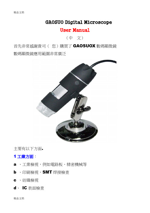
GAOSUO Digital MicroscopeUser Manual(中文)首先非常感謝貴司(您)購買了GAOSUOX數碼顯微鏡數碼顯微鏡應用範圍非常廣泛主要有以下方面.1工業方面:a 、工業檢視,例如電路板、精密機械等b 、印刷檢視,SMT焊接檢查c 、紡織檢視d、IC表面檢查2美容方面A、皮膚檢視B、發根檢視C、紅外理療(特定產品)3生物應用A、微生物觀察B、動物切片觀察4其他A、擴視器,協助視障人士閱讀B、寶石鑒定GAOSUO數碼顯微鏡使用簡單,只需要同電腦USB介面連接就可以使用,本產品還配備測量軟體,可做簡單的量測,非常的方便。
為了更詳細的介紹本產品,敬請耐心的閱讀產下面的產品介紹,使用方法,注意事項。
規格:傳感器:高性能感光晶片主控晶片:專用主控16Bit DSP放大倍率:1X ~ 500X拍照/錄影:內置輔助光源:8顆白光LED燈靜態解析度:640x480 最高可達1600x1200(可按需定制)數碼變焦:多段式成像距離:手動調節0~無限遠影像解析度:標準640*480固定底座:萬向金屬底座(可選配用升降支架)光盤:內含驅動,測量軟體,說明書支援系統:WIN XP/VISTA;WIN 7 32位和64位電源:USB(5V DC)電腦介面:USB 2.0 & USB 1.1 相容動態幀數: 30f/s Under 600 LUX Brightness照度範圍:0 ~ 30000LUX線控可調硬體需求:奔騰主頻700M Hz或以上;1G硬碟CD ROM 光碟機;64MB 記憶體支援語言:中文(簡)中文(繁體)英(其他語言需要定制)產品顏色:磨砂黑色,其他顏色可定制主體尺寸112 mm ( 長) 33 mm ( 外徑)單機包裝重量380g安全警告及注意事項1. 勿自行拆解本產品,以避免靜電擊穿精密晶片。
2. 勿用酒精等有機溶劑清潔產品。
3. 勿用手指觸摸鏡頭,以免表面造成刮痕和贓汙。
数字显微镜使用方法说明书

数字显微镜使用方法说明书尊敬的用户,欢迎您购买并使用我们的数字显微镜。
为了帮助您更好地使用该产品,我们特别提供了数字显微镜的使用方法说明书。
请您仔细阅读以下内容,并按照步骤进行操作。
1. 准备工作在开始使用数字显微镜之前,您需要准备以下物品:- 数字显微镜主机- 显微镜平台- 标本样品- 电源适配器- 连接线- 电脑或手机设备2. 连接设备将数字显微镜主机与电脑或手机设备连接,可通过USB接口或者Wi-Fi连接两种方式。
连接完成后,请确保设备之间的连接稳定。
3. 开启电源使用电源适配器将数字显微镜主机与电源连接,并将电源适配器插入电源插座。
此时,您可以通过按下电源按钮开启数字显微镜的电源。
4. 打开软件根据您的设备类型,打开相关的软件应用程序。
选择连接到数字显微镜的设备并确认连接成功。
5. 放置标本将准备好的标本样品放置在显微镜平台上,并调整好位置。
确保标本没有杂物,并尽量使其平整。
6. 调节放大倍率在软件界面上,您可以找到放大倍率的选项。
根据需要,选择适当的放大倍率。
请注意,较高的放大倍率可能导致画面模糊,因此请根据您的需求进行选择。
7. 聚焦通过调节显微镜主机上的焦距调节器或在软件界面上使用聚焦功能,将标本的细节调至最清晰。
您可以通过观察显微镜或软件界面上的画面来判断聚焦的效果。
8. 观察与拍摄确认标本已合理聚焦后,您可以通过显微镜或软件界面来观察标本的显微细节。
如果需要,您还可以使用软件提供的拍摄功能,保存显微图像。
9. 调节亮度和对比度在观察或拍摄时,您可能需要调节图像的亮度和对比度,以获得更清晰的显示效果。
在软件界面上,您可以找到相关的亮度和对比度调节选项。
10. 关机与存储使用完成后,请按下数字显微镜主机上的关机按钮,将其关闭。
同时,将标本样本从显微镜平台上取下,并将数字显微镜主机等物品妥善存放。
注意事项:- 使用过程中,请避免碰撞、摔落或强烈震动。
- 清洁数字显微镜时,请使用柔软的干净布轻轻擦拭,避免使用任何溶剂类物品。
微型显微镜使用说明书

微型显微镜使用说明书欢迎使用微型显微镜!本说明书将介绍如何正确操作、维护和存储微型显微镜,以获得最佳的观察效果和延长设备寿命。
1. 产品概述微型显微镜是一种适用于学术研究、工业检测和日常观察的便携式显微镜。
它具有高清晰度和放大倍数,可直观地观察细微结构和细胞组织。
2. 器材准备在使用之前,请确保以下物品已准备就绪:- 微型显微镜主机- 电池或充电器(根据型号而定)- 可替换镜头(可选)- 盖玻片、载玻片和移动舞台- 定标尺或标尺(根据需要)3. 启动和关闭插入电池或连接充电器以启动设备。
按下电源按钮开机,并且在使用结束后,长按电源按钮进行关机。
4. 变焦调节使用显微镜上的滚轮或按钮控制变焦,根据需要调整放大倍数。
5. 光源调节微型显微镜通常配备内置光源,可通过旋转光源开关或调节光源亮度按钮来调整照明强度和角度。
6. 样品观察将待观察的样品放置在盖玻片上,轻轻按压载玻片以确保样品贴合。
将载玻片放置在移动舞台上,然后用舞台控制按钮使样品位于视野中心。
7. 焦距调节使用焦距微调钮或旋转手柄,使观察的样品图像变得清晰可见。
8. 图像拍摄和录制部分微型显微镜具备拍照和录像功能。
按下相应按钮或通过连接电脑来进行图像和视频的捕捉。
9. 存储和维护在使用完微型显微镜后,及时将设备清洁干燥,并将其存放在专用盒子中,避免灰尘和湿气的侵蚀。
定期检查和更换电池或维护充电器以确保设备正常工作。
10. 安全使用- 不要直视太阳或其他强光源,以免对眼睛造成伤害。
- 小心处理显微镜,避免碰撞或摔落,以免损坏设备。
- 使用时保持平稳,避免快速转动或突然停止,以避免样品或设备的意外伤害。
11. 故障排除以下是常见的故障及其解决方法:- 显示屏无法正常显示:确认电池或充电器是否正常连接,重新启动设备。
- 图像模糊或不清晰:调整焦距,确保样品和载玻片表面的清洁。
- 光源不亮或亮度不足:检查电池电量或充电状态,调节光源亮度。
12. 总结本说明书介绍了微型显微镜的正确使用方法,包括器材准备、启动和关闭、样品观察、图像拍摄和录制、存储和维护、安全使用以及故障排除等。
EcoBlue 生物显微镜产品说明书

EC.1001MONOCULAR MODEL EC.1001355 (h) x 170 (w) x 220 (d) | 2.48 kg BINOCULAR MODEL EC.1152onocular, binocular and trinocular models with WF10x/18 mm he eyepieces of the binocular and trinocular models are suppliedI L L U M I N AT I O N• M onocular models are supplied with a 1 W LED illumination with integrated power supply• B inocular and trinocular models are supplied with an adjustable 1 W NeoLED™ illumination system for increased light output• M odels EC.1151, EC.1152, EC.1153, EC.1652 and EC.1653 are supplied with rechargeable batteries and an external 100-240 V battery charger/mains adapter• T he models for polarization are standard supplied with a 20 W halogen illumination for correct color rendering and an integrated 100-240 V power supply; on special requestThe models for polarization are supplied with H-LED illumination for correct color rendering (very similar to the halogen light spectrum) and an integrated 100-240 V power supply. However 20 W halogen illumination is also available on special requestPAC K AG I N G• S upplied with power cord or mains adapter-charger, dust cover, spare fuse, user manual and 5 ml immersion oil for models with an S100x objective • M odels for polarization with a halogen illumination are supplied with blue filter, models with LED illumination are supplied with white filter• All packed in a polystyrene box DIGI TAL MODEL SC A M E R A• A ll the EcoBlue digital microscopes are equipped with a 3.2 MP CMOS USB-2 camera• M aximum resolution 2048 ×1635, 24 bits color depth, up to 10 frames per second• D elivered with the ImageFocus Plus software, USB-2 cable and a micrometer 1mm/100 slide• Warranty for the camera is 2 yearsSO F T WA R E• Delivered with ImageFocus Plus for capturing of images and videos• T he capture and analysis ImageFocus Plus software allows to save images in .jpg, .tif or .bmp formats and videos in .avi format• I mages can be annotated and measurements can be performed on live or captured images• C ompatible with Windows 7, 8 and 10, both 32 and 64 bits configurations • A Mac OS version is also available• Free updates can be downloaded from our websiteEC.1001-P-HLEDEC.1152(1) Integrated mechanical stage with 70 x 27 mm X-Y travel range(2) With an external 100-240 V battery charger/mains adapter(3) P olarization models can also be supplied with S60x or S100x objectives;replace second digit of model number with resp. 6 or 1, e.g. EC.1602-P-HLED, EC.1103-P-HLED, etc ACCESSOR IES AND SPAR E PAR TSE Y E P I E C E SAE.5571 Wide field eyepiece WF 5x/18 mmEC.6010 HWF 10x/18 mm eyepieceEC.6010-M Wide field HWF 10x/18 mm eyepiece with micrometer EC.6015 HWF 15x/12 mm eyepieceEC.6110 HWF 10x/18 mm eyepiece with micrometerEC.6099 Pair of eyecupsO B J E C T I V E SEC.7004 Achromatic 4x/0.10 objective. Working distance 37.5 mm EC.7010 Achromatic 10x/0.25 objective. Working distance 6.8 mm EC.7020 Achromatic 20x/0.40 objective. Working distance 2.0 mm EC.7040 Achromatic S40x/0.65 objective. Working distance 0.6 mm EC.7060 Achromatic S60x/0.85 objective. Working distance 0.185 mm EC.7000 A chromatic S100x/1.25 oil immersion objective.Working distance 0.195 mmP O L A R I Z AT I O N AT TAC H M E N T SAE.5150 S imple polarization attachment. Polarizer 18 mm diameter mounted in eyepiece, analyzer 32 mm diameter placed infilter holderAE.5152 S imple polarization attachment. Analyzer mounted over eyepiece and analyzer 32 mm diameter in filter holder AE.5153 S imple polarization attachment. Analyzer mounted over eyepiece and 360° rotatable polarizer on table AE.5154 Polarizer in mount, to place over eyepieceAE.5155-P P olarizaton kit with analyzer in slider and small rotatable (100 x65 mm) stage with two clamps to be mounted on plain stage ofEcoBlue. Polarization filter to be mounted on the lamphouse AE.5158 P olarization kit with analyzer in slider and small rotatable(75 x 50 mm) stage with two clamps to be mounted on X-Y stageof EcoBlue. 360° Rotatable polarization filter to be mounted onthe lamphouseAE.5158-P P olarization kit with analyzer in slider and small rotatable(100 x 65 mm) stage with two clamps to be mounted on plainstage of EcoBlue. 360° Rotatable polarization filter to be mountedon the lamphouseC A M E R A ACC E SS O R I E SAE.5130 U niversal SLR camera adapter with 2x projection lens for23.2 mm tubes. Need T2 adapterAE.5025 T2 a dapter for Nikon D digital SLR camerasAE.5040 T2 adapter for Canon EOS digital SLR cameraOther T2 adapters on requestM I SC E L L A N E O U SEC.9500 A ttachable mechanical X-Y stage, translationrange 51 x 26 mm, with double Vernier andseparate horizontal adjustments knobsEuromexMicroscopenbv•Papenkamp20•6836BDArnhem•TheNetherlands•T+31(0)263232211•F+31(0)263232833•****************•EC.1005EC.9710 Frosted white filter EC.9981 1 W LED replacement unit EC.9991 1 W NeoLED™ replacement unit EC.9994 1 W H-LED replacement unit EC.9975 E xternal mains/charger adapter100-240 Vac/5 Vdc (50/60 Hz)AE.5511 Discussion head AE.5202 Blue filter, diameter 32 mm AE.5203 Yellow filter, diameter 32 mm AE.5204 Neutral grey filter, diameter 32 mm AE.5205 Green filter, diameter 32 mm AE.5207 Blue filter plexiglass, diameter 32 mm SL.1379 S pare halogen 20 W 12 V bulbfor polarization modelsAE.9918 Nylon microscope bag (25 x 39 x 19 cm)D I SP O SA B LE SPB.5157-W M icroscope slides 76 x 26 mm, ground edges.White frosted side, 50 pieces per packPB.5157-B M icroscope slides 76 x 26 mm, ground edges.Blue frosted side, 50 pieces per packPB.5155 Microscope slides 76 x 26 mm, ground edges, 50 pieces PB.5165 Cover glasses 18 x 18 mm, thickness 0.13-0.17 mm, 100 pieces PB.5168 Cover glasses 22 x 22 mm, thickness 0.13-0.17 mm, 100 pieces PB.5245 Lens cleaning paper, 100 sheets per pack PB.5255 Immersion oil, n = 1.482 (25 ml) PB.5274 Isopropyl alcohol 99% (200 ml)PB.5275 C leaning kit: lens cleaning fluid, lint free lens tissue, brush,air blower, cotton swabsPB.5276 M icroscope maintenance and servicing kit, 16pcs: cleaningbrush, 6 pcs screwdriver set, air blower, 3 pcs Allen key, 1.5, 2, 2.5 mm, lens cleaning fluid 20 ml, cleaning cloth 140 x 140 mm, 100 pcs Lens tissue sheets, tube of maintenance grease, 10 ml bottle of oil, packed in a nice toolbox。
Celestron MicroSpin 数字显微镜说明书

INSTRUCTION MANUAL MODEL # 441141. INTRODUCTIONThank you for purchasing the Celestron MicroSpin™ Digital Microscope. Please read this instruction manual carefully before using this product and retain it for future reference. With proper care and maintenance, your microscope will provide you years of service.The MicroSpin Digital Microscope differs from a traditional optical microscope. Instead of an eyepiece, MicroSpin’s internal camera sensor acts as a 10x eyepiece. The microscope connects to your computer via the USB 2.0 cable and the magnified image of your specimen is displayed on your monitor. The integrated objective wheel has 6 objective lenses with powers from 10x to 60x, resulting in 100x to 600x final magnification.*Y ou can capture and save still images and video to your computer using the included MicroSpin Digital Capture software.*NOTE: The final magnification you can achieve depends on the size of your monitor—the larger the monitor, the higher the magnification.Windows OS• W indows 8 (32 bit or 64 bit)/Windows 7 (32 bit or 64 bit)/Windows XP SP2, SP3 systems/Windows Vista (32 bit or 64 bit)• CD-ROM drive• CPU speed P4-1.8 GHz or above• Minimum 512 MB RAM• Minimum 8OO MB available hard disk space• Available USB 2.0 port Mac OS• M ac OS X 10.4.8-Mac OS X systems 10.10.x • C PU speed Power PC G3/G4/G5 or Intel-based • C D-ROM drive• M inimum 128 MB RAM• M inimum 800 MB available hard disk space • A vailable USB 2.0 portSPECIFICATIONSMagnification Powers100x, 200x, 300x, 400x, 500x, 600xSensor 2 MP CMOSImage File Format JPEGVideo Capturing Resolution320 x 240, 640 x 480Effective observation area Objective lens 100x = 3.6 x 2.70 mmObjective lens 200x = 1.8 x 1.40 mmObjective lens 300x = 1.2 x 0.90 mmObjective lens 400x = 0.9 x 0.70 mmObjective lens 500x = 0.7 x 0.50 mmObjective lens 600x = 0.6 x 0.45 mmOS systems supported Windows 8/7/XP/VistaMac OSX 10.4.8 and upConnection T ype USBPower5V, 140mAh (through USB cable connected to computer) Illuminators Upper and lower LEDSize8.19in. x 5.12in. x 6.02in. / 208mm x 130mm x 153mm Weight0.77 lbs / 350 gPARTS1.Shutter trigger button2. Objective wheel3. Objective lenses (6)4. Stage and lower illuminator LED5. Illuminator selector switch6. Upper iIluminator LED7. Stage clips8. Focus knob9. USB cableIN THE BOX58967MicroSpin DigitalMicroscope (1)(3) B lank specimen slides(1) MicroSpin Digital Capture software(3) B lank specimen slide cover slips (1) Tweezers(1) Eye dropper (1) Q uick Set-up Guide(1) P repared slide with cotton swatchCONNECTING THE DEVICE• C onnect the device to the computer by plugging the USB 2.0 cable into an open USB port on your PC.STARTING THE DIGITAL CAPTURE SOFTWAREWindows OSLaunch the MicroSpin Digital Capture software by double clicking the desktop icon from the desktop, or from the start menu {Start > All Programs > MicroSpin Digital Capture > MicroSpin Digital Capture}.Mac OS1. I nsert the supplied MicroSpin Digital Capture CD to the computer’s CD-ROM drive.2. D ouble click the “MicroSpin Digital Capture.dmg” icon within the CD.3. D rag the MicroSpin Digital Capture Viewer icon into the Mac OSLaunch the MicroSpin Digital Capture software by double clicking the icon in the Applications menu.Windows OS1. I nsert the supplied MicroSpin Digital Capture CD to the computer’s CD-ROM drive.2. D ouble click the “MicroSpin Digital Capture.exe” icon within the CD.3. F ollow the MicroSpin Digital Capture software wizard to 2. GETTING STARTEDSOFTWARE INSTALLATIONMicroSpin Digital CaptureMicroSpin Digital CaptureSELECTING A LIGHT SOURCE• T his digital microscope contains an upper (6) and lower (4) LED illuminator. Once connected to your computer, one of the lights will turn on. Press the illuminator selector switch (5) to toggle between the illuminators.SYSTEM SETTINGS MENUThe first time the MicroSpin Digital Capture software is started, the default settings will be loaded, and you may change these settings manually in the system settings menu.Device - Choose your video streaming device (in this case the MicroSpin Digital Microscope).Resolution - Change the capture resolution settings here. Timed shot setup - Setup automatic image capture here.Movie setup - Choose your video resolution here (640x480 or 320x240).Save setting - Choose the destination folder on your computer where all images and video will be nguage setting - The language of the MicroSpin Digital Capture software can be changed using this setting.Advanced settings - C licking the “More…” button on the right of the system settings menu will allow you to manually adjust all ofthe image settings.These may include:- Brightness - Contrast - Hue - Saturation - SharpnessNOTE: These settings available may vary depending on your operating system.Saved files - W ith the MicroSpin Digital Capture software open, you can locate the saved files folder by clicking the “More… “ buttonlocated on the left of the main window.Full screen - T o activate full screen mode, click the full screen button located in the bottom right hand comer of the MicroSpin DigitalCapture software window. - T o exit full screen mode, either double click on the screen, or press the “Esc” button on the keyboard.Video Record/StopInformationPower offSettings iconShutter trigger Timer 3. USING THE DIGITAL CAPTURE SOFTWAREMAIN WINDOW ICONS- Gamma - White Balance - Backlight comp- GainCARE AND MAINTENANCE• Keep the device dry and protect it from water or vapor.• Do not leave your device in a place with an extreme high or low temperature.• Do not touch the device with wet hands. Doing so may damage the device or cause an electric shock to the user.• Do not use or store the device in dusty, dirty areas as its moving parts may be damaged.• D o not use harsh chemicals, cleaning solvents or strong detergents to clean the device. Instead, wipe it with a soft cloth slightly dampened in a mild soap-and-water solution.WARNING• Do not look into the digital microscope’s illuminators. Doing so may cause permanent eye damage.• Do not attempt to open or dismantle the digital microscope.LEGAL INFORMATIONThis document is published without any warranty. While the information provided is believed to be accurate, it may include errors or inaccuracies. In no event shall the manufacturer or its distributors be liable for incidental or consequential damages of any nature, including but not limited to loss of profits or commercial loss, arising out of the use of the information in this document. Intel is a trademark of Intel Corp. In the U.S. and other countries. Mac, Mac OS and OS X are trademarks of Apple Inc., registered In the U.S. and other countries.WARRANTYY our MicroSpin Digital Microscope has a two year limited warranty. Please visit the Celestron website for detailedinformation on all Celestron microscopes:© 2015 Celestron • All rights reserved. • 2835 Columbia Street • Torrance, CA 90503 U.S.A.Telephone: 1(800) 421-9649 • Printed in China 2013FCC Note: This equipment has been tested and found to comply with the limits for a Class B digital device, pursuant to part 15 of the FCC Rules. These limits are designed to provide reasonable protection againstharmful interference in a residential installation. This equipment generates, uses, and can radiate radio frequency energy and, if not installed and used in accordance with the instructions, may cause harmfulinterference to radio communications. However, there is no guarantee that interference will not occur in a particular installation. If this equipment does cause harmful interference to radio or television reception,which can be determined by turning the equipment off and on, the user is encouraged to try to correct the interference by one or more of the following measures:+ Reorient or relocate the receiving antenna.+ Increase the separation between the equipment and receiver.+C onnect the equipment into an outlet on a circuit different from that to which the receiver is connected.+ Consult the dealer or an experienced radio/TV technician for help.This product is designed and intended for use by those 14 years of age and older. Product design and specifications are subject to change without prior notification.。
(整理)电子显微镜使用说明书

GAOSUO Digital MicroscopeUser Manual(中文)首先非常感謝貴司(您)購買了GAOSUOX數碼顯微鏡數碼顯微鏡應用範圍非常廣泛主要有以下方面.1工業方面:a 、工業檢視,例如電路板、精密機械等b 、印刷檢視,SMT焊接檢查c 、紡織檢視d、IC表面檢查2美容方面A、皮膚檢視B、發根檢視C、紅外理療(特定產品)3生物應用A、微生物觀察B、動物切片觀察4其他A、擴視器,協助視障人士閱讀B、寶石鑒定GAOSUO數碼顯微鏡使用簡單,只需要同電腦USB介面連接就可以使用,本產品還配備測量軟體,可做簡單的量測,非常的方便。
為了更詳細的介紹本產品,敬請耐心的閱讀產下面的產品介紹,使用方法,注意事項。
規格:傳感器:高性能感光晶片主控晶片:專用主控16Bit DSP放大倍率:1X ~ 500X拍照/錄影:內置輔助光源:8顆白光LED燈靜態解析度:640x480 最高可達1600x1200(可按需定制)數碼變焦:多段式成像距離:手動調節0~無限遠影像解析度:標準640*480固定底座:萬向金屬底座(可選配用升降支架)光盤:內含驅動,測量軟體,說明書支援系統:WIN XP/VISTA;WIN 7 32位和64位電源:USB(5V DC)電腦介面:USB 2.0 & USB 1.1 相容動態幀數: 30f/s Under 600 LUX Brightness照度範圍:0 ~ 30000LUX線控可調硬體需求:奔騰主頻700M Hz或以上;1G硬碟CD ROM 光碟機;64MB 記憶體支援語言:中文(簡)中文(繁體)英(其他語言需要定制)產品顏色:磨砂黑色,其他顏色可定制主體尺寸112 mm ( 長) 33 mm ( 外徑)單機包裝重量380g安全警告及注意事項1. 勿自行拆解本產品,以避免靜電擊穿精密晶片。
2. 勿用酒精等有機溶劑清潔產品。
3. 勿用手指觸摸鏡頭,以免表面造成刮痕和贓汙。
数字显微镜使用说明书

数字显微镜使用说明书一、产品概述数字显微镜是一种现代化的显微镜设备,旨在通过数字化技术来观察微小物体。
本说明书将向您介绍数字显微镜的使用方法和基本操作。
二、产品组成1. 数字显微镜主机:包括显示屏、图像传感器、光源等组件。
2. 目镜:用于观察样本的镜头。
3. 物镜:放置在目镜下方的显微镜镜头,用于放大样本。
4. 保护罩:用于保护数字显微镜主机免受外部环境的影响。
5. 支架:支撑数字显微镜主机和样品的框架。
三、使用方法1. 准备工作在使用数字显微镜之前,请确保以下准备工作已完成:- 将数字显微镜主机插入电源,并确保显示屏亮起。
- 确保目镜和物镜的表面清洁,以获得更清晰的观察效果。
- 将样本放置在支架上,并确保样本位置稳定。
2. 打开数字显微镜按下电源按钮,数字显微镜主机将开始运行。
在显示屏上将出现显微镜图像。
3. 调节焦距通过转动物镜,可以调节样本的焦距。
根据需要,使样本在显示屏上清晰可见。
4. 调节光源数字显微镜通常配有可调节光源。
根据需要,使用控制按钮来调整光源的亮度和颜色温度,以获得最佳的观察效果。
5. 拍摄图像和录制视频如果您需要记录观察结果,数字显微镜通常提供图像和视频录制功能。
使用相应的按钮来拍摄图像或录制视频,并保存在指定的存储设备上。
6. 调整放大倍率数字显微镜通常具有可调节的放大倍率。
您可以使用相应的按钮或滑杆来增加或减小放大倍率,以获得所需的观察效果。
7. 切换观察模式某些数字显微镜还支持不同的观察模式,如干扰对比、亮场和暗场观察。
根据需要,通过按下相应的按钮来切换观察模式。
四、注意事项1. 使用前,请仔细阅读本说明书,并按照正确的方法操作数字显微镜。
2. 使用过程中,尽量避免触摸或刮擦目镜和物镜的表面。
3. 使用完成后,请将数字显微镜主机和附件妥善保管,避免损坏或丢失。
4. 清洁数字显微镜时,请仅使用柔软的干净布进行擦拭,避免使用化学溶剂或粗糙的材料。
五、故障排除如果在使用数字显微镜时遇到问题,如图像模糊、无法启动等情况,建议您尝试以下解决方法:1. 检查电源连接是否正常,确保数字显微镜主机已插入电源并开启。
3B SCIENTIFIC BS-200 实验室显微镜说明书.pdf_1718717388.9668

3B SCIENTIFIC ®PHYSICS1Microscopio di laboratorio BS-200 1005455Istruzioni d’uso08/13 ALF1 Oculare2 Tubo3 Revolver portaobiettivi4 Guida per oggetti5 Tavolino portaoggetti6 Regolatore di condensatore (nonvisibile)7 Condensatore con diaframma a iride 8 Illuminazione9 Regolatore d’illuminazione (nonvisibile) 10 Base11 Scomparto lampada 12 Interruttore di rete13 Regolazione macrometrica e microme-trica con freno di arresto14 Vite di fissaggio del condensatore 15 Avanzamento coassiale del tavolinoportaoggetti 16 Stativo17 Vite di arresto del tavolino18 Vite di fissaggio della testata del mi-croscopio1. Norme di sicurezza•L’allacciamento elettrico del microscopio può essere effettuato solo ad una presa col-legata a terra.Attenzione! La lampada si riscalda durante l’uso. Pericolo di ustioni!• Non toccare la lampada durante e al terminede l’uso del microscopio.2. Descrizione, datiIl microscopio di laboratorio BS-200 consente l’osservazione bidimensionale di oggetti (sezioni sottili di piante o animali) con ingrandimento da 40 a 1000 volte.Stativo: Stativo interamente in metallo robusto e antiribaltamento, focalizzazione attraverso manopole di regolazione coassiali applicate su entrami i lati dello stativo per ingranaggi fini e grossolani con giunto a frizioneTubo: Visione binoculare, tubo inclinato a 30°,testata girevole a 360°, distanza interoculare regolabile tra 50 mm e 76 mm, compensazione diottrica ±5 per entrambi gli oculariOculare: Coppia di oculari PL10x 18 mm con ottica infinita e "High Eye Point"Obiettivo: Revolver portaobiettivi inclinato al-l'indietro con obiettivi infiniti plano acromatici 4x, 10x, 40xS e 100xS (immersione olio) Ingrandimento: 40x, 100x, 400x, 1000xTavolino portaoggetti: Piatto mobile x-y, 150 x 140 mm 2, con guida per oggetti e manopole di regolazione coassiali verticali rispetto al tavolino portaoggetti, campo di regolazione 50 x 76mm 2 Illuminazione: Lampada alogena regolabile da 6 V, 20 W, trasformatore incorporato per una tensione di rete da 90 a 240 VCondensatore: Condensatore N.A.1,25 con diaframma a iride, messa a fuoco tramite ingra-naggio a cremaglieraDimensioni: ca. 320 x 200 x 400 mm³ Peso: ca. 6,7 kg3. Disimballo e assemblaggioIl microscopio viene fornito in un cartone in Styropor.•Aprire con precauzione il contenitore una volta rimosso il nastro adesivo. Durante taleoperazione prestare attenzione affinché i pezzi dell’ottica (obiettivi e oculari) non ca-dano.•Per evitare la formazione di condensa sui componenti ottici lasciare il microscopio nel-la confezione finché non abbia raggiunto latemperatura ambiente.•Estrarre il microscopio con entrambe le mani (una mano sul braccio dello stativo e una sulpiede) e collocarlo su una superficie piana. •Gli obiettivi sono confezionati in piccole sca-tole separate. Essi devono essere avvitati nelle aperture della piastra portarevolver inordine progressivo, cominciando dal lato posteriore e in senso orario a partire dall’obiettivo con il fattore di ingrandimentominore fino a quello con l’ingrandimento maggiore.•Inserire il condensatore. A tale scopo, por-tare il tavolino portaoggetti nella posizionepiù alta, collocare il condensatore sul sup-porto e fissarlo la vite di bloccaggio.•Quindi collocare la testata del microscopio sul braccio e fissarla con la vite di bloccag-gio. Inserire gli oculari nel tubo.4. Comandi4.1 Indicazioni generali•Collocare il microscopio su un tavolo dalla superficie piana.•Collocare l'oggetto da osservare al centro del tavolino portaoggetti e bloccarlo nella guida.•Collegare il cavo di rete e attivare l’illuminazione.•Spostare il supporto portaoggetti sul percor-so dei raggi luminosi in modo che questi loilluminino chiaramente.•Regolare la distanza interoculare finché non sarà visibile un unico cerchio luminoso. •Adattare agli occhi il potere diottrico.•Per ottenere un contrasto elevato, impostare l'illuminazione posteriore attraverso il dia-framma ad iride e l’illuminazione regolabile. •Ruotare l’obiettivo con l’ingrandimento mi-nimo fino a portarlo sul percorso dei raggiluminosi. Il raggiungimento della corretta posizione viene segnalato dallo scatto dell’obiettivo. Nota: È opportuno cominciare con l’ingrandimento minimo per poter riconoscere dapprima i dettagli macroscopici delle strutture.Il passaggio a fattori di ingrandimento maggiori avviene attraverso la rotazione del revolver fino all’inserimento dell’obiettivo desiderato. Quandosi utilizza l’obiettivo 100x lubrificare con olio il tavolino portaoggetti.Il valore di ingrandimento viene ottenuto dal prodotto dei fattori di ingrandimento dell’ocularee dell’obiettivo.•Con il freno di arresto impostare la tensione adatta del sistema di messa a fuoco.•Con la manopola di regolazione macrome-trica mettere a fuoco il preparato, ancorasfuocato; prestare attenzione, durante taleoperazione, affinché l’obiettivo non vada atoccare il supporto portaoggetti. (rischio didanneggiamento)•Quindi regolare la definizione dell’immagine con la regolazione micrometrica.•Per l'uso di filtri colorati, sistemare il filtro direttamente sull'alloggiamento della lam-pada.•Utilizzando l’azionamento coassiale del piat-to mobile è possibile spostare l’oggetto daosservare nel punto desiderato.•Dopo l’uso spegnere immediatamente la lampada.•Il microscopio non deve entrare in contatto con sostanze liquide.•Non sottoporre il microscopio a sollecitazioni meccaniche.•Non toccare con le dita le parti ottiche del microscopio.•In caso di danneggiamento o di difetti del microscopio non cercare di effettuare la ripa-razione autonomamente.4.2 Sostituzione della lampada e del fusibile4.2.1 Sostituzione della lampada•Disconnettere l’alimentazione elettrica, e-strarre la spina e lasciar raffreddare il micro-scopio.•Sfilare l'attacco della lampada dallo scom-parto della stessa.•Per sostituire la lampada alogena, utilizzare un panno o simile. Non toccare la lampadacon le dita.•Estrarre la lampada alogena e inserire quel-la nuova.•Richiudere lo scomparto lampada.4.2.2 Sostituzione del fusibile•Disconnettere l’alimentazione elettrica ed estrarre assolutamente la spina.23B Scientific GmbH • Rudorffweg 8 • 21031 Amburgo • Germania • Con riserva di modifiche tecniche © Copyright 2013 3B Scientific GmbH•Svitare il portafusibili sul lato posteriore del microscopio con un oggetto piatto (ad es. un cacciavite).•Sostituire il fusibile e riavvitare il supporto.5. Conservazione, pulizia, smaltimento• Conservare il microscopio in un luogo pulito, asciutto e privo di polvere.• Durante il periodo di non utilizzo coprire se pre il microscopio con la custodia antipolvere. •Non esporre il microscopio a temperature inferiori a 0°C e superiori a 40°, né ad un’umidità relativa superiore all’85%.• Prima di effettuare lavori di cura o manuten-zione è necessario staccare sempre la spina. • Non impiegare detergenti o soluzioni ag-gressive per la pulizia del microscopio.• Non separare gli obiettivi e gli oculari per effettuarne la pulizia.•In caso di sporco notevole ripulire il micro-scopio con un panno morbido e un poco di etanolo.• Pulire le componenti ottiche con un panno morbido per lenti.• Smaltire l'imballo presso i centri di raccolta e riciclaggio locali.•Non gettare l'apparecchio nei rifiuti domestici. Per lo smaltimento delle appare-cchiature elettriche, rispet-tare le disposizioni vigenti a livello locale.。
星特朗 迷你手持式数码显微镜 说明书

使用说明书#44301目录简介 (2)参数 (2)部件 (3)显微镜所含标准附件 (3)显微镜的安装 (4)显微镜的操作 (4)使用显微镜观测和/或成像 (5)安装数码显微镜套装软件(DMS) (5)使用数码显微镜套装软件(DMS) (6)保养、维护 (7)保修条款 (8)1祝贺您正确地选择并购买了星特朗显微镜产品系列。
您新购买的这款显微镜是我们精心研制的精密光学仪器,精选优质材料,经久耐用,在最大程度上免除了您维护的烦恼。
本款显微镜几乎适用于传统显微镜的所有适用领域: 发烧友、教育工作者、医学实验室、工业检测、工程应用、教师、学生、科学应用、医生办公、公安机关、政府检验、以及用户的各类自行运用。
在使用本款显微镜之前,请仔细阅读本说明书,熟悉产品的功能和操作,以便让您使用起来更容易。
本手册中所涉及的各部件请参见显微镜图示。
此款显微镜支持15倍至30倍的放大率(与19”显示器连用)。
特别适合于检验立体物件,比如钱币、邮票、岩石、文物、昆虫、植物、皮肤、宝石、电路板、各种材料以及其它各类物件。
您也可以检验标本。
通过附带的软件,您可以在Win7、XP及Vista系统中观察放大的影像、采集视频或拍摄快照。
同时,此款显微镜还与Win7、XP及Vista操作系统中常用的图像抓取软件兼容。
如果您使用的是苹果电脑,您可以采集视频和快照,但您需要兼容苹果系统的成像/照片抓取软件(比如iChat与Photo Booth软件的组合运用等)。
您还可能会用到CD/DVD光驱以及USB接口。
注意:产品是为13岁或以上人员设计和使用的。
参数#44301参数倍率15倍至30倍 使用19” 显示器USB数据线 2.0接口电脑供电照明器6个白色LED灯泡数码相机130万像素CMOS传感器--1280x1024像素(经插值运算后快照可高达500万像素)重量及尺寸 2.9盎司 (82克) 3.5"x 1.25"(89 mm x 32 mm)23•带USB 数据线的数码照相机—内置• LED 照明器—内置• CD 光盘—数码显微镜软件套装• 使用手册显微镜所含标准附件快照按钮调焦环显微镜USB 数据线41. 小心地将显微镜取出,放置在桌子、办公桌或其他平稳表面上。
数字显微镜操作说明书

数字显微镜操作说明书一、简介数字显微镜是一种现代化的显微镜设备,利用数字技术对样本进行放大观察和记录。
本操作说明书旨在帮助用户正确操作数字显微镜,并获得最佳的观察效果。
二、操作步骤以下是数字显微镜的操作步骤:1. 开启电源和图像系统a) 确保数字显微镜已连接到稳定的电源供应。
b) 打开图像系统并启动电源开关。
2. 放置样本a) 准备一片待观察的样本,并将其放置在显微镜平台上。
b) 调节样本的位置,使其位于物镜下方的中心位置。
3. 调节镜头a) 使用像差调节旋钮,确保镜头清晰度最佳。
b) 如果需要改变放大倍数,使用倍率切换旋钮进行调整。
4. 调节照明a) 调节照明亮度,确保样本得到适当的光照。
b) 根据需要,选择透射光或反射光模式。
5. 观察样本a) 使用目镜观察样本,并通过调节焦距和调光装置,确保观察到清晰的图像。
b) 通过调节样本平台,移动样本以获得全面的观察。
6. 图像捕捉和记录a) 如需捕捉样本图像,使用数字显微镜的图像捕捉功能。
b) 如果需要记录观察结果,将图像导出到计算机或存储设备中。
7. 关闭数字显微镜a) 在使用完毕后,断开电源供应。
b) 清理样本、物镜和目镜,并储存数字显微镜在干燥、清洁的环境中。
三、注意事项在操作数字显微镜时,需注意以下事项:1. 尽量避免直接接触显微镜镜头和物镜,以免造成损坏或影响观测质量。
2. 定期清洁和维护数字显微镜,以确保其正常工作和延长使用寿命。
3. 避免在潮湿或有强磁场的环境中使用数字显微镜,以免影响其性能。
4. 在使用数字显微镜时,要保持平稳的操作,避免震动和碰撞。
四、故障排除如果数字显微镜出现故障或异常情况,请尝试以下解决方法:1. 检查电源供应是否正常连接。
2. 检查图像系统设置是否正确。
3. 清洁镜头和物镜,确保无尘或污渍。
4. 尝试重新启动数字显微镜,看是否能够解决问题。
5. 如仍无法解决故障,请联系售后服务人员进行维修或更换设备。
五、结语本操作说明书介绍了数字显微镜的基本操作步骤,希望能够帮助用户正确使用数字显微镜,获得高质量的观察结果。
显微镜简易使用手册

显微镜使用说明显微镜使用前的注意事项1安装完成后,确认显微镜的高度,建议购买可调节高度的坐椅,使得观察者和显微镜高度保持一致.2放置显微镜的桌子下方,应尽量为空置的位置,方便将腿放入.显微镜应尽量向前摆放,这样观察时腰和颈椎可以保持垂直状态,降低因为长时间观察导致的身体疲劳,并可以有效降低腰和颈椎疾病的发生概率.3使用者应当明白基本的光学原理,知道如何调节科勒照明,孔径光阑,相差,DIC,荧光等4使用者应会使用照相软件,知道如何调节至最佳状态.使用时的注意事项1 低档显微镜开关时卤素灯电压一般应调至最低。
2 转换物镜时,应旋转物镜架,不要用手直接转物镜。
3 荧光光源汞灯由于使用寿命短(大约200小时) ,为保证尽可能的延长使用时间和安全,汞灯电源开 /关之间的隔时间必须大于三十分钟。
4 显微镜的各光学部件应调节到位(如, DIC 插件,荧光滤片,物镜等),以免影响显微镜的正常工作。
如果不到位 ,那么观察时通常会看到月牙形阴影 .5 使用油镜时应使用专业用油,不要用其它介质(如香柏油),以免损伤镜头。
每次使用完油镜后,需将油镜擦拭干净(物镜及玻片)。
(正置显微镜,油滴在样品上。
倒置显微镜,油直接滴在物镜上。
)6 不要用手直接触摸光学部件的表面(如物镜,荧光模块,目镜等),以免留下手指印在上面,影响观察效果。
7 在拆卸全自动显微镜的聚光镜,荧光滤片盒(倒置显微镜)等部件时,应在断电的情况下进行操作。
8 因为各款显微镜的有效工作距离/特性等各有不同,使用前应充分了解所用仪器的特性及观测范围,以免因不恰当的操作,对显微镜及其配件造成损害。
9 调焦首先应该是让物镜向远离样品的方向调节,这样可以充分保证物镜的安全提示:1,观测样品时(特别是金相样品),切不可将物镜撞及样品,以免将物镜镜头压碎。
2,移动显微镜时,应做到小心轻放,以免因震动而对光学部件及光路造成损害,影响显微镜的使用。
(建议移动显微镜时,先将各光学部件拆除,移位后,再重新安装。
Wi-Fi数字显微镜说明书

913-2503
Raccommended Products
913-2500
ENGLISH
913-2516
Dimensions: (W)8cm x (L)15cm x (H)25cm The 913-2503 articulated desktop stand holds the Mic-Fi microscope with a flexible gooseneck arm that can be bent and twisted into many positions. This convenient and versatile stand is compatible with all Mic-Fi handheld microscopes, which allow the user with hands free stable operations of the scope.
HOW TO INSTALL APPS (Example) For iPad and iPhone: search “Mic-Fi” in App Store to download an install the app. For Android’s Smart-Phone and Tablet: search “Mic-Fi” in Google Play to download an install the app
Field of view (y) 29 15 10 7.5 6 5 4 3 2 1.5
Unit: mm
RS, Professionally Approved Products, gives you professional quality parts across all products categories. Our range has been testified by engineers as giving comparable quality to that of the leading brands without paying a premium price.
显微镜使用说明书

显微镜使用说明书一、前言显微镜是一种科学研究和生物观察的重要工具,它可以放大微小物体,使我们能够观察到肉眼无法看清的细节。
本使用说明书旨在向用户提供关于显微镜的基本操作指南,帮助用户正确使用显微镜并获取高质量的观察结果。
二、显微镜的组成1. 目镜:位于显微镜顶部的镜片,用于观察物体。
2. 物镜:位于显微镜底部的镜片,用于放大物体。
3. 镜身:连接目镜和物镜的管状结构,用于支撑和调节显微镜。
4. 台座:显微镜的底部,提供稳定的支撑平台。
5. 焦距调节轮:用于调节物镜与物体的距离,以获得清晰的观察效果。
6. 光源:提供光线照明,使样本能够被观察到。
三、显微镜的使用步骤1. 准备工作a. 将显微镜放置在平稳的桌面上,并确保底部台座稳固。
b. 打开光源,调节光线亮度,以便能够清晰地观察样本。
c. 拿起显微镜时,要轻轻握住镜身,避免触摸目镜和物镜。
2. 调节目镜焦距a. 将样本放置在显微镜台座上,并将其固定。
b. 通过旋转焦距调节轮,调节目镜与样本的距离,直到观察到清晰的图像。
3. 切换物镜a. 选择合适的物镜,通常从低倍物镜开始,逐渐切换到高倍物镜以获取更高放大倍数。
b. 注意,在切换物镜时要小心避免物镜接触到样本,以免损坏样本或物镜。
4. 调节光源a. 通过光源调节开关调节光线亮度,以获得适当的照明强度。
b. 对于透射式显微镜,可以使用光圈调节器调节光线的角度和方向,以获得最佳的观察效果。
5. 观察样本a. 将目镜对准样本,并通过移动显微镜的镜身,使样本位于视野中心。
b. 使用焦距调节轮逐渐调节焦距,直到样本变得清晰可见。
c. 注意,当调节焦距时要小心移动镜身,避免接触到样本。
四、注意事项1. 使用显微镜时要小心轻拿轻放,避免碰撞和摔落。
2. 在观察不同样本之前,应清洁物镜和目镜,以避免污染和交叉感染。
3. 在观察过程中,避免用手指直接触摸样本,以免留下指纹或损坏样本。
4. 使用适当的放大倍数观察样本,过高的放大倍数可能导致图像模糊或失真。
显微镜说明书

铰链式双目数码头(选配件)
BA400主体可选配铰链式双目数码头,成为数码显微镜DM-BA400-C (配300万像素数码系统)。 随机配备功能强大的图像处理软件。(详见软件简介)
03
DM-BA400-C
普通视频分辨率: 768 x 576(PAL) 640 x 480(NTSC)
DM-BA400-C分辨率: 2048 x 1536
目镜
内定位五孔物镜转换器,朝镜臂 内安装,使操作更为方便。
矩形机械移动载物台
WF PL10X目镜的标准视场数为 22mm,超大的视野使搜索更迅 速,观察更容易。视度可调,保 证系统齐焦。高眼点设计使观察 时无须摘下眼镜。可安装测量和 计算用的各种测微尺。 每个目镜都配有可翻叠橡胶眼罩, 可有效地防止外界光线的干扰。
物料编号
1300700102231 1300700101011 1300700100841 1300200100271 1300200100281 1300200100291 1300700901803 1300700903231 1300700902373 1300700902363 1300700207861 1300700206671 1300700207521 1300700207741 1300700207911 1300700206531 1300700206621 1300700207461 1300700207661 1300700206471 1300700200341 1300700200621 1300700201651 1300700902501 1300700902491 1300700902061 1300700903781 1300700903791 1300700902041 1300401300541 1300300900411 1300300900421 1300701001881 1300700902231 1300701000221 1300701000231 1300704200531 1300700400641 1300700902261 1300700902211 1300701001261 1300700902821 1300700902831 1300700902431 1300700901521 1300700902381 1300700902401 1300200901551 1300201000481 1300201000461 1300201000451 1300200900011 1300200901611 1300606400011
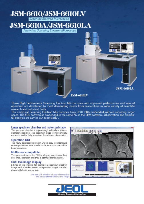
新一代高性能扫描电子显微镜(SEM-EDX)操作手册说明书

Large specimen chamber and motorized stageThe specimen chamber is large enough to handle a 200mmdiameter specimen. The specimen stage is mechanicallyeucentric and is fully motorized for efficient observation.Operation GUIThe newly developed operation GUI is easy to understandso that you do not have to refer to the instruction manual forbasic operations.Multi-user compatibleYou can customize the GUI to display only icons theyuse. Thus, operation efficiency is optimized for each user.Dual live image display2 kinds of live images, for example a secondary electronimage and a backscattered composition image, are dis-played at fall size side by side.These High Performance Scanning Electron Microscopes with improved performance and ease of operation are developed to meet demanding needs from researchers in wide variety of scientific research and industrial fields.The analytical Scanning Electron Microscopes have JEOL EDS embedded without requiring larger space. The EDS software is embedded in the same PC as the SEM software. Observation and elemen-tal analysis are carried out seamlessly .Scanning Electron MicroscopeAnalytical Scanning Electron MicroscopeThe new GUI with live display of secondaryand backscattered electron live imageJSM-6610LAJSM-6610LVMaintenance is easyThe pre-centered filament makes it simple to replace a filament. The centering of a filament in the electron gun is done automat-ically. Thus the best performance is always reproduced.Built-in animated movies help you understand these procedures.SEM Display EDS DisplayThe analytical scanning electron microscopes have 2 monitors, one for observation and the other for elemental analysis. OneA variety of measurement functions•Australia / JEOL (AUSTRALASIA) Pty. Ltd., Unit 9, 750-752 Pittwater Road Brookvale, N.S.W. 2100 Australia •Belgium / JEOL (EUROPE) B.V., Planet II, Building B Leuvensesteenweg 542, B-1930Zaventem Belgium •Canada / JEOL CANADA, INC. (Represented by Soquelec, Ltd.), 5757 Cavendish Boulevard, Suite 540, Montreal, Quebec H4W 2W8, Canada •China / JEOL LTD., BEIJING OFFICE,Room B1110/11, Wantong New World Plaza No. 2 Fuchengmenwai Street, Xicheng District, Beijing 100037, P.R.China •Egypt / JEOL SERIVCE BUREAU, 3rd Fl. Nile Center Bldg., Nawal Street, Dokki,(Cairo), Egypt •Germany / JEOL (GERMANY) GmbH, Oskar-Von-Miller-Strasse 1A, 85386 Eching, Germany •Great Britain & Ireland / JEOL (U.K.) LTD., JEOL House, Silver Court, Watchmead, Welwyn Garden City, Herts AL7 1LT, U.K.•Italy / JEOL (ITALIA) S.p.A.,Centro Direzionale Green Office Via dei Tulipani, 1 20090 Pieve Emanuele (MI) Italy •Korea / JEOL KOREA LTD., Dongwoo Bldg. 7F, 458-5,Gil-Dong, Gangdong-Gu, Seoul, 134-010, Korea •Malaysia / JEOL(MALAYSIA) SDN.BHD.(359011-M), 205, Block A, Mezzanine Floor, Kelana Business Center, 97, Jalan SS 7/2, Kelana Jaya, 47301 Petaling Jaya, Selangor, Malaysia •Mexico / JEOL DE MEXICO S.A. DE C.V., Av. Amsterdam #46 DEPS. 402 Col Hipodromo, 06100, Mexico D.F. Mexico •Scandinavia / JEOL (SKAN DIN AVISKA) A.B.,Hammarbacken 6A, Box 716, 191 27 Sollentuna Sweden •Singapore / JEOL ASIA PTE. LTD., 29 International Business Park #04-02A Acer Building, Tower B Singapore 609923•Taiwan / JIE DONG CO.,LTD., 7F, 112, Chung Hsiao East Road, Section 1, Taipei, Taiwan 10023 Republic of China •The Netherlands / JEOL (EUROPE) B.V., Lireweg 4, NL-2153 PH Nieuw-Vennep, The Netherlands •USA / JEOL USA, INC., 11 Dearborn Road, Peabody, MA 01960, U.S.A.•Please confirm other territories by the web site.1-2 Musashino 3-chome Akishima Tokyo 196-8558 Japan Sales Division 1(042)528-3381 6(042)528-3386/No. 1301G848C Printed in Japan, Kp。
数码显微诊断软件使用说明书

医疗内窥镜数码影像系统使用说明第一章系统启动与环境设置一采集影像前的准备工作---------------------------------------------------------3二文档约定---------------------------------------------------------------------------3三汉字的输入------------------------------------------------------------------------4四硬件操作-----------------------------------------------------------------------------4五软件安装---------------------------------------------------------------------------9六启动软件--------------------------------------------------------------------------12七软件设置和诊断医生登记-----------------------------------------------------13 第二章高分辨率影像采集----------------------------------------------14第三章生成显微诊断报告及保存和打印-----------------------------19第四章查阅诊断报告及打印-------------------------------------------20第五章数码显微镜技术参数及系统需求一技术参数---------------------------------------------------------------------------24二系统需求---------------------------------------------------------------------------25 第六章数码显微诊断系统软硬件使用注意事项一附加说明----------------------------------------------------------------------------25二安全警告及注意事项------------------------------------------------------------26三免责声明---------------------------------------------------------------------------26第一章系统启动与环境设置一、采集影像前的准备工作1:由于数码显微镜是用于观察患者病变部位的上皮及血管病理形态的异常变化,所以检查前应尽量减少对检查部位的刺激与干扰。
WITEC RamanMicro 200 散射谱微显微镜说明书

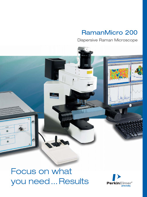
RamanMicro 200Dispersive Raman Microscope Focus on whatyou need...Results2RamanMicro 200 – an affordable dispersive Raman microscopy system that makes routine sample analysis and chemical imaging accessible to any laboratory.Faster, simplerspectroscopy and chemical imagingResults in secondsUnlike most Raman microscopes, the RamanMicro ™200 is permanently aligned and ready to use so you can focus on your results. Simply switch on and capture the information you need in secondswithout need for manual adjustments. With both manual and motorized sampling stations, the RamanMicro 200 can accom-modate a wide range of sample types and allows you to get de-tailed spectral analysis or chemical images quickly and easily.Chemical image of a poorly blended pharmaceutical tablet.Chemical image of a correctly blended pharmaceu-tical tablet.Choose manual or motorized sample stagesBoth manual and motorized stages can handle slide-based samples, while the motorized stage comes in standard and large formats to accommodate a variety of accessories such as tablet and powder holders, multi-well plates and hot cells.Add a Raman probe for outstanding flexibilityThe RamanMicro 200F includes a fiber optic probe that adds remote sampling, ‘point and identify’, bulk sample analysis and reaction monitoring to the RamanMicro 200 functionality. Software control allows for a quick switch between probe and micro-scope without the need for manual intervention.Maximum sampling versatilityProbes for:•Hand-held/non-contact •Liquid immersion •Reaction monitoring •Pilot production• Elevated temperature and pressure analysis (3000 psi and 500 ˚C)Fiber optic probe performing sample identifica-tion through large amber bottle.The same convenient tablet and powder kits, sample holders and accessories are used by both theRamanMicro 200 and PerkinElmer’s Spotlight ™FT-IR Microscope.A wide range of sampling stages allows you to analyze almost any sample type.Fast and easy assisted samplevisualization and spectrum acquisition A video alignment camera is featured for sample visualization. It assists with sample positioning and focusing, particularly useful when analyzing small or non-homogenous samples.The Visible Image Survey provides a large field of view when using a motorized stage. It raster-scans the sample giving a much larger image than the camera’s field of view. Raman maps or line scans can be overlaid onto this visible image.Advanced and intuitive software for simple, automateddata acquisition and analysisThe RamanMicro 200 uses PerkinElmer®Spectrum™software* for data acquisition and as a processing interface. This intuitive package includes all the required data manipulation algorithms as well as many novel automated features. Spectral library searching and library building software are provided, with access to a wide range of libraries.Quantitative analysis can beperformed using SpectrumQUANT+™chemometrics dataprocessing interface and chemicalimages can be generated inseconds with SpectrumIMAGE™chemical imaging interface,using peak intensity, chemi-map, COMPARE™correlationor quantitative chemometricmodels.Automate your dataacquisition andprocessing•Auto exposure timecalculated – simply typein the total analysis time•Auto spectral subtract –removes spectral contri-butions from host materialin real time•Auto baseline correction – removes the sloping base-line sometimes caused by fluorescence contamination of Raman spectra•Auto photobleaching – for significantly improved spectral quality even in samples that would otherwise yield low quality data*Common across PerkinElmer’s Raman, FT-IR and NIR product lines Push-button chemical mappingUsing a motorized stage, the sample can be point mapped.The unique Show Structure function auto-mates chemometric analysis on a spectral map by identifying the individual chemical constituents. By applying a colorimetric intensity to each constituent, the sample’s chemical distribution can be visualized.5Every compound has its own unique Raman spectrum,providing a virtual fingerprint for identification. Iden-tifying, characterizing and investigating the structures of a wide range of material types are easier than ever using the RamanMicro 200. When it comes to non-destructive sample analysis and identification Raman spectroscopy is second-to-none, but traditional spec-trometers are typically complex instruments requiring constant realignment and are difficult to use. The RamanStation ™dispersive Raman system from PerkinElmer combines this great ease of sampling with unparalleled ease-of-use. The RamanMicro 200 is equipped with a range of novel technologies such as fluorescence removal, automated spectral subtrac-tion and automated generation of chemical images.Our broad range of spectral libraries allows for rapid sample identification at the click of a buttonNon-destructivesample analysis and identification©2007 PerkinElmer, Inc. All rights reserved. The PerkinElmer logo and design are registered trademarks of PerkinElmer, Inc. COMPARE , QUANT+, RamanMicro,RamanStation, Spectrum, SpectrumIMAGE and Spotlight are trademarks and PerkinElmer is a registered trademark of PerkinElmer, Inc. or its subsidiaries, in theUnited States and other countries. All other trademarks not owned by PerkinElmer, Inc. or its subsidiaries that are depicted herein are the property of their respective owners. PerkinElmer reserves the right to change this document at any time without notice and disclaims liability for editorial, pictorial or typographical errors.007928A_01Printed in USAPerkinElmer, Inc.940 Winter StreetWaltham, MA 02451 USA Phone: (800) 762-4000 or (+1) 203-925-4602Figure on front cover. RamanMicro 200 with large motorized stage and joystick. SpectrumIMAGE software is shown displaying a chemicalimage and a spectrum from within the image.PerkinElmer: Exceedingexpectations for over 60 yearsWe provide the skills and capabilities to deliver solutions that enable laboratories like yours to be more productive and efficient. Our solutions are customized to meet your individual needs, and can address issues ranging from asset management to technical training of your personnel to equipment moves. We also provide solutions for multi-vendor environments, including preventative maintenance,validation, repair and compliance.Working with you, we’ll make sure that your laboratory achieves its goals.That’s precisely our business.PerkinElmer manufactures and supports the broadest range of instruments, reagents, and consumables in the industry –giving us unparalleled knowledge and depth of expertise.With over 60 years of experience, PerkinElmer is a company you can count on to be there when you need us. We have the largest and most experienced service force in the industry.Our 1,200 factory-trained and certified engineers have an average of 15 years of experience maintaining leading-edge scientific equipment including preventative maintenance, validation support, and instrument repair, along with the training and technical support you have come to rely upon.For a complete listing of our global offices, visit /lasofficesE-mail us at ***************************or go to /raman。
- 1、下载文档前请自行甄别文档内容的完整性,平台不提供额外的编辑、内容补充、找答案等附加服务。
- 2、"仅部分预览"的文档,不可在线预览部分如存在完整性等问题,可反馈申请退款(可完整预览的文档不适用该条件!)。
- 3、如文档侵犯您的权益,请联系客服反馈,我们会尽快为您处理(人工客服工作时间:9:00-18:30)。
非常感谢贵司(您)购买了S06-200X无线(WIFI)数码显微镜,WIFI无线数码显微镜主要针对便携式移动设备带WIFI信号的平板电脑和手机。
非常便利。
无线数码显微镜应用范围非常广泛,主要有以下方面.
1工业方面:
a工业检视,例如电路板、精密机械等
b印刷检视,SMT焊接检查
c纺织检视
d IC表面检查
2美容方面
a皮肤检视
b发根检视
C红外理疗(特定产品)
3生物应用
a微生物观察b
动物切片观察
4其它
a扩视器,协助视障人士阅读
b宝石鉴定
……
S06无线数码显微镜弥补了有线数码显微镜的距离限制,能够在空间
10~30m的环境内,在IOS系统的平板和手机(如Ipad 和Iphone),andriod 系统的平板和手机上使用,非常的方便。
为了更详细的介绍本产品,敬请耐心的阅读产的产品介绍,使用方法,注意事项。
H o T ech
0录
首页 (11)
前言 (12)
目录 (13)
各部位介绍 (13)
产品规格 (14)
使用说明 (14)
安全警告及注意事项 (19)
配件说明 (19)
各部位说明
1
3
2
4
5
7
6
①DC5V插座②开关盒调光旋钮③充电和WIFI信号指示灯④调焦滚轮⑤
倍率刻度⑥镜头⑦辅助光源灯罩
H o T ech
规格:
传感器:高性能外国进口感光芯片
主控芯片:专用主控DSP
放大倍率:5X ~ 200X
拍照/录像:应用程序内置辅助
光源: 4 ~ 8颗白光LED灯
静态分辨率:640*480,可根据自己的要求做放大和缩小处理
成像距离:手动调节0~40mm,其他距离可定制影像分辨率:640*480,可根据自己的要求做放大和缩小处理固定底
座:特制底座
0盘:含应用软件及说明书
支持系统:基于IOS系统和andriod系统的平板电脑和手机
电源:内置1000mA自保护锂电池
应用程序:hotviewer
动态帧数: QVGA 30f/s Under 600 LUX Brightness
VGA 20 f/s Under 600 LUX Brightness 照度范围:0 ~ 30000LUX线控可调
硬件需求:主频700M Hz以上,1G硬盘CD ROM光驱;
64MB 内存支持语
言:英文(其它语言需要定制)
产品颜色:深蓝色主体尺寸:
142mm (L)* 37mm ( R)
单机净重:100g
使用说明:
1 打开电源开关,将光线调整到最亮
开关和调光旋钮
WIFI 指示灯
图1图2 2LED绿色指示灯开始亮起,3~5S 后,LED灯开始闪烁,代表WIFI信号发射正常
3 针对IOS系统(IPAD)为列,做如下说明:
1 NetcamViewer 软件下载
2 在APP store 中,搜索栏中输入NetcamViewer
3安装后,应用程序面会出现如下图标
4 点击NetcamViewer,进行一些参数设置
用户名同密码一致,都是HOT
全部设置完成后,点击右上方按钮就完成设置了
深圳浩特尔电子技术有限公司
4进行WIFI信号连接,点击Settings
3~5S后在Network中选中SSID为HOT
SSID:HOT
SSID:HOT
5 WIFI 信号连接后,点击应用图面上的,就可以正常使用了
如下图
3WIFI信号连接后,如果不想下载AP软件NetcamView,可直接在Safari 中,IP地址栏中直接输入10.10.1.1:8196,就可以再网页中直接浏览图像。
如果需要输入用户名和密码的,直接输入HOT
4针对android系统的平台,将光盘中的AP软件HotViewer,直接安装到相应的平板电脑或手机中应用程序免会出现相应的图标。
使用方式同之前一致.打开显微
镜开光---将光线亮度调到最大----左侧LED灯开始闪烁,标示信号正常
深圳浩特尔电子技术有限公司
了.设置--Wifi--SSID为HOT,连接。
然后直接点击应用程序的图标就可以正常使用了
安全警告及注意事项
1.勿拆解本产品,以避免静电击穿精密芯片。
2.勿用酒精等有机溶剂清洁产品。
3.勿用手指触摸镜头,以免表面造成刮痕和脏污
4.户外使用时应避免高温和高湿环境中,防电子器件短路。
本产品不具有防水功能,请应避免淋雨和进水。
5.本产品的使用和存储温适度范围:0°C ~ 40°C,相对湿度:45%RH ~
85%RH
6.若不慎使异物或水分/液体进入数产品内部,立即断开电源并送至维
修中心检修,切勿自行处理。
7设备测量精度仅供参照,由测量误差引起的纠纷与本产品无关。
8本产品有效距离在10~30m之内。
配件说明
1.无线(WIFI)数码显微镜(1台)
2.专用底座一台
3.充电器一台
4 光盘(安装说明书)
5. 保修卡一份,快速使用指南一份。
