黄色肉芽肿性胆囊炎的影像表现及病理学特征
黄色肉芽肿性胆囊炎6例CT诊断分析

肿大淋 巴结 1 例。 T误诊为胆囊癌 4 , C 例 一般慢性胆 囊 炎伴 结 石 1 。 例 6例均行 胆囊切 除术 。 体病理 : 大 胆囊壁 不规则 增厚 . 最厚达 25c . m伴有溃疡形成 。 内均可见境界 壁 不清黄棕色小结节、 黏膜粗糙 。 镜下检查 : 壁内见结节 状 肉芽肿性结构 .中央为炎性坏死组织伴有淋 巴细 胞、 单核细胞浸润 , 外层为大量吞 噬脂质 的泡沫样组 织 细胞 浸润 。
3 讨 论
X C是 一 种少 见 、 良性 而有 破 坏 性 的 胆囊 慢 性 G 炎症 [, 引 常与胆囊结石并存 , 它见于外科切除胆囊 的 05 1 %[ .%~ . 。男 女 均 可 发 病 , 病 年 龄 以 5 ~ 0岁 8 发 O6 多见 , 本组 病 例平 均 年 龄 5 2岁 , 女 比例 相 同 。该 病 男 病 因 目前 尚不 完 全清 楚 。一 般认 为 是 由 于结石 嵌 顿 、 胆汁淤积等因素的共同作用导致罗一 阿窦破裂 。胆汁 由此溢人胆囊壁 . 引起 以巨噬细胞浸润为主的炎症反 应 。 汁 中的 脂 质 和胆 固醇 被 巨 噬 细胞 吞 噬 。 成 泡 胆 形 沫细 胞 和多 核 巨细胞 .并 聚集 成 肉芽肿 性病 变 。 病 本 病理特征为胆囊壁炎性破坏 区大量泡沫细胞 、 巴细 淋 胞 、 核 细 胞 聚集 . 形 成 的病灶 有 较 多 的炎 症 与 坏 单 新 死表现 . 而老病灶 主要为纤维组织伴有少量泡沫细胞 浸润 。文献 [ ] ・ 报道 X C中有 1% - 5 G 0 伴发腺癌 。 本组无 此类病例。 G X C的临床表现无特异性 。 与一般 的胆囊 炎类似 , 主要为腹痛 、 呕吐 、 发热 、 黄疸等 。 故易误诊为 般慢性胆囊炎伴结石 . 本病例有 5 例表现为反复发 作的慢性胆囊炎病史 。 其中 1 例误诊 。 31 X C的 C . G T征 象 平 扫多 为胆囊 壁 不 均 匀增 厚 。 根 据 病 变 累及 是 否 超 过 胆囊 周 长 的 5%分 为 局 限 型 0 和 弥漫 型 两种 。 以弥 漫型 多见 。 本病 例 弥漫 型 4例 , 局 限型 2 。囊壁内可见多个大小不等低 密度结节影 。 例 即壁 内黄色 肉芽肿 .作为该病特征性表现有助于诊 断。 本组 4 (/ ) 例 4 6 出现该征象 。 如数 目较多且邻近呈 融合状 .T则表现为增厚 的胆囊壁呈分隔状改变。 c 常 伴胆囊结石 。 本组 5 。倪炯等[ 例 6 ] 报道 1 例 X C中 5 G 发现 2 例钙化 , 本组无类似病例。增强扫描增厚 的胆
胆囊炎影像诊断

2021年7月5日6时31分
第 22 页
三 急性胆囊炎
三 急性胆囊炎
基本知识
胆囊急性感染,以50~69年龄段居多。 临床:
高热、畏寒、腹痛、肌卫、Murphy征阳性。 发病机理:
结石性胆囊炎(90%):与胆囊管结石嵌顿有关。 非结石性胆囊炎(10%):严重创伤或长期禁食,胆囊扩张导致胆 囊壁缺血。 易患因素: 胆石症,油腻食物,营养过度。
2021年7月5日6时31分
第 15 页
一 检查方法
MRI
胆囊壁: 菲薄呈细线状。
胆汁: T1WI:胆汁多呈低信号,郁积的胆汁为等信号或稍高信号。 T2WI:多为极高信号,少数为略高信号。
2021年7月5日6时31分
第 16 页
一 检查方法
直接多轴面成像
MRI
2021年7月5日6时31分
第 17 页
2021年7月5日6时31分
第 43 页
四 慢性胆囊炎
影像检查
CT/MRI 1、胆囊体积可显著萎缩或呈“葫芦状”。 2、胆囊壁光滑或增厚(充盈良好时≥3mm) ,呈软组织密度,可有钙 化,重度T2WI上,胆囊壁信号比邻近脂肪信号低。 3、胆囊周围无明显水肿/积液。 4、动态增强,黏膜和肌层早期强化,黏膜下层(纤维化)延迟强化。 5、应用促胰素后收缩能力减低。 6、胆囊浓缩功能差 鉴别诊断:
黄色肉芽肿性胆囊炎( xanthogranulomatous cholecystitis ) 弥漫性腺肌瘤病( Diffuse Adenomyomatosis) 局灶性腺肌瘤病( Focal Adenomyomatosis) 瓷胆囊(Porcelain Gallbladder) Mirizzi 综合征 胆囊憩室炎 胆固醇沉着症/胆固醇息肉 胆囊壁反应性增厚Reactive Thickening of the Gallbladder Wall 胆囊壁静脉曲张Varices in the Gallbladder Wall
21例黄色肉芽肿性胆囊炎的诊断

山东医药2023 年第 63 卷第 3 期21例黄色肉芽肿性胆囊炎的诊断杨志鹏1,谷建斌2,张玉斌2,王岩2,薛菲2,杨玉华31 河北医科大学,石家庄 050000;2 石家庄市人民医院肝胆外科;3 石家庄市人民医院病理科摘要:目的 总结黄色肉芽肿性胆囊炎的有效诊断方法。
方法 对21例黄色肉芽肿性胆囊炎患者的临床表现、实验室检查、影像学特征及病理结果进行回顾性分析。
结果 21例患者中,8例出现皮肤巩膜黄染,10例Murphy征阳性;16例白细胞计数偏高,9例ALT偏高,7例AST偏高,8例TBIL偏高;血清肿瘤标记物中,4例患者CA19-9>1 000.00 U/mL,4例CA125及1例CEA超出正常范围;术前影像学检查诊断为胆囊结石17例,胆总管结石10例,发现胆道结石性炎症、胆囊壁弥漫增厚、胆囊壁内存在低密度结节、胆囊壁强化后可见“夹心饼干征”且胆囊内黏膜线完整,以及胆囊邻近组织器官存在炎性浸润等;5例患者术中发现胆囊与周围组织黏连较为紧密,对周围组织器官侵袭较严重,送快速冰冻病理检查,均除外胆囊癌。
术后19例病理结果为黄色肉芽肿性胆囊炎,1例为黄色肉芽肿性胆囊炎合并胆囊腺肌症,1例为黄色肉芽肿性胆囊炎合并胆囊癌及胆管癌。
结论 影像学检查发现胆道结石性炎症、胆囊壁弥漫增厚、胆囊壁内存在低密度结节、胆囊壁强化后可见“夹心饼干征”且胆囊内黏膜线完整,以及胆囊邻近组织器官存在炎性浸润等可考虑为黄色肉芽肿性胆囊炎,术中冰冻活组织检查或术后病理检查可明确诊断。
关键词:黄色肉芽肿性胆囊炎;胆囊炎;胆囊结石doi:10.3969/j.issn.1002-266X.2023.03.021中图分类号:R657.4 文献标志码:A 文章编号:1002-266X(2023)03-0088-03黄色肉芽肿性胆囊炎(XGC)是一种以胆囊壁内炎性瘤样变为特征的胆囊炎,其发病率占所有胆囊炎的1.46%~5.0%[1],在临床上比较罕见。
黄色肉芽肿性胆囊炎的影像学表现研究

取术前行常规超声及超声造影检查的XGC 14例,同时行增强CT检查的9例,分析图像特点,总结其特征表现。结果
14例病灶胆囊壁均增厚,13例伴囊腔内结石,1例伴胆总管结石。超声造影4例病灶增厚的囊壁动脉期出现小灶性不增
强区,9例胆囊黏膜线完整清晰,6例浆膜线完整清晰,4例胆囊旁肝实质有减退区。增强CT组与超声造影组囊壁动脉期
The study on the imaging findings of xanthogranulomatous cholecystitis
SHEN Haiyun, KONG Wentao, XUE Haiyan, HAN Hao, WU Min Department of Ultrasound, Nanjing Drum Tower Hospital, Naming University, Nanjing 210008, P. R* China
[Abstract] Objective To investigate the diagnostic value of contrast-enhanced ultrasound for xanthogranulomatous cholecysti tis (XGC). Methods There were 14 cases of XGC who underwent preoperative ultrasound and contrast-enhanced ultrasound
(CEUS) examination in our hospital from February 2016 to April 2020 and 9 cases who underwent contrast-enhanced CT examina tion at the same time. The images were analyzed to summarize the performance o£ XGC. Results All 14 gallbladder walls were thickened, 13 cases were with intracystic stones, 1 case was with bile duct stones. There were 9 cases that the gallbladder muco sal line was continuous and 6 cases that the gallbladder serous line was intact and clear. And the liver parenchyma adjacent to the gallbladder was with focal reduction area in 4 cases. There was significant difference of the degree of gallbladder enhancement be tween group contrast-enhanced CT and contrast-enhanced ultrasound. The diagnostic accuracy of the contrast-enhanced ultrasound was higher than contrast-enhanced CT. Conclusion Contrast-enhanced ultrasound has more advantages in the observation of the enhancement characteristics of the arterial phase of gallbladder lesions, and has more diagnostic value for XGC. [Key words] Xanthogranulomatous cholecystitis ; Contrast-enhanced ultrasound ; Tomography, X-ray computed
黄色肉芽肿性胆囊炎影像诊断及手术治疗

黄色肉芽肿性胆囊炎影像诊断及手术治疗黄色肉芽肿性胆囊炎(xanthogranulomatous cholecystitis,XGC)是一种极少见但临床明显的胆囊疾病,其病理特征为胆囊壁局部或全壁出现黄色肉芽肿性炎性改变。
本文旨在探讨XGC的影像诊断及手术治疗。
一、影像诊断XGC影像学上表现为胆囊壁局部或全壁增厚,病变区呈均质或不均形低密度或中等密度影像,可见胆囊内低密度的黏液、结石等。
胆囊管扩张或腹腔周围淋巴结增大径向是XGC常见的CT表现之一。
在磁共振成像(MRI)中,寻常胆囊炎表现为胆囊白色结构。
XGC表现为胆囊局部或全层不均匀增厚,可以看到不连续的低信号变化区,以及胆囊壁增厚与增强的不匀性,具有高度特异性。
同时,MRI可以检测到胆囊周围淋巴结的增大和胆囊区域的转移,具有极高的准确性和敏感性,是诊断XGC的关键检查方法之一。
二、手术治疗对于XGC患者,一般推荐对症治疗或保守治疗。
对于常规治疗无效或合并有胆囊结石及其他并发症的患者,需要考虑手术治疗。
手术治疗的主要方式为胆囊切除,包括开放胆囊切除和腹腔镜胆囊切除两种方式。
对于大部分XGC患者来说,由于胆囊局部弥漫性病变切除困难,因此开放胆囊切除是常规选择。
开放胆囊切除手术切口较大,创伤较大,术后疼痛、恢复时间长,但是在XGC的治疗中其切除率高、疗效显著,是目前最常用的XGC手术方法。
近年来,随着微创技术的发展,腹腔镜胆囊切除对于轻、中度XGC患者来说仍是一种可行的选择。
与开放手术相比,腹腔镜胆囊切除手术损伤较小、恢复时间短、疼痛轻,但是手术难度大、操作要求高,对于病变程度较严重的患者效果较差,仍存在术后胆囊切缺失率较高等风险,因此选择要慎重。
总之,XGC是一种罕见但临床症状明显的胆囊疾病,在影像学和手术治疗中仍然面临一定的挑战。
合适的影像学检查可以对XGC进行准确的诊断,手术治疗的选择则应根据患者病情、治疗目的和外科医生经验来决定。
要提醒的是,XGC对于胆囊及周围脏器的转移和复发风险较高,因此术后患者还需进行持续随访和治疗。
黄色肉芽肿性胆囊炎的病理诊断、腹腔镜胆囊切除术及护理
geo i n染 色 、 mii 应 ]可 证 实 为 胆 色 素 。 1 s G ln反 l ”, 2例 同 时 伴 有 胆 汁性 肉 芽 肿 、 噬 胆 汁 的 多 核 巨 细 胞 及 大 量 胆 固 醇 裂 隙 , 5 答 4
免 疫 组 织化 学 染 色 泡 沫 样 细 胞 及 巨噬 细 胞 AA + ) C 6 ( ) 胆 囊 黏 膜 及 R—A 窦 上 皮 E T( 、 D 8 + ; MA( ) C + ) C A( ) 腹腔 镜 组 + 、 K( 、 E 一 。
的手 术 时 间 、 院天 数 明显 短 于开 腹 组 。[  ̄ ] GC是 一 种 良性 而有 破 坏 性 的 特 殊 类型 胆 囊 炎 , 要 表 现 为 胆 囊 壁 不 同程 度 增 厚 , 住 结 X 主 常伴 有胆 囊 结 石 或 胆 管 结 石 , 临床 诊 断 困难 , 诊 有 赖 于病 理 诊 断 , C 可作 为 治 疗 X C 的 首选 方 法 , 确 L G 高质 量 的 护 理 是 L 成 功 的 重 C
质 中 可 见 棕 黄 色 色 素 颗 粒 , P u s n蓝 、 A 、 l i 经 r si a P S A ia c n蓝 、 a V n—
的炎 性 病 变 , 胆 囊 慢 性 炎 症 的 特 殊 类 型 l 。 X C 的 临 床 表 是 _ 2 ] G 现及 超 声 表 现 难 以 与 胆 囊 炎 胆 石 症 鉴 别 _ 。当 X C 的 胆 囊 壁 3 ] G
全 科 护 理 20 0 9年 1月 第 7卷 第 1期 中旬 版 ( 总第 1 1 ) 3 期
常见胆囊炎性病变的影像学诊断
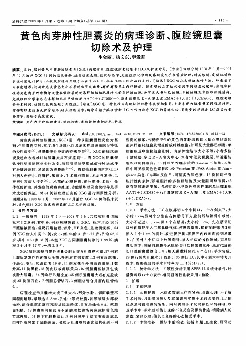
常见胆囊炎性病变的影像学诊断 1、急性单纯性
胆囊增大(≥5cm),胆囊壁厚、水肿。
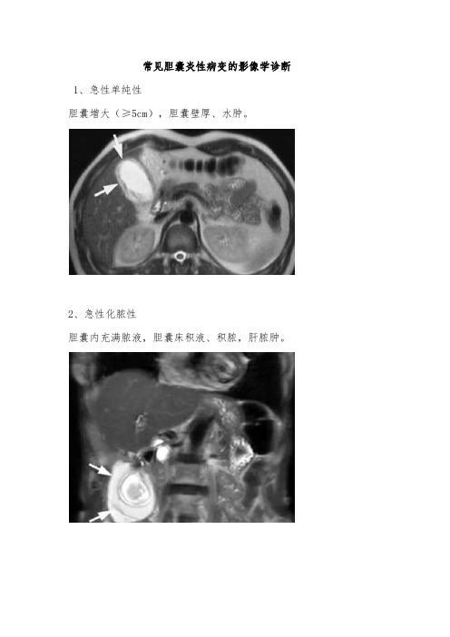
2、急性化脓性
胆囊内充满脓液,胆囊床积液、积脓,肝脓肿。
3、急性坏疽性
胆囊缺血、坏疽、穿孔,胆系内/胆系旁积气(气肿性胆囊炎),胆囊内积血(出血性胆囊炎)。
4.慢性胆囊炎
体积可显著萎缩或呈“葫芦状”;胆囊壁光滑或增厚,可有钙化;胆
囊周围无明显水肿/积液。
5.黄色肉芽肿性胆囊炎
胆囊壁明显增厚,通常弥漫性。
胆囊壁不均匀强化。
6.胆囊腺肌症
胆囊壁增厚+假憩室形成(胆囊壁平滑肌增生相关的罗-阿氏窦)
7.瓷胆囊
胆囊壁连续性、断续性壳样钙化。
除钙化外,还见于纤维化、胆囊壁内积气。
8.Mirizzi 综合征Ⅰ型
胆囊颈/胆囊管结石导致的胆总管外侧局限性贝壳样狭窄。
9.Mirizzi 综合征Ⅱ型
胆囊管-CHD连接处,尤其胆囊管内显示结石,没有胆总管侧方光滑压迹。
10.气肿性胆囊炎
胆囊壁内、胆囊腔内积气
11.钙胆汁
胆汁密度弥漫性增高
12.胆囊憩室周围炎
积液、水肿、胆囊壁增厚等急性感染表现,可见到1~多个憩室。
13.胆固醇息肉。
15例黄色肉芽肿性胆囊炎患者的诊疗报告

降的双硫仑样反应患者可应用升压药进行治疗 , 一般数小时内就能缓
1 黄色 肉芽肿 性胆囊炎 患者 的诊 疗报告 5例
汪 杰
( 长沙市八 医 院 湖南 长沙 400 ) 1 1 0
【 摘要 】 目的: 探讨黄色 肉芽肿性胆囊炎( n hg au0 aosc o cs i s XC的诊 断与治疗方法。 x t0 rn 1m t u h l ytt , G ) a e i 方法: 回顾性分析2 0 0 5年8月~ 0 1 2 1
3 急 救 及 护理
人 们一 旦 出现 双硫 仑 样 反应 , 及时停 药 和 停用 含 乙醇 制 品 , 应 轻者
可 自行缓解 。 较重者需进行吸氧及对症治疗。 治疗上可通过洗胃来排 除 胃内 乙醇 , 少 身体对 乙醇 的 吸收 , 减 同时 可静 注地 塞 米松 或 肌注 纳 洛 酮 , 静 脉输 葡萄 糖液 、 维生索 C , 行护 肝 治疗 , 进 乙醇 代谢 和 排泄 。 等 以进 促 伴 有 心绞 痛 的双硫 仑 样反 应患 者 需进 行改 善冠 脉 循环 的治 疗 , 有 血压 下 伴
《 求医问药 Y  ̄P刊 S e e i l dA k h Me i n 2 1 F l ekM d c An s T e d c e 0 2年第 1 卷 第 4 a i 0 期
1 01
双硫仑样反应的临床表现多样 , 程度不一 , 与饮酒种类、 饮酒量和饮 酒时间密切相关。 一般情况下, 在服药期间饮用白酒且饮酒量大 , 临床 其 表 现就 复 杂 , 情程 度 也就越 重 , 者 可 出现 胸 闷、 病 患 气性胆 囊炎 ; 囊切除 ; 断; 胆 诊 治疗 【中图分类号 】 6 74 R 5 . 【文献标 识码 】B 【 文章编号 】 7 — 5 3 2 1 0 — 1 l 0 1 2 2 2 ( 0 4 0 0 一 2 6 2)
黄色肉芽肿性胆囊炎的影像诊断分析
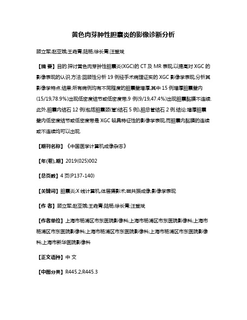
黄色肉芽肿性胆囊炎的影像诊断分析顾立军;赵亚娥;王垚青;陆杨;徐长青;汪登斌【摘要】目的:探讨黄色肉芽肿性胆囊炎(XGC)的CT及MR表现,以提高对XGC的影像表现的认识.方法:回顾性分析19例经手术病理证实的XGC影像学表现,分析其影像学特点.结果:所有病例均有不同程度的胆囊壁增厚,其中15例增厚胆囊壁内(15/19,78.9%)出现低密度结节或低密度带,9例(9/19,47.4%)出现胆囊黏膜不连续.此外,胆囊内结石12例(包括胆囊颈(管)结石5例),胆总管结石2例.结论:增厚胆囊壁内低密度结节或低密度带是XGC较具特征性的影像学表现,而胆囊内黏膜的连续或不连续均可以出现.【期刊名称】《中国医学计算机成像杂志》【年(卷),期】2019(025)002【总页数】4页(P137-140)【关键词】胆囊炎;X线计算机,体层摄影术;磁共振成像;影像学表现【作者】顾立军;赵亚娥;王垚青;陆杨;徐长青;汪登斌【作者单位】上海市杨浦区市东医院影像科;上海市杨浦区市东医院影像科;上海市杨浦区市东医院影像科;上海市杨浦区市东医院影像科;上海市杨浦区市东医院影像科;上海市新华医院影像科【正文语种】中文【中图分类】R445.2;R445.3黄色肉芽肿性胆囊炎(XGC)是慢性胆囊炎的一种变型,通常由于胆囊出口梗阻导致的胆囊内压力增高,胆汁中的脂质进入胆囊壁结缔组织中,引起的慢性炎症反应,以胆囊壁内胆汁性肉芽肿的形成并伴有重度增生性纤维化及大量泡沫样细胞为特征,病变发展具有一定的破坏性,因此与胆囊癌表现部分重叠、相似,术前较易误诊,所以正确的术前诊断可以避免侵袭性手术,也对患者预后产生影响[1]。
笔者回顾性分析19例经手术病理证实的XGC患者的临床及影像资料,以期提高该病的术前诊断水平。
方法1.临床资料收集本院2012年1月至2018年6月经病理证实、资料完整的XGC患者19例,其中男11例,女8例;年龄30~86岁,中位年龄61岁。
黄色肉芽肿性胆囊炎CT诊断ppt课件
增厚的胆囊壁内显示低密度结节,增强后无强化,门静脉期显著。 通常无门腔间隙、肝门及腹腔动脉周围淋巴结肿大,无肝脏转移等恶性
肿瘤的影像学表现。但可有胆囊床炎性浸润。 增厚胆囊壁内低密度结节是XGC的特异性CT表现。
29
谢谢!
30
1
黄色肉芽肿性胆囊炎 (xanthogranulomatous cholecystitis, XGC)是一种少见特殊类型的胆囊炎性疾病,在胆
囊
1976年Christensen和Ishak首先以纤维黄色 肉芽肿性炎症报道描述了此病变,其后亦有学 者将此病变命名为胆囊蜡质或蜡质样组织细胞 肉芽肿和胆汁性肉芽肿性胆囊炎.该病在发病率 低,仅占胆囊炎症性疾病的0.7%~13.2%,其 中以中老年人多见,男女比例不一.
2
XGC的病因及发病机制目前尚不十分清楚, 但绝大部分病人伴有胆石症,说明胆囊结石 在其发病中至关重要,另外XGC部分病人伴 有高脂血症和2型糖尿病,说明该病可能与 代谢紊乱有关。
3
多数学者认为胆道梗阻合并细菌感染是导致该病发生的关 键因素。胆囊结石等因素引起胆囊壁黏膜溃疡,局灶微小 脓肿形成,并使T淋巴细胞受刺激而发生细胞免疫反应; 胆汁及黏蛋白沿破裂(当存在胆道阻塞使得壁内压力升高 所致)的罗-阿氏窦(Rokitansky-Aschoff窦)或黏膜溃 疡病灶不断渗入胆囊壁,导致其慢性炎症;巨噬细胞聚集, 吞噬胆汁中的胆固醇、磷脂及胆色素等,形成富含脂质的 特异性泡沫细胞(即黄瘤细胞);随病程进展,纤维组织 增生,逐渐形成炎性肉芽肿,进而胆囊壁出现局灶或弥漫 性增厚,形成XGC,病程进一步进展,胆囊与肝及周围脏 器粘连,甚至导致胆囊内瘘、胆囊坏疽或穿孔。因此, XGC的形成是间质组织对胆汁外渗的反应,这一点由脂质 和脂褐质在泡沫细胞内予以证实。
黄色肉芽肿性胆囊炎31例临床病理分析

Th l ia nfsain o eci c l n ma i tt f e o XGC sl ec rncc oe ytt . wa k ho i h lc sii sCT x mi t ns o d t ik n coe yt ai i s e a n i h we h t c e h lc si p r a o e h c s e
ma u hl yti X C) i re rv u n e t dn idsaeMehd Th t-n ae f G o t sco cs t e is( G , nod rOi o e r dr a i o t s i s. to s t mp o u sn g f h e i yo e sso C r c X
l f mmain o h alld e .icreain 0 ac lsa dc oet i. ti a kn fd y d g a uo tu fet n a t fteg l a d r n c rt fc uu n h l a s I id 0 da e rn lmao safci o b a o l ss s o
壁增厚或结节/ 块及与周围器官粘连或内瘘形成, 月 中 而误诊为癌。病理大体见胆囊壁黄色结节或肿块, 镜下见急
慢性炎症背景上有特征性泡沫细 胞 肉芽肿 肿性病变 。
【 词】 胆囊炎 , 肉芽肿性 ; 免疫组织化学 关键 黄色
Xa t o r n lma o h lc sii : n c p t oo ia t d f 3 a e n h g a u o t m c oe y tt a di io a h lg c ls u y o 1 c s s s 删 Y h me , HI Li n , a — i Z a L, ・
黄色肉芽肿性胆囊炎的MRI表现特点

M RI i agng e t e o nt g a m i f a ur s f xa ho r nul m at us c l c tts o o ho e ysii
H UA G N Zi x n . CH EA — ig Gt n ue .LU Ch n l. S t g- n a a g- i OXG Bi n
符合 其 病 理 特点 ?
【 键 词】 胆 囊 炎 ; 共振 关 磁 【 图 分 类号 】 R 7 .l R 4 . 中 5 5 ; 4 52 6 【 献标 识 码 】 A 文 【 章编 号 】 1 0 一 0 2 2 1 )2 0 5 — 4 文 0 8 l6 ( 0 0 1 — 8 6 0
f s hn s i 1 ih a n ri ' h l dl 1 0 1 hn ) We t ia Hopt .Sc u n C a est,C el l 0 4 .C ia )  ̄ 6
A s at bt c:Obet e O ivsgt R et e o xnhgau( t seoeyti X C .Meh d :A rt set e r jc v :T net a M Ifa rs u ma is( G ) to s e opci r v
中断 4例 。 并胆 囊 结 l 合 5例 : 昕 病 例 均 出现 肝 实 质动 脉 期 一 过性 强 化 , 近 肝 实 质 出现 肝 脓 肿 7例 , 十 指 肠 粘连 6例 。 邻 与 二 结论 : 胆囊 壁 不 均匀 增 厚 、 内出 现结 节 、 鼙 黏膜 线 完 整行 明 显强 化 、 近 肝 匝 动 脉 期一 过 强 化 是 X C较 常 见 的影 像 表 现 ,| 邻 G J
性增 厚 , 为 局灶 性 增 厚 l 弥漫 件 增 厚 的病 例 中均 匀件 增 厚 2例 , 均 匀增 厚 1 例 1 患 者 出 现胆 囊 壁 内结 节 , 小 2例 3例 l 0例 大
黄色肉芽肿性胆囊炎1例ppt课件
Murphy’s征(-)。 ❖ 实验室检查:CA-199(200.2u/ml)明显升高,白细胞
、中性粒细胞升高。
12Leabharlann 3456
???
7
病理结果
❖ 1、胆囊及肝脏:黄色肉芽肿性胆囊炎累及肝脏。 ❖ 2、(第十二组)淋巴结反应性增生。
11
讨论
❖ 黄色肉芽肿性胆囊炎肉眼观表现为边界不清、黄色肿块侵及 胆囊壁,组织学上由泡沫细胞、淋巴细胞、浆细胞、多核白 细胞、纤维母细胞和异体巨细胞构成。黄色肉芽肿性胆囊炎 的影像学表现为胆囊壁增厚,可以是结节状,也可以是脂肪 界面消失。超声检查,黄色肉芽肿性胆囊炎表现为在增厚的 胆囊壁内出现低回声条带或代表脓肿灶的结节,其他表现有 黏膜线中断、胆囊周围积液、胆囊石和肝内胆管扩张等。
12
讨论
❖ CT可以有效显示黄色肉芽肿性胆囊炎对临近器官的侵犯、 周围组织的浸润情况。虽然黄色肉芽肿性胆囊炎是一种良性 的炎性过程,但可与胆囊癌、胆管癌并存,此外,与胆囊癌 的影像学表现类似也使二者在术前鉴别困难。
13
此课件下载可自行编辑修改,供参考! 感谢您的支持,我们努力做得更好!
10
讨论
❖ 黄色肉芽肿性胆囊炎是一种罕见的慢性胆囊炎,与胆囊结石和慢 性感染有关。好发于60~70岁女性,临床和影像学表现均与恶 性肿瘤相似。患者常常有慢性胆囊炎的症状和体征:右上腹痛、 呕吐、白细胞升高和Murphy征阳性等。约半数患者体检于右 上腹可及明显肿块,有触痛。32%的患者有并发症:穿孔、脓 肿形成、皮肤或十二指肠漏、侵犯肝脏、结肠或临近组织等。虽 然黄色肉芽肿性胆囊炎的发生机制尚不明确,但是胆汁渗入胆囊 壁在疾病的发展过程中具有一定作用。
黄色肉芽肿性胆囊炎(英)

ll S u r g
一 199 4
,
17 9 ( 8 ) 一 2 4 9 一 2 5 2
、
(英 ) / C h
1 7 9 (6
w a i J I一
…
//
J
Am
e
o
ll s
u
r
ge o
n
s
பைடு நூலகம்
一 19 9 4
,
黄 色 肉 芽肿 性 胆 囊 炎 ( X G C ) 是 一 种 少 见 而有破 坏性 的 胆 囊炎性病变
,
良性
。
)
o
64 1 一
645
:
不 同 于慢 性胆 囊 炎
”
、
最
”
T
d
;
a
n i
将 胆 管囊 肿 分 为 下列 几 型
,
I
.
型
1 型 B 型
单一 肝
,
初在
“
19
0 7
年 称 为 纤 维 性 黄色 肉 芽 肿
”
。
“
“
胆囊 假 瘤 和
。
外囊肿
1 型 n
;
; 肝外十 二 指肠 上 方憩室
,
胆总 多发 为 r
。
管 引流
伴有胆 管缺损时 有时 需用 胆 囊 壁 瓣 修补
o
因素
应 予注 意
总之
,
,
在 n A 型 胆 管囊 肿的 治 疗 中
一
有 学 者 认为 胆 管修 补 后 胆 管 狭 窄 率 和 死 亡 率 较 高 建 议 行 胆 总 管 十 二指 肠 吻 合 术 或 吻合术
。
故
肝 叶 切 除 是 不必 要 的
黄色肉芽肿性胆囊炎与胆囊腺肌症的影像诊断与鉴别诊断【32页】

GBA临床表现&病理
> 无特异;一般病程较缓慢,多数表现为上腹部反复发作的胀 痛或不适、恶心、厌油腻食物。 > 少数完全没有症状,体检时由B超或CT检查发现。
✓ 黏膜增生肥厚,RA窦数目增多、扩大成囊状、穿至肌层深部, 窦与胆囊腔之间有管道相连,形成假性憩室。 ✓ 肌层明显增生,导致胆囊壁显著增厚,囊腔变窄。 ✓ 假性憩室中可充满胆汁,形成结石。
> 胆囊壁正常或略增厚,体部多见,低而扁平,部分呈乳头状突出。 > 窄基底,或有蒂,表面不规则,增强明显强化(胆固醇息肉无强化)。
27
I 鉴别诊断—胆囊癌
> 囊壁局限性增厚,常为不规则或结节状增厚。 > 肿块较大时可使胆囊腔闭塞或胆囊形态消失。 > 黏膜线破坏,内壁不光整。 > 增强扫描:动脉期明显强化,门静脉期和实质期仍较明显。 > 常直接侵犯肝脏,形成无分界的肿块影,肝胆界面消失。
> 常有肝脏内转移灶,大多病例有肝门区、腹腔和腹膜后淋巴结肿大。
28
I Case:M,65Y,发现胆囊结节1周
I
29
XGC小结
> 胆囊壁常弥漫增厚,胆囊腔变小而不闭塞。 > 增强后黏膜线完整明显强化。
> 无强化低密度结节、“夹心饼干征”或“三明治征”。 > 累及肝脏而肝胆界面存在。 > 含有脂质信号。 > 常伴有胆石症。
→随病程进展,纤维组织大量增生,炎症机化形成黄色斑块样肉芽肿。
6
I XGC病理特征
✓大体病理:胆囊壁增厚,黏膜线多完整, 胆囊壁内结节灶内见胶冻状物。
✓显微镜下:胆囊壁正常结构不同程度的 破坏,胆囊壁内见大小不等结节性肉芽肿 性结构:中央为坏死组织伴有淋巴细胞、 单核细胞及中性粒细胞浸润;外层为大量 吞噬脂质的泡沫样组织细胞及增生的纤维 母细胞,并见大量淋巴细胞、浆细胞等慢 性炎症细胞浸润。
40例黄色肉芽肿性胆囊炎患者的临床病理诊断报告

crnc n a ma r cl , otnmu iula dg t e s n bol t C n ls n G os l id cdb ei a ao h i if m ty el T uo hn cet a l df rba s o cui sX Ci p sby n ue yt f mm tn, o l o s ei n c la i s . o S i h n l i
胞 、异物 巨细胞等组成。结论 XG C可能是 由胆 囊炎症 、结石嵌顿和胆 汁淤积 因素所诱导 ,与免疫反应有 关的迟发性 肉芽肿病
变 , 少数 病 例 , 可伴 发 腺 癌 ,应 予 注 意 。
【 词】黄 色肉芽肿性胆 囊炎;临床病理 ;诊 断 关键
中 图分 类 号 :R 5 . 67 4 文 献 标 识 码 :A 文章 编 号 :10 — 6 3 (0 8 0 — 0 1 0 0625 20) 803— 2
Cl ia too yDig o i o nh g a uo ao sCh lc sis A Re o ̄o 0Ca e i c l h lg a n ss f n Pa Xa to r n lm tu oe y t i: p
2 001i i o pia. s lsUlr s n ce a nain h we nl g me to al a e ,  ̄e ul h c n so ewal nd g lso e . o s n t sh s t Re u t ta o i x mi to s o de a e n fg lbldd r i g a t i k es ft l alt n s Gr s— h 1 r r h a
(hr e pe s i l f a c o gCt, i u nPo ic , 3 0 0 C ia T i P o l ’S pt n h n i Sc a r n e 6 7 0 , hn ) d Ho a o N y h v
黄色肉芽肿性胆囊炎影像诊断及手术治疗

黄色肉芽肿性胆囊炎影像诊断及手术治疗【摘要】黄色肉芽肿性胆囊炎是一种常见的胆囊疾病,临床上常见的表现为黄疸、发热、腹痛等症状。
影像诊断方法包括超声、CT、MRI等,能够准确诊断病变和评估病情。
手术治疗是治疗黄色肉芽肿性胆囊炎的主要方法,根据病情选择开腹手术或腹腔镜手术。
术后护理需注意严格控制饮食、密切观察病情变化和并发症的发生。
预防措施也很重要,如注意饮食结构、避免饮酒等。
综合治疗是关键,及时诊断、有效手术治疗和细心护理能够提高患者的康复率和生活质量。
要保持良好的生活习惯和定期体检,预防黄色肉芽肿性胆囊炎的发生。
【关键词】黄色肉芽肿性胆囊炎、影像诊断、手术治疗、术后护理、并发症、预防措施、综合治疗。
1. 引言1.1 黄色肉芽肿性胆囊炎概述黄色肉芽肿性胆囊炎是一种罕见的胆囊疾病,主要发生在中年及老年人群中。
该疾病的特点是胆囊内黏膜丘疹肿胀,并伴有大量黄色脓肿和黏液样物质。
黄色肉芽肿性胆囊炎的发病机制尚不明确,有可能与胆囊慢性炎症、胆石、胆囊寄生虫等因素有关。
黄色肉芽肿性胆囊炎的临床症状主要包括右上腹疼痛、恶心、呕吐、发热等,严重的情况下甚至会出现黄疸和腹部压痛。
诊断该疾病通常需要通过影像学检查,如超声、CT或MRI等,结合临床症状和实验室检查结果进行综合分析。
治疗黄色肉芽肿性胆囊炎的主要方法是手术切除胆囊,术后需严格控制饮食、定期复查并注意并发症的监测。
预防该疾病的关键在于保持良好的生活习惯,避免过度饮酒、多吃高脂肪食物等。
综合治疗是控制和预防黄色肉芽肿性胆囊炎的有效手段。
2. 正文2.1 影像诊断方法黄色肉芽肿性胆囊炎是一种罕见但严重的胆囊疾病,影像诊断在其诊断和治疗中起着至关重要的作用。
常用的影像学检查方法包括超声、CT、MRI等,其中超声是最常见且最经济实惠的检查方法。
超声可以显示胆囊壁增厚、内腔积液、结石等特征性表现,对于诊断黄色肉芽肿性胆囊炎有较高的敏感性和特异性。
CT检查可以更清晰地显示胆囊的结构和病变,有助于判断病变的范围和程度。
- 1、下载文档前请自行甄别文档内容的完整性,平台不提供额外的编辑、内容补充、找答案等附加服务。
- 2、"仅部分预览"的文档,不可在线预览部分如存在完整性等问题,可反馈申请退款(可完整预览的文档不适用该条件!)。
- 3、如文档侵犯您的权益,请联系客服反馈,我们会尽快为您处理(人工客服工作时间:9:00-18:30)。
不仅能反映病情严重程度,且可观察疗效,指导临床用药。
综上所述,本研究表明,脓毒症患者体内早期即存在血小板的异常活化,活化程度与患者病情的严重程度呈正相关,为脓毒症的发病机制及治疗提供了一定的理论和实验依据。
动态监测CD62p、CD63及PLT的变化趋势,以期对脓毒症患儿的微循环功能障碍治疗提供新的治疗依据。
但是,由于本实验样本量偏小,对于CD62p、CD63及PLT在不同程度感染的脓毒症患者中表达有所差异,需要更深入研究证实。
此外,上述生物学指标必须与临床紧密联系,在临床表现的基础上发挥其辅助诊断价值,才能参考文献[1] L evi M.Platelets at a crossroad of pathogenic pathways in sepsis[J].J Thromb Haemost,2004,2(12):2094-2095.[2] L evi M,Van der Poll T,Buller HR.Bidirectional relation between inflammation and coagulation[J].Circulation,2004,109(22):2698-2704. [3] S edlmayr P,Grobhaupt B,Muniean W.Flow cytometric detection of intracellular Platelet antigens[J].Cytometry,1996,23(4):284-289.[4] D e l l i n g e r R P,L e v y M M,C a r l e t J M,e t a l.S u v i v i n g S e p s i s Campaign:international guidelines for management of severe sepsis and septic shock:2008[J].Crit Care Med,2008,36(1):296-327.[5] A lt E,Amann Vesti BR,Madl C,et al.Platelet aggregation and blood rheology in severe sepsis/septic shock:relation to the Sepsis related Organ Failure Assessment(SOFA) score[J].Clin Hemorheol Microcirc,2004,30(2):107-115.[6] G oto S,Sakai H,Keda Y,et al.Arterial thrombosis in heart failure[J].Lancet,1998,83(9):1345-1348.[7] 庞毅,梁河涛,余世禄,等.川崎病患儿血小板膜糖蛋白变化的动态观察[J].中华血液学杂志,2002,23(3):134-137.[8] O gawa S,Sheeniwas R,Bret T,et al.The effect of hypoxia on capillary endothelial cell funetion:mobulation of barrier and coagulant function[J].Br J Haematol,1994,75(4):517-524.[9] C hen Y,Davis Gorman G,Watson RR,et al.Platelet CD62p Expression and Microparticle Formation in Murine Acquired Immune Deficience Syndrome and Chronic Ethanol Consumption[J].Alcohol& Alcoholism,2003,38(1):25-30.[10] L eo R,Pratico D,Iuliano L,et al.Platelet Activation by Superoxide Anion and Hydroxyl Radicals Intrin-sically Generated by Platelets that had Undergone Anoxia and than Reoxygenated[J].Circulation,1997,95(4):885-891.[11] W arkentin TE,Aird WC,Rand JH.Platelets endotherlial interaction: sepsis,HIT and antiphospholipid syndrom[J].Hematology,2003(1):497-519.黄色肉芽肿性胆囊炎的影像表现及病理学特征陈雪华 熊雯[摘要] 目的 分析黄色肉芽肿性胆囊炎患者的不同影像学检查特点,并总结患者的病理学特征。
方法 回顾性分析2010年2月-2013年3月收治的黄色肉芽肿性胆囊炎患者35例的临床资料。
所有患者的资料完整可靠,并在手术前进行了超声诊断、CT诊断和MRI诊断,在手术后进行病理诊断。
观察总结患者的不同影像学检查特点,比较不同影像学检查方法的诊断准确率。
结果 35例患者使用超声诊断2例,使用CT诊断4例,MRI诊断5例,3种方法诊断准确率比较,差异无统计学意义;超声、CT和MRI诊断与病理诊断差异有统计学意义(P<0.05)。
结论 黄色肉芽肿性胆囊炎患者的影像学检查均缺乏特异性表现,需要临床结合病理诊断方式,以提高诊断准确率。
[关键词] 胆囊炎;黄色肉芽肿性胆囊炎;病理;影像[Abstract] Objective To analyze different imaging characteristics of patients with xanthogranulomatous cholecystitis and summarize the patient's pathological features. Methods Clinical data of 35 patients with xanthogranulomatous cholecystitis were retrospectively analyzed from February 2010 to March 2013 in the hospital. All patients had complete and reliable information, before surgery ultrasound, CT and MRI diagnosis were performed and after surgery pathological diagnosis were done. different imaging characteristics were observed, different imaging methods for diagnostic accuracy were compared. Results Among 35 patients,two cases used ultrasound, four cases used CT diagnosis, five cases used MRI diagnosis, The diagnostic accuracy compared three methods was not significantly different; ultrasound, CT and MRI diagnosis and pathological diagnosis were significantly different,P<0.05. Conclusion Xanthogranulomatous cholecystitis patients have lack of specific imaging performance ,need combined with clinical pathological diagnosis to improve diagnostic accuracy.[Key words] Cholecystitis; Xanthogranulomatous cholecystitis; Pathology;Images黄色肉芽肿性胆囊炎是临床较为少见的一种胆囊慢性炎性疾病[1],为良性病变,但具有破坏性,占胆囊炎的0.16%~13%[2]。
目前多认为黄色肉芽肿性胆囊炎是由结石导致的梗阻,进而引起胆囊黏膜的溃疡形成,出现罗—阿窦破裂[3],进而导致胆汁深入到胆囊壁,形成组织细胞的增生性反应。
黄色肉芽肿性胆囊炎在手术前的诊断较为困难,多会由于胆囊壁的厚度不均匀和结节的增生,导致鉴别难、确诊难[4]。
为更好地提高对黄色肉芽肿性胆囊炎患者的诊断准确率,本院对35例黄色肉芽肿性胆囊炎患者的影像学检查结果进行了总结,并观察其病理特点,报道如下。
1 资料与方法1.1 一般资料 回顾性分析2010年2月-2013年3月常德市第一人民医院收治的黄色肉芽肿性胆囊炎患者35例的临床资料。
其中男11例,女24例。
患者年龄为47~74岁,平均年龄为(64±6)岁。
患者入院时临床症状:右上腹反复疼痛35例,腹胀21例,恶心呕吐12例,食欲不振9例,腹泻8例,发热7例。
患者病程为7个月~10年,平均病程为(5.63±1.02)年。
体格检查结果:有右上腹压痛35例,右上腹反跳痛19例,胆囊区触及包块15例。
患者均经过手术后病理证实为黄色肉芽肿性胆囊炎。
1.2 方法 患者均进行手术前的影像学检查和手术后的病理检查。
其中影像学检查包括超声检查、CT检查和MRI检查。
作者单位:湖南415000 常德市第一人民医院 (陈雪华 熊雯)doi:10.3969/j.issn.1009-4393.2014.2.002超声诊断:使用美国GE公司生产的超声诊断仪,探头的频率为3.5~5.0MHz,首先进行常规的胆囊和胆道系统检查,随后观察患者的胆囊大小和形态,观察胆囊壁及回声,是否有结石,是否有胆道扩张及肝实质的分界情况。
CT检查:使用CT平扫、动态增强扫描的方法。
使用GE 64排螺旋CT扫描仪,扫描前患者做常规的肠道准备。
扫描范围为患者的中上腹。
层厚选择5~7mm,层距为5~7mm,层距为5mm。
在常规平扫结束后,对患者进行增强扫描,对比剂使用碘海醇,注射速度为3.0ml/s。