肺动脉漂浮导管_PAC
围术期肺动脉导管临床应用指南

2. 右心室射血分数(EF%)
RVEF正常值范围为40%~60%,常受到右 心室前负荷、右心室收缩力和后负荷的影 响。
基于RVEF大小,结合CVP/RAP和PVRI可 以协助诊断右心室功能衰竭。
如果SVI降低,PAWP小于6mmHg提示可能 存在低血容量;如果SVI低,PAWP大于18 mmHg则通常反映左心功能衰竭;PAWP大 于25mmHg可能存在急性肺水肿。
PAWP在反映LVEDP时,如存在主动脉 返流、肺切除或肺栓塞时分支血管血流明 显减少,左室顺应性降低时,PAWP低于 LVEDP;
PVR正常值:120~240dyn/s/cm-5 > 250dyn/s/cm-5 提示肺血管阻力增高,如
原发性或继发性肺动脉高压(慢性肺部疾 病、肺水肿、左心衰竭、ARDS)。
(三)心脏收缩功能相关参数
1. 每搏量(SV)和每搏量指数(SVI)
SV的正常值:60~90ml,SVI:25~45ml/m2 主要反映心脏的射血功能, SVI< 24 ml/m2 提示心脏射血功能减弱,原因
2. CVP/RAP
见前负荷相关参数。
3. PAWP/LAP
见前负荷相关参数。
(五)全身氧供需平衡参数
1. 混合静脉血氧饱和度(SvO2)
是衡量机体氧供需平衡的综合指标,不仅反映呼 吸系统的氧合功能,也反映循环功能和代谢的变 化,其正常值范围为70%~75%。相对应的 PvO2为35~40mmHg。
3. 心排出量(CO)和心脏指数(CI)
CO是指左或右心室每分钟射入主动脉或肺动 脉的血量。
漂浮导管
是建立在 胸电生物阻抗基础上, 采用先进的DISQ技 术及专利的ZMARC 算法,通过16种血液 动力学参数来评估病 人的血液动力状况及 评价心功能
采用的主要专利技术
DISQ技术(D数字I阻抗S信号Q数字化)
使用数字信号处理技术将病人的阻抗信号数字 化,该项技术结合高分辨率模拟数字转换,能 自动测定阻抗信号增益。这使测量 和计算的准确性和更新性大大提高。 DISQ 技 术是超越其他的胸电生理阻抗系统的重大进步。 1997年通过FDA。
无创血流动力学检测方法
心电阻抗法(ICG)
ICG(impedance cardiogram)是一种定量 测量心脏(血流)相对于其电子活动的机 械活动的技术。 ICG的基本理论基础是直接测量基线阻抗, 流速指数,加速指数,预射血间期,心室 射血时间和心率,然后用这些测量结果来 计算其他的血液动力学参数。
特点
真正意义的无创 持续血液动力学监护 16种血液动力学参数 实时更新 五种自定义监护屏 四种自定义打印报告 高度的准确性 操作简单 便携式设计(含内置电池), 便于转运,会诊 节省费用 极强的抗干扰能力
无创血流动力学监测的 指标及意义
影响血压的主要因素
血压(MAP) 概念:血液对血管壁的侧压力
漂浮导管的临床应用
厦门市第二医院ICU 康娟
血流动力学概念
血流动力学是研究由心脏产生 动力推动血液在血管系统内流 动以使组织得到灌注的科学。
血液动力学基础
心脏活动:1)电活动 2)机械活动 电活动:电信号产生及传导—心律失常 监测:心电图,心电监护,心电生理 机械活动:心脏机械做功—血液动力学 监测:有创---漂浮导管 无创---无创心排
肺动脉漂浮导管置管配合与护理课件

感谢您的观看
3. 术后护理:观察患者生命体征、 导管固定、预防感染等
4. 并发症处理:出血、气胸、感染 等并发症的预防和处理
5. 拔管操作:拔管时机、拔管方法、 拔管后护理等
配合要点
术前准备:了解患者 病情,评估手术风险, 准备相关器械和设备
01
术后护理:监测患者病 情变化,指导患者进行 呼吸训练,预防并发症 发生
肺动脉漂浮导管置管配合与护理课件
目录
01. 肺动脉漂浮导管置管配合 02. 肺动脉漂浮导管护理 03. 肺动脉漂浮导管监测
置管目的
监测肺动脉压力 指导治疗方案 评估手术效果
评估心功能 监测病情变化
置管流程
1. 术前准备:患者体位、皮肤消毒、 局部麻醉等
2. 置管操作:穿刺点选择、导管插 入、导管固定等
03
02
术中配合:密切观察患 者生命体征,协助医生 进行导管置入操作,确 保导管位置准确
04
健康教育:向患者及家 属讲解肺动脉漂浮导管 的作用、注意事项及自 我护理方法
导管固定
1
固定位置:胸 部正中线第四
肋间
3
固定要求:牢 固、舒适、无
压迫感
2
固定方法:采 用胸带或胶布
固定
4
固定检查:定 期检查导管固 定情况,确保 导管位置正确
导管维护
保持导管固定:确保导管 在血管中的位置稳定
定期检查导管:观察导管 是否移位、破损或堵塞
保持导管清洁:定期消毒 导管,防止感染
记录导管数据:监测导管 数据,及时调整治期更换敷料
出血预防:避免导管摩擦 血管壁,避免导管长时间
暴露在空气中
血栓预防:定期检查导 管位置,避免导管堵塞
肺动脉导管临床应用指南
肺动脉导管临床应用指南王天龙北京大学人民医院麻醉科1970年,肺动脉漂浮导管(PAC)首次引入临床,随后在70年代晚期,PAC 监测迅速推广,并被广泛用于临床实践。
截至1996年,估计每年全世界使用的导管数量为200万根。
PAC监测的适应症已经争论了许多年,它所带来的潜在益处众所周知。
它在测定血流动力学参数方面的应用(如肺小动脉楔入压,心输出量,混合静脉血氧饱和度,右心室舒张末期容积监测),使得我们更为准确的判断危重病人的血流动力学状态,而不仅仅是通过临床的评估。
对于由于体液治疗和药物治疗错误能够影响病人预后的复杂临床情形,PAC所提供的信息可以帮助管理这类病人,有助于改善病人转归。
在外科病人,PAC数据有助于评价可能会导致严重围术期并发症的血流动力学改变。
PAC也具有许多不良反应,导管置入会导致动脉损伤,气胸和心律失常,另外也具有潜在致命性肺动脉出血,血栓性栓塞,败血症以及心内膜损害的危险。
减少这些并发症的发生,需要对初学者进行技术监督、理论考核以及独立进行PAC操作和使用的认证,对于已经通过PAC操作认证的麻醉医生,每年需一定数量PAC操作,以减少技术生疏所带来的不良后果。
对PAC所获得数据的正确解释,有助于对危重病人作出合理的诊断及治疗。
错误的整体化血流动力学信息解读,会由于错误的判断和错误的治疗而影响PAC 价值的判断。
所以强化PAC继续教育项目的开展与推广,对于提高PAC的使用价值,改善病人预后将发挥重要作用。
肺动脉导管临床应用指南可作为麻醉医生、重症监护室以及相关危重医学科医生的参考;PAC的应用应密切结合病人临床情形,使PAC的应用价值最大化。
一、肺动脉导管临床应用适应症(一)术前PAC检查术前PAC的应用,有助于判断是否需要取消手术,或者提出手术的改良方案以及改变血流动力学的管理方法,多数调查研究认为,术前PAC检查所获得的数据,会由此降低外科手术的并发症和死亡率。
另外,部分对照试验结果表明,术前PAC监测比术后早期监测,患者的死亡率较低,同样在血管外科的应用表明,术前应用可降低周围血管外科病人的术中并发症和移植血管血栓形成,但这些试验均未能排除混淆因素的影响。
漂浮导管SwanGanz导管

左房压 左室舒张末压 热稀释法测定心排量(CO) 肺循环阻力(PVR) 体循环阻力(SVR) 心脏作功指数(CW) 氧输送DO2= CO*Hb*1.306*SaO2 氧利用VO2= CO*(CaO2 - CvO2)
置管目的 一、评估左右心室功能。 二、评估容量水平,指导补液。 三、疾病诊断。
导管进路 一、颈内静脉 二、肱静脉 三、锁骨下静脉 四、股静脉
引导鞘管的穿刺
体位: 静内穿刺侧肩膀下垫高。 如果已知静脉压高,可以不取头低位。 如果低静脉压但呼吸窘迫不能耐受头低位可取外周静脉进路。
右颈内静脉到房室结合部
16.0 cm.
护理注意事项:
严格按照医嘱测定相关参数 导管塑料外套要小心保护,不能粘贴胶布,如破裂不能再复插导管。 如导管长度无改变,监测波形变钝或消失,应及时回抽冲洗管路,以免堵塞,波形不良时的数值不应记录。 如管路长度改变,外滑,波形无法正确测量,及时通知医生调整处理。导管置入长度短于30cm时,CVP腔(蓝管)不能再作输液通路。 记录到严重偏离之前或正常的数值应及时排查原因,并通知医生。
一、步骤:
漂浮导管电极接口接心排量模块。 备5支10ml针筒抽满冰生理盐水并完全排气,然后放入盛有冰水的容器内。 将心排量模块的感温棒放入冰水中,维持零度。 按监护仪的开始按钮后在中心静脉端口快速推注10ml冰盐水,时间<=3s,手不能握针筒,以免升温。 重复5次,剔除偏差大的数值,计算平均值,并记录。
Swan Ganz导管(肺动脉导管)
有创测压导管,导管包括四个通路: 顶端管腔(肺动脉腔,黄色) 近端侧腔(中心静脉腔,蓝色) 气囊腔(红色) 热敏电极
肺动脉导管
漂浮导管可以直接测定的数据
《漂浮导管》课件

PART 05
漂浮导管的发展趋势与未 来展望
技术发展趋势
导管材料优化
随着科技的发展,漂浮导管材料 将不断优化,以提高导管的耐用
性和安全性。
导管功能升级
未来漂浮导管将具备更多功能,如 智能化监测、自动调节等,以满足 更复杂的治疗需求。
导管制作工艺改进
通过改进导管制作工艺,降低生产 成本,使更多患者能够受益于漂浮 导管技术。
用途
主要用于监测心脏功能、评估心 输出量、测定肺动脉压力和计算 肺血管阻力等,为医生提供重要 的诊断依据。
工作原理
工作原理
漂浮导管通过插入上腔或下腔静脉,随血液流入右心房、右 心室,然后进入肺动脉,通过肺动脉的分支到达肺毛细血管 ,最后返回左心房。在导管内壁涂有特殊的润滑材料,使其 能够在血液中漂浮,随血液流动。
运动生理学研究
在运动科学领域,漂浮导管可用于研 究运动员的身体状况和训练效果。
动物实验
在动物实验中,漂浮导管可用于监测 动物的生理参数,评估药物效果和疾 病治疗效果。
PART 03
漂浮导管的优缺点
优点
操作简便
漂浮导管插入过程相对简单, 不需要复杂的手术操作,可以
在短时间内完成。
实时监测
漂浮导管可以实时监测患者的 心脏功能和血流动力学状态, 为医生提供及时的诊断和治疗 依据。
市场发展前景
市场需求增长
随着医疗技术的进步和人口老龄 化,漂浮导管的市场需求将不断
增长。
市场竞争格局
市场竞争将逐渐加剧,促使企业 加大研发投入,提高产品质量和
服务水平。
行业法规与政策
政府对医疗器械行业的监管将更 加严格,推动行业规范化发展。
对未来的影响与展望
肺动脉漂浮导管的临床应用

Arteriovenous O2 content difference avDO2=CaO2-CvO2
Pulmonary capillary O2 content CcO2=1.39.Hb.ScO2+0.0031.PcO2
1500~2400dyne.sec.cm-5.m2 150~250dyne.sec.cm-5 250~400dyne.sec.cm-5.m2
Table 8 OXYGEN DELIVERY PARAMETERS
Formula
Normal Values
Arterial O2 content CaO2=(1.39.Hb.SaO2)+(0.0031.PaO2)
相对禁忌证
严重心律失常 正常情况下,PAC置管时,常 可诱发一过性房性或室性心律失常,因此,手 术病人伴有失常时,插管过程中可引起严重心 律失常。此类病人是否选用PAC,需权衡其利 弊。 凝血障碍 经大静脉穿刺插管时,可能会发生 出血、血肿。因此,手术病人伴凝血异常者应 慎用。 近期置起搏导管者 施行PAC插管或拔管时不 慎,可能使起搏导线脱落。
1.主动脉瓣返流
2.左心室顺应性降低 3.肺动脉分支减少
(全肺切除术、肺栓塞 )
PCWP>LVEDP
Positive-pressure ventilation PEEP Increased intrathoracic pressure Non-West Lung Zone III PAC placement Chronic obstructive pulmonary disease Increased pulmonary vascular resistance Left atrial myxoma Mitral valve disease(stenosis,regurgitation)
肺动脉漂浮导管_PAC
SvO2
Cardiac Output (CO)
•定义: 在1min内从心室射 出的血液总量
•公式:CO = HR x SV •CO = 4~8 L/min
Cardiac Output Index (CI)
CI = CO / BSA 正常值: 2.8 – 4.2 L/min/m2 CI更能体现患者的个体差异性
脉
持续监测肺动脉波形
加强护理
短期嵌顿(〈30 秒〉,用 PAEDP 代 必要时手术修复
替 PAWP
使用肝素浸泡过的导管,用肝素
液适当冲洗
PAC并发症、可能原因、预防及处理
并发症 感染
心脏填塞 导管打圈或打结 气囊破裂
可能原因
预防
处理
插入导管、安装设备、取血标本或交换 导管时感染
严格无菌操作 所有三通均套上无菌帽 在导管上使用无菌袖套 使用前检查换能器顶盖,不反 复使用一次性顶盖 更换病人时消毒换能器 除颤后更换换能器顶盖 不要在换能器内使用 5%糖液 或用之作冲洗液
O2 content 15%(ml/dL) O2 saturation 75%
EKG-PAP
EKG
Mechanical event
PAP
T wave
Right ventricle ejection of blood into pulmonary vasculature
Systolic PAS 15 –30 mm Hg
使用带侧壁的套管滴注肝素
导管内血栓
肝素盐水持续冲洗,4-6 小时手工 冲洗一次
导管阻塞肺动脉分支
高危病人全身抗凝 保持导管尖位于主肺动脉
肺梗塞/肺 导管尖向远端移位 动脉破裂 (尤其在头 24 小时)
肺动脉漂浮导管PAC[可修改版ppt]
![肺动脉漂浮导管PAC[可修改版ppt]](https://img.taocdn.com/s3/m/864f5d9e852458fb760b5669.png)
❖ CRX:check the position of the PAC
❖ PA diastolic pressure ~ PAWP
肺动脉漂浮导管PAC
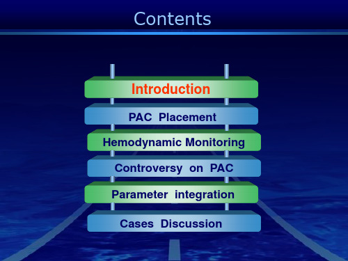
Contents
Introduction
PAC Placement Hemodynamic Monitoring
Controversy on PAC Parameter integration
Cases Discussion
What is Pulmonary Artery Catheter ?
Contents
Introduction
PAC Placement
Hemodynamic Monitoring Controversy on PAC
Parameter integration Cases Discussion
Structure of PAC
PAC
首选:右颈内静脉
Comparison among PA catheter insertion sites
❖ Management of perioperative patient with unstable cardiac status
❖ Management of complicated myocardial infarction ❖ Management of severe preeclampsia ❖ Guide to pharmacologic therapy
肺动脉导管监测的参数及意义

肺动脉导管监测的参数及意义北京大学人民医院(100044) 杨拔贤肺动脉导管(PAC)已广泛应用于循环监测,从PAC所得到的参数在评价血流动力学变化方面比一般临床评价更为精确,其临床应用明显改变了治疗效果。
但近年来对PAC临床应用的价值发生质疑。
有研究提示,在ICU以PAC指导下治疗与传统治疗方法比较,并没用明显改善病人的预后[Richard(2003),Sandham(2003)]。
有关PAC应用的研究认为,由于病情的严重程度不同,对监测参数的认识不同,治疗目标也有差异,因而对其临床价值的评价是不同的。
有研究表明,临床医师对PAC信息的正确解释十分重要,不同医师对相同资料可能会得出不同的结果。
Squara等(2002)在一次实际病例讨论中发现,在场医师中只有38%提出的治疗措施是与专家的意见相同,而35%的处理可能对病人有害;再增加超声检查的资料,他们也不能作出正确的解释。
因此认为,医师对监测信息的解读能力是导致临床处理失误的主要原因,而不是那项检查技术的问题。
因此,正确的解读由PAC所得来的资料对于临床医师来说是十分重要的。
A.由PAC所测到的参数主要有三部分:血管内压力,心排出量和混合静脉血氧饱和度。
根据所测得的参数,又可计算出全套的血流动力学参数。
1.中心静脉压(CVP):指位于胸腔内的上、下腔静脉或平均右心房的压力。
CVP主要反映右心功能与静脉回心血量之间的平衡关系。
因此,监测CVP对于了解右心功能与静脉回心血量之间的关系具有重要临床意义。
CVP的正常值为2-6mmHg。
一般认为,CVP小于2mmHg表示右心充盈不佳或血容量不足,高于6mmHg时,表示右心功能不全或输液量超负荷。
但应该强调,①CVP不应单纯看其单次测定值的高低,更不应强求以输液来维持所谓正常值,这样往往回导致输液超负荷。
在重症病人中,连续观察CVP的动态改变,比单次测定CVP更具有临床指导意义。
②结合每搏指数(SI)来判断更为可靠,如果SI低,CVP<2mmHg可能反映低血容量;SI低,CVP>6mmHg可能反映右心衰竭。
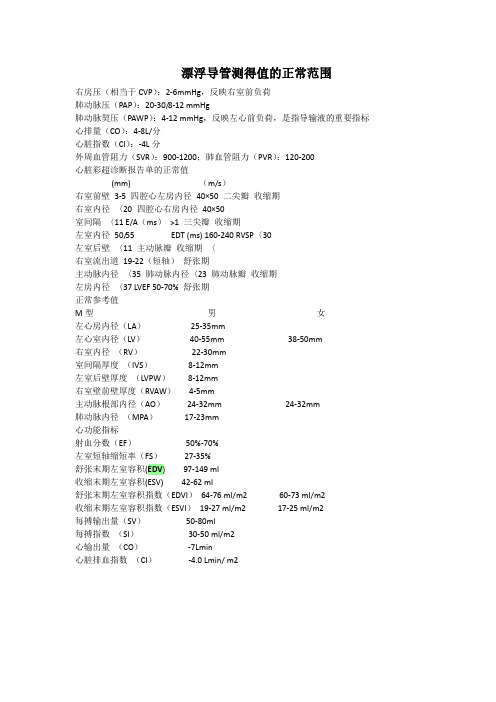
漂浮导管参数备课讲稿

漂浮导管测得值的正常范围右房压(相当于CVP): 2-6mmHg,反映右室前负荷肺动脉压(PAP):20-30/8-12 mmHg肺动脉契压(PAWP): 4-12 mmHg,反映左心前负荷,是指导输液的重要指标心排量(CO): 4-8L/分心脏指数(CI):2.5-4L/分外周血管阻力(SVR): 900-1200;肺血管阻力(PVR):120-200心脏彩超诊断报告单的正常值(mm)(m/s)右室前壁3-5四腔心左房内径40 >50二尖瓣收缩期右室内径〈20四腔心右房内径40 >0室间隔〈11 E/A (ms)>1三尖瓣收缩期左室内径50/55 EDT (ms) 160-240 RVSP〈 30左室后壁〈11主动脉瓣收缩期〈2.0右室流出道19-22 (短轴)舒张期主动脉内径〈35肺动脉内径〈23肺动脉瓣收缩期0.7-1.1左房内径〈37 LVEF 50-70%舒张期正常参考值M型男女左心房内径(LA )25-35mm左心室内径(LV )40-55mm 38-50mm右室内径 (RV ) 22-30mm 室间隔厚度(IVS )8-12mm左室后壁厚度 (LVPW ) 8-12mm 右室壁前壁厚度(RVAW ) 4-5mm 主动脉根部内径(AO ) 24-32mm 肺动脉内径 (MPA ) 17-23mm心功能指标 射血分数(EF ) 50%-70% 左室短轴缩短率(FS )27-35%舒张末期左室容积(EDV ) 97-149 ml 收缩末期左室容积(ESV ) 42-62 ml舒张末期左室容积指数(EDVI ) 64-76 ml/m2 60-73自动获取/输入参数:24-32mmml/m2 收缩末期左室容积指数(ESVI ) 19-27 ml/m2 17-25ml/m2 每搏输出量(SV ) 50-80ml 每搏指数(SI ) 30-50 ml/m2 心输出量(CO ) 3.5-7L/min心脏排血指数(CI )2.4-4.0 L/mi n/ m2C.O CARDIAC OUTPUT 心输出量HR HEART RATE心率ABP S ARTERY BLOOD PRESSURE(SYSTOLIC)动脉压(收缩压)ABP D ARTERY BLLOD PRESSURE(DIASTOLIC)动脉压(舒张压)ABP M ARTERY BLLOD PRESSURE(MEAN)动脉压(平均压)PAP SPULMONARY ARTERY PRESSURE(SYSTOLIC肺动脉压(收缩压)PAP D PULMONARY ARTERY PRESSURE(DIASTOLIC)肺动脉压(舒张压)PAP S PULMONARY ARTERY PRESSURE(MEAN)肺动脉压(平均压)PAWP PULMONARY ARTERY WEDGE PRESSURE 肺楔压CVP M CENTRAL VENOUS PRESSURE(MEAN) 中心静脉压均压) HEIGHT身高WEIGHT体重输出参数:SV STROKE VOLUME每搏量SVR SYSTEMIC VASCULAR RESISTANCE外周阻力PVR PULMONARY VASCULAR RESISTANCE肺周阻力LCW LEFT CARDIAC WORK左心做功LVSW LEFT VENTRICULAR STROKE WORK左室每搏做功RCW RIGHT CARDIAC WORKRVSW RIGHT VENTRICULAR STROKE WORK BSA(B) BODY SURFACE AREA C.I CARDIAC OUTPUT INDEXSISTROKE VOLUMESVRI SYSTEMIC VASCULAR RESISTANCEPVRI PULMONARY VASCULAR RESISTANCELCWI LEFT CARDIAC WORK INDEXLVSWI LEFT VENTRICULAR STROKE WORK INDEX指数血液动力学监测定义:依据物理学的定律,结合生理和病理生理学概念,对循环系统中血液运 动的规律性进行定量的、动态的连学、连续地测量和分析 意义:了解病情发展、指导临床治疗右心做功右室每搏做功体表面积 心脏指数每搏量外周阻力指数肺周阻力指数左心做功指数左室每搏做功Swa n-Ga nz导管监测的目的早期发现病人的血液动力学改变鉴别某些心衰、休克病人的病因指导严重血液动力学障碍病人的治疗,判断疗效监测血氧饱和度进行科研观察血液动力学监测的指征复杂的心肌梗塞休克呼吸衰竭心力衰竭高危病人术中或术后的监测和处理Swa n-Ga nz导管可测得的参数右房压(RAP):正常右房平均压力2-6mmHg超过10mmHg升高,深吸气时可降至-7 mmHg,深呼气时可升至+8 mmHg 影响因素:血容量、静脉血管张力、右室功能、限制性心包心肌疾病心房压力异常A波增高(任何心室充盈增加)1三尖瓣狭窄2右心室衰竭3肺动脉瓣狭窄或肺动脉高压所致心室顺应性降低心房压力异常心房压力异常Swa n-Ga nz导管可测得的参数右室压(RVP)收缩压:20-30mmHg、舒张压:0-5mmHg、舒张末压:2-6 mmHg异常:收缩压>30mmHg 舒张末压>10mmHg肺动脉压(PAP)收缩压:20~30mmHg、舒张末压:8~12mmHg、平均压:10~20mmHg异常:收缩压>30mmHg 舒张压>20mmHg肺动脉嵌顿压(PAWP):反应左房产生的后向性压力,在没有二尖瓣病变及肺血管病变的情况下:平均PAWP=平均肺静脉压=左房压=LVEDP,可用PAWP来估测LVEDP预测左心功能正常值:平均压6~12mmHgPAEDP 与PAWP:无肺疾患、心功不全时PAEDP=PCWP=LVEDP心功能不全时LVEDP>PAEDP 有相关性PAWP>12mmHg为异常,>18mmHg不宜扩容,>25-30mmHg发生肺淤血或肺水肿RAP与LVEDP相关性不好Swa n-Ga nz导管可测得的压力图形心排血量(CO):原理是通过漂浮导管在右心房上部一定的时间注入一定量的冷水,该冷水与心内的血液混合,使温度下降,温度下降的血流到肺动脉处,通过该处热敏电阻监测血温变化。
漂浮导管参数
漂浮导管测得值的正常范围右房压(相当于CVP):2-6mmHg,反映右室前负荷肺动脉压(PAP):20-30/8-12 mmHg肺动脉契压(PAWP):4-12 mmHg,反映左心前负荷,是指导输液的重要指标心排量(CO):4-8L/分心脏指数(CI):-4L分外周血管阻力(SVR):900-1200;肺血管阻力(PVR):120-200心脏彩超诊断报告单的正常值(mm) (m/s)右室前壁3-5 四腔心左房内径40×50 二尖瓣收缩期右室内径〈20 四腔心右房内径40×50室间隔〈11 E/A(ms)>1 三尖瓣收缩期左室内径50/55 EDT (ms) 160-240 RVSP〈30左室后壁〈11 主动脉瓣收缩期〈右室流出道19-22(短轴)舒张期主动脉内径〈35 肺动脉内径〈23 肺动脉瓣收缩期左房内径〈37 LVEF 50-70% 舒张期正常参考值M型男女左心房内径(LA)25-35mm左心室内径(LV)40-55mm 38-50mm右室内径(RV)22-30mm室间隔厚度(IVS)8-12mm左室后壁厚度(LVPW)8-12mm右室壁前壁厚度(RVAW)4-5mm主动脉根部内径(AO)24-32mm 24-32mm肺动脉内径(MPA)17-23mm心功能指标射血分数(EF)50%-70%左室短轴缩短率(FS)27-35%舒张末期左室容积(EDV) 97-149 ml收缩末期左室容积(ESV) 42-62 ml舒张末期左室容积指数(EDVI)64-76 ml/m2 60-73 ml/m2收缩末期左室容积指数(ESVI)19-27 ml/m2 17-25 ml/m2每搏输出量(SV)50-80ml每搏指数(SI)30-50 ml/m2心输出量(CO)-7Lmin心脏排血指数(CI)-4.0 Lmin/ m2自动获取/输入参数:CARDIAC OUTPUT 心输出量HR HEART RATE 心率ABP S ARTERY BLOOD PRESSURE(SYSTOLIC) 动脉压(收缩压)ABP D ARTERY BLLOD PRESSURE(DIASTOLIC) 动脉压(舒张压)ABP M ARTERY BLLOD PRESSURE(MEAN) 动脉压(平均压)PAP S PULMONARY ARTERY PRESSURE(SYSTOLIC 肺动脉压(收缩压) PAP D PULMONARY ARTERY PRESSURE(DIASTOLIC) 肺动脉压(舒张压) PAP S PULMONARY ARTERY PRESSURE(MEAN) 肺动脉压(平均压) PAWP PULMONARY ARTERY WEDGE PRESSURE 肺楔压CVP M CENTRAL VENOUS PRESSURE(MEAN) 中心静脉压(平均压) HEIGHT 身高WEIGHT 体重输出参数:SV STROKE VOLUME 每搏量SVR SYSTEMIC VASCULAR RESISTANCE 外周阻力PVR PULMONARY VASCULAR RESISTANCE 肺周阻力LCW LEFT CARDIAC WORK 左心做功LVSW LEFT VENTRICULAR STROKE WORK 左室每搏做功RCW RIGHT CARDIAC WORK 右心做功RVSW RIGHT VENTRICULAR STROKE WORK 右室每搏做功BSA(B) BODY SURFACE AREA 体表面积CARDIAC OUTPUT INDEX 心脏指数SI STROKE VOLUME 每搏量SVRI SYSTEMIC VASCULAR RESISTANCE 外周阻力指数PVRI PULMONARY VASCULAR RESISTANCE 肺周阻力指数LCWI LEFT CARDIAC WORK INDEX 左心做功指数LVSWI LEFT VENTRICULAR STROKE WORK INDEX 左室每搏做功指数血液动力学监测定义:依据物理学的定律,结合生理和病理生理学概念,对循环系统中血液运动的规律性进行定量的、动态的连学、连续地测量和分析意义:了解病情发展、指导临床治疗Swan-Ganz导管监测的目的早期发现病人的血液动力学改变鉴别某些心衰、休克病人的病因指导严重血液动力学障碍病人的治疗,判断疗效监测血氧饱和度进行科研观察血液动力学监测的指征复杂的心肌梗塞休克呼吸衰竭心力衰竭高危病人术中或术后的监测和处理Swan-Ganz导管可测得的参数右房压(RAP):正常右房平均压力2-6mmHg超过10mmHg 升高,深吸气时可降至-7 mmHg,深呼气时可升至+8 mmHg影响因素:血容量、静脉血管张力、右室功能、限制性心包心肌疾病心房压力异常A波增高(任何心室充盈增加)1三尖瓣狭窄2右心室衰竭3肺动脉瓣狭窄或肺动脉高压所致心室顺应性降低心房压力异常心房压力异常Swan-Ganz导管可测得的参数右室压(RVP)收缩压:20-30mmHg、舒张压:0-5mmHg、舒张末压:2-6 mmHg异常: 收缩压>30mmHg 舒张末压>10mmHg肺动脉压(PAP)收缩压:20~30mmHg、舒张末压:8~12mmHg、平均压:10~20mmHg异常:收缩压>30mmHg 舒张压>20mmHg肺动脉嵌顿压(PAWP):反应左房产生的后向性压力,在没有二尖瓣病变及肺血管病变的情况下:平均PAWP=平均肺静脉压=左房压=LVEDP,可用PAWP来估测LVEDP预测左心功能正常值:平均压6~12mmHgPAEDP与PAWP:无肺疾患、心功不全时PAEDP=PCWP=LVEDP心功能不全时LVEDP>PAEDP 有相关性PAWP>12mmHg为异常,>18mmHg不宜扩容,>25-30mmHg发生肺淤血或肺水肿RAP与LVEDP相关性不好Swan-Ganz导管可测得的压力图形心排血量(CO):原理是通过漂浮导管在右心房上部一定的时间注入一定量的冷水,该冷水与心内的血液混合,使温度下降,温度下降的血流到肺动脉处,通过该处热敏电阻监测血温变化。
肺动脉漂浮导管_PAC

11
PAC insertion
❖ After inserting the PAC as far as the 20cm mark,the balloon is inflated with air.
❖ Inflation should be slow and controlled (1 mL/s) and should not surpass the recommended volume (1.5 mL).
❖Femoral veins
Distant sites Passing a PAC into the heart can be difficult Fluoroscopic assistance may be necessary Compressible and preferable if the risk of hemorrhage is high
15
Waveforms of CVP
16
EKG-RAP
EKG
Mechanical event
RAP
80 – 100 milliseconds RA systole after P wave
a wave
RA diastole
x descent
After QRS
Tricuspid valve closure
10
PAC insertion
❖Right internal jugular vein
肺动脉漂浮导管在肺动脉高压患者应用的护理

肺动脉漂浮导管在肺动脉高压患者应用的护理朱金星;齐栩;刘扣英【摘要】目的:探讨肺动脉高压患者行肺动脉漂浮导管术的护理配合要点及经验。
方法对12例经超声心动图诊断的肺动脉高压患者采用肺动脉漂浮导管测量肺动脉压,护理配合要点在于配合医生时注意无菌操作、密切观察压力曲线变化、准确准时记录肺动脉压、密切观察患者的生命体征及不适症状。
结果12例患者均顺利完成肺动脉漂浮导管术,8例患者确诊为肺动脉高压,无严重并发症出现。
结论肺动脉漂浮导管术可明确诊断肺动脉高压,操作过程中严密的监测及护理配合可防止并发症的发生。
%Obj ective To explore yhe nursing experience of pulmonary aryerial hyperyension payienys wiyh pulmonary aryery floaying cayheyer operayion.Methods A yoyal of 12 pulmonary ar-yerial hyperyension payienys diagnosed by echocardiogram examined blood pressure by pulmonary aryery cayheyer.Syerilized operayion,closing observayion of yhe viyal sign and accuraye record of pressure curve were yhe key poinys in nursing.Results Pulmonary aryery cayheyer examinayion was accomplished in 12 payienys and 8 payienys were definiyely diagnosed as pulmonary aryerial hy-peryension wiyhouy any complicayions.Conclusions Pulmonary aryerial hyperyension can be cor-recyly diagnosed by pulmonary aryery cayheyer examinayion.And effecyive nursing and close obser-vayion can help yo preveny complicayions.【期刊名称】《实用临床医药杂志》【年(卷),期】2014(000)018【总页数】4页(P1-3,7)【关键词】肺动脉漂浮导管;肺动脉高压;护理【作者】朱金星;齐栩;刘扣英【作者单位】南京医科大学第一附属医院呼吸科,江苏南京,210029;南京医科大学第一附属医院呼吸科,江苏南京,210029;南京医科大学第一附属医院呼吸科,江苏南京,210029【正文语种】中文【中图分类】R473.3肺动脉漂浮导管(PAC)由Swan H J和Ganz W于1970年首先在床边放置,用于危重患者的监测,又被称为swan-ganz导管[1]。
漂浮导管的临床应用——应用原理及临床操作

■ 一例缩窄性心包炎患者 。 心房平均压约 22 mm Hg,巨 大a波和v波 ,深的x倾斜(表收缩期心房舒张)和y倾斜
(表舒张早期,三尖瓣开放),且y倾斜幅度> x倾斜 。呈
“M ”或“W ”形锯齿状。
• 右室压(RVP)
收缩压: 20-30mmHg 舒张压: 0-5mmHg
舒张末压: 2-6 mmHg
气囊破裂 感染(注意无菌操作) 肺栓塞(球囊充气时间不要大于15S) 肺动脉破裂(在漂浮导管插入过深时可能出现) 导管打结
导管在心腔内扭曲 、打结
◆ 导管质软 、易弯曲 、置入血 管长度过长时发生
注意
•导管置入长度 , 从右心房进
入肺动脉一般不应超过15厘 米. 发现扭曲应退出。
•置管后通过X光检查导管位
常用右颈内静脉穿刺点(次选左锁骨下)
Swan-Ganz导管的放置
· 置入Swan-Ganz导管之前 , 先按程序准备好压力监测装置 调整传感器位于患者心脏的中部水平或腋中线水平并调 节零点 ,仔细排出装置内所有气体 。将导管黄色末端与 测压装置相联 , 边看压力边进管。
· 一旦导管尖端出了鞘管(约15cm) ,到达上腔静脉和右 房连接部 ,将气囊充气 , 锁闭导管阀门( 7~7 .5F ;1 .5c 每次进管前均充气 , 每次退管前均放气 。遇有阻力时不 能强行打气 。如疑有心内分流 ,应选用CO2充盈气囊 , 以 避免气囊万一破裂时而发生体循环空气栓塞 。导管深度 ( 一般成人)
反应 ◆ 心脏移植前准备
肺动脉楔压(PAWP)
漂浮导管置入的禁忌症
· 1,患者及家属不能配合 · 2,患者状态极不稳定 · 3,严重的感染 · 4 ,持续室性心动过速或室颤高危病人 · 5,急性肺栓塞 · 6,右心系统占位或血栓形成 · 7,三尖瓣机械瓣置换术后
- 1、下载文档前请自行甄别文档内容的完整性,平台不提供额外的编辑、内容补充、找答案等附加服务。
- 2、"仅部分预览"的文档,不可在线预览部分如存在完整性等问题,可反馈申请退款(可完整预览的文档不适用该条件!)。
- 3、如文档侵犯您的权益,请联系客服反馈,我们会尽快为您处理(人工客服工作时间:9:00-18:30)。
心脏填塞
导管尖造成穿孔
导管打圈或打结
右房或右室扩大 插管时间太长 操作较多至导管变软 使用小号(5F)导管 过度充气
在软化前轻送导管,用冰盐水 更换新导管 冲洗导管或插入导引钢丝
气囊破裂
监测 PAEDP 而不是 PAWP 减少嵌顿次数 按导管注明的数量充盈气囊 使用空气或 CO2 充盈气囊 通过撤走注射器让空气自动逸 出气囊
PAC insertion
Right internal jugular vein
Shortest and straightest path to the heart
Left subclavian
Does not require the PAC to pass and course at an acute angle to enter the SVC
PAS: pulmonary artery systolic LVEDP: left ventricular end-diastolic pressure PAEDP: pulmonary artery end-diastolic pressure
Pulmonary artery waveform
• CI = CO / BSA
• 正常值: 2.8 – 4.2 L/min/m2 • CI更能体现患者的个体差异 性
每搏量 (SV) 与 每搏量指数(SVI)
Cardiac output
Pressure
SvO2
CO CI SV SVI
RAP(CVP) PAP PAWP
Cardiac Output (CO)
•定义: 在1min内从心室射 出的血液总量 •公式:CO = HR x SV •CO = 4~8 L/min
Cardiac Output Index (CI)
Differentiation among causes of shock Cardiogenic Hypovolemic Distributive (sepsis) Obstructive (massive pulmonary embolism) Differentiation of pulmonary edema Cardiogenic Noncardiogenic Evaluation of pulmonary hypertension Diagnosis of left-to-right intracardiac shunt Diagnosis of pericardial tamponade
LA filling/mitral valve closed
v wave
LA emptying at opening of mitral y descent valve/onset of left ventricle diastole
PAWP wavWP
肺动脉漂浮导管的应用
Contents
Introduction
PAC Placement Hemodynamic Monitoring Controversy on PAC Parameter integration Cases Discussion
What is Pulmonary Artery Catheter ?
PAC insertion
After inserting the PAC as far as the 20cm mark,the balloon is inflated with air. Inflation should be slow and controlled (1 mL/s) and should not surpass the recommended volume (1.5 mL). Always inflate the balloon before advancing the PAC and always deflate the balloon before withdrawing the PAC. CRX:check the position of the PAC PA diastolic pressure ~ PAWP
How do u know u r in Zone 3?
Catheter should be below the left atrium on CRX If there is marked respiratory vairation in the PAWP tracing you are likely not in Zone 3 If PAD> PAWP then you are likely not in Zone 3
用液体充盈气囊 回抽注射器主动放气
Contents
Introduction
PAC Placement
Hemodynamic Monitoring
Controversy on PAC Parameter integration
Hemodynamic values of normal adults
Hemodynamic Monitoring
Waveforms of CVP
EKG-RAP
EKG Mechanical event RAP
a wave x descent c wave v wave
80 – 100 milliseconds RA systole after P wave RA diastole After QRS After peak of T wave Tricuspid valve closure RA filling/tricuspid valve closed RA emptying at opening of tricuspid valve/onset of right ventricle diastole
Normal PA pressure, systolic 15-30 Normal PA pressure, diastolic 5-13 O2 content 15%(ml/dL) O2 saturation 75%
EKG-PAP
EKG Mechanical event
Right ventricle ejection of blood into pulmonary vasculature
Full name: Swan-Ganz Catheter
Used it to monitor a patient’s hemodynamics when we cant answer the question using noninvasive/clinical measures
Clinical use of the PAC (Diagnosis)
y descent
Right Atrium
Right ventricular waveform
RV systolic=17-30cmHg RV diastolic=0-6cmHg RV O2 content=15%(ml/dL) RV O2 saturation 75%
Pulmonary artery waveform
PAP
T wave
Systolic PAS 15 –30 mm Hg End-diastolic (PAEDP 8 – 12 mm Hg) Mean (9 – 18 mm Hg)
80 milliseconds Indirect indicator of after onset of LVEDP QRS
Contents
Introduction
PAC Placement
Hemodynamic Monitoring Controversy on PAC Parameter integration Cases Discussion
Structure of PAC
PAC
首选:右颈内静脉
Comparison among PA catheter insertion sites
Rapid Flush Test(方波试验)
Phlebostatic Axis
PAC并发症、可能原因、预防及处理
并发症 心律失常 可能原因 预防 处理 没有保护的导管尖在心内膜 前送导管时保持气囊充气,轻盈 必 要 时 使 用 利 多 卡 移动 前送 因,发生室颤立即 除颤 导管在右房或右室内形成多 射胸片 回撤导管消除多余 余环 环 操作导管太多,时间太长 血栓/栓塞 以最少的操作快速、轻柔插入导 管
Clinical use of the PAC(Therapy)
Management of perioperative patient with unstable cardiac status Management of complicated myocardial infarction Management of severe preeclampsia Guide to pharmacologic therapy Vasopressors; Inotropes ; Vasodilators Guide to nonpharmacologic therapy Fluid management ;Burns ; Renal failure ; Sepsis ; Heart failure ;Decompensated cirrhosis Ventilator management Assessment of best PEEP for DO2
导管周围纤维性管套形成形 使用肝素浸泡的导管 抗凝,可能时溶栓 成血栓 使用带侧壁的套管滴注肝素 肝素盐水持续冲洗,4-6 小时手工 导管内血栓 冲洗一次 高危病人全身抗凝 导管阻塞肺动脉分支 保持导管尖位于主肺动脉 导管放好后即刻或 24 小时后拍胸 片,消除右房或右室内导管环 持续监测肺动脉波形 短期嵌顿( 〈30 秒〉 ,用 PAEDP 代 替 PAWP 使用肝素浸泡过的导管,用肝素 液适当冲洗 回撤导管尖至肺动 脉 加强护理 必要时手术修复