神经解剖学英文课件-脑干的内部结构英文课件
合集下载
系统解剖 英文版 神经系统 脑干Brain stem
Pons ➢ Attached to 5th to 8th cranial n. ➢ Divided into the basilar part and tegmentum
Midbrain ➢ Attached to the 3rd and 4th cranial n. ➢ Divided into the cerebral peduncle and tectum
Brachium of inferior colliculus 下丘臂
Fourth ventricle 第四脑室
Central canal →fourth ventricle →mesencephalic aqueduct→third ventricle
Crus cerebri 大脑脚底 Interpeduncular fossa 脚间窝
Oculomotor nerve emerges on the medial side of crus cerebri Posterior perforated substance 后穿质
★ Dorsal Surface of Midbrain
Superior colliculus 上丘 Constitute the centers for visual reflexes
Inferior colliculus 下丘 Associated with auditory pathway
Brachium of superior colliculus 上丘臂
➢ Inferior cerebellar peduncle 小脑下脚
➢ Obex 闩
Upper portion: forms the lower half of rhomboid fossa 菱形窝
脑干课件.

髓
橄榄(olive),下橄榄核
橄榄后沟,舌咽神经、迷走神经、副神经的根丝
上部,菱形窝下半
背面
下部
薄束结节(gracile tubercle) 楔束结节(cuneate tubercle) 小脑下脚(inferior cerebellar peduncle)
10
•
整体形态
脑桥基底部,上下缘 菱形窝上半 脑桥小脑角(cerebellopontine angle)
界沟 )
前庭区 前庭神经核
外侧部
听结节 蜗背侧核
上端,蓝斑(locus ceruleus) 蓝斑核,为含色素的NA能神经元
12
•
上:间脑视束
整体形态 大脑脚底、四叠体、中脑水管
下:脑桥
中
大脑脚底,锥体束 腹侧面
脑
脚间窝(interpeducular fossa) ,后穿质、动神经
上丘,视觉反射中枢 上丘臂 外侧膝状体
后→ 1个正中孔median aperture ,Magendie孔
16
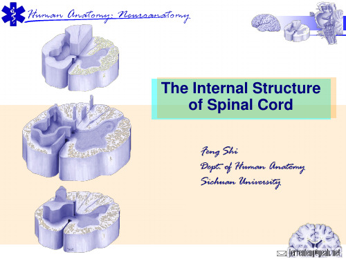
脊髓的内部结构
前角(柱)
中间带,侧角(柱)T~L3
• 灰质 H形
后角(柱)
中央灰质
灰质前连合 灰质后连合
• 中央管central canal 终池
前索anterior funiculus
侧索
• 白质 后索
白质前连合
• 网状结构
低 级 中 结构 枢
功能
脊髓
分化少、节段性、连外周
脑干
简单、连续成柱
成核团、延续似成柱
不同断面结构相同
不同断面不同形态
传导功能、反射功能
皮 质
结构
红核、黑质、蓝斑、臂旁核、脑桥核、上橄榄核、下橄榄核、网 状结构等。
脑干内部结构解剖学教学PPT课件

21
3. 一般内脏运动柱:
general visceral motor column(GVM)
位于躯体运动柱的外侧,靠近界沟, 自上而下有4个核团:
涎核(superior salivatory nucleus) 下泌涎核(inferior salivatory nucleus) 迷走神经背核(dorsal nucleus of vagus nerve)。
10
●功能相同的脑神经核,在脑干内有规 律地排列成纵行的细胞柱,称脑神经核 机能柱。 ●同一机能柱内的脑神经核多是不连续 的。 ●一般和特殊内脏感觉核实为一个机能 柱,均为孤束核。 ●故每侧脑干实为6个脑神经功能柱。
11
●它们在脑干中有一定的排列关系: 感觉柱位于界沟的外侧 运动柱位于界沟的内侧 内脏相关的机能柱靠近界沟 躯体相关的机能柱远离界沟
25
5. 一般躯体感觉柱:
general somatic afferent column(GSA)
位于内脏感觉柱的腹外侧。自上 而下为3个核团:
三叉神经中脑核
(mesencephalic nucleus of trigeminal nerve)
17
(1)三叉神经运动核: 位于脑桥中部,发出纤维行向腹
外侧,构成三叉神经运动根,加入下 颌神经,支配咀嚼肌、二腹肌前腹、 下颌舌骨肌、腭帆张肌、鼓膜张肌。
18
(2)面神经核: 位于脑桥下部,上橄榄核背外侧。
面神经核发出的纤维行向背内侧,从内 侧绕展神经核背方,行向腹外侧,经延 髓脑桥沟出脑。支配面肌、颈阔肌、茎 突舌骨肌和镫骨肌和二腹肌后腹。
14
(1)动眼神经核:位于中脑上丘阶段, 中脑水管腹侧。 支配下直肌、内直肌、 下斜肌、上直肌。动眼神经核发出的纤 维经大脑脚底内侧出脑,组成动眼神经。
脑干内部结构脑神经非脑神经核团课件
下丘 IV
小脑上脚 菱形窝上半
rhomboid fossa
菱形窝下半 小脑下脚 楔束结节 gracile tubercle 薄束结节 cuneate tubercle
脑干内部结构脑神经非脑神经核团
髓纹
4
第三脑室 大脑导水管
顶
第四脑室 底
小脑上脚、上髓帆 下髓帆、第四脑室脉络组织 菱形窝
脊髓中央管
蛛网膜下腔
名称
位置 相关脑N 分布区
动眼N副核 上丘水平 动眼N 睫状肌、瞳孔括约肌 上泌涎核 脑桥下部 面N 泪腺舌下腺下颌下腺 下泌涎核 延髓上部 舌咽N 腮腺 迷走N背核 X △深方 迷走N 颈、胸、腹 大部脏器
脑干内部结构脑神经非脑神经核团
25
脑干内部结构脑神经非脑神经核团
26
一般内脏运动柱
1、动眼神经副核accessory nuc.of oculomotor 位于中脑上丘平面 支配瞳孔括约肌、睫状肌 损伤表现为瞳孔散大
顶盖上区
中脑部称为顶盖 上丘
室 腔 顶部:的
下丘 脑桥的顶部为上、下髓帆
后 方
延髓上部的顶为第四脑室脉络丛 和脉络组织
延髓下部的顶为后索及 薄、楔束核
室腔部:中脑水管、第四脑室、 延髓下部的中央管
被盖:室腔前方的广大区域
基底部:中脑的大脑脚底、脑桥部 的基底和延髓的锥体
脑干内部结构脑神经非脑神经核团
脑干内部结构脑神经非脑神经核团
37
(二)非脑神经核:属于中继核团
1 延髓的非脑神经核:
1)薄束核与楔束核
位于延髓薄束结节和楔束结节的深方,接受来自薄束与楔束的纤维, 发出纤维弓形绕过中央灰质,在正中线交叉形成内侧丘系交叉,在 中线两侧后继续上升形成内侧丘系。
系统解剖学7脑干小脑课件
名 称 位置 相关脑N 分布区
动眼N核 上丘 动眼N 5块
oculomotor n. 水平
眼外肌
滑车N核 下丘 滑车N 上斜肌 trochlear n. 水平
展N核 面N丘 展N 外直肌 abducent n. 水平
舌下N核 舌下N 舌下N 舌肌 hypoglossal n. △深方
特殊内脏运动柱
脑干内部结构的分区
室管:中脑水管 第四脑室 中央管
灰质 :脑N核 非脑N核白质:四系两束 网状源自构脊髓内部结构• 分区
中央管
posterior horn 后角
后索
网状结构
灰质 gray matter
中间带
前角、后角、
中间带、侧角、
灰质连合
白质 white matter 前索、外侧索 、后索、白质 前连合
下 丘 蜗N核 上 丘 视束, 脑
下丘臂,上丘 Ⅲ,Ⅳ,Ⅵ核
下丘, 脊髓 顶盖脊髓束
脑干的非脑cNsoul核pliecruilours
名 称 传入纤维自 传出纤维至
薄束核 薄束
内侧丘系
楔束核 楔束
内侧丘系
下橄榄核 大脑,脊髓 小脑
上橄榄核 蜗N核 外侧丘系
脑桥核 大脑皮质 小脑中脚
下 丘 蜗N核 下丘臂,上丘 上 丘 视束, 脑 III、IV、VI核
背核 深方
大部脏器
内脏感觉柱
the visceral sensory nuclei
名称 位置 相关脑N 分布
孤束核 延髓
头部
VII 舌粘膜
IX 味蕾
尾部
IX 舌咽喉和
X 胸腹腔脏器
孤孤 束叔 头头 尾尾 部步
一般躯体感觉柱
人体解剖学脑干-PPT
等处发出的纤维,后经小脑下脚终止于小脑皮质。在小 脑对运动的控制特别是对运动的学习和记忆起重要作用。
下页
系统解剖:脑干(brain stem)
内部结构
脑干外形
脑干内部结构
(3)脑桥核: 居脑桥基底部的纤维束之间,由若干群细胞构成, 为大脑皮质与小脑皮质之间的中继站。 (4)红核:
脑干功能 居上丘平面的被盖部,呈圆柱状。主要接受来自小 脑及大脑皮质的传入纤维,并发出红核脊髓束。与 躯体运动(屈肌的运动)有关。
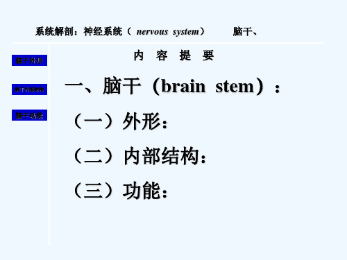
系统解剖:神经系统( nervous system) 脑干、
脑干外形
内容提要
脑干内部结构 一、脑干(brain stem):
脑干功能 (一)外形:
(二)内部结构:
(三)功能:
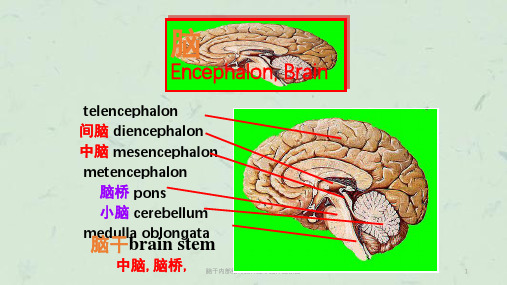
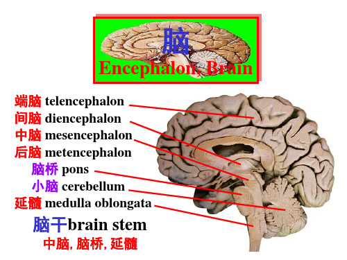
脑的分部
脑 端脑
的 区
间脑
分 小脑
中脑
脑干 脑桥
延髓
一、脑干的位置和外形
位于颅后窝,脊髓 和间脑之间,小脑腹侧。
系统解剖:脑干(brain stem)
脑干内部结构
脑干功能
内部结构
返回
系统解剖:脑干(brain stem)
脑干外形
脑干内部结构
脑干功能
内部结构
蜗神经核
返回
系统解剖:脑干(brain stem)
脑干外形
脑干内部结构
脑干功能
内部结构
下图
返回
系统解剖:脑干(brain stem)
脑干外形
脑干内部结构
脑干功能
内部结构
下图
返回
系统解剖:脑干(brain stem)
与脑相连的脑神经部位
脑干外形
脑干内部结构
脑干功能
端脑—Ⅰ 间脑—Ⅱ 中脑 —Ⅲ、Ⅳ 脑桥—Ⅴ、Ⅵ、Ⅶ、Ⅷ 延髓—Ⅸ、Ⅹ、Ⅺ、Ⅻ
最新系统解剖英文版神经系统脊髓Spinalcord精品课件
第十六页,共61页。
★脊髓(jǐ suǐ)圆锥Conus medullaris-the lower pyramid end of spinal cord
★终丝Filum terminale-a delicate filament, about 20 cm in length, prolonged downward from the apex of the conus medullaris to the back of the first segment of the coccyx, continuous above with that of the pia mater.
to Vertebral Numbers
Spinal segments
Vertebral levels (spines)
C1~C4 C5~T4
Lie opposite the corresponding vertebrae
One lower in number than corresponding vertebrae
Brain stem脑干
第三页,共61页。
Major Divisions of the Peripheral Nervous System
脑神经(shénjīng)Cranial nerves (12 pairs) 脊神经(shénjīng)Spinal nerves (31 pairs) 内脏神经(shénjīng)Visceral nerves Visceral sensory nerve Visceral motor nerve Sympathetic part 交感神经(shénjīng) Parasympathetic part 副交感神经
第十一页,共61页。
神经解剖学英文课件-脊髓的内部结构英文课件
Conscious Long Tracts
▪ descending tract - corticospinal tract
▪ ascending tracts - fasciculi gracilis & cuneatus - spinothalamic tract
1st Sensory Neuron
The Anterior Horn
▪α motor neuron – extrafusal muscle fibres
(voluntary movement)
▪γ motor neuron – intrafusal muscle fibers (muscle tonus)
▪ Renshaw cell:
▪ nucleus propris (laminae Ⅲ & Ⅳ) of spinal cord
▪ nucleus thoracicus (lamina Ⅶ) thoracic region
Functions of Nuclei in Posterior Horn
▪ marginal layer & substantia gelatinosa: pain, temperature, crude touch & pressure - spinothalamic tract ▪ nucleus propris: discriminative touch, vibratory sense & conscious muscle joint sense - fasciculi gracilis & cuneatus ▪ nucleus thoracicus: proprioceptive information to cerebellum - spinocerebellar tract
sx007-脑干 ppt课件

☆ 2.背侧面 Dorsal surface
1)Lower portion: ①薄结节 Gracile tubercle → produced by
underlying gracile nucleus at lower angle of fourth ventricle.
②楔结节 Cuneate tubercle → marks site of cuneate nucleus lateral and adjacent to Gracile tubercle. ③小脑下脚 Inferior
⑨前庭区 Vestibular area →
is a triangular field lateral to sulcus limitans and overlies vestibular nuclei.
⑩听结节 Acoustic tubercle
→ lies lateral angle of vestibular area overlying dorsal cochlear nucleus.
Brain stem is stalk like part of brain, which connects cerebrum, cerebellum with spinal cord.
Optic tract serves as rostral boundary of brain stem, which separates it from diencephalon.
①髓纹 Striae medullares →
divide rhomboid fossa into pontine and medullary parts.
②正中沟 Median sulcus → divide rhomboid fossa into symmetrical halves. ③内侧隆起 Medial eminence → a ridge on each side of median sulcus. ④界沟 Sulcus limitans →
解剖学《脑干》课件
动眼神经核
Nucleus of trochlear n.
滑车神经核
Nucleus of abducent n.
展神经核
Nucleus of hypoglossal motor nuclei 特殊内脏运动核
Motor nucleus of trigeminal n.
小脑上脚
面神经丘 前庭区
舌下神经三角 迷走神经三角
Fourth ventricle 第四脑室
Central canal →fourth ventricle →mesencephalic aqueduct→third ventricle
Position
Situated ventral to cerebellum, and dorsal to pons and cranial half of medulla
Features
Median sulcus 正中沟 Sulcus limitans 界沟 Vestibular area前庭区
overlies vestibular nuclei
Acoustic tubercle听结节
overlying dorsal cochlear nucleus
Medial eminence 内侧隆起 Striae medullares 髓纹
Trochlear nerve 滑车神经
Midbrain 中脑
Ventral surface
Crus cerebri 大脑脚底 Interpeduncular fossa
脚间窝 oculomotor nerves
emerge from medial of crus cerebri
Posterior perforated substance 后穿质
系统解剖 英文版 神经系统 脑干.Brain stem

Fourth ventricle 第四脑室
Central canal →fourth ventricle →mesencephalic aqueduct→third ventricle
★ Position situated ventral to cerebellum dorsal to pons and cranial half of medulla Boundaries Floor: Rhomboid Fossa 菱形窝 Roof
Gross Appearance of Brain Stem
★ Gross Appearance of Brain Stem
Medulla oblongata Attached to the 9th to 12th cranial n. Divided into an open(superior) and a closed (inferior) part Pons Attached to 5th to 8th cranial n. Divided into the basilar part and tegmentum Midbrain Attached to the 3rd and 4th cranial n. Divided into the cerebral peduncle and tectum
Subarachnoid space
Tela choroidea and choroid plexus 脉络丛和脉络组织
Internal structure of the brain Stem
Gray matter Cranial nerve nuclei 脑神经核 Non-cranial nerve nuclei 非脑神经核 White matter Reticular formation of brain stem 脑干网状结构 Central canal
- 1、下载文档前请自行甄别文档内容的完整性,平台不提供额外的编辑、内容补充、找答案等附加服务。
- 2、"仅部分预览"的文档,不可在线预览部分如存在完整性等问题,可反馈申请退款(可完整预览的文档不适用该条件!)。
- 3、如文档侵犯您的权益,请联系客服反馈,我们会尽快为您处理(人工客服工作时间:9:00-18:30)。
GSE: Oculomotor Nucleus – at the level of the superior colliculus of midbrain Trochlear Nucleus – at the level of the inferior colliculus of the midbrain Abducent Nucleus – beneath the fascial colliculus Hypoglossal Nucleus – beneath the hypoglossal trigone
▪ Midbrain
- corpora quadrigemina - pineal body
▪ Medulla Oblongata
- gracile tubercle - cuneate tubercle - trigeminal tubercle
The Continuation of Brainstem & Spinal Cord
Ventral View
▪ Medulla Oblongata
- anterior medial fissure - pyramid - olive - pyramidal decussation
▪ Pons
- basilar sulcus - basilar part - middle cerebellar peduncle
Location of the Cranial Nerve Nuclei
▪ superior colliculus ▪ inferior colliculus ▪ facial colliculus ▪ hypoglossal triangle
▪ vagal triangle ▪ striae medullares ▪ sulcus limitans ▪ vestibular area
(Rhomboid Fossa & Fourth Ventricle)
Grey Matter of Brainstem: Cranial Nerve Nuclei
Development of Spinal Cord & Brainstem Sulcus Limitans
The Arrangement of the Cranial Nerves during the Fetal Development
▪ GSE column (SE column) ▪ SVE column (branchial efferent column) ▪ GVE column – parasympathicus ▪ GVA column – subconscious sensation ▪ SVA column – taste sense ▪ GSA column – conscious sensation ▪ SSA column – audition & posture
(mesencephalic, principal / main & spinal) nuclei
▪ SSA: vestibular & cochlear nuclei
Motor Cranial Nerve Nuclei
▪ GSE: oculmotor, trochlear, abducent & hypoglossal nuclei ▪ SVE: trigeminal motor, facial motor, ambiguus nuclei (& accessory spinal) ▪ GVE: accessory oculmotor, sup. & inf. salivatory & dorsal motor vagus
Motor & Sensory Columns of Spinal Cord & Brainstem
SE SVE GVE
SVA GVA GSA SSA
Brainstem
Spinal cord
SE VE
VA
SA
Components of Cranial Nerve Nuclei in Brainstem
Medially
GSE(SE)
GVE
SVE (branchial motor)
VA (GVA & SVA)
SSABiblioteka GSALaterally
▪ GSE: oculmotor, trochlear,
abducent & hypoglossal nuclei
▪ SVE: trigeminal motor, facial
nuclei
Sensory Cranial Nerve Nuclei
▪ VA: solitary nuclei ▪ GSA: trigeminal sensory (mesencephalic, principal / main & spinal)
nuclei
▪ SSA: vestibular & cochlear nuclei
▪ Midbrain
- cerebral peduncle - interpeduncular fossa - posterior perforated substance
External Features of Brainstem
Dorsal View
▪ Rhomboid Fossa
- dorsal median sulcus - striae medullares - sulcus limitans - medial eminence - fascial colliculus - vestibular area - hypoglossal trigone - vagal trigone
The Internal Structure of Brainstem
OUTLINE
▪ Gray Matter - Cranial Nerve Nuclei - Relay Nuclei
▪ White Matter - Ascending Tracts - Descending Tracts
▪ Reticular Formation ▪ Transverse Sections
motor, ambiguus & accessory spinal nuclei
▪ GVE: accessory oculmotor, sup.
& inf. salivatory & dorsal motor vagus nuclei
▪ VA: solitary nuclei ▪ GSA: trigeminal sensory