微生物翻译1
微生物学细菌中英翻译及促生素概论
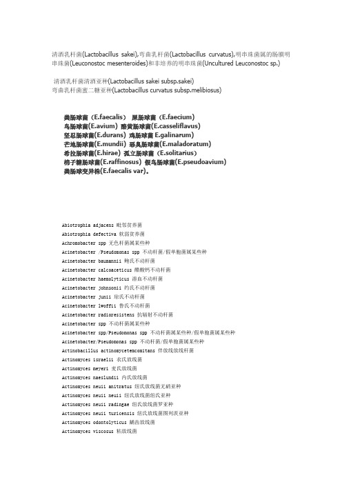
清酒乳杆菌(Lactobacillus sakei),弯曲乳杆菌(Lactobacillus curvatus),明串珠菌属的肠膜明串珠菌(Leuconostoc mesenteroides)和非培养的明串珠菌(Uncultured Leuconostoc sp.)清酒乳杆菌清酒亚种(Lactobacillus sakei subsp.sakei)弯曲乳杆菌蜜二糖亚种(Lactobacillus curvatus subsp.melibiosus)粪肠球菌(E.faecalis)屎肠球菌(E.faecium)鸟肠球菌(E.avium) 酪黄肠球菌(E.casseliflavus)坚忍肠球菌(E.durans) 鸡肠球菌E.galinarum)芒地肠球菌(E.mundii) 恶臭肠球菌(E.maladoratum)希拉肠球菌(E.hirae) 孤立肠球菌(E.solitarius)棉子糖肠球菌(E.raffinosus) 假鸟肠球菌(E.pseudoavium)粪肠球变异株(E.faecalis var)。
Abiotrophia adjacens 毗邻贫养菌Abiotrophia defectiva 软弱贫养菌Achromobacter spp 无色杆菌属某些种Acinetobacter /Pseudomonas spp 不动杆菌/假单胞菌属某些种Acinetobacter baumannii 鲍氏不动杆菌Acinetobacter calcoaceticus 醋酸钙不动杆菌Acinetobacter haemolyticus 溶血不动杆菌Acinetobacter johnsonii 约氏不动杆菌Acinetobacter junii 琼氏不动杆菌Acinetobacter lwoffii 鲁氏不动杆菌Acinetobacter radioresistens 抗辐射不动杆菌Acinetobacter spp 不动杆菌属某些种Acinetobacter spp/Pseudomonas spp 不动杆菌属某些种/假单胞菌属某些种Acinetobacter/Pseudomonas spp 不动杆菌/假单胞菌属某些种Actinobacillus actinomycetemcomitans 伴放线放线杆菌Actinomyces israelii 衣氏放线菌Actinomyces meyeri 麦氏放线菌Actinomyces naeslundii 内氏放线菌Actinomyces neuii anitratus 纽氏放线菌无硝亚种Actinomyces neuii neuii 纽氏放线菌纽氏亚种Actinomyces neuii radingae 纽氏放线菌罗亚种Actinomyces neuii turicensis 纽氏放线菌图列茨亚种Actinomyces odontolyticus 龋齿放线菌Actinomyces viscosus 粘放线菌Aeromonas caviae 豚鼠气单胞菌Aeromonas hydrophila 嗜水气单胞菌Aeromonas hydrophila gr.嗜水气单胞菌群Aeromonas salmonicida achromogenes 杀鲑气单胞菌无色亚种Aeromonas salmonicida masoucida 杀鲑气单胞菌杀日本鲑亚种Aeromonas salmonicida salmonicida 杀鲑气单胞菌杀鲑亚种Aeromonas sobria 温和气单胞菌Agrobacterium radiobacter 放射形土壤杆菌Alcaligenes denitrificans 反硝化产碱菌Alcaligenes faecalis 粪产碱菌Alcaligenes spp 产碱菌属某些种Alcaligenes xylosoxidans 木糖氧化产碱菌Alloiococcus otitis 耳炎差异球菌Anaerobiospirllum succiniproducens 产琥珀酸厌氧螺菌Arachnia propionica 丙酸蛛菌Arcanobacterium bernardiae 伯纳德隐秘杆菌Arcanobacterium haemolyticum 溶血隐秘杆菌Arcanobacterium pyogenes 化脓隐秘杆菌Arcobacter cryaerohoilus 嗜低温弓形杆菌Arthrobacter spp 节杆菌属某些种Debaryomyces polymorphus 多形德巴利酵母菌Dermabacter hominis 人皮肤杆菌Dermacoccus nishinomiyaensis 西宫皮肤球菌Dietzia spp 迪茨菌属某些种Edwardsiella hoshinae 保科爱德华菌Edwardsiella tarda 迟钝爱德华菌Eikenella corrodens 啮蚀艾肯菌Enterobacter aerogenes 产气肠杆菌Enterobacter amnigenus 河生肠杆菌Enterobacter asburiae 阿氏肠杆菌Enterobacter cancerogenus 生癌肠杆菌Enterobacter cloacae 阴沟肠杆菌Enterobacter gergoviae 日沟维肠杆菌Enterobacter intermedius 中间肠杆菌Enterobacter sakazakii 阪崎肠杆菌Enterobacter spp 肠杆菌属某些种Enterococcus avium 鸟肠球菌Enterococcus casselifavus 铅黄肠球菌Enterococcus durans 耐久肠球菌Enterococcus faecalis 粪肠球菌Enterococcus faecium 屎肠球菌Enterococcus gallinarum 鹑鸡肠球菌Enterococcus saccharolyticus 解糖肠球菌Erwinia spp 欧文菌属某些种Erysipelothrix rhusiopathiae 猪红斑丹毒丝菌Escherichia coli 大肠埃希菌Escherichia fergusonii 费格森埃希菌Escherichia hermannii 赫氏埃希菌Escherichia vulneris 伤口埃希菌Eubacterium aerofaciens 产气真杆菌Eubacterium lentum 迟缓真杆菌Eubacterium limosum 粘液真杆菌Ewingella americana 美洲爱文菌促生素:概论简介发酵乳形态的促生素历史要追溯回数千年前,但直到本世纪初期才根据科学原理进行科学性研究。
微生物学细菌中英翻译及促生素概论
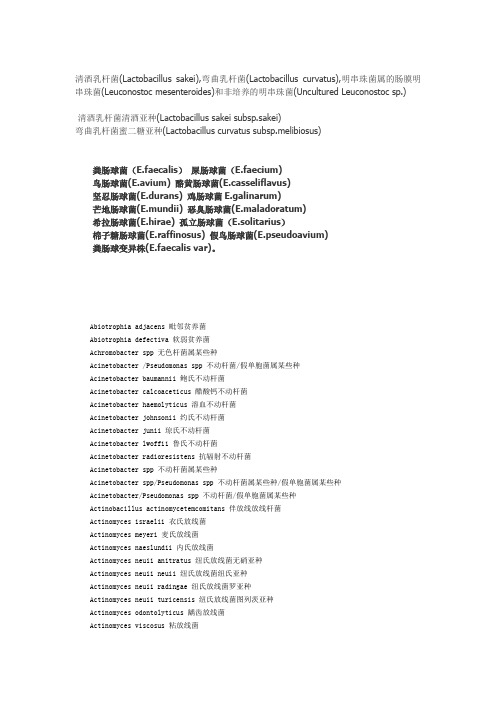
清酒乳杆菌(Lactobacillus sakei),弯曲乳杆菌(Lactobacillus curvatus),明串珠菌属的肠膜明串珠菌(Leuconostoc mesenteroides)和非培养的明串珠菌(Uncultured Leuconostoc sp.)清酒乳杆菌清酒亚种(Lactobacillus sakei subsp.sakei)弯曲乳杆菌蜜二糖亚种(Lactobacillus curvatus subsp.melibiosus)粪肠球菌(E.faecalis)屎肠球菌(E.faecium)鸟肠球菌(E.avium) 酪黄肠球菌(E.casseliflavus)坚忍肠球菌(E.durans) 鸡肠球菌E.galinarum)芒地肠球菌(E.mundii) 恶臭肠球菌(E.maladoratum)希拉肠球菌(E.hirae) 孤立肠球菌(E.solitarius)棉子糖肠球菌(E.raffinosus) 假鸟肠球菌(E.pseudoavium)粪肠球变异株(E.faecalis var)。
Abiotrophia adjacens 毗邻贫养菌Abiotrophia defectiva 软弱贫养菌Achromobacter spp 无色杆菌属某些种Acinetobacter /Pseudomonas spp 不动杆菌/假单胞菌属某些种Acinetobacter baumannii 鲍氏不动杆菌Acinetobacter calcoaceticus 醋酸钙不动杆菌Acinetobacter haemolyticus 溶血不动杆菌Acinetobacter johnsonii 约氏不动杆菌Acinetobacter junii 琼氏不动杆菌Acinetobacter lwoffii 鲁氏不动杆菌Acinetobacter radioresistens 抗辐射不动杆菌Acinetobacter spp 不动杆菌属某些种Acinetobacter spp/Pseudomonas spp 不动杆菌属某些种/假单胞菌属某些种Acinetobacter/Pseudomonas spp 不动杆菌/假单胞菌属某些种Actinobacillus actinomycetemcomitans 伴放线放线杆菌Actinomyces israelii 衣氏放线菌Actinomyces meyeri 麦氏放线菌Actinomyces naeslundii 内氏放线菌Actinomyces neuii anitratus 纽氏放线菌无硝亚种Actinomyces neuii neuii 纽氏放线菌纽氏亚种Actinomyces neuii radingae 纽氏放线菌罗亚种Actinomyces neuii turicensis 纽氏放线菌图列茨亚种Actinomyces odontolyticus 龋齿放线菌Actinomyces viscosus 粘放线菌Aeromonas caviae 豚鼠气单胞菌Aeromonas hydrophila 嗜水气单胞菌Aeromonas hydrophila gr.嗜水气单胞菌群Aeromonas salmonicida achromogenes 杀鲑气单胞菌无色亚种Aeromonas salmonicida masoucida 杀鲑气单胞菌杀日本鲑亚种Aeromonas salmonicida salmonicida 杀鲑气单胞菌杀鲑亚种Aeromonas sobria 温和气单胞菌Agrobacterium radiobacter 放射形土壤杆菌Alcaligenes denitrificans 反硝化产碱菌Alcaligenes faecalis 粪产碱菌Alcaligenes spp 产碱菌属某些种Alcaligenes xylosoxidans 木糖氧化产碱菌Alloiococcus otitis 耳炎差异球菌Anaerobiospirllum succiniproducens 产琥珀酸厌氧螺菌Arachnia propionica 丙酸蛛菌Arcanobacterium bernardiae 伯纳德隐秘杆菌Arcanobacterium haemolyticum 溶血隐秘杆菌Arcanobacterium pyogenes 化脓隐秘杆菌Arcobacter cryaerohoilus 嗜低温弓形杆菌Arthrobacter spp 节杆菌属某些种Debaryomyces polymorphus 多形德巴利酵母菌Dermabacter hominis 人皮肤杆菌Dermacoccus nishinomiyaensis 西宫皮肤球菌Dietzia spp 迪茨菌属某些种Edwardsiella hoshinae 保科爱德华菌Edwardsiella tarda 迟钝爱德华菌Eikenella corrodens 啮蚀艾肯菌Enterobacter aerogenes 产气肠杆菌Enterobacter amnigenus 河生肠杆菌Enterobacter asburiae 阿氏肠杆菌Enterobacter cancerogenus 生癌肠杆菌Enterobacter cloacae 阴沟肠杆菌Enterobacter gergoviae 日沟维肠杆菌Enterobacter intermedius 中间肠杆菌Enterobacter sakazakii 阪崎肠杆菌Enterobacter spp 肠杆菌属某些种Enterococcus avium 鸟肠球菌Enterococcus casselifavus 铅黄肠球菌Enterococcus durans 耐久肠球菌Enterococcus faecalis 粪肠球菌Enterococcus faecium 屎肠球菌Enterococcus gallinarum 鹑鸡肠球菌Enterococcus saccharolyticus 解糖肠球菌Erwinia spp 欧文菌属某些种Erysipelothrix rhusiopathiae 猪红斑丹毒丝菌Escherichia coli 大肠埃希菌Escherichia fergusonii 费格森埃希菌Escherichia hermannii 赫氏埃希菌Escherichia vulneris 伤口埃希菌Eubacterium aerofaciens 产气真杆菌Eubacterium lentum 迟缓真杆菌Eubacterium limosum 粘液真杆菌Ewingella americana 美洲爱文菌促生素:概论简介发酵乳形态的促生素历史要追溯回数千年前,但直到本世纪初期才根据科学原理进行科学性研究。
医学常用微生物学名词英汉翻译
医学常用微生物学名词英汉翻译微生物学作为医学领域中的重要学科,涉及到许多微生物学名词的英汉翻译。
准确的翻译对于医学研究和临床实践都具有重要意义。
以下是一些常用微生物学名词的英汉翻译示例。
1. Bacteria - 细菌Bacteria are single-celled microorganisms that can be found in various habitats.2. Virus - 病毒Viruses are infectious agents that can cause diseases in humans, animals, and plants.3. Fungi - 真菌Fungi are a group of organisms that include yeasts, molds, and mushrooms.4. Protozoa - 原生动物Protozoa are single-celled eukaryotic microorganisms that can be found in water and soil.5. Parasite - 寄生虫Parasites are organisms that live in or on another organism (host) and obtain nutrients from the host.6. Antibiotic - 抗生素Antibiotics are substances that can inhibit the growth of or destroy bacteria.7. Antimicrobial - 抗菌剂Antimicrobials are substances that can inhibit the growth of or destroy microorganisms, including bacteria, viruses, fungi, and protozoa.8. Pathogen - 病原体Pathogens are microorganisms that can cause diseases in their hosts.9. Pathogenesis - 致病机制Pathogenesis refers to the process by which a pathogen causes disease in an organism.10. Immunization - 免疫Immunization is the process of inducing immunity against a particular disease through vaccination.11. Contagious - 传染的Contagious refers to a disease that can be transmitted from one person to another through direct or indirect contact.12. Sterilization - 杀菌Sterilization is the process of completely removing or destroying all microorganisms, including bacteria, viruses, fungi, and spores.13. Disinfection - 消毒Disinfection is the process of eliminating or reducing the number of pathogenic microorganisms on surfaces or objects.14. Culture - 培养Culture refers to the process of growing microorganisms in a controlled environment for research or diagnostic purposes.15. Resistance - 耐药性Resistance refers to the ability of microorganisms to withstand the effects of antimicrobial drugs.以上是一些医学常用微生物学名词的英汉翻译示例。
考研-微生物名词解释
8,11,27,351.炭疽芽孢杆菌(bacillus thuringiensis):属于需氧芽孢杆菌属,能引起牛、羊、马等动物及人类的炭疽病。
这种细菌通常以内孢子之形态出现在土壤中,并可借此状态存活数十年之久,一旦由牲畜摄入,孢子便开始在动物体内大量复制,最后造成死亡,随后于尸体中仍能继续繁殖,当细菌将宿主养分用尽,又可重回睡眠态的孢子。
链霉菌属(Streptomyces)拉丁字母(拉丁字母翻译成汉语):(07年,15年)放线菌目的一科。
基内菌丝不断,气生菌丝通常发育良好,形成长的孢子丝。
孢子不能转动,外鞘上常有疣、刺或毛发等状饰物。
2.地衣(3):真菌和光合生物(绿藻或蓝细菌)之间稳定而又互利的共生联合体,真菌是主要成员,其形态及后代的繁殖菌依靠真菌。
也就是说地衣是一类专化性的特殊真菌。
3.子囊孢子(3):子囊菌亚门的真菌产生于子囊中经减数分裂后形成有性孢子,子囊指的是两性细胞接触以后形成的囊状结构。
4.球状体(P27)5.生长因子(07,13,15)(P47)6古生菌(07,13): .古菌(11,14)古生菌:又称为古细菌(archaeobacteria)或称古菌生物按流行的3域分类观点可分为古生菌域、细菌域以及真核生物域。
古生菌和细菌属于原核生物,它们没有完整的细胞核。
真核生物包括动植物、霉菌等。
古生菌是地球进化早期产生的微生物,能适应极端环境,例如在强酸下生长的Thermoplasma (最佳生长pH=2)还有能在沸水中生长的Pyrobolus。
古生菌成为和细菌域、真核生物域并驾齐驱的三大类生物之一。
7.抗代谢物(P87)8.免疫酶蛋白9.bacillus thuringiensis (09,10): 炭疽芽孢杆菌10.底物水平磷酸化(P70)11.命名模式12.类病毒(P103)13.次级代谢(09,14)(P71)14.蓝细菌(拉丁文) :Cyanobacteria,或蓝绿藻。
(P182)曾被称为蓝藻或蓝绿藻,是一类分布很广、含有叶绿素a,无鞭毛,能够以水作为供氢体和电子供体、通过光合作用将光能转变成化学能、同化CO2为有机物的光合细菌。
微生物英文文献及翻译—翻译

A/O法活性污泥中氨氧化菌群落的动态与分布摘要:我们研究了在厌氧—好氧序批式反应器(SBR)中氨氧化菌群落(AOB)和亚硝酸盐氧化菌群落(NOB)的结构活性和分布。
在研究过程中,分子生物技术和微型技术被用于识别和鉴定这些微生物。
污泥微粒中的氨氧化菌群落结构大体上与初始的接种污泥中的结构不同。
与颗粒形成一起,由于过程条件中生物选择的压力,AOB的多样性下降了。
DGGE测序表明,亚硝化菌依然存在,这是因为它们能迅速的适应固定以对抗洗涤行为。
DGGE更进一步的分析揭露了较大的微粒对更多的AOB种类在反应器中的生存有好处。
在SBR反应器中有很多大小不一的微粒共存,颗粒的直径影响这AOB和NOB的分布。
中小微粒(直径<0.6mm)不能限制氧在所有污泥空间的传输。
大颗粒(直径>0.9mm)可以使含氧量降低从而限制NOB的生长。
所有这些研究提供了未来对AOB微粒系统机制可能性研究的支持。
关键词:氨氧化菌(AOB),污泥微粒,菌落发展,微粒大小,硝化菌分布,发育多样性1.简介在浓度足够高的条件下,氨在水环境中对水生生物有毒,并且对富营养化有贡献。
因此,废水中氨的生物降解和去除是废水处理工程的基本功能。
硝化反应,将氨通过硝化转化为硝酸盐,是去除氨的一个重要途径。
这是分两步组成的,由氨氧化和亚硝酸盐氧化细菌完成。
好氧氨氧化一般是第一步,硝化反应的限制步骤:然而,这是废水中氨去除的本质。
对16S rRNA的对比分析显示,大多数活性污泥里的氨氧化菌系统的跟ß-变形菌有关联。
然而,一系列的研究表明,在氨氧化菌的不同代和不同系有生理和生态区别,而且环境因素例如处理常量,溶解氧,盐度,pH,自由氨例子浓度会影响氨氧化菌的种类。
因此,废水处理中氨氧化菌的生理活动和平衡对废水处理系统的设计和运行是至关重要的。
由于这个原因,对氨氧化菌生态和微生物学更深一层的了解对加强处理效果是必须的。
当今,有几个进阶技术在废水生物处理系统中被用作鉴别、刻画微生物种类的有价值的工具。
微生物英文文献及翻译—原文

微生物英文文献及翻译—原文本期为微生物学的第二讲,主要讨论炭疽和蛔虫病这两种既往常见而当今社会较为罕见的疾病。
炭疽是由炭疽杆菌所致的一种人畜共患的急性传染病。
人因接触病畜及其产品及食用病畜的肉类而发生感染。
临床上主要表现为皮肤坏死、溃疡、焦痂和周围组织广泛水肿及毒血症症状;似蚓蛔线虫简称蛔虫,是人体内最常见的寄生虫之一。
成虫寄生于小肠,可引起蛔虫病。
其幼虫能在人体内移行,引起内脏幼虫移行症。
案例分析Case 1:A local craftsman who makes garments from the hides of goats visits his physician because over the past few days he has developed several black lesions on his hands and arms. The lesions are not painful, but he is alarmed by their appearance. He is afebrile and his physical examination is unremarkable.案例1:一名使用鹿皮做皮衣的当地木匠来就医,主诉过去几天中手掌和手臂上出现几个黑色皮肤损害。
皮损无痛,但是外观较为骇人。
患者无发热,体检无异常发现。
1. What is the most likely diagnosis?Cutaneous anthrax, caused by Bacillus anthracis. The skin lesions are painless and dark or charred ulcerations known as black eschar. It is classically transmitted by contact with thehide of a goat at the site of a minor open wound.皮肤炭疽:由炭疽杆菌引起,皮损通常无痛、黑色或称为焦痂样溃疡。
EP5.0微生物检测翻译

EP5.0微生物检测翻译版本下为EP5.0微生物检测2.6.12和2.6.13的中译本,不足之处请各位大侠指正未消毒产品的微生物检测以下所要描述的试验可定量检测出有氧条件下嗜温性细菌及菌类的数目。
这个试验首先旨在于确定专题论文中的物质是否符合药典中关于微生物的要求。
当用于此目的时可参考下面的说明,包括取样的数目及以结果的解释如下。
这个测试还可用于药典中抗菌剂保存的功效(5.1.3)。
他们可以用于进一步监测原料质量或用于制备药品的微生物质量(5.1.4)的相关项目。
当用于这些目的时,例如制造者用于监测原料或成品或工艺验证,测试的产品包括取样的数目,结果的解释等都需要制造者与有能力的权威机构达成共识。
在避免产品偶然污染的条件下进行检测。
避免污染的进行的预防措施不能影响到试验中显示出的微生物。
如果要检测的产品有抗菌微生物的活动必须使其中立。
如果灭活剂用于此目的他们的功效及无毒性对抗微生物就会显现出来。
通过膜过滤方法确定活菌总数目,或用药典中所描述中平板计数法。
当没有其它方法用于计算细菌数目时,可用最大或然数法。
方法的选择基于几个因素,例如产品的性质,微生物的期待值。
选择任何方法都要经过适当的验证。
当联合就用5.1.3和5.1.4时,平板计数法,表面铺展法和膜过滤法可能用到。
样品的制备取样计划产品取样必须依照一个较好定义的取样计划。
取样计划主要取决于例如批量,与不可接受高污染产品的健康危害因素,产品的物性及污染的预期水平等因素。
除另有描述,考虑到以上预防措施一般用制备或取10g或10ml物质做试验。
如果必要的话,也可取其它量,用足够数量的容器将这样品混匀,取决于待检测物质的性质。
适用于同种性质的产品的取样计划与微生物的分布是一个问题,是三级取样计划。
在这种情况下每批取五个样品并分别调查。
这三级分别是:1. 可接受样品,例如,样品每克或每毫升包含的少于m集落形成单位,m表示相关专著中规定的限度。
2. 边际样品,例如多于mCFU,但每克或每毫升少于10mCFU3. 缺陷试样,例如包含多于10mCFU水溶性产品在pH=7.0缓冲氯化钠胨中或其它恰当液体中溶解或稀释10g或10ml产品.一般十个稀释液中制备一个。
大三专业英语作业(微生物翻译)

微生物的范围难以界定促使罗杰Dtanier提议,这个领域不仅要从它研究对象的
大小,而且要从它研究的方法技巧等方面来定义。
Add you title
一个微生物学家首先 把一个特定的微生物
A microbiologist usually first isolates a specific microoorganism from a population and then cultures it.
不清楚地生物体的研究,也就是研究微小生物。
Add you title
Its subjects are
viruses,bacteria,many
它研究的对象有病毒,细 菌,各种藻类,菌类和原 生动物。
algae and fungi,and
protozoa.
Add you title
Yet other members of these groups,particularly some of the algae and fungi,are larger and quite visible.
text
text text
余的显微镜使用者----荷
兰的列文虎克
Part one
Add Your Text Leeuwenhoek earned his living as a draper and haberdasher ,but spent much of his spare time
constructing simple microscopes lamo Fracastoro 认为疾病是由看不见得生活生
disease was caused by
invisible living creatures.
微生物英文文献及翻译—翻译
A/O法活性污泥中氨氧化菌群落的动态与分布摘要:我们研究了在厌氧—好氧序批式反应器(SBR)中氨氧化菌群落(AOB)和亚硝酸盐氧化菌群落(NOB)的结构活性和分布。
在研究过程中,分子生物技术和微型技术被用于识别和鉴定这些微生物。
污泥微粒中的氨氧化菌群落结构大体上与初始的接种污泥中的结构不同。
与颗粒形成一起,由于过程条件中生物选择的压力,AOB的多样性下降了。
DGGE测序表明,亚硝化菌依然存在,这是因为它们能迅速的适应固定以对抗洗涤行为。
DGGE更进一步的分析揭露了较大的微粒对更多的AOB种类在反应器中的生存有好处。
在SBR反应器中有很多大小不一的微粒共存,颗粒的直径影响这AOB和NOB的分布。
中小微粒(直径<0.6mm)不能限制氧在所有污泥空间的传输。
大颗粒(直径>0.9mm)可以使含氧量降低从而限制NOB的生长。
所有这些研究提供了未来对AOB微粒系统机制可能性研究的支持。
关键词:氨氧化菌(AOB),污泥微粒,菌落发展,微粒大小,硝化菌分布,发育多样性•简介在浓度足够高的条件下,氨在水环境中对水生生物有毒,并且对富营养化有贡献。
因此,废水中氨的生物降解和去除是废水处理工程的基本功能。
硝化反应,将氨通过硝化转化为硝酸盐,是去除氨的一个重要途径。
这是分两步组成的,由氨氧化和亚硝酸盐氧化细菌完成。
好氧氨氧化一般是第一步,硝化反应的限制步骤:然而,这是废水中氨去除的本质。
对16S rRNA的对比分析显示,大多数活性污泥里的氨氧化菌系统的跟ß-变形菌有关联。
然而,一系列的研究表明,在氨氧化菌的不同代和不同系有生理和生态区别,而且环境因素例如处理常量,溶解氧,盐度,pH,自由氨例子浓度会影响氨氧化菌的种类。
因此,废水处理中氨氧化菌的生理活动和平衡对废水处理系统的设计和运行是至关重要的。
由于这个原因,对氨氧化菌生态和微生物学更深一层的了解对加强处理效果是必须的。
当今,有几个进阶技术在废水生物处理系统中被用作鉴别、刻画微生物种类的有价值的工具。
微生物学细菌中英翻译及促生素概论
清酒乳杆菌(Lactobacillus sakei),弯曲乳杆菌(Lactobacillus curvatus),明串珠菌属的肠膜明串珠菌(Leuconostoc mesenteroides)和非培养的明串珠菌(Uncultured Leuconostoc sp.)清酒乳杆菌清酒亚种(Lactobacillus sakei subsp.sakei)弯曲乳杆菌蜜二糖亚种(Lactobacillus curvatus subsp.melibiosus)粪肠球菌(E.faecalis)屎肠球菌(E.faecium)鸟肠球菌(E.avium) 酪黄肠球菌(E.casseliflavus)坚忍肠球菌(E.durans) 鸡肠球菌E.galinarum)芒地肠球菌(E.mundii) 恶臭肠球菌(E.maladoratum)希拉肠球菌(E.hirae) 孤立肠球菌(E.solitarius)棉子糖肠球菌(E.raffinosus) 假鸟肠球菌(E.pseudoavium)粪肠球变异株(E.faecalis var)。
Abiotrophia adjacens 毗邻贫养菌Abiotrophia defectiva 软弱贫养菌Achromobacter spp 无色杆菌属某些种Acinetobacter /Pseudomonas spp 不动杆菌/假单胞菌属某些种Acinetobacter baumannii 鲍氏不动杆菌Acinetobacter calcoaceticus 醋酸钙不动杆菌Acinetobacter haemolyticus 溶血不动杆菌Acinetobacter johnsonii 约氏不动杆菌Acinetobacter junii 琼氏不动杆菌Acinetobacter lwoffii 鲁氏不动杆菌Acinetobacter radioresistens 抗辐射不动杆菌Acinetobacter spp 不动杆菌属某些种Acinetobacter spp/Pseudomonas spp 不动杆菌属某些种/假单胞菌属某些种Acinetobacter/Pseudomonas spp 不动杆菌/假单胞菌属某些种Actinobacillus actinomycetemcomitans 伴放线放线杆菌Actinomyces israelii 衣氏放线菌Actinomyces meyeri 麦氏放线菌Actinomyces naeslundii 内氏放线菌Actinomyces neuii anitratus 纽氏放线菌无硝亚种Actinomyces neuii neuii 纽氏放线菌纽氏亚种Actinomyces neuii radingae 纽氏放线菌罗亚种Actinomyces neuii turicensis 纽氏放线菌图列茨亚种Actinomyces odontolyticus 龋齿放线菌Actinomyces viscosus 粘放线菌Aeromonas caviae 豚鼠气单胞菌Aeromonas hydrophila 嗜水气单胞菌Aeromonas hydrophila gr.嗜水气单胞菌群Aeromonas salmonicida achromogenes 杀鲑气单胞菌无色亚种Aeromonas salmonicida masoucida 杀鲑气单胞菌杀日本鲑亚种Aeromonas salmonicida salmonicida 杀鲑气单胞菌杀鲑亚种Aeromonas sobria 温和气单胞菌Agrobacterium radiobacter 放射形土壤杆菌Alcaligenes denitrificans 反硝化产碱菌Alcaligenes faecalis 粪产碱菌Alcaligenes spp 产碱菌属某些种Alcaligenes xylosoxidans 木糖氧化产碱菌Alloiococcus otitis 耳炎差异球菌Anaerobiospirllum succiniproducens 产琥珀酸厌氧螺菌Arachnia propionica 丙酸蛛菌Arcanobacterium bernardiae 伯纳德隐秘杆菌Arcanobacterium haemolyticum 溶血隐秘杆菌Arcanobacterium pyogenes 化脓隐秘杆菌Arcobacter cryaerohoilus 嗜低温弓形杆菌Arthrobacter spp 节杆菌属某些种Debaryomyces polymorphus 多形德巴利酵母菌Dermabacter hominis 人皮肤杆菌Dermacoccus nishinomiyaensis 西宫皮肤球菌Dietzia spp 迪茨菌属某些种Edwardsiella hoshinae 保科爱德华菌Edwardsiella tarda 迟钝爱德华菌Eikenella corrodens 啮蚀艾肯菌Enterobacter aerogenes 产气肠杆菌Enterobacter amnigenus 河生肠杆菌Enterobacter asburiae 阿氏肠杆菌Enterobacter cancerogenus 生癌肠杆菌Enterobacter cloacae 阴沟肠杆菌Enterobacter gergoviae 日沟维肠杆菌Enterobacter intermedius 中间肠杆菌Enterobacter sakazakii 阪崎肠杆菌Enterobacter spp 肠杆菌属某些种Enterococcus avium 鸟肠球菌Enterococcus casselifavus 铅黄肠球菌Enterococcus durans 耐久肠球菌Enterococcus faecalis 粪肠球菌Enterococcus faecium 屎肠球菌Enterococcus gallinarum 鹑鸡肠球菌Enterococcus saccharolyticus 解糖肠球菌Erwinia spp 欧文菌属某些种Erysipelothrix rhusiopathiae 猪红斑丹毒丝菌Escherichia coli 大肠埃希菌Escherichia fergusonii 费格森埃希菌Escherichia hermannii 赫氏埃希菌Escherichia vulneris 伤口埃希菌Eubacterium aerofaciens 产气真杆菌Eubacterium lentum 迟缓真杆菌Eubacterium limosum 粘液真杆菌Ewingella americana 美洲爱文菌促生素:概论简介发酵乳形态的促生素历史要追溯回数千年前,但直到本世纪初期才根据科学原理进行科学性研究。
中国药典微生物检测翻译稿Appendix XI J Microbial Limit Test Metho...
Appendix XI J Microbial Limit Test MethodThe tests for microbial limit are designed for non-sterile products and their APIs, excipients to determine the microbial contamination, including tests for bacteria count, mold count and the test for specified micro-organisms.The microbial limit test should be performed in the 100-Class clean area with one-direction air flow introduced in, which settled in the 10000-Class clean environment. And all the tests should strictly comply with sterile technique to protect recontamination. The cleanliness of the one-direction air flow area, working bench and the environment should be validated based upon current national criteria on Test Method for Airborne Particles, Airborne Microbe and Settling Microbe in Clean Room (area) of the Pharmaceutical Industry regularly.If the substances to be examined have surface-active agents, neutralizing agents or inactivators, their efficacy and absence of influence for micro-organisms must be demonstrated.The culturing temperature for bacteria is 30~35˚C; for molds and yeasts 23~28˚C; and for specified micro-organisms 35~37˚C,unless otherwise prescribed.The results should be reported in 1g, 1ml, 10g, 10ml or 10cm2.Test AmountTest amount is the amount of the sample to be examined once (in g, ml or cm2).The test amount is normally 10g or 10ml, unless otherwise prescribed for chemical films 100 cm2; for expensive or micro-packaged drugs, reduced test amount is acceptable. The sample to be examined for Salmonella another 10g or 10ml is required.While testing, sample from more than 2 minimum packages, for films at least 4 sheets.Generally speaking, randomly sample 3 times of the test amount (more than two minimum packages).Preparation of Test SolutionThe suitable method for test solution preparation depends upon the physical and biological characteristics of the substance. If the preparation introduces water bath for heating, the temperature shouldn’t exceed 45˚C. As soon as the test solution is prepared, the culture medium should be added within 1 hour.Unless otherwise prescribed, the common preparation methods are presented as follows.1.Liquid productsTake 10ml of the sample, add in sterile buffered sodium chloride-peptone solution pH 7.0 to 100ml, and mix well to obtain 1 in 10 dilution. For oils, add in suitable quantity of sterile polysorbate 80 to evenly-disperse the test solution; and for water-soluble liquid products, mixed sample concentrate can be used as test solution.2.Solid, semisolid or viscous productsTake 10g of the sample, add in sterile buffered sodium chloride-peptone solution pH 7.0 to 100ml, and mix well with homogenizer or other suitable methods, to obtain 1 in 10 dilution. Add in suitable quantity of sterile polysorbate 80 and properly heat with water bath to disperse the sample evenly.3.Products prepared by special procedures(1) Water-insoluble productsMethod 1 Take 5g (or 5ml) of the sample, and add to the beaker in which there is dissolved sterile mixture(temperature not more than 45˚C) containing 5g of Tween 80, 3g of glyceryl monostearate and 10g of polysorbate 80. Stir to be mass with sterile glass rod. Then slowly add in 45˚C sterile buffered sodium chloride-peptone solution pH 7.0 to 100ml, while stirring. Emulsify completely to obtain 1 in 20 dilution.Method 2 Take 10g of the sample, and add to the suitable container in which there are 20ml of sterile isopropyl myristate (refer to the Sterility Test in Appendix XI H Sterility Test for preparation methods) and sterile glass balls. If necessary, increase the amount of isopropyl myristate used. Shake and vibrate to dissolve completely. Then add in 45˚C sterile buffered sodium chloride-peptone solution pH 7.0 to 100ml, vibrate for 5~10 minutes to extract, and stand still to laminate the water and oil. Take the water layer as 1 in 10 dilution.(2) Films productsTake 100 cm2 of the sample, cut into pieces, add in 100ml of sterile buffered sodium chloride-peptone solution pH 7.0 (If necessary increase the amount added), soak, and vibrate to obtain 1 in 10 dilution.(3) Enteric or colonic targeting productsTake 10g of the sample, add in sterile phosphate buffer solution pH 6.8 ( for enteric targeting products) or sterile phosphate buffer solution pH 7.6 ( for colonic targeting products) to 100ml, and place in 45˚C water bath. Shake and vibrate to dissolve, and obtain 1 in 10 dilution.(4) Aerosol and Spray productsTake specified amount of the sample, freeze in freezing room for about 1hour. Take out and sterilize the open part of the sample quickly. Drill a small hole with sterile steel awl in the sterilize part, and settle in room temperature. Gently rotate the container to release all the propellants slowly. Then suck all th products out with sterile syringe, add to the suitable quantity of sterile buffered sodium chloride-peptone solution pH 7.0 (If water-insoluble component exists, add in suitable quantity of sterile polysorbate 80), and mix well. Take the sample equals to contain 10g or 10ml of the products, dilute to obtain 1 in 10 dilution(5) Products with antimicrobial activityIf the products to be examined have antimicrobial activity, the test should be performed after removing the activity. Common methods following:①Culture medium dilutionTransfer specified amount of the test solution to a great quantity of culture medium to reduce the sample content per unit volume, till the antimicrobial activity is removed. When performing bacteria, molds and yeasts count, take 2ml of test solution with the same dilution level. Transfer each 1ml of the test solution to different dishes with equal quantity, pour in agar culture medium, mix well, allow to solidify, incubate and count. The sum of the number of Colony forming Units from each dish with the 1 ml test solution transferred to is namely the CFU per ml test solution. Calculate the average CFU for each ml, and report CFU following the counting rule of plate-count method; When performing the test for specified micro-organisms, increase the amount of enrichment medium used.②Micro-organisms accumulating by Centrifuging and precipitatingTake a quantity of the test solution, centrifuge at 3000r /min for 20 minutes (if precipitate exists, centrifuge at 500r/min for 5 minutes firstly, and then take the supernatant to further centrifuge), discard the supernatant, take 2ml of the accumulated solution at the bottom, and add in diluent to original volume.③Membrane filtrationRefer to the Membrane filtration in the Section of Bacteria, Molds and Yeasts Counts.④NeutralizationFor the products containing mercurial, arsenics or antiseptics which inhibit micro-organisms, proper neutralizing agents or inactivating agents can be chose to remove their antimicrobial activity. The neutralizing agents or inactivating agentscan add in the diluent or culture medium used.Bacteria, Molds and Yeasts CountValidation of the Count MethodsWhile establishing the test methods of the microbial limit for the product, the validation of the bacteria, molds and yeasts count methods should be performed to assure that the applied method is suitable for testing the bacteria, molds and yeasts in the product. If the components in product or the test conditions are changed, which will possibly influence the test results, the method should be revalidated.Perform the validation following the methods and requirements in Preparation of Test Solution and Bacteria, Molds and Yeasts Count. For each of the micro-organism listed, separate tests for recovery should be performed.StrainsThe strain used to validate are not more than 5 passages (the freeze-dry strain removed from the microbiological culture collection center is defined as 0 generation), and maintained using suitable culture maintenance techniques to ensure their biological characteristics.Escherichia coli [CMCC (B) 44 102]Staphylococcus aureus [CMCC (B) 26 003]Bacillus subtilis[CMCC (B) 63 501]Candida albicans[CMCC (F) 98 001]Aspergillus niger[CMCC (F) 98 003]Preparation of Test StrainsInoculate the Nutrient broth or Nutrient Agar with the fresh culture materials of Escherichia coli, Saphylococcus aureus, Bacillus subtilis , and incubate for 18~24 hours; Inoculate the Modified Martin Broth or Modified Martin Agar with the fresh culture materials of Candida albicans, and incubate for 24~48 hours. Use 0.9% sterile sodium chloride solution, prepare the test suspension containing 50~100 CFU per ml with the above culture materials. Inoculate the Modified Martin Agar slant with the fresh culture materials of Aspergillus niger, and incubate for 5~7 days. Add in 3~5ml of 0.9% sterile sodium chloride solution to wash out the spores. Then suck the spores suspension (use the sterile capillary with thin sterile cotton or gauze at its port to filter mycelium) to sterile test tube, and prepare the test suspension containing 50~100 CFU per ml using 0.9% sterile sodium chloride solution.Validation MethodAt least three separate parallel tests should be performed for validation, and calculatethe recovery for each micro-organism for each test respectively.(1)Test groupPlate count method: take 1ml of test solution with the possible minimum dilution level and 50~100 CFU of test micro-organism, add to the dish respectively, immediately pour agar medium in. 2 parallel dishes are prepared for each micro-organism, and calculate the CFU following the plate-count method. Membrane filtration method: take specified quantity of test solution with the possible minimum dilution level, filter, rinse and add 50~100 CFU of micro-organism into the final diluent used to rinse, filer, and calculate the CFU following the membrane filtration method.(2)Micro-organism groupTo determine the number of CFU for the test micro-organism added.(3)Test control groupTake specified quantity of test solution, and determine the number of CFU in test solution.(4)Diluent control groupIf dispersing,emulsifying, neutralizing, centrifuging or membrane filtering are introduced in the preparation of test solution, diluent control group should be added to observe the influence on the micro-organisms. Use the chosen diluent in place of test solution, add in test micro-organism to obtain the final strain concentration of 50~100 CFU per ml. Determine the number of CFU with the same preparation methods and CFU count methods as the test group.Interpretation of the ResultsIn the three separate parallel tests, the recovery for diluent control group (the percentage for mean counts of diluent control group of the results obtained with micro-organism group) should be all not less than 70%. If the recovery for test group (mean counts of test group minus the results obtained with test control group, and the percentage for this value of the results from micro-organism group) should be all not less than 70%, the test solution preparation methods and count methods can be used to test bacteria, molds and yeasts counts in product; If any result obtained from test group is less than 70%, Culture medium dilution, Micro-organisms accumulating by Centrifuging and precipitating, Membrane filtration, Neutralization or a combination of the above measures should be used to remove the antimicrobial activity, and revalidate the methods.The validation can be performed while the bacteria, molds and yeasts counts are testing.Test MethodsThe test methods include plate-count method and membrane filtration method.. When testing, use the validated count method to test the bacteria, molds and yeasts counts.Take the well-mixed test solution prepared with validated methods, and dilute with sterile buffered sodium chloride-peptone solution pH 7.0 to obtain 1 in 10, 1 in 102 and 1 in 103 dilutions.1. Plate-count methodUsing this method, 2~3 suitable test solutions with consecutive dilution level should be chosen.For dishes 90mm in diameter, add to the sterile dish 1ml of the test solution and 15~20ml of Nutrient Agar Medium or Rose Bengal Medium or Yeast Extract Peptone Dextrose Agar Medium at not more than 45˚C, mix well, allow to solidify and incubate upside down. Prepare for each medium at least 2 dishes for each level of dilution.Negative ControlAdd to the sterile dish 1ml of the chosen diluent and the medium, allow to solidify and incubate upside down. Prepare for each medium 2dishes. There must be no growth of micro-organisms.Incubating and CountingUnless otherwise prescribed, incubate the bacteria for 48 hours, count the CFU every day, and normally report the results at 48 hours; incubate the molds and yeasts for 72 hours, count the CFU every day, and normally report the results at 48 hour; prolong the incubating period to 5~7 days if necessary, count and report then. The plates on which the colonies grow up and appear laminar are unsuitable to count. After counting, calculate the mean number of CFU for each dilution level, and report the results following the rule. If the numbers of CFU on the two plates for the same dilution level are not less than 15, the difference between plates shouldn’t be more than 1 time.Generally, Nutrient Agar Medium is used for bacteria counting; Rose Bengal Medium for molds and yeasts; Yeast Extract Peptone Dextrose Agar Medium for yeasts. Under special conditions, if colonies of molds and yeasts are detected on Nutrient Agar Medium, or colonies of bacteria on Rose Bengal Medium, they are counted separately. And then compare the colonies number of molds and yeasts detected on Nutrient Agar Medium, or colonies of bacteria on Rose Bengal Medium to that of molds and yeasts on Rose Bengal Medium or bacteria on Nutrient Agar Medium, and take the higher results.For the liquid products containing honey, bee milk, use Rose Bengal Medium to determine molds, and Yeast Extract Peptone Dextrose Agar Medium to determine yeasts, and the calculate the sum.CFU Count Reporting RuleIt is acceptable to chose the dilution level with 30~300 mean CFU of bacteria, yeasts, 30~100 of molds to be the criteria to report the results (keep two effective numbers).(1) If the number of CFU count for only one dilution level conforms to the previous described criteria, multiply the mean number of CFU count by dilution factor to report the results.(2) If the numbers of CFU counts for two dilution levels conform to the previous described criteria, the results depend on the ratio (multiply the number of CFU count by dilution factor and then calculate the quotient of the value from higher dilution level to that from lower one). If the ratio is not more than 2, multiply the numbers of CFU counts by dilution factor, and then calculate the average as the reported results. If the ratio is more than 2 but not more than 5, multiply the number of CFU count obtained from the lower dilution level by dilution factor to report the results. If the ratio is more than 5, or the colonies from higher dilution level are more or equal to that from lower one, which is abnormal, firstly find out the cause and then perform the testing. Revalidate the methods if necessary.(3) If the mean number of CFU count for each dilution level is less than 30, multiply the number of CFU count obtained from the lowest dilution level by dilution factor to report the results.(4) if no growth of micro-organisms on the plate for each dilution level, or only the growth on the plate for lowest dilution level but the mean number of CFU count is less than 1, multiply <1 by dilution factor to report the results.2. membrane filtration methodUse membrane filters having a nominal pore size not greater than 0.45μm, and diameter about 50mm. The type of filter material is chosen such that the bacteria-retaining efficiency is not affected by the components of the sample to be examined and the diluent chosen. Before using, sterilize the filters and membranes properly. The integrality of the used membrane before and after filtering should be ensured. For the water-soluble sample, filter a small quantity of diluent to moisten the membrane. For oil sample, the membranes and filters should be dry enough. To obtain the maximum filtering efficiency of the membrane, please ensure that the test solution and diluent cover the whole surface of the membrane. After filtering test solution, rinse the membrane with diluent. Each time 100ml of diluent is used for each membrane. The total filtration amount for each membrane should be controlled to protect the micro-organisms from injury.Take the quantity of test solution which equals to 1g or 1ml of sample on each filtration membrane, add to suitable quantity of diluent, mix well and filter. If the micro-organisms in 1g or 1ml of sample is too much, choose the 1ml of the test solution with suitable dilution level, and filter. Rinse the filtration membrane with sterile buffered sodium chloride-peptone solution pH 7.0, the methods should follow the related section in Validation. After rinsing, transfer the membrane to the Nutrient Agar Medium or Rose Bengal Medium or Yeast Extract Peptone Dextrose Agar Medium plates with the micro-organisms side up. Then incubate. For each medium, at least one membrane is used.Negative ControlTake 1ml of the chosen diluent, perform the testing as previous described for membrane filtration as negative control. There must be no growth of micro-organisms.Incubating and CountingIncubating conditions and counting methods are the same as the plate-count methods. Not more than 100 CFU on each membrane.CFU Count Reporting RuleReport the results for the number of CFU in 1g or 1ml of the sample; if there is no growth on the membrane, report as <1(for each membrane, filer 1g or 1ml of the sample), or <1 multiplied by the dilution factor.Test for Specified Micro-organismsValidation of Test Method for Specified Micro-organismsWhile establishing the test methods of the microbial limit for the product, the validation of test for specified micro-organism should be performed to assure that the applied method is suitable for testing the specified micro-organism in the product. If the components in product or the test conditions are changed, which will possibly influence the test results, the method should be revalidated.While validating, select the corresponding validating strain according to the specified micro-organism listed in the criteria of Microbial Limit. For example, to validate coliform group Escherichia coli is used as validating strain. Validation should be performed according to the rules of test solution preparation and specified micro-organism test methods and the following requirements.StrainsThe requirements is the same as that in the validation of Bacteria, Molds and Yeasts Count method.Escherichia coli [CMCC(B) 44 102]Staphylococcus aureus [CMCC (B) 26 003]Salmonella paratyphi B [CMCC (B) 50 094]Pseudomonas aeruginosa [CMCC (B) 10 104]Clostridium sporogenes [CMCC (B) 64 941]Preparation of Test StrainsInoculate the Nutrient broth or Nutrient Agar Medium with the fresh culture materials of Escherichia coli, Staphylococcus aureus, Salmonella paratyphi B, Pseudomonas aeruginosa, and Fluid Thioglycollate Medium with the fresh culture materials of Clostridium sporogenes. Incubate for 18~24 hours; Use 0.9% sterile sodium chloride solution, prepare the test suspension containing 10~100 CFU per ml.Validation Method(1)Test groupTake specified quantity of the test solution and 10~100 CFU test micro-organism, add to the enrichment medium, determine according to the corresponding test methods for specified micro-organism. Using membrane filtration method, take specified quantity of test solution, filter, rinse and add micro-organism into the final diluent used to rinse, filer, then add in enrichment medium or take out the membrane and transfer to enrichment medium.(2)Negative micro-organism control groupThe purpose is to validate the specificity of the method. Detailed procedures are the same as test group, to validate E. coli , Coliform, Salmonella, S. aureus is used as negative control; to validate S. aureus, P. aeruginosa, Clostridium, E. coli is used. There must be no growth of the negative micro-organisms.Interpretation of the ResultsThere must be no growth of the negative micro-organisms in the negative micro-organism control group. If there is growth in the test group, the test solution preparation and specified micro-organism test methods can be performed; if there is no growth, Culture medium dilution, Micro-organisms accumulating by Centrifuging and precipitating, Membrane filtration, Neutralization or a combination of the above measures should be used to remove the antimicrobial activity, and revalidate the methods.The validation can be performed while the specified micro-organisms are testing. Test methodsTest for specified micro-organism should be performed with the validated methods, and the actual used quantity of enrichment medium should be also according to the validation.Positive Control TestPositive control should be tested, while performing the test for specified micro-organisms in the products. The inoculums’volume is 10~100 CFU, the procedures are the same as the test for specified micro-organisms. There must be growth in positive control test.Negative Control TestTake 10ml of the diluent, perform the testing as test for specified micro-organisms. There must be no growth in negative control test.(1) Escherichia coliTake 10ml of the test solution (equal to 1g, 1ml, 10cm2of the sample), inoculate directly or after treating to Lactose Bile Medium, and incubate for 18~24hours. Prolong to 48 hours if necessary.Inoculate 0.2ml of above culture materials to the test tube containing 5ml of MUG medium, incubate, and observe under 366nm ultraviolet light at 5-hour, 24-hour. Meanwhile use the MUG medium without inoculation as control. If the medium show fluorescence, record as MUG positive; or MUG negative. After observation, add several drops of Indole TS along the tube wall, if the rosy red appears, record as Indole positive; the original color of the TS remains, record as Indole negative. The results for control must be MUG negative and Indole negative.MUG positive and Indole positive, indicate the presence of E. coli in the sample; MUG negative and Indole negative indicate the absence of E. coli; if MUG positive and Indole negative, or MUG negative and Indole positive, inoculate and scratch the culture materials from Lactose Bile Medium to plate with Eosin Methylene Blue Agar or MacConkey agar medium, and incubate or 18~24 hours.If there is no growth of micro-organism on the plate, or the colonies characteristic on the plate not conform to those listed in Table 1, then indicate the absence of E. coli in the sample. If the characteristic conform to or are similar to those listed in Table 1, isolating, purification, dyeing for microscope observation and other suitable biochemical tests should be used to confirm if the micro-organism is E. coli.(2) ColiformTake three tubes containing suitable quantity (not less than 10ml) of Lactose Bile Medium, add in 1ml of 1 in 10 diluted test solution, 1 in 100 diluted test solution (containing 0.01g or 0.01ml of sample), 1 in 1000 diluted test solution (containing 0.001g or 0.001ml of sample) respectively, add 1ml of diluent into another Lactose Bile Medium tube as negative control. Incubate for 18~24 hours.If no growths of micro-organism in Lactose Bile Medium tube, or the growths of micro-organism without acid and gas released can indicate the absence of Coliform; if acid and gas can be detected, inoculate and scratch the culture materials from tubes to the plates with Eosin Methylene Blue Agar or MacConkey agar medium, and incubate or 18~24 hours.If there is no growth of micro-organism on the plate, or the colonies characteristic on the plate not conform to those listed in Table 2 or it is belong to Non- Gram-negative no-spores rod bacteria, then indicate the absence of E. coli in the sample. If the characteristic conform to or are similar to those listed in Table 1, and be determined as Gram-negative rod, the confirmation test should be performed.Confirmation TestProvoke 4~5 doubted colonies from the isolation medium as previous described, inoculate to Lactose Fermentation Tubes, and incubate for 24~48hours. If acid and gas can be detected, can indicate the presence of Coliform, or the absence of Coliform.Based upon the tubes number detected Coliform, and report the Coliform CFU numbers in 1g or 1ml of the sample following Table 3.(3) SalmonellaTransfer 10g or 10ml of sample, directly or after treating to the suitable quantity (not less than 200ml) of Nutrient broth , mix well with homogenizer or other suitable method, and incubate for 18~24 hours.Inoculate 1 ml of the above culture materials to 10ml of TTB medium, and incubate for 18~24houre. Then separately inoculate and scratch to the plated with DHL (or SS ) Medium and MacC (or EMB) Medium, and incubate for 18~24 hours ( prolong to 40~48 hours if necessary). If there is no growth of micro-organism on the plate, or the colonies characteristic on the plate not conform to those listed in Table 4, then indicate the absence of Salmonella.If the characteristic conform to or are similar to those listed in Table 4, with inoculating needle provoke 2~3 colonies and to TSI deep slant, both inoculate to the surface and puncture in the deep slant. Incubate for 18~24 hours, if no red appears on the slant and no yellow appears at the bottom; or yellow appears on the slant and no black appears at the bottom, then indicate the absence of Salmonella in the sample. Otherwise, perform suitable biochemical tests and serum coagulation tests to confirm if the micro-organism is Salmonella.(4) Pseudomonas aeruginosaTake 10ml of the test solution (equal to 1g, 1ml, 10cm2of the sample), inoculate directly or after treating to suitable quantity (not less than 100ml) of Lactose Bile Medium, and incubate for 18~24hours. Inoculate and scratch the above culturematerials to the plate with Cetrimide Agar Medium and incubate for 18~24hours.The typical colony for Pseudomonas aeruginosa is flat, amorphous, the edge is diffused and the surface is moist, grey-white. There is blue-green pigment diffused around. If there is no growth of micro-organism on the plate, or the colonies characteristic on the plate not conform to those listed above, then can indicate the absence of Pseudomonas aeruginosa in the sample. If the characteristic conform to or are similar to those listed above, provoke 2~3 colonies and inoculate to Nutrient Agar slant, and incubate for 18~24 hours. Take the slant culture materials, Gram-dye, observe by microscope and perform Oxidase test.Oxidase TestPlace clean filter paper into the plate. Take the slant culture materials and spread on the paper. Drip fresh-prepared 1 % N,N-Dimethyl-p-Phenylenediamine Dihydrochloride TS. If the culture materials appear pink and then turn purple red gradually, recognize as Oxidase test positive, or the negative.If the materials is the non Gram negative no-spores rod bacteria or the Oxidase Test shows negative, then can indicate the absence of Pseudomonas aeruginosa in the sample. Otherwise Pyocyanin Test should be performed.Pyocyanin TestInoculate the slant culture materials on the slant with the PDP agar medium, incubate for 24 hours, add 3~5ml of Chloroform into the tube, stir medium to pieces, shake and mix well. Stand still for a while, transfer the chloroform phase into another tube, add in 1ml of 1 mol/L hydrochloric acid, shake, stand still for a while and observe. If the hydrochloric acid solution appears pink, recognize as the Pyocyanin test positive, or the negative. Meanwhile prepare a slant of PDP agar medium without inoculation with the same methods as negative control, of which the results should be negative.If the above doubted micro-organism is Gram-negative rod, and both the Oxidase test and Pyocyanin test shows positive, then can indicate the presence of Pseudomonas aeruginosa. If the above doubted micro-organism is Gram-negative rod, and both the Oxidase test and Pyocyanin test shows negative, then suitable biochemical tests should be performed to confirm if the micro-organism is Pseudomonas aeruginosa. (5) Staphylococcus aureusTake 10ml of the test solution (equal to 1g, 1ml, 10cm2of the sample), inoculate directly or after treating to suitable quantity (not less than 100ml) of Sodium (Potassium) Tellurite broth (or Nutrient broth) Medium, and incubate for 18~24hours, prolong to 48hours if necessary. Inoculate and scratch the above culture materials to the plate with Egg Yolk Salt Agar Medium (or Mannitol Salt Agar Medium) and incubate for 24~72hours. If there is no growth of micro-organism on the plate, or the colonies characteristic on the plate not conform to those listed in Table 5, then indicate the absence of Staphylococcus aureus in the sample.。
微生物学名词解释三

第七章1,遗传型(genotype):又称基因型,指某一生物个体所含有的全部遗传因子及基因组所携带的遗传信息。
2,表型(phenotype):指某一生物体所具有的一切外表特征和内在特征的总和,是其遗传型再合适环境条件下通过代谢和发育而得到的具体体现,所以它与遗传型不同是一种现实性,具体性状3,变异(variation):指生物体在某种外因或内因的作用下所引起的遗传物质结构或数量的改变,亦称遗传型的改变,其特点是在群体中只以极低的概率一般为10-5到10-10出现,性状变化幅度大且变化后的新性状是稳定的可遗传的。
4,饰交(modification);是指外表的修饰性改变,一种不涉及遗传物质结构改变而只发生在转录、翻译水平上的表情变化,其特点是整个群体中的几乎每一个体都发生同样变化,形状变化的幅度,因其遗传物质未变,故饰变是不遗传的,例如黏质沙雷氏菌在25℃下培养时会产生深红色的灵杆菌素,把菌落染成鲜血状,可是当培养在37℃下时,此菌群体中的一切个体都不产色素。
5.核基因组:不论真核生物的细胞和或原核生物细胞的核区都是该微生物遗传信息的最主要负荷者,被称为核基因组和染色体组,或简称基因组。
6,卡巴颗粒:是草履虫放毒者品系中的,是一类属于杀手杆菌属的共生细菌。
7. 2um质粒:又称2μm环状体,存在于酿酒酵母的细胞核中,但不与核基因组整合,长6300bp,每个酵母细胞核中约含30个2μm质粒。
8,单倍体(heploid):如果一个细胞中只有一套染色体就称单倍体。
在自然界中存在的微生物多数都是单倍体,而高等动植物只有其生殖细胞才是单倍体。
9.二倍体(diploid):一个细胞中含有两套功能相同的染色体。
只有少数微生物如酿酒酵母的营养细胞以及由两个单倍体性细胞通过结合形成的合子等少数细胞才是双倍体,而高等动植物的体细胞都是双倍体。
在原核生物中通过转化转导或结合等过程而获得外源染色体片段时,只能形成一种不稳定的称作部分双倍体的细胞。
医学微生物学名词解释汇总

医学微生物学名词解释汇总1.微生物:指存在于自然界的一大群体形微小、结构简单、肉眼直接看不见,必须借助光学显微镜或电子显微镜放大数百倍、数千倍,甚至数万倍才能观察到的微小生物。
2.病原微生物:少数具有致病性,能引起人类和动、植物病毒害的微生物。
3.脂多糖(LPS):指G-菌脂质双层的外层及其向细胞外伸出的部分,由脂质A、核心多糖和特异多糖三部分组成,又称为G-菌的内毒素。
4.*细菌细胞壁缺陷型(细菌L-型):细菌细胞壁的肽聚糖结构受到理化或生物因素的直接破坏或抑制其合成,这种细胞壁受损但在高渗环境下仍可存活的细菌称为细菌细胞壁缺陷型。
5.原生质体:G+菌细胞壁缺失后,原生质仅被一层细胞膜包住,称为原生质体。
6.原生质球:G-菌肽聚糖层受损后尚有外膜保护,称为原生质球。
7.中介体:细菌部分细胞膜内陷、折叠、卷曲形成的囊状物称为中介体。
中介体的形成,有效地扩大了细胞膜面积,相应地增加了酶的含量和能量的产生,其功能类似于真核细胞的线粒体,故亦称为拟线粒体。
8.*质粒:指细菌染色体外的遗传物质,存在于细胞质中,为闭合环状的双链DNA,带有遗传信息,控制细菌某些特定的遗传性状,能独立自行复制,并随细菌分裂转移到子代细胞中。
9.荚膜:某些细菌细胞壁外包绕的一层本质为多糖或蛋白质多聚体的黏液性物质牢固地与细胞壁结合,厚度≥0.2μm,边界明显者称为荚膜。
10.鞭毛:附着于菌体上的细长并呈波状弯曲的丝状物称为鞭毛,是细菌的运动器官。
11.菌毛:许多G-菌和少数G+菌菌体表面存在着的一种直的、比鞭毛更细、更短的丝状物。
12.芽胞:某些细菌在一定的环境条件下,胞质脱水浓缩,在菌体内部形成一个圆形或卵圆形小体,是细菌的休眠形式。
13.兼性厌氧菌:兼有需氧呼吸和无氧发酵两种功能,不论在有氧或无氧环境中都能生长的细菌。
14.*专性厌氧菌:缺乏完善的呼吸酶系统,利用氧以外的其他物质作为受氢体,只能在低氧分压或无氧环境中进行发酵的细菌;当有游离氧存在时,不但不能利用分子氧,且还将受其毒害,甚至死亡。
微生物英文文献及翻译—原文
Dynamic and distribution of ammonia-oxidizing bacteria communities during sludge granulation in an anaerobic e aerobic sequencing batch reactorZhang Bin a ,b ,Chen Zhe a ,b ,Qiu Zhigang a ,b ,Jin Min a ,b ,Chen Zhiqiang a ,b ,Chen Zhaoli a ,b ,Li Junwen a ,b ,Wang Xuan c ,*,Wang Jingfeng a ,b ,**aInstitute of Hygiene and Environmental Medicine,Academy of Military Medical Sciences,Tianjin 300050,PR China bTianjin Key Laboratory of Risk Assessment and Control for Environment and Food Safety,Tianjin 300050,PR China cTianjin Key Laboratory of Hollow Fiber Membrane Material and Membrane Process,Institute of Biological and Chemical Engineering,Tianjin Polytechnical University,Tianjin 300160,PR Chinaa r t i c l e i n f oArticle history:Received 30June 2011Received in revised form 10September 2011Accepted 10September 2011Available online xxx Keywords:Ammonia-oxidizing bacteria Granular sludgeCommunity development Granule sizeNitrifying bacteria distribution Phylogenetic diversitya b s t r a c tThe structure dynamic of ammonia-oxidizing bacteria (AOB)community and the distribution of AOB and nitrite-oxidizing bacteria (NOB)in granular sludge from an anaerobic e aerobic sequencing batch reactor (SBR)were investigated.A combination of process studies,molecular biotechniques and microscale techniques were employed to identify and characterize these organisms.The AOB community structure in granules was substantially different from that of the initial pattern of the inoculants sludge.Along with granules formation,the AOB diversity declined due to the selection pressure imposed by process conditions.Denaturing gradient gel electrophoresis (DGGE)and sequencing results demonstrated that most of Nitrosomonas in the inoculating sludge were remained because of their ability to rapidly adapt to the settling e washing out action.Furthermore,DGGE analysis revealed that larger granules benefit more AOB species surviving in the reactor.In the SBR were various size granules coexisted,granule diameter affected the distribution range of AOB and NOB.Small and medium granules (d <0.6mm)cannot restrict oxygen mass transfer in all spaces of the rger granules (d >0.9mm)can result in smaller aerobic volume fraction and inhibition of NOB growth.All these observations provide support to future studies on the mechanisms responsible for the AOB in granules systems.ª2011Elsevier Ltd.All rights reserved.1.IntroductionAt sufficiently high levels,ammonia in aquatic environments can be toxic to aquatic life and can contribute to eutrophica-tion.Accordingly,biodegradation and elimination of ammonia in wastewater are the primary functions of thewastewater treatment process.Nitrification,the conversion of ammonia to nitrate via nitrite,is an important way to remove ammonia nitrogen.It is a two-step process catalyzed by ammonia-oxidizing and nitrite-oxidizing bacteria (AOB and NOB).Aerobic ammonia-oxidation is often the first,rate-limiting step of nitrification;however,it is essential for the*Corresponding author .**Corresponding author.Institute of Hygiene and Environmental Medicine,Academy of Military Medical Sciences,Tianjin 300050,PR China.Tel.:+862284655498;fax:+862223328809.E-mail addresses:wangxuan0116@ (W.Xuan),jingfengwang@ (W.Jingfeng).Available online atjournal homepage:/locate/watresw a t e r r e s e a r c h x x x (2011)1e 100043-1354/$e see front matter ª2011Elsevier Ltd.All rights reserved.doi:10.1016/j.watres.2011.09.026removal of ammonia from the wastewater(Prosser and Nicol, 2008).Comparative analyses of16S rRNA sequences have revealed that most AOB in activated sludge are phylogeneti-cally closely related to the clade of b-Proteobacteria (Kowalchuk and Stephen,2001).However,a number of studies have suggested that there are physiological and ecological differences between different AOB genera and lineages,and that environmental factors such as process parameter,dis-solved oxygen,salinity,pH,and concentrations of free ammonia can impact certain species of AOB(Erguder et al., 2008;Kim et al.,2006;Koops and Pommerening-Ro¨ser,2001; Kowalchuk and Stephen,2001;Shi et al.,2010).Therefore, the physiological activity and abundance of AOB in waste-water processing is critical in the design and operation of waste treatment systems.For this reason,a better under-standing of the ecology and microbiology of AOB in waste-water treatment systems is necessary to enhance treatment performance.Recently,several developed techniques have served as valuable tools for the characterization of microbial diversity in biological wastewater treatment systems(Li et al., 2008;Yin and Xu,2009).Currently,the application of molec-ular biotechniques can provide clarification of the ammonia-oxidizing community in detail(Haseborg et al.,2010;Tawan et al.,2005;Vlaeminck et al.,2010).In recent years,the aerobic granular sludge process has become an attractive alternative to conventional processes for wastewater treatment mainly due to its cell immobilization strategy(de Bruin et al.,2004;Liu et al.,2009;Schwarzenbeck et al.,2005;Schwarzenbeck et al.,2004a,b;Xavier et al.,2007). Granules have a more tightly compact structure(Li et al.,2008; Liu and Tay,2008;Wang et al.,2004)and rapid settling velocity (Kong et al.,2009;Lemaire et al.,2008).Therefore,granular sludge systems have a higher mixed liquid suspended sludge (MLSS)concentration and longer solid retention times(SRT) than conventional activated sludge systems.Longer SRT can provide enough time for the growth of organisms that require a long generation time(e.g.,AOB).Some studies have indicated that nitrifying granules can be cultivated with ammonia-rich inorganic wastewater and the diameter of granules was small (Shi et al.,2010;Tsuneda et al.,2003).Other researchers reported that larger granules have been developed with the synthetic organic wastewater in sequencing batch reactors(SBRs)(Li et al., 2008;Liu and Tay,2008).The diverse populations of microor-ganisms that coexist in granules remove the chemical oxygen demand(COD),nitrogen and phosphate(de Kreuk et al.,2005). However,for larger granules with a particle diameter greater than0.6mm,an outer aerobic shell and an inner anaerobic zone coexist because of restricted oxygen diffusion to the granule core.These properties of granular sludge suggest that the inner environment of granules is unfavorable to AOB growth.Some research has shown that particle size and density induced the different distribution and dominance of AOB,NOB and anam-mox(Winkler et al.,2011b).Although a number of studies have been conducted to assess the ecology and microbiology of AOB in wastewater treatment systems,the information on the dynamics,distribution,and quantification of AOB communities during sludge granulation is still limited up to now.To address these concerns,the main objective of the present work was to investigate the population dynamics of AOB communities during the development of seedingflocs into granules,and the distribution of AOB and NOB in different size granules from an anaerobic e aerobic SBR.A combination of process studies,molecular biotechniques and microscale techniques were employed to identify and char-acterize these organisms.Based on these approaches,we demonstrate the differences in both AOB community evolu-tion and composition of theflocs and granules co-existing in the SBR and further elucidate the relationship between distribution of nitrifying bacteria and granule size.It is ex-pected that the work would be useful to better understand the mechanisms responsible for the AOB in granules and apply them for optimal control and management strategies of granulation systems.2.Material and methods2.1.Reactor set-up and operationThe granules were cultivated in a lab-scale SBR with an effective volume of4L.The effective diameter and height of the reactor was10cm and51cm,respectively.The hydraulic retention time was set at8h.Activated sludge from a full-scale sewage treat-ment plant(Jizhuangzi Sewage Treatment Works,Tianjin, China)was used as the seed sludge for the reactor at an initial sludge concentration of3876mg LÀ1in MLSS.The reactor was operated on6-h cycles,consisting of2-min influent feeding,90-min anaerobic phase(mixing),240-min aeration phase and5-min effluent discharge periods.The sludge settling time was reduced gradually from10to5min after80SBR cycles in20days, and only particles with a settling velocity higher than4.5m hÀ1 were retained in the reactor.The composition of the influent media were NaAc(450mg LÀ1),NH4Cl(100mg LÀ1),(NH4)2SO4 (10mg LÀ1),KH2PO4(20mg LÀ1),MgSO4$7H2O(50mg LÀ1),KCl (20mg LÀ1),CaCl2(20mg LÀ1),FeSO4$7H2O(1mg LÀ1),pH7.0e7.5, and0.1mL LÀ1trace element solution(Li et al.,2007).Analytical methods-The total organic carbon(TOC),NHþ4e N, NOÀ2e N,NOÀ3e N,total nitrogen(TN),total phosphate(TP) concentration,mixed liquid suspended solids(MLSS) concentration,and sludge volume index at10min(SVI10)were measured regularly according to the standard methods (APHA-AWWA-WEF,2005).Sludge size distribution was determined by the sieving method(Laguna et al.,1999).Screening was performed with four stainless steel sieves of5cm diameter having respective mesh openings of0.9,0.6,0.45,and0.2mm.A100mL volume of sludge from the reactor was sampled with a calibrated cylinder and then deposited on the0.9mm mesh sieve.The sample was subsequently washed with distilled water and particles less than0.9mm in diameter passed through this sieve to the sieves with smaller openings.The washing procedure was repeated several times to separate the gran-ules.The granules collected on the different screens were recovered by backwashing with distilled water.Each fraction was collected in a different beaker andfiltered on quantitative filter paper to determine the total suspended solid(TSS).Once the amount of total suspended solid(TSS)retained on each sieve was acquired,it was reasonable to determine for each class of size(<0.2,[0.2e0.45],[0.45e0.6],[0.6e0.9],>0.9mm) the percentage of the total weight that they represent.w a t e r r e s e a r c h x x x(2011)1e10 22.2.DNA extraction and nested PCR e DGGEThe sludge from approximately8mg of MLSS was transferred into a1.5-mL Eppendorf tube and then centrifuged at14,000g for10min.The supernatant was removed,and the pellet was added to1mL of sodium phosphate buffer solution and aseptically mixed with a sterilized pestle in order to detach granules.Genomic DNA was extracted from the pellets using E.Z.N.A.äSoil DNA kit(D5625-01,Omega Bio-tek Inc.,USA).To amplify ammonia-oxidizer specific16S rRNA for dena-turing gradient gel electrophoresis(DGGE),a nested PCR approach was performed as described previously(Zhang et al., 2010).30m l of nested PCR amplicons(with5m l6Âloading buffer)were loaded and separated by DGGE on polyacrylamide gels(8%,37.5:1acrylamide e bisacrylamide)with a linear gradient of35%e55%denaturant(100%denaturant¼7M urea plus40%formamide).The gel was run for6.5h at140V in 1ÂTAE buffer(40mM Tris-acetate,20mM sodium acetate, 1mM Na2EDTA,pH7.4)maintained at60 C(DCodeäUniversal Mutation Detection System,Bio-Rad,Hercules,CA, USA).After electrophoresis,silver-staining and development of the gels were performed as described by Sanguinetti et al. (1994).These were followed by air-drying and scanning with a gel imaging analysis system(Image Quant350,GE Inc.,USA). The gel images were analyzed with the software Quantity One,version4.31(Bio-rad).Dice index(Cs)of pair wise community similarity was calculated to evaluate the similarity of the AOB community among DGGE lanes(LaPara et al.,2002).This index ranges from0%(no common band)to100%(identical band patterns) with the assistance of Quantity One.The Shannon diversity index(H)was used to measure the microbial diversity that takes into account the richness and proportion of each species in a population.H was calculatedusing the following equation:H¼ÀPn iNlogn iN,where n i/Nis the proportion of community made up by species i(bright-ness of the band i/total brightness of all bands in the lane).Dendrograms relating band pattern similarities were automatically calculated without band weighting(consider-ation of band density)by the unweighted pair group method with arithmetic mean(UPGMA)algorithms in the Quantity One software.Prominent DGGE bands were excised and dissolved in30m L Milli-Q water overnight,at4 C.DNA was recovered from the gel by freeze e thawing thrice.Cloning and sequencing of the target DNA fragments were conducted following the estab-lished method(Zhang et al.,2010).2.3.Distribution of nitrifying bacteriaThree classes of size([0.2e0.45],[0.45e0.6],>0.9mm)were chosen on day180for FISH analysis in order to investigate the spatial distribution characteristics of AOB and NOB in granules.2mg sludge samples werefixed in4%para-formaldehyde solution for16e24h at4 C and then washed twice with sodium phosphate buffer;the samples were dehydrated in50%,80%and100%ethanol for10min each. Ethanol in the granules was then completely replaced by xylene by serial immersion in ethanol-xylene solutions of3:1, 1:1,and1:3by volume andfinally in100%xylene,for10min periods at room temperature.Subsequently,the granules were embedded in paraffin(m.p.56e58 C)by serial immer-sion in1:1xylene-paraffin for30min at60 C,followed by 100%paraffin.After solidification in paraffin,8-m m-thick sections were prepared and placed on gelatin-coated micro-scopic slides.Paraffin was removed by immersing the slide in xylene and ethanol for30min each,followed by air-drying of the slides.The three oligonucleotide probes were used for hybridiza-tion(Downing and Nerenberg,2008):FITC-labeled Nso190, which targets the majority of AOB;TRITC-labeled NIT3,which targets Nitrobacter sp.;TRITC-labeled NSR1156,which targets Nitrospira sp.All probe sequences,their hybridization condi-tions,and washing conditions are given in Table1.Oligonu-cleotides were synthesized andfluorescently labeled with fluorochomes by Takara,Inc.(Dalian,China).Hybridizations were performed at46 C for2h with a hybridization buffer(0.9M NaCl,formamide at the percentage shown in Table1,20mM Tris/HCl,pH8.0,0.01% SDS)containing each labeled probe(5ng m LÀ1).After hybrid-ization,unbound oligonucleotides were removed by a strin-gent washing step at48 C for15min in washing buffer containing the same components as the hybridization buffer except for the probes.For detection of all DNA,4,6-diamidino-2-phenylindole (DAPI)was diluted with methanol to afinal concentration of1ng m LÀ1.Cover the slides with DAPI e methanol and incubate for15min at37 C.The slides were subsequently washed once with methanol,rinsed briefly with ddH2O and immediately air-dried.Vectashield(Vector Laboratories)was used to prevent photo bleaching.The hybridization images were captured using a confocal laser scanning microscope (CLSM,Zeiss710).A total of10images were captured for each probe at each class of size.The representative images were selected andfinal image evaluation was done in Adobe PhotoShop.w a t e r r e s e a r c h x x x(2011)1e1033.Results3.1.SBR performance and granule characteristicsDuring the startup period,the reactor removed TOC and NH 4þ-N efficiently.98%of NH 4þ-N and 100%of TOC were removed from the influent by day 3and day 5respectively (Figs.S2,S3,Supporting information ).Removal of TN and TP were lower during this period (Figs.S3,S4,Supporting information ),though the removal of TP gradually improved to 100%removal by day 33(Fig.S4,Supporting information ).To determine the sludge volume index of granular sludge,a settling time of 10min was chosen instead of 30min,because granular sludge has a similar SVI after 60min and after 5min of settling (Schwarzenbeck et al.,2004b ).The SVI 10of the inoculating sludge was 108.2mL g À1.The changing patterns of MLSS and SVI 10in the continuous operation of the SBR are illustrated in Fig.1.The sludge settleability increased markedly during the set-up period.Fig.2reflects the slow andgradual process of sludge granulation,i.e.,from flocculentsludge to granules.3.2.DGGE analysis:AOB communities structure changes during sludge granulationThe results of nested PCR were shown in Fig.S1.The well-resolved DGGE bands were obtained at the representative points throughout the GSBR operation and the patterns revealed that the structure of the AOB communities was dynamic during sludge granulation and stabilization (Fig.3).The community structure at the end of experiment was different from that of the initial pattern of the seed sludge.The AOB communities on day 1showed 40%similarity only to that at the end of the GSBR operation (Table S1,Supporting information ),indicating the considerable difference of AOB communities structures between inoculated sludge and granular sludge.Biodiversity based on the DGGE patterns was analyzed by calculating the Shannon diversity index H as204060801001201401254159738494104115125135147160172188Time (d)S V I 10 (m L .g -1)10002000300040005000600070008000900010000M L S S (m g .L -1)Fig.1e Change in biomass content and SVI 10during whole operation.SVI,sludge volume index;MLSS,mixed liquid suspendedsolids.Fig.2e Variation in granule size distribution in the sludge during operation.d,particle diameter;TSS,total suspended solids.w a t e r r e s e a r c h x x x (2011)1e 104shown in Fig.S5.In the phase of sludge inoculation (before day 38),H decreased remarkably (from 0.94to 0.75)due to the absence of some species in the reactor.Though several dominant species (bands2,7,10,11)in the inoculating sludge were preserved,many bands disappeared or weakened (bands 3,4,6,8,13,14,15).After day 45,the diversity index tended to be stable and showed small fluctuation (from 0.72to 0.82).Banding pattern similarity was analyzed by applying UPGMA (Fig.4)algorithms.The UPGMA analysis showed three groups with intragroup similarity at approximately 67%e 78%and intergroup similarity at 44e 62%.Generally,the clustering followed the time course;and the algorithms showed a closer clustering of groups II and III.In the analysis,group I was associated with sludge inoculation and washout,group IIwithFig.3e DGGE profile of the AOB communities in the SBR during the sludge granulation process (lane labels along the top show the sampling time (days)from startup of the bioreactor).The major bands were labeled with the numbers (bands 1e15).Fig.4e UPGMA analysis dendrograms of AOB community DGGE banding patterns,showing schematics of banding patterns.Roman numerals indicate major clusters.w a t e r r e s e a r c h x x x (2011)1e 105startup sludge granulation and decreasing SVI 10,and group III with a stable system and excellent biomass settleability.In Fig.3,the locations of the predominant bands were excised from the gel.DNA in these bands were reamplified,cloned and sequenced.The comparative analysis of these partial 16S rRNA sequences (Table 2and Fig.S6)revealed the phylogenetic affiliation of 13sequences retrieved.The majority of the bacteria in seed sludge grouped with members of Nitrosomonas and Nitrosospira .Along with sludge granula-tion,most of Nitrosomonas (Bands 2,5,7,9,10,11)were remained or eventually became dominant in GSBR;however,all of Nitrosospira (Bands 6,13,15)were gradually eliminated from the reactor.3.3.Distribution of AOB and NOB in different sized granulesFISH was performed on the granule sections mainly to deter-mine the location of AOB and NOB within the different size classes of granules,and the images were not further analyzed for quantification of cell counts.As shown in Fig.6,in small granules (0.2mm <d <0.45mm),AOB located mainly in the outer part of granular space,whereas NOB were detected only in the core of granules.In medium granules (0.45mm <d <0.6mm),AOB distributed evenly throughout the whole granular space,whereas NOB still existed in the inner part.In the larger granules (d >0.9mm),AOB and NOB were mostly located in the surface area of the granules,and moreover,NOB became rare.4.Discussion4.1.Relationship between granule formation and reactor performanceAfter day 32,the SVI 10stabilized at 20e 35mL g À1,which is very low compared to the values measured for activated sludge (100e 150mL g À1).However,the size distribution of the granules measured on day 32(Fig.2)indicated that only 22%of the biomass was made of granular sludge with diameter largerthan 0.2mm.These results suggest that sludge settleability increased prior to granule formation and was not affected by different particle sizes in the sludge during the GSBR operation.It was observed,however,that the diameter of the granules fluctuated over longer durations.The large granules tended to destabilize due to endogenous respiration,and broke into smaller granules that could seed the formation of large granules again.Pochana and Keller reported that physically broken sludge flocs contribute to lower denitrification rates,due to their reduced anoxic zone (Pochana and Keller,1999).Therefore,TN removal efficiency raises fluctuantly throughout the experiment.Some previous research had demonstrated that bigger,more dense granules favored the enrichment of PAO (Winkler et al.,2011a ).Hence,after day 77,removal efficiency of TP was higher and relatively stable because the granules mass fraction was over 90%and more larger granules formed.4.2.Relationship between AOB communities dynamic and sludge granulationFor granule formation,a short settling time was set,and only particles with a settling velocity higher than 4.5m h À1were retained in the reactor.Moreover,as shown in Fig.1,the variation in SVI 10was greater before day 41(from 108.2mL g À1e 34.1mL g À1).During this phase,large amounts of biomass could not survive in the reactor.A clear shift in pop-ulations was evident,with 58%similarity between days 8and 18(Table S1).In the SBR system fed with acetate-based synthetic wastewater,heterotrophic bacteria can produce much larger amounts of extracellular polysaccharides than autotrophic bacteria (Tsuneda et al.,2003).Some researchers found that microorganisms in high shear environments adhered by extracellular polymeric substances (EPS)to resist the damage of suspended cells by environmental forces (Trinet et al.,1991).Additionally,it had been proved that the dominant heterotrophic species in the inoculating sludge were preserved throughout the process in our previous research (Zhang et al.,2011).It is well known that AOB are chemoau-totrophic and slow-growing;accordingly,numerous AOBw a t e r r e s e a r c h x x x (2011)1e 106populations that cannot become big and dense enough to settle fast were washed out from the system.As a result,the variation in AOB was remarkable in the period of sludge inoculation,and the diversity index of population decreased rapidly.After day 45,AOB communities’structure became stable due to the improvement of sludge settleability and the retention of more biomass.These results suggest that the short settling time (selection pressure)apparently stressed the biomass,leading to a violent dynamic of AOB communities.Further,these results suggest that certain populations may have been responsible for the operational success of the GSBR and were able to persist despite the large fluctuations in pop-ulation similarity.This bacterial population instability,coupled with a generally acceptable bioreactor performance,is congruent with the results obtained from a membrane biore-actor (MBR)for graywater treatment (Stamper et al.,2003).Nitrosomonas e like and Nitrosospira e like populations are the dominant AOB populations in wastewater treatment systems (Kowalchuk and Stephen,2001).A few previous studies revealed that the predominant populations in AOB communities are different in various wastewater treatment processes (Tawan et al.,2005;Thomas et al.,2010).Some researchers found that the community was dominated by AOB from the genus Nitrosospira in MBRs (Zhang et al.,2010),whereas Nitrosomonas sp.is the predominant population in biofilter sludge (Yin and Xu,2009).In the currentstudy,Fig.5e DGGE profile of the AOB communities in different size of granules (lane labels along the top show the range of particle diameter (d,mm)).Values along the bottom indicate the Shannon diversity index (H ).Bands labeled with the numbers were consistent with the bands in Fig.3.w a t e r r e s e a r c h x x x (2011)1e 107sequence analysis revealed that selection pressure evidently effect on the survival of Nitrosospira in granular sludge.Almost all of Nitrosospira were washed out initially and had no chance to evolve with the environmental changes.However,some members of Nitrosomonas sp.have been shown to produce more amounts of EPS than Nitrosospira ,especially under limited ammonia conditions (Stehr et al.,1995);and this feature has also been observed for other members of the same lineage.Accordingly,these EPS are helpful to communicate cells with each other and granulate sludge (Adav et al.,2008).Therefore,most of Nitrosomonas could adapt to this challenge (to become big and dense enough to settle fast)and were retained in the reactor.At the end of reactor operation (day 180),granules with different particle size were sieved.The effects of variation in granules size on the composition of the AOBcommunitiesFig.6e Micrographs of FISH performed on three size classes of granule sections.DAPI stain micrographs (A,D,G);AOB appear as green fluorescence (B,E,H),and NOB appear as red fluorescence (C,F,I).Bar [100m m in (A)e (C)and (G)e (I).d,particle diameter.(For interpretation of the references to colour in this figure legend,the reader is referred to the web version of this article.)w a t e r r e s e a r c h x x x (2011)1e 108were investigated.As shown in Fig.5,AOB communities structures in different size of granules were varied.Although several predominant bands(bands2,5,11)were present in all samples,only bands3and6appeared in the granules with diameters larger than0.6mm.Additionally,bands7and10 were intense in the granules larger than0.45mm.According to Table2,it can be clearly indicated that Nitrosospira could be retained merely in the granules larger than0.6mm.Therefore, Nitrosospira was not present at a high level in Fig.3due to the lower proportion of larger granules(d>0.6mm)in TSS along with reactor operation.DGGE analysis also revealed that larger granules had a greater microbial diversity than smaller ones. This result also demonstrates that more organisms can survive in larger granules as a result of more space,which can provide the suitable environment for the growth of microbes(Fig.6).4.3.Effect of variance in particle size on the distribution of AOB and NOB in granulesAlthough an influence of granule size has been observed in experiments and simulations for simultaneous N-and P-removal(de Kreuk et al.,2007),the effect of granule size on the distribution of different biomass species need be revealed further with the assistance of visible experimental results, especially in the same granular sludge reactors.Related studies on the diversity of bacterial communities in granular sludge often focus on the distribution of important functional bacteria populations in single-size granules(Matsumoto et al., 2010).In the present study,different size granules were sieved,and the distribution patterns of AOB and NOB were explored.In the nitrification processes considered,AOB and NOB compete for space and oxygen in the granules(Volcke et al.,2010).Since ammonium oxidizers have a higheroxygen affinity(K AOBO2<K NOBO2)and accumulate more rapidly inthe reactor than nitrite oxidizers(Volcke et al.,2010),NOB are located just below the layer of AOB,where still some oxygen is present and allows ready access to the nitrite produced.In smaller granules,the location boundaries of the both biomass species were distinct due to the limited existence space provided by granules for both microorganism’s growth.AOB exist outside of the granules where oxygen and ammonia are present.Medium granules can provide broader space for microbe multiplying;accordingly,AOB spread out in the whole granules.This result also confirms that oxygen could penetrate deep into the granule’s core without restriction when particle diameter is less than0.6mm.Some mathematic model also supposed that NOBs are favored to grow in smaller granules because of the higher fractional aerobic volume (Volcke et al.,2010).As shown in the results of the batch experiments(Zhang et al.,2011),nitrite accumulation temporarily occurred,accompanied by the more large gran-ules(d>0.9mm)forming.This phenomenon can be attrib-uted to the increased ammonium surface load associated with larger granules and smaller aerobic volume fraction,resulting in outcompetes of NOB.It also suggests that the core areas of large granules(d>0.9mm)could provide anoxic environment for the growth of anaerobic denitrificans(such as Tb.deni-trificans or Tb.thioparus in Fig.S7,Supporting information).As shown in Fig.2and Fig.S3,the removal efficiency of total nitrogen increased with formation of larger granules.5.ConclusionsThe variation in AOB communities’structure was remarkable during sludge inoculation,and the diversity index of pop-ulation decreased rapidly.Most of Nitrosomonas in the inocu-lating sludge were retained because of their capability to rapidly adapt to the settling e washing out action.DGGE anal-ysis also revealed that larger granules had greater AOB diversity than that of smaller ones.Oxygen penetration was not restricted in the granules of less than0.6mm particle diameter.However,the larger granules(d>0.9mm)can result in the smaller aerobic volume fraction and inhibition of NOB growth.Henceforth,further studies on controlling and opti-mizing distribution of granule size could be beneficial to the nitrogen removal and expansive application of granular sludge technology.AcknowledgmentsThis work was supported by grants from the National Natural Science Foundation of China(No.51108456,50908227)and the National High Technology Research and Development Program of China(No.2009AA06Z312).Appendix.Supplementary dataSupplementary data associated with this article can be found in online version at doi:10.1016/j.watres.2011.09.026.r e f e r e n c e sAdav,S.S.,Lee, D.J.,Show,K.Y.,2008.Aerobic granular sludge:recent advances.Biotechnology Advances26,411e423.APHA-AWWA-WEF,2005.Standard Methods for the Examination of Water and Wastewater,first ed.American Public Health Association/American Water Works Association/WaterEnvironment Federation,Washington,DC.de Bruin,L.M.,de Kreuk,M.,van der Roest,H.F.,Uijterlinde,C., van Loosdrecht,M.C.M.,2004.Aerobic granular sludgetechnology:an alternative to activated sludge?Water Science and Technology49,1e7.de Kreuk,M.,Heijnen,J.J.,van Loosdrecht,M.C.M.,2005.Simultaneous COD,nitrogen,and phosphate removal byaerobic granular sludge.Biotechnology and Bioengineering90, 761e769.de Kreuk,M.,Picioreanu,C.,Hosseini,M.,Xavier,J.B.,van Loosdrecht,M.C.M.,2007.Kinetic model of a granular sludge SBR:influences on nutrient removal.Biotechnology andBioengineering97,801e815.Downing,L.S.,Nerenberg,R.,2008.Total nitrogen removal ina hybrid,membrane-aerated activated sludge process.WaterResearch42,3697e3708.Erguder,T.H.,Boon,N.,Vlaeminck,S.E.,Verstraete,W.,2008.Partial nitrification achieved by pulse sulfide doses ina sequential batch reactor.Environmental Science andTechnology42,8715e8720.w a t e r r e s e a r c h x x x(2011)1e109。
医学微生物学名词解释
医学微生物学名词解释1.微生物(microorganism):指存在于自然界的一大群体形微小、结构简单、肉眼直接看不见,必须借助光学显微镜或电子显微镜放大数百倍、数千倍,甚至数万倍才能观察到的微小生物。
2.病原微生物(pathogenic microbes):少数具有致病性,能引起人类和动、植物病毒害的微生物。
3.细菌L型(bacterial L form):细菌细胞壁的肽聚糖结构受到理化或生物因素的直接破坏或合成被抑制,这种细胞壁受损但在高渗环境下仍可存活并具有一定致病性的细菌称为细菌细胞壁缺陷型。
4.中介体(mesosome):细菌部分细胞膜内陷、折叠、卷曲形成的囊状物称为中介体。
5.质粒(plasmid):指带有遗传信息,控制细菌某些特定遗传性状的闭合环状双链DNA,是存在于细胞质中,染色体外的遗传物质,能独立自行复制,并随细菌分裂转移到子代细胞中。
6.荚膜(capsule):某些细菌细胞壁外包绕的一层本质为多糖或蛋白质多聚体的黏液性物质牢固地与细胞壁结合,厚度≥0.2μm,边界明显者称为荚膜。
7.鞭毛(flagellum):附着于菌体上的细长并呈波状弯曲的丝状物称为鞭毛,是细菌的运动器官。
8.菌毛(pilus):许多G-菌和少数G+菌菌体表面存在着的一种直的、比鞭毛更细、更短的丝状物。
9.芽胞(spore):某些细菌在一定的环境条件下,胞质脱水浓缩,在菌体内部形成一个圆形或卵圆形小体,是细菌的休眠形式。
10.专性厌氧菌(obligate anaerobe):缺乏完善的呼吸酶系统,利用氧以外的其他物质作为受氢体,只能在低氧分压或无氧环境中进行发酵的细菌;当有游离氧存在时,不但不能利用分子氧,且还将受其毒害,甚至死亡。
11.热原质(pyrogen):细菌合成的一种注入人体或动物体内能引起发热反应的物质,又称致热原。
12.细菌素:某些菌株产生的一类具有抗菌作用,仅对与产生菌有亲缘关系的细菌有杀伤作用的蛋白质,称为细菌素。
微生物名词解释
一、写出下列名词解释的中文翻译及作出解释Gram positive bacteria 革兰氏阳性菌:细菌经革兰氏染色染色后最终染成紫色的菌2.parasporal crystal 伴胞晶体:少数芽孢杆菌,在形成芽孢的同时,会在芽孢旁形成一颗菱形,方形,或不规则形的碱溶性蛋白质晶体称为半胞晶体colony 菌落:当单个细菌细胞或者一小堆同种细胞接种到固体培养基表面,当它占有一定的发展空间并处于适宜的培养条件下时,该细胞就会迅速生长繁殖并形成细胞堆,此即菌落。
菌落:单个细胞接种到固体培养基上,经过一段时间的培养,就会在培养基表面形成肉眼可见的微生物群体,即为菌落。
life cycle ,生命周期:指的是上一代生物个体经过一系列的生长,发育阶段而产生下一代个体的全部过程。
capsule荚膜:包被于某些细菌细胞壁外的一层厚度不定的透明胶状物质endospore芽孢:某些细菌在其生长发育的后期,在细胞内形成的一个圆形或椭圆形,厚壁,含水量低,抗逆性强的休眠构造。
.芽孢:某些细菌在其生长发育后期,在细胞内形成的一个圆形或椭圆形、壁厚 抗逆性强的休眠构造。
芽孢:菌体产生的内生孢子,机体为度过不良的环境而使原生质浓缩失水得到的产物,并非有性或无性繁殖体。
fed-batch culture ;continuous culture colony ;cloning连续培养:是按特定的控制方式以一定的速度加入新鲜培养基和放出培养物的培养方法。
其中微生物的生长速度稳定;补料分批培养是一种介于分批培养和连续培养之间的培养方式。
它是在分批培养的过程中,间歇或连续地补加新鲜培养基的培养方法。
其中微生物的生长速度并不一定稳定。
菌落:指单个细胞在有限空间中发展成肉眼可见的细胞堆。
cloning指从同一祖先产生同一DNA(目的基因)分子群体或细胞群体的过程。
或指将外源基因引入到宿主菌体内,并使其大量扩增的过程。
complete medium ;minimal medium完全培养基:是能满足一切营养缺陷型菌株营养要求的培养基。
微生物功能基因组学 翻译

The polar clusters. R. sphaeroides cells typically have a chemotaxis cluster at each pole (FIG. 3). These clusters comprise the transmembrane chemoreceptors and the cheop2-encoded proteins CheA2, CheW2 , CheW3 and CheR2. Cluster formation depends on CheA2, CheW2, CheW3 and the chemoreceptors. The cluster senses the periplasmic concentration of chemoeffectors and controls the rate at which CheA2 autophosphorylates. CheA2-P is capable of phosphorylating all of the R. sphaeroides chemotaxis response regulators, meaning that the polar cluster can influence the phosphorylation levels of all of the chemotaxis response regulators in the cell.The cytoplasmic cluster. The cytoplasmic cluster comprises the cytoplasmic chemoreceptors and the cheop3-encoded proteins CheA3, CheA4, CheW4and CheR3(ReF.73). Cluster formation requires the cytoplasmic chemoreceptors along with CheW4 and either CheA3 or CheA4 (ReF.83). As with the polar chemo-taxis clusters, there are >1,000 chemoreceptors and associated chemosensory proteins in the cytoplasmic cluster. The arrangement of the proteins is unknown, and it is unclear how they form a quaternary complex in the absence of a membrane scaffold. Newly divided wild-type cells typically have a single cytoplasmic cluster located approximately at mid-cell. Unlike the polar chemotaxis clusters, which are automatically segregated at cell division (as each daughter cell inherits one pole from the parent), the cytoplasmic cluster requires a spe-cialized mechanism of protein segregation to ensure that each daughter cell inherits a cluster (FIG. 3) . Before cell division, the single cluster located at mid-cell seems to become two clusters that migrate to about the one-quarter and three-quarter cell positions, one at each position, ensuring that each daughter cell inherits a single cluster. This intriguing mechanism uses PpfA, a protein encoded in cheop3 that is a homologue of the ParA family type I DNA-partitioning ATPases (which are involved in plas-mid and chromosome partitioning). PpfA controls the number and positioning of cytoplasmic clusters. ΔppfA mutant cells have aberrant positioning of the cytoplas-mic cluster and rarely contain more than a single cluster; the two daughter cells inherit either a single cluster or no cluster at all. Although the daughter cell lacking a cluster eventually synthesizes a new one, the delay could explain the reduced chemotaxis phenotype of the ΔppfA mutant.The cytoplasmic cluster, which is believed to sense the metabolic state of the cell, controls the activity of the two unusual CheA homologues, CheA3and CheA4. CheA4is an N-terminally truncated CheA homologue that lacks the P1 and P2 domains but retains the P3–P5 domains, while CheA 3 has a P1 and a P5 domain separated by a novel 794 amino acid sequence that has phosphatase activity. On stimulation, CheA4phosphorylates the P1 domain of CheA3on His51. Unlike CheA2-P, which can phosphorylate all eight of the chemotaxis responseregulators, CheA3-P acts as a phosphodonor for only CheY1, CheY6 and CheB2 (ReF.75). CheY6and CheB2 are encoded by cheop3 and are essential for normal chemo-taxis; however, CheY1 is encoded by cheop1 and is not expressed in wild-type cells under laboratory conditions. The cytoplasmic cluster is therefore capable of directly influencing the phosphorylation state of only two of the five chemotaxis response regulators that are nor-mally expressed in wild-type cells, CheY6 and CheB2. In addition to its ability to act as a phosphodonor to these response regulators, CheA3 shows specific phosphatase activity towards CheY6-P; this activity depends on the novel 794 amino acid sequence in CheA3(ReF.88). CheA3and CheA4have different receptor-binding (P5) domains;therefore, independent regulation of the kinase activity of CheA 4 and the phosphatase activity of CheA3 could be crucial for this pathway.The cytoplasmic cluster, which is believed to sense the metabolic state of the cell, controls the activity of the two unusual CheA homologues, CheA3 and CheA4. CheA4is an N-terminally truncated CheA homologue that lacks the P1 and P2 domains but retains the P3–P5 domains, while CheA3 has a P1 and a P5 domain separated by a novel 794 amino acid sequence in CheA3(ReF.88).CheA3 and CheA4have different receptor-binding (P5) domains; therefore, independent regulation of the kinase activity of CheA4 and the phosphatase activity of CheA 3 could be crucial for this pathway.Role of CheY. Wild-type R. sphaeroides expresses three of its six CheY proteins under laboratory conditions: CheY3, CheY4 and CheY6. CheY6 is essential for chemotaxis, but there is redundancy between CheY3 and CheY4, with only one being required for chemotaxis. In the absence of chemotaxis proteins, the flagellar motor rotates con-tinuously. CheY6-P is capable of stopping the flagellar motor alone, but without CheY3 or CheY4, it is unable to support chemotaxis. All three of these CheY homo-logues can bind the FliM component of the Fla1 flagellar motor in vitro, but the effects of CheY3-P and CheY4-P binding on flagellar rotation are unknown.。
微生物名词解释(2)
100.分解代谢(catabolism) 也称产能代谢,生物氧化,是指大分子物质在细胞内降解成小分子物质,并产生能量的过程。
101.合成代谢(anabolism) 是指利用小分子物质在细胞内合成复杂大分子物质,并消耗能量的过程。
102.糖酵解(glycolysis) 无氧条件下,异养生物降解葡萄糖生成两个丙酮酸并产生能量的过程。
是葡萄糖分解代谢的共同途径。
103.发酵(fermentation) 是指微生物细胞将有机物氧化释放的电子直接交给底物本身未完全氧化的某种中间产物,同时释放能量并产生各种不同的代谢产物的过程。
104.底物水平磷酸化(substrate—level phosphorylation) 发酵过程中往往伴随着一些高能化合物的生成,这些高能化合物可以直接偶联ATP或GTP的生成。
105.呼吸(respiration) 微生物在降解底物的过程中,将释放出的电子交给NAD(P)’、FAD或FMN等电子载体,再经电子传递系统传给外源电子受体,从而生成水或其他还原型产物并释放出能量的过程。
以分子氧作为最终电子受体的称为有氧呼吸(aerobic respiration),以氧化型化合物作为最终电子受体的称为无氧呼吸(anaerobic respiration)。
106.电子传递系统(electron transport system) 一系列膜相关电子载体,把电子传递给最终的电子受体,除了泛醌之外,电子载体在膜上的排列顺序为还原电位最负到最正。
一般电子传递系统的组成及电子传递方向为:NAD(P)一FP(黄素蛋白)一Fes(铁硫蛋白)一CoQ(辅酶Q)一cytb_Cytc_Cyt aCyta3。
107.氧化磷酸化(oxidative phosphorylation) 在糖酵解和三羧酸循环过程中,形成的NAD(P)H和FADH:,通过电子传递系统将电子传递给电子受体(氧或其他氧化性化合物),同时偶联ATP合成的生物过程。
细菌名称翻译
Corynebacterium jeikeium 杰氏棒杆菌
Corynebacterium kutscher 库氏棒杆菌
Corynebacterium macginleyi 麦氏棒杆菌
1 细菌分类的 层次和其他 生物相同, 也是界、门 、纲、目、 科、属、 种,以属和 种为主要。
2 种(Species): 是细菌分类 的基本单位 。生物学性 状基本相同 的细菌群体 构成一个菌 种。
3 属(genus): 性状相近关 系密切的若 干菌种组成 属。
4 亚种 (Subspecies) 和变种 (Variety):同 一菌种的各 个细菌,虽 然性状基本 相同,但在 某些方面可 能有一些差 异,因此在 菌种内部还 可根据这些 差异而进一 步分组。
5 差异较明显 的称亚种和 变种。
6 差异微小的 则称型 (type)
Acinetobacter haemolyticus 溶血不动杆菌 Acinetobacter johnsonii 约氏不动杆菌
Acinetobacter junii 琼氏不动杆菌
Acinetobacter lwoffii 鲁氏不动杆菌 Acinetobacter radioresistens 抗辐射不动杆菌 Acinetobacter spp 不动杆菌属某些种
Corynebacterium minutissimum 极小棒杆菌 Corynebacterium pilosum 多毛棒杆菌 Corynebacterium propinquum 丙酸棒杆菌 Corynebacterium pseudodiphtheriticum 假白喉棒杆菌 Corynebacterium pseudotuberculosis 假结核棒杆菌
- 1、下载文档前请自行甄别文档内容的完整性,平台不提供额外的编辑、内容补充、找答案等附加服务。
- 2、"仅部分预览"的文档,不可在线预览部分如存在完整性等问题,可反馈申请退款(可完整预览的文档不适用该条件!)。
- 3、如文档侵犯您的权益,请联系客服反馈,我们会尽快为您处理(人工客服工作时间:9:00-18:30)。
微生物翻译
201111202919尹炫植
Synopsis
Microbiology is the study of microorganisms, which are tiny organisms too small to be seen without the aid of a microscope. The family of microorganisms includes prokaryotes, eukaryotes, and viruses. In general, microorganisms are small, simple, organisms that grow rapidly. Prokaryotes are single-cell organisms, such as bacteria, that have no real nucleus and do not contain membrane-enclosed organelles. Eukaryotes, such as algae, fungi and protozoa, have a real nucleus and
membrane-enclosed organelles. Viruses are tiny, complex molecules composed of protein and nucleic acid, that cannot replicate independently off their host cells.
The study of microbiology provides an excellent foundation for understanding cell function in higher organisms. Knowledge of microbiology is necessary in
problem-solving and dealing with practical issues in medicine, agriculture, industry, and environmental studies.
In this chapter we will introduce the study of microbiology as a scientific discipline and review the major historical developments in the field. The chapter concludes with a discussion of the important role that microbiology plays in the life sciences.
微生物学是研究不借助显微镜看不到的微小的生物学科。
微生物的家庭中包括原核生物、真核生物和病毒。
一般来说,微生物微小,简单,生长迅速。
原核生物是单细胞生物,没有真正的细胞核和不含有细胞膜的细胞器,如细菌。
真核生物, 有一个真正的细胞核和细胞膜封闭的细胞器,如藻类、真菌和原生动物。
病毒是由微小的,复杂的蛋白质与核酸分子组成,不能独立于他们的宿主细胞生存,复制。
微生物的研究为高等生物细胞的功能的理解提供了一个良好的基础。
微生物学的知识在解决医学、农业、工业、和环境研究方面问题和处理实际问题过程中是必要的。
在这一章中, 我们将微生物学作为一门学科介绍学习并复习这一领域的主
要历史发展。
本章结尾讨论微生物学在生命科学中的重要作用。
