内科学教学课件:Intestinal tuberculosis Tuberculous
合集下载
内科学PPT课件 肺结核TB 呼吸系统疾病
手术治疗
适应症
直径≥3cm的结核球与肺癌难鉴别时 复治单侧纤维厚壁空洞,内科治疗 无效,持续排菌。 单侧毁损肺伴支气管扩张, 合并支气管胸膜瘘或结核性脓胸
预防
➢控制传染源,切断 ➢传染途径,增强免疫力,降低易
感性。
相关疾病
➢HIV/AIDS ➢肝炎 ➢糖尿病 ➢矽肺
结核控制策略与措施
➢ 全程督导化疗 ➢ 病例报告与转诊 ➢ 病例登记与管理 ➢ 卡介苗注射 ➢ 预防性化疗:高危人群,单药异烟肼6-9个月;或异烟
肼、利福平3个月;异烟肼、利福喷丁每周一次,3个 月
病历分析
患者,女,23岁,发热 、咳嗽、盗汗、乏力月余, 一天前突然咯血200 m l 平素身体健康,无心脏病及支 气管扩张病史。其妹妹患肺结核,正在治疗中。 体格检查:T38℃ P100次/分 R20次/分 BP 120/80mmHg. 发育正常,营养中等,消瘦,表浅淋巴 结未触及,左上肺及肩胛间区可闻及小水泡音,心脏听 诊无异常,腹平软,肝脾未触及。胸片:
复治
排菌病人须做药敏试验,总疗程应 稍长, 12--18个月;
复治涂阳敏感方案:2HRZES/610HRE
或者2H3R3Z3E3S3 / 6-10H3R3E3
耐多药结核
➢ MDR-TB:至少耐利福平、异烟肼 ➢ XDR-TB:除了耐利福平、异烟肼,还耐二线抗结核药
耐多药结核治疗原则
➢ 药敏; ➢ 避免只选一种新药; ➢ 至少含4种二线敏感药; ➢ 至少包括:吡嗪酰胺、喹诺酮、卡那霉素、阿
占全球病例40%,非洲24%,印度、中国、南非等国家 集中了全球80%的结核病例,被WHO列为结核病高危险、 高负担国家。
我国疫情
➢高患病率 •中青年患病率高
内科学课件 肺结核
fixed, irregular, gaping, and incompeten
Straight arrow show Conical and shrunken cecum;curved arrow show the narrowing of the terminal ileum
Colonoscopy
结肠镜检查
Ulceration
Granuloma
Hypertrophic
肥厚的
After resection
切割术
Imaging features
Ultrasonography, CT, MRI Asymmetric bowel wall thickening 不对称性肠壁肥厚 Ascites 腹水 Inflammatory mass of bowel wall Narrowing of the terminal ileum with thickening and
腹腔镜检查
in the peritoneum or other parts Laparotomy 破腹手术 Capsule endoscopy 胶囊内窥镜 enteroscopy
肠镜
Diagnosis & differential diagnosis
Diagnostic criteria
Younger patients presented with extraintestinal TB
● poor appetite 食欲
● anemia
Local symptoms ● abdominal distension 腹胀 ● ascites 腹水 ● mass 肿块 ● abdominal pain ● diarrhea & constipation ● complication 痢疾 便秘
内科学肺结核PPT课件

细胞和成纤维细胞组成。
3. 干酪样坏死
肉眼观察呈淡黄色,类似奶酪,所以称为干 酪样坏死。
精选ppt课件最新
24
病理学
病理变化转归
① 治疗后渗出性病灶完全吸收。 ② 治疗后病灶纤维化或钙化。 ③ 形成小硬结病灶。 ④ 恶化:病灶液化、形成空洞、沿支气
管播散。
精选ppt课件最新
25
低倍
中倍
精选ppt课件最新
抗酸性
把结核分枝杆菌又称为抗酸菌(acid-fast bacilli, AFB)。
抗酸菌(AFB)包括:
结核分枝杆菌 非结核分枝杆菌(MOTT) 麻风杆菌
抗酸菌几乎成为结核分枝杆菌的代名词。
精选ppt课件最新
7
抗酸杆菌(AFB)
肺组织切片抗酸染色
精选ppt课件最新
8
结核分枝杆菌(M.Tuberculosis)
抑制剂等降低了人体免疫力,容易感染发病, 或引起原来已经稳定的病灶重新活动。
精选ppt课件最新
13
结核病在人群中的传播
4. 影响传染性的因素
患者排菌量多少; 空间含菌微滴密度; 通风情况; 与患者接触密切程度和时间长短; 个体免疫力状况。
5. 化学治疗对结核病传染性的影响
化学治疗后肺结核患者痰菌明显减少、致病力 明显减弱。
病率低。
精选ppt课件最新
5
结核分枝杆菌(M.Tuberculosis)
1. 分型
结核分枝杆菌分为人型、牛型、非 洲型和鼠型。
对人类致病的主要是人型结核分枝 杆菌。
精选ppt课件最新
6
结核分枝杆菌(M.Tuberculosis)
2. 生物学特性
➢ 多形性
典型的结核分枝杆菌是细长稍弯两端圆形的杆菌,但 在痰标本中可表现为多种形态。
内科学教学课件

稳定期:患者咳、痰、气短等症 状稳定或轻微。
鉴别诊断
一、支气管哮喘 二、支气管扩张 三、肺结核(lung tuberculosis) 四、支气管肺癌 (lung cancer) 五、弥漫性泛细支气管炎 六、其他原因的肺气肿
并发症
一、自发性气胸 (spontaneous pneumothorax )
如每年发病不足三个月,而有明确的客 观检查依据(如X线、肺功能等)亦可作出 诊断。
鉴别诊断
一、咳嗽变异性哮喘 二、嗜酸细胞性支气管炎 三、肺结核(lung tuberculosis) 四、肺癌 (lung cancer) 五、肺间质纤维化 六、支气管扩张
实验室检查
血常规 胸部X线片或胸部CT 肺功能
【Diagnose】
一、慢支的病史(History of chronic bronchitis)
二、肺气肿的临床症状及体征 (Clinical symptom and sign )
三、胸部X线表现(X-ray )
四、肺功能的检查(examination of pulmonary function)
概述(overview)
慢性阻塞性肺疾病(COPD)是一组气流 受限为特征的肺部疾病,气流受限不完全可 逆呈进行性发展,与肺部对有害气体和有害 颗粒的异常炎症反应有关。 慢性阻塞性肺疾病急性加重期(AECOPD)
肺功能检查对确定气流受限有重要意义。 COPD与慢性支气管炎和肺气肿密切相关。 当慢性支气管炎和肺气肿患者肺功能检查出现气流 受限并且不完全可逆时可诊断.如果只有慢支或肺 气肿而没有气流受限,则不能诊断。
cough) 氨溴索,甘草片等药物。 (三)解痉、平喘 (relieve convulsion,
stop gasping )氨茶碱,沙丁胺醇等。
鉴别诊断
一、支气管哮喘 二、支气管扩张 三、肺结核(lung tuberculosis) 四、支气管肺癌 (lung cancer) 五、弥漫性泛细支气管炎 六、其他原因的肺气肿
并发症
一、自发性气胸 (spontaneous pneumothorax )
如每年发病不足三个月,而有明确的客 观检查依据(如X线、肺功能等)亦可作出 诊断。
鉴别诊断
一、咳嗽变异性哮喘 二、嗜酸细胞性支气管炎 三、肺结核(lung tuberculosis) 四、肺癌 (lung cancer) 五、肺间质纤维化 六、支气管扩张
实验室检查
血常规 胸部X线片或胸部CT 肺功能
【Diagnose】
一、慢支的病史(History of chronic bronchitis)
二、肺气肿的临床症状及体征 (Clinical symptom and sign )
三、胸部X线表现(X-ray )
四、肺功能的检查(examination of pulmonary function)
概述(overview)
慢性阻塞性肺疾病(COPD)是一组气流 受限为特征的肺部疾病,气流受限不完全可 逆呈进行性发展,与肺部对有害气体和有害 颗粒的异常炎症反应有关。 慢性阻塞性肺疾病急性加重期(AECOPD)
肺功能检查对确定气流受限有重要意义。 COPD与慢性支气管炎和肺气肿密切相关。 当慢性支气管炎和肺气肿患者肺功能检查出现气流 受限并且不完全可逆时可诊断.如果只有慢支或肺 气肿而没有气流受限,则不能诊断。
cough) 氨溴索,甘草片等药物。 (三)解痉、平喘 (relieve convulsion,
stop gasping )氨茶碱,沙丁胺醇等。
内科学教学课件:Tubulointerstitial disease
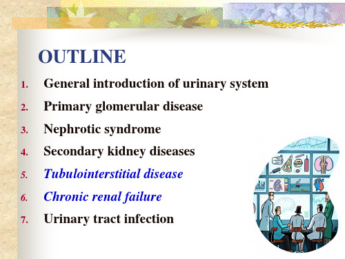
B
Others- Dermatologic abnormalities
Clinical manifestations (in uremia)
1) Disturbances in homeostasis Water and Sodium
Dehydration---hypernatremia, rare. Retention of water ---dilutional hyponatremia, common.
weight protein. ❖ Renal glycosuria, aminoaciduria ❖ Defects in urinary acidification (renal tubular acidosis) and
concentrating ability
❖ Renal dysfunction (CRF)
Prednisolone treatment ??
Chronic interstitial nephritis(CIN)
❖ Usually with chronic medication and drug exposure
❖ Insidious onset, polyuria, nocturia ❖ Mild or moderate proteinuria, predominantly low-molecular-
2) Cardiovascular system and lung Hypertension: could be malignant HTN Heart failure: hypertension + high blood volume +
cardiomyopathy. --- Emergency.
内科学教学课件:英文班尿路感染
4: B-ultrasound to exclude obstruction and urolithiasis
Diagnosis
True bacterial urine:
1) mid-stream clean-catch bacteria count≥105/ml, excluding false positive
bacteria placed are rapidly cleared, the antibacterial properties of urine and the bladder mucosa , high urea concentration and high osmolarity .
Precipitating factors
Urinary Tract Infection
Definition
UTI is the urinary infective disease which is causing by many different microorganisms growing and breeding in urinary tract often occur in old man ,worst immune, urinary deformity and woman in child-bearing period
2) puncture urinary bladder can find bacteria count
Differential diagnosis
1: chronic glomerulonephritis 2: Renal tuberculosis 3: Urethral syndrome: not find true
2: a: Urine bacteria culture: midstream clean-catch bacteria count ≥ 10 5/ml. puncture urinary bladder find bacteria count.
内科学 肾小球肾炎 PPT课件
ppt课件 14
炎症介质
• • • • • • • • 补体:C5b-9 凝血纤溶系统因子 血管活性胺:ET、AⅡ 生物活性肽:TGF-β 、PDGF、IL 生物活性酯 细胞粘附分子 活性氧 活性氮 :NO
ppt课件 15
非免疫机制的作用
• 血液动力学改变,“高滤过”学说,促 进肾小球硬化; • 大量蛋白尿加重肾小管间质损伤; • 高脂血症加重肾小球损伤
• 是以急进性肾衰竭综合征为表现的一组 疾病 • 其特征:起病类似急性肾炎综合症,肾 功能在几周内急剧坏转,常在早期出现 少尿性急性肾衰竭。 • 病理类型:新月体性肾炎。
ppt课件
30
慢性肾小球肾炎
Chronic glomerulonephtitis
ppt课件
31
慢性肾炎的定义
• 起病缓慢,以蛋白尿、血尿、高血压和 水肿为基本表现,病情迁延,病变缓慢 进展,可有不同程度的肾功能减退,最 终将发展为慢性肾衰竭的一组肾小球病。 • 典型表现:肾炎综合征+慢性肾功能减退
ppt课件
20
蛋白尿的定义
• 正常人尿蛋白排出量低于150 mg /24小 时尿,其中包括白蛋白、免疫球蛋白和 其他血浆蛋白成分,用尿常规的定性试 验测不出 • 蛋白尿的定义是尿蛋白排出量超过150 mg /24小时尿
ppt课件
21
微量白蛋白尿的定义
• 正常尿白蛋白排出量低于30 mg /24小时尿, 尿蛋白排出量大于300 mg/24小时尿才能用尿 常规的定性试验测出,故微量白蛋白尿的定义 为30~300 mg/24小时尿。 • 正常尿白蛋白排泄率低于20 mg/min,微量白 蛋白尿的第二个定义是白蛋白尿排泄率20~200 mg/min • 微量白蛋白尿的第三个定义是尿白蛋白/肌酐 比率:男17 -250mg/g,女25-355mg/g
内科学PPT课件 肺结核TB 呼吸系统疾病
源自病理渗出、增殖、变质
增殖
结核结节
特征性病理改变
变质----干酪性坏死
转归
吸收消散 钙化 纤维化
形成空洞
痊愈
恶化
转归
临床特征
结核中毒症状
发热---午后低热多见 盗汗---睡眠中出汗 纳差 乏力 消瘦 其他---妇女可有月经失调
呼吸系统症状
咳嗽---多干咳,合并支气管内膜结 核,刺激性咳嗽;
主要成分,变态反应;多糖:血清免疫反应。 病灶中的菌群:休眠菌,生长繁殖菌 自然耐药与继发耐药: 多耐药产生:2种以上药耐药
流行病学
➢ 传染源:痰涂阳性者 ➢ 传播途径:飞沫最主要,消化道、皮肤 ➢ 易感人群:婴幼儿、老年人、免疫抑制剂使用者,慢性病
患者
传染源
传 播 途 径
免疫与变态反应
成人:PPD 1U(+++)---有活动性结核病灶 PPD 5U(+)---有结核感染
<3岁儿童: 5U PPD(+++)---应视为有新感染活 动
阴性意义
(1) 无结核感染 (2) 变态反应前期 (3) 免疫系统暂时受到抑制 (4) 免疫功能缺陷
ɤ-干扰素释放试验
➢通过特异性抗原ESAT-6,GFP-10与全 血细胞共同孵育,检测ɤ-干扰素或计 数分泌ɤ-干扰素的T淋巴细胞,
↘ ↗
结果判断--48~72h测硬节直径
判断
阴性 (一) 弱阳性 (+) 阳性 (++) 强阳性 (+++)
无或硬结d<5mm 硬结d 5~9mm 硬结d 10~19mm 硬结d >20mm,或局
部水疱,坏死者
阳性---临床意义
结核菌素试验阳性对儿童、青少年结核诊断有参考意 义;结核菌素试验阳性,不能区分结核菌感染或卡介苗接 种;WHO推荐PPD试验。
肠结核的临床表现 ppt课件

疗
ppt课件
21
预
后
早期诊断 及时治疗 预后良好
ppt课件
22
内科学消化系统疾病
结核性腹膜炎
tuberculous peritonitis
ppt课件
23
目标要求
掌握:结核性腹膜炎的临床表现、并发症、诊 断、鉴别诊断和治疗。
了解:本病的病因和发病机制、病理分型、预 后和预防。
ppt课件
ppt课件
5
病因和发病机制
四、发病条件:
入侵的结核杆菌数量多、毒力强;
人体免疫功能低下、肠壁局部抵抗力削弱。
ppt课件
6
病因和发病机制
根据人体对结核杆菌的免疫力和变态反应不
同分为:
1、溃疡型肠结核:渗出 干酪样坏死 溃疡
2、增生型肠结核:肉芽肿和纤维组织增生
3、混合型肠结核:二者兼有。
ppt课件
ppt课件 13
实验室和辅助检查
三、结肠镜检查 充血、水肿, 溃疡(环形、边缘呈鼠咬状), 炎性息肉, 肠腔狭窄等。
活检:干酪样坏死性肉芽肿 结核杆菌(AFB)
ppt课件 14
诊
依据:
断
1.青壮年,有肠外结核(主要是肺结核);
2.腹痛、腹泻,腹部肿块;
3.不明原因的肠梗阻;
4.结核中毒症状;
6.PPD或T-SPOD强阳性。
ppt课件 34
诊
断
诊断困难者,可行腹腔镜检查或诊断性抗结核 治疗(2-4周有效可诊断)。
ppt课件
35
鉴别诊断
1.以腹水为主要表现者:肝硬化腹水、癌性腹水 、其他疾病引起腹水。 2.以腹部肿块为主要表现者:腹部肿瘤。
内科17 intestinal tuberculosis 2014
• Active pulmonary tuberculosis
▪ Complications
• Intestinal obstruction
• Intestinal fistula
• Tuberculosis peritonitis(结核性腹膜炎)
• Bleeding Not common
精选2021版课件
(多重耐药结核分枝杆菌) • Immunosuppressive agents
(免疫抑制剂)
▪ Tuberculosis is back !!
精选2021版课件
3
Etiology and Pathogenesis
▪ Pathogene
• Bacillus Tuberculosis(人型结核分枝杆菌)
11
Laboratory Tests and Imaging studies
▪ Laboratory Tests
• Anemia
• Erythrocyte sedimentation rate (ESR,血沉)
• Mushy stool
• Tuberculin test(结核菌素试验) ++ Protein purified derivative tuberculin (PPD)
Intestinal Tuberculosis
(肠结核)
Chen Xin Jie Department of Gastroenterology
Zhu Jiang Hospital
精选2021版课件
1
Useful words
• Bacillus tuberculosis(人型结核分枝杆菌) • Mycobacterium bovis(牛型结核分枝杆菌) • ileocecal region(回盲部) • Intestinal fistula(肠瘘) • Abscess(脓肿) • Erythrocyte sedimentation rate (ESR,血沉) • Exploratory laparotomy(剖腹探查)
- 1、下载文档前请自行甄别文档内容的完整性,平台不提供额外的编辑、内容补充、找答案等附加服务。
- 2、"仅部分预览"的文档,不可在线预览部分如存在完整性等问题,可反馈申请退款(可完整预览的文档不适用该条件!)。
- 3、如文档侵犯您的权益,请联系客服反馈,我们会尽快为您处理(人工客服工作时间:9:00-18:30)。
Pathology
according to the balance between the bacterium and patient immune
• Exudation • Caseation • Ulceration • Hypertrophy
Pathology
1.Ulcerative form
X-ray test
Barium meal or enema Irritable sign ( Stierlin sign ) , Ulceration, Nodular thickening of mucosal folds, Short annular strictures
CT Enterography,CTE MRE
Diagnosis
1. Abdominal symptom of TB + toxemia of TB 2. X-ray 3. Endoscopy 4. extra bowel TB & PPD / T-spot
• Therapeutic trial of antituberculosis treatment for 2~6 weeks
.
Etiology & Pathogenesis
• Pathogen is Mycobacterium tuberculosis • Routes of infection:
oral ,swallow infected sputum , main route
blood,miliary tuberculosis; surrounding spread,peritoneal, pelvic cavity
Endoscopy
Congestion, Edema Ulceration perpendicular to the long axis of the bowel inflammatory polyps stricture caseating granulomas
endoscopy
• Endoscopie Capsulaire • Enteroscopy
inflammation of bowel wall and regional lymph tissue
↓
caseous necrosis
↓ ulceration
↓ extensive fibrosis
↓ Strictures and fistulae
Pathology
1.Ulcerative form
Chapter 6
Intestinal tuberculosis &
Tuberculous peritonitis
Section Ⅰ Intestinal tuberculosis
Mycobacterium tuberculosis caused special infection of intestine
• Laparotomy
irritable bowel condition
Clinic features
1. abdominal pain 2. Diarrhea and constipation 3. Palpable mass Hypertrophic type in the right iliac fossa
Hale Waihona Puke Clinic features
Complication
• Obstruction • Fistulization or penetration • Intestinal hemorrhage • Perforation
Laboratory test
blood :anemia ,ESR↑ tuberculin test:PPD/T-spot
Etiology & Pathogenesis
Why Ileocecal region is a common TB site : 1. a long stasis
2. abundant lymphatic tissue
Pathogenesis: Infection of TB + strong toxicity + large amount + weakened immune condition + reduced local resistibility
1. abdominal pain right iliac fossa(髂窝) pain
referred pain of superior belly or peripheral umbilicus ; tenderness in that region
Clinic features
1. abdominal pain 2. Diarrhea and constipation • Diarrhea : ulcerative type • Constipation: hypertrophic type • Diarrhea alternated with constipation:
1. abdominal pain 2. Diarrhea and constipation 3. Palpable mass
4. systemic and extra bowel symptoms
fever night sweats weight loss, Anemia
Malnutrition Pulmonary TB
2.Hypertrophic form • Commonly in cecum • granuloma , fiber hyperproliferation→ a rigid thickening of the bowel wall→
Strictures → obstruction
Pathology
1.Ulcerative form
2.Hypertrophic form 3.Ulcerohypertrophic form
a subtype , a combination of features of the ulcerative and hypertrophic forms.
Clinic features