鼻咽纤维血管瘤的影像表现及临床
鼻咽癌与鼻咽血管纤维瘤的影像诊断-精品医学课件

鼻咽癌的淋巴引流:
咽后淋巴结:上界颅底,下界舌骨体上缘, 前界咽黏膜下筋膜,后界锥前肌,外侧界 颈内动脉内缘,内侧界为中线。
鼻咽癌最易累及咽后及Ⅱ区,容易累及Ⅲ、 Ⅳ、Ⅴ区,极少累及ⅠB区,罕见累及ⅠA 区。
淋巴结转移
M分期
M0 无远处转移 M1 有远处转移(包括颈部以下淋巴结转移)
远处转移
缘)、咽旁间隙 T3 侵犯颅底、翼内肌 T4 侵犯颅神经、鼻窦、翼外肌及以外的咀嚼肌间隙、颅内(海绵
窦、脑膜等)
局限于鼻咽
肿 瘤 侵 犯 鼻 腔 、 口 咽 、 咽 旁 间 隙
侵犯翼内肌
颅内侵犯
N分期
N0 影像学检查及体检无淋巴结转移 N1a 咽后淋巴结转移 N1b 单侧Ⅰb、Ⅱ、Ⅲ、Ⅴa区转移淋巴结且直径≤3CM N2 双侧Ⅰb、Ⅱ、Ⅲ、Ⅴa区转移淋巴结;或直径> 3CM; 或淋巴结包膜外侵犯 N3 Ⅳ、Ⅴb区转移淋巴结
颈部淋巴结
咽后淋巴结(PR) 颏下淋巴结(ⅠA) 颌下淋巴结(ⅠB) 颈内静脉淋巴结上组(ⅡA) 二腹肌下淋巴结(ⅡB) 颈内静脉淋巴结中组(Ⅲ) 颈内静脉淋巴结下组(Ⅳ) 颈后三角淋巴结(ⅤA) 锁骨上淋巴结(ⅤB) 气管周围淋巴结(Ⅵ) 上纵膈淋巴结(Ⅶ)
信号更高 。 MRI对放疗后的评价:鼻咽癌对放疗敏感,放疗后出现鼻咽腔扩大,咽隐窝
变深,肌肉萎缩变性,粘膜萎缩的征象。
鼻咽部肿块
T1WI
T2WI
C+
分泌性中耳炎
上颌窦炎
骨 质 破 坏
放 疗 后 评 价
鼻咽癌的TNM分期
T分期 ቤተ መጻሕፍቲ ባይዱ1 局限于鼻咽 T2 侵犯至鼻腔(超过上颌窦后壁连线)、口咽(超过C2椎体下
鼻咽部纤维血管瘤
窦后壁受压前移、塑形, 上颌窦腔
上颌窦、筛窦、翼腭窝及颞下窝、
缩小, 但上颌窦后壁骨质无破坏
颅底侵犯,累及海绵窦,病变范围 整理ppt 较广泛
五、影像表现:
(一)CT表现:
1.软组织肿块:鼻咽部等或稍高密度软组织肿块,外缘 光滑锐利,强化明显、向周围组织浸润生长。
CT增强扫描:病灶明显强化,肿块广泛累及左 整理侧pp翼t 腭窝、颞下窝、鼻后孔、上颌窦后壁、后
整理ppt
二、病理:
起源:枕骨斜坡底部、蝶骨体及翼突内侧的骨膜, 向下突入鼻咽并向前生长,经后鼻孔进入同侧鼻腔。 内镜检查:瘤体大小不一,呈类圆形、椭圆形或不 规则形,表面为粉红色、暗红色,可有扩张的血管。
肿瘤由丰富的血管组织和纤维组织基质构成。血管 壁薄,缺乏弹性,易引起大出血,较大的肿瘤可以 压迫(或破坏)邻近骨质,侵入鼻窦、眼眶、翼腭 窝,故本瘤虽属良性,但具有侵袭性。
鼻咽纤维血管瘤
王莉莉 2014.05.26
整理ppt
鼻咽的解剖
鼻咽又称上咽,位于颅底 与软腭之间,大小较恒定, 前后径约2cm,高约4cm。 鼻咽前壁经后鼻孔与鼻腔 想通,向下与口咽连续。
咽隐窝:咽口上方有一隆 起部分称咽鼓管圆枕,其 后上方与咽后壁之间的凹 陷称为咽隐窝。
咽扁桃体:鼻咽顶部和后 壁移行相连,呈倾斜的圆 拱形,常合称顶后壁,其 粘膜下有丰富的淋巴组织, 称咽扁桃体,即腺样体, 在婴幼儿较发达,6-7岁 后开始萎缩。
整理ppt
男性,14岁,肿瘤为分叶状软组织影,呈等T1、 长T2信号,增强后明显强化。
整理ppt
整理ppt
男,19岁,右侧后鼻 孔翼腭窝分叶状软组 织影,呈等T1、长T2 信号,病灶明显强化 ,可见到迂曲条状流 空信号。
鼻咽纤维血管瘤

鼻咽纤维血管瘤鼻咽纤维血管瘤鼻咽纤维血管瘤是鼻咽部最常见的良性肿瘤,与一般纤维瘤不同,为致密结缔组织、大量弹性纤维和血管组成,常发生于10~25岁青年男性,女性少见,故又名“男性青春期出血性鼻咽血管纤维瘤”。
是发生于鼻咽部易出血的良性肿瘤,占H&N肿瘤0.5%,多见于男性,平均15岁(7-19岁),>25岁很少发病。
病因1. 激素学说:由于患者以青春男性占明显比例,故有人认为可能与男性激素过剩引起内分泌功能紊乱而致病。
2. 骨外膜或胚源性纤维软骨、枕骨和蝶骨底的结缔组织反应性增生;3. 上颌动脉终末支的非嗜铬副神经节细胞学说。
鼻咽纤维血管瘤的病理:肿瘤血供主要来自颌内动脉和咽升动脉,也可来自颈内动脉的眼动脉与脑膜支,以及椎动脉系统的分支。
肿瘤起源于枕骨底部、蝶骨体及翼突内侧的骨膜。
瘤体由胶原纤维及多核成纤维细胞组成网状基质,其间分布大量管壁薄且无收缩能力的血管,这种血管受损后极易出血。
肿瘤的生长与扩展途径:起源于蝶腭区,在鼻咽粘膜下生长。
大肿瘤常被压迫成分叶状或哑铃型。
肿瘤有侵袭邻近结构的倾向,可通过蝶腭孔播散至翼颌裂、翼突和颞下窝,也可长入鼻腔、鼻窦、鼻咽部、蝶窦和斜坡,还可指形扩展到眼眶和中颅窝(鞍旁)其时,肿瘤只会压迫海绵窦而不会穿破窦壁。
鼻咽纤维血管瘤的临床表现:1.出血:阵发性鼻腔或口腔出血,且常为病人首诊主诉。
初期出血为间断发生,逐渐发展为不易制止的大出血。
反复出血可导致失血性贫血。
合并有感染或有溃疡者,出血更为严重。
2.鼻塞:鼻塞初为单侧,进行性加重。
肿瘤继续长大,可致双侧鼻塞。
肿瘤堵塞后鼻孔并侵入鼻腔,引起一侧或双侧鼻塞,常伴有流鼻涕,闭塞性鼻音,嗅觉减退等。
3.其他症状:由于瘤体不断增长引起邻近骨质压迫吸收和相应器官的功能障碍,肿瘤侵入邻近结构则出现相应症状。
肿瘤增大后压迫咽鼓管咽口,可致耳闷塞、耳鸣、听力障碍,并可致中耳炎。
压迫阻塞鼻窦自然开口可引起鼻窦炎。
如侵入眼眶,则出现眼球突出,视神经受压,视力下降;侵入翼腭窝引起面颊部隆起;侵入鼻腔可引起外鼻畸形;侵入颅内压迫神经,引起头痛及脑神经麻痹。
鼻咽血管纤维瘤

三、影像表现
1.软组织肿块:分叶状或不规则,CT呈等或稍高密度,密度较均匀, 外缘光滑锐利;MRI T1WI呈中等或稍高信号、T2WI呈明显高信号, 内部可掺杂低信号,与肿瘤富含血管及其与纤维成分比例有关;明 显强化(超过100HU)、向周围组织浸润生长。侵犯翼腭窝最常见。
2.压迫性骨质破坏:骨质受压,吸收破坏 3.颅内侵犯:颅内肿块与颅外肿块密度一致, 高于脑实质;病灶与
体征:鼻根部变宽呈蛙状鼻、眼球突出、运动障碍、鼓膜及听 力改变。
鼻镜可以检查肿瘤的大小、色泽、表面血管情况。鼻咽部触诊 要谨慎小心,一般不作活检,以免发生严重出血。
按Chandler标准进行分期: I 期:肿瘤局限于鼻咽部; Ⅱ期:肿瘤扩展至鼻腔和蝶窦; Ⅲ期:肿瘤扩展至上颌窦、筛窦、翼腭窝、颞下窝、眼 眶(眶上裂、眶下裂、眶内),颊部; Ⅳ期:肿瘤侵入颅内。
四、鉴别诊断
鼻咽癌:最常发生于中年人,回缩性血涕是其典型的早期 临床表现之一,影像检查见鼻咽部浸润性肿块,边界不清, 侵蚀性骨质破坏明显,增强扫描呈轻中度强化,颈部淋巴 结肿大往往为初诊的首发症状。
淋巴瘤:以青壮年多见,病变侵犯范围较广,增强扫描呈 轻度强化,骨质破坏少见,转移常见,如转移到皮肤、胃 肠道、肝、淋巴结等。
鼻咽血管纤瘤
一、概述
鼻咽血管纤维瘤:又称男性青春期出血性鼻咽血管纤维瘤,为鼻 咽部最常见的良性肿瘤,好发于10-25岁青年男性(19:1)。起 源:枕骨斜坡底部、蝶骨体及翼突内侧的骨膜,向下突入鼻咽并 向前生长,经后鼻孔进入同侧鼻腔。故本瘤虽属良性,但具有侵 袭性。
病因:尚不明确,可能与性激素、发育异常、炎症刺激等因素有 关。
分期
chandler I 期: 肿瘤局限于鼻咽 部。
鼻咽部纤维血管瘤

通过栓塞肿瘤供血动脉,使肿瘤缺血坏死,适 用于位置较深、手术难度较大的患者。
3
放疗
通过放射线照射肿瘤,使肿瘤组织坏死,适用 于无法手术或术后辅助治疗的患者。
治疗过程与护理
治疗过程
根据肿瘤大小、位置、与周围组织 关系等因素制定个体化治疗方案, 通常采用多学科联合治疗。
术前准备
完善相关检查,对患者进行心理疏 导,制定应急预案。
06
研究进展与前沿
基础研究进展
分子生物学研究
研究发现,鼻咽部纤维血管瘤的发生发展与一系列基因的变异和调控有关。 例如,研究发现,某些致癌基因的变异会导致鼻咽部纤维血管瘤的发生,而 另一些基因的变异则可能导致肿瘤细胞的增殖和迁移。
细胞生物学研究
鼻咽部纤维血管瘤的细胞生物学研究主要集中在探讨肿瘤细胞的增殖、分化 、凋亡和迁移等方面。研究发现,某些细胞因子和信号转导通路在肿瘤的生 物学行为中发挥重要作用。
病理学检查
组织病理学检查
通过对病变组织进行病理学检查,可以确定肿瘤的性质和组织来源,从而明确诊 断鼻咽部纤维血管瘤。
免疫组织化学检查
免疫组织化学检查可以检测肿瘤细胞的特定蛋白质,有助于确定肿瘤的性质和分 化程度。
04
治疗与预后
治疗方法
1 2
传统手术
包括鼻内镜手术、颅底显微手术等,适用于肿 瘤体积较小、位置较浅的患者。
感染
颅内并发症
长期鼻出血和肿瘤压迫可能导致局部感染, 严重时可引起菌血症、败血症等。
肿瘤侵犯颅底并破裂可导致脑脊液鼻漏、颅 内感染等严重并发症。
与其他疾病的鉴别诊断
鼻炎
鼻咽部纤维血管瘤与鼻炎症状相似,但鼻炎一般 无出血症状。
上颌窦炎
鼻咽部血管瘤(鼻咽纤维血管瘤)临床路径(全文)
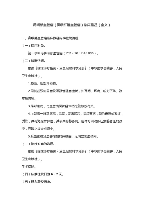
鼻咽部血管瘤(鼻咽纤维血管瘤)临床路径(全文)一、鼻咽部血管瘤临床路径标准住院流程(一)适用对象。
第一诊断为鼻咽部血管瘤(ICD-10:D18.006)。
(二)诊断依据。
根据《临床诊疗指南-耳鼻咽喉科学分册》(中华医学会编著,人民卫生出版社)。
1.咯血、咽部异物感。
2.同侧或双侧鼻塞及咽鼓管阻塞症状,如耳闭、耳痛、听力下降、鼓室积液等。
3.局部疼痛,与血管表面神经末梢比较敏感有关。
4.血管瘤一般基底宽,无蒂,表面隆起,呈结节状,颜色青蓝或紫红,质软,具有海绵样弹性,其表面有静脉网。
瘤体可因动脉压或静脉压的改变,而随之增大或缩小。
5.系血管成分显著增加的纤维瘤,无明显出血倾向。
(三)治疗方案的选择。
根据《临床诊疗指南-耳鼻咽喉科学分册》(中华医学会编著,人民卫生出版社)。
手术切除。
(四)标准住院日为6-7天。
(五)进入路径标准。
1.第一诊断必须符合ICD-10:D18.006鼻咽部血管瘤疾病编码。
2.影像学证据表明瘤体仅局限于鼻咽部,未生长进入咽旁间隙、鞍区、翼颚窝等颅底结构。
3.当患者同时具有其他疾病诊断,但住院期间不需特殊处理,也不影响第一诊断的临床路径流程实施时,可以进入路径。
(六)住院期间检查项目。
1.必需的检查项目:(1)血常规、尿常规;(2)肝肾功能、电解质、血糖、凝血功能;(3)感染性疾病筛查(乙肝、丙肝、梅毒、艾滋等);(4)影像学检查(鼻咽部CT、MRI);(5)鼻内镜;(6)胸片、心电图;2.根据患者情况可选择的检查项目:(1)影像学检查(MRA、CTA)。
(七)治疗方案与药物选择。
1.手术治疗。
(1)麻醉。
采用气管内插管全身麻醉,控制性低血压麻醉可以减少术中出血。
(2)体位。
仰卧位,肩部垫高,头后仰,低于肩部平面。
(3)放置开口器,将口张开。
(4)切口呈马蹄形,自一侧第二磨牙开始沿牙龈内侧距龈缘0.5mm,向前延至切牙孔后方,弯向对侧第二磨牙。
(5)沿切口将黏骨膜自硬腭骨板上分离直达硬腭后缘。
鼻咽纤维血管瘤的CT诊断与鉴别诊断

参考文献:
1.刘庆.何晓峰.王江云.庞桦进.杨旸 青少年鼻咽部纤维血管瘤的影像诊断及介 入治疗 [J] -西部医学2011(12) 2.刘继远.余志强 鼻咽纤维血管瘤研究进展 [J] -国际耳鼻咽喉头颈外科杂志 2012(4) 3.胡章勇,王宏清,贾明辉. 鼻咽纤维血管瘤的CT和MRI诊断[J].医学影像学杂 志,2010,(02) 4.陈晓丽,王振常,鲜军舫. 鼻咽纤维血管瘤的CT和MRI诊断[J]. 实用放射学杂 志. 2007(01) 5.魏小宾,刘宝良. CT对鼻咽纤维血管瘤的诊断和应用价值[J]. 海南医学院学 报. 2010(10) 6.吕明明,范新东. 47例青少年鼻咽纤维血管瘤的临床及影像学特征分析[J]. 中国口腔颌面外科杂志. 2010(05) 7.吴 刚. 200 例鼻咽癌 MRI 影像分析[J]. 中国中西医结合影像学杂志. 8.杨敏洁,魏雪峰. 鼻咽癌的影像学诊断分析比较[J]. 中国医药指南. 2010(08) 9.程南生. 鼻咽癌的CT诊断和鉴别诊断[J]. 中国中西医结合耳鼻咽喉科杂志. 2006(05) 10.谢敏,张国华. 鼻咽部淋巴瘤的CT诊断[J]. 中国现代医学杂志. 2011(07)
• 侵犯范围及部位:同前
CT表现 CT findings
• 骨质改变: 相邻骨质多表现为不同程度受压、孔 缝扩大和吸收改变,少数可见骨质破坏, 虫蚀状
CT表现 CT findings
• 继发表现: 中耳乳突炎、副鼻窦炎、突眼等
病例展示 cases discussion
伴 间 断 鼻 出 血持 续 月鼻 余塞 2
力 减 退
月 余 右 , 侧 头 耳 痛 鸣 月, 余听 3
case1 F 24 5
case1
case1
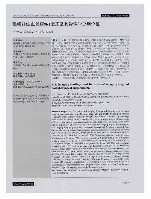
鼻咽纤维血管瘤MRI表现及其影像学分期价值
C i J g R sn m g g 2 1 , o , o5 hn Ma n eo I a i , 0 2 V l N n 3
富 血 供 肿 瘤 , 好 发 于 l ~2 岁 青 春 期 男 性 。病 理 上 3 0 5 . MRI 鼻咽纤维血管瘤术前分期应 用及 比较 2 在 肿 瘤 通 常 由错 综 复 杂 血 管 网 和 纤 维 基 质 以 不 同 比例 MR 断 层 影 像 对 于 邻 近 鼻 咽 解 剖 深 在 、 结 构 复 I 构 成 ,根据 血管 成分 和纤 维成分 在肿 瘤组 织 中所 占 杂颅 底等 结构 显示 比较全 面 。MR 多方位 多参数 成 I
比例 ,可分 别称 其为血 管 纤维瘤 或纤 维血 管瘤 。镜 像 、 软 组 织 分辨 率 高 ,可 以显 示海 绵 窦 、颅 内垂 下 血管 内膜 一般 为单层 内皮细胞 ,基 底为 不完 整 的 体 、视交 叉、硬 脑膜 、翼管神 经 、圆孔 内结构有 无 平滑 肌 层 ,纤维样 基质 可从 致密 的纤 维区到 水肿 或 累及 ,与邻 近鼻 腔鼻 窦的慢性 炎症及 息 肉有 区别 。 黏 液样 结缔组 织 ,其 内细胞 呈梭形 或星 形 ,但核 的 已有 的研究 认为 ,鼻 咽纤维血 管瘤 的生长特 性大 多
31 . MRI 表现及 术前诊 断 鼻 咽纤维 血管 瘤 由血管和 纤 维组织 以不 同 比例 构成 ,肿瘤 由富含 胶 原和纤 维原 细胞 的纤维 基质 、 不 同 口径 的血 管排 列形 成 ,排 列 的血管 缺乏肌 层 、
分 期准确 性有 一定程 度提高 。 同样 对于 原发组 与手 术 后复发 组分 期判断 ,两组 中影像 分期 与手术 分期 不一致病例主要在R d o siI 期 ,原发组有3 ak w k I Ia 例影
鼻咽纤维血管瘤的临床与CT诊断分析
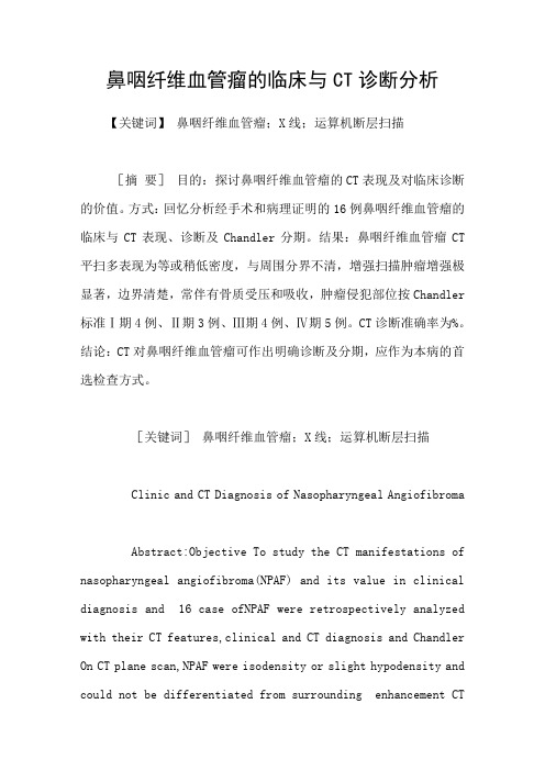
鼻咽纤维血管瘤的临床与CT诊断分析【关键词】鼻咽纤维血管瘤;X线;运算机断层扫描[摘要]目的:探讨鼻咽纤维血管瘤的CT表现及对临床诊断的价值。
方式:回忆分析经手术和病理证明的16例鼻咽纤维血管瘤的临床与CT表现、诊断及Chandler分期。
结果:鼻咽纤维血管瘤CT 平扫多表现为等或稍低密度,与周围分界不清,增强扫描肿瘤增强极显著,边界清楚,常伴有骨质受压和吸收,肿瘤侵犯部位按Chandler 标准Ⅰ期4例、Ⅱ期3例、Ⅲ期4例、Ⅳ期5例。
CT诊断准确率为%。
结论:CT对鼻咽纤维血管瘤可作出明确诊断及分期,应作为本病的首选检查方式。
[关键词]鼻咽纤维血管瘤;X线;运算机断层扫描Clinic and CT Diagnosis of Nasopharyngeal AngiofibromaAbstract:Objective To study the CT manifestations of nasopharyngeal angiofibroma(NPAF) and its value in clinical diagnosis and 16 case ofNPAF were retrospectively analyzed with their CT features,clinical and CT diagnosis and Chandler On CT plane scan,NPAF were isodensity or slight hypodensity and could not be differentiated from surrounding enhancement CTscan,NPAF were grossly enhanced and could be clearly differentiated from surrounding case showed either slight or remarkable pressure and bone destruction over the base of to Chandler classification,there were 4 cases of stage Ⅰ,3 cases of stage Ⅱ,4 cases of stage Ⅲ ang 5 cases of stage Ⅳ.CT diagnosis accurate rate is %.Conclusion CT can provide accurate diagnosis and stage for NPAF and has high value in surgical can be used as the first choice of examination for NPAF.Key words:Nasopharyngeal angifibroma;Xray;Computed tomography鼻咽纤维血管瘤(nasopharyngeal angiofibroma,NPAF)又名男性青春期出血性纤维瘤,是鼻咽顶部后鼻孔区最多见的良性肿瘤。
鼻咽纤维血管瘤的影像表现及临床 ppt课件
Fig. 2 Magnetic resonance, saggital T1-weighted image after contrast administration.
南华大学附属第一医院
版权所有
Enhancement on CT and MRI as well as signal-void areas on MR images, typical for high flow vessels (Fig. 2). Arteriography revealed abundant vascularity with main blood supply from the internal maxillary artery.
南华大学附属第一医院
版权所有
Abstract
Nasopharyngeal angiofibroma (NA) is a rare,vascular tumor affecting dolescent males. Due to aggressive local
growth, skull base location and risk of profound hemorrhage,
embedded in fibrous stroma. The abundant vascular component is responsible for excessive bleeding during surgery or following biopsies. It also contributes to certain characteristic radiological features of NAs, including strong contrast enhancement on CT and
鼻咽纤维血管瘤的影像表现
• Saylam 等发现增殖细胞核抗原、血管内皮生长因子 (VEGF)和转化生长因子β (TGF-β) 可能参与 血管性增殖。VEGF 和TGF-β 会从基质产生大量 成纤维细胞,从而形成鼻咽纤维血管瘤的病理基础。
分型
CT 和MRI 影像分期参照Radkowski( 1996) 临床标准: Ⅰa: 局限于鼻腔和/或鼻咽穹窿部; Ⅰb:扩展入一个或多个鼻窦; Ⅱa: 少部分侵入翼腭窝; Ⅱb: 整个翼腭窝受侵犯,上颌窦后壁前移,眶骨侵蚀、上颌动脉移位; Ⅱc: 颞下窝和/或颊部受侵犯或侵入翼板后方; Ⅲa: 颅底受侵( 中颅窝/翼突根部) ,小部分颅内扩展; Ⅲb: 颅底受侵,广泛颅内扩展伴有或不伴有海绵窦受侵。
连续;在病变边缘或内部可见高密度影 • MRI整体信号混杂,T2WI上内部的不均匀高信号为低信号围绕,增强后呈结
节状、斑片状的强化
鉴别诊断——内翻乳头状瘤
• 好发于40岁以上男性,50-70岁发病率最高高 •好发部位:单侧,肿瘤的生生发中心多位于中鼻道鼻腔外侧壁及 上颌窦 •局灶性骨质增生与SNIP的起源之间有很大的一致性;而骨质破坏 则与肿瘤恶变相关 •特性脑 征:在T2WI或增强T1WI上,病变内部结构多呈较规整 的栅栏状
增强明显强化。 • 椒盐征是鼻咽纤维血管瘤的特征性表现。
谢谢
• 供血动脉主要来自颈外动脉分支, 如上颌动脉和咽升动脉等。
• 肿瘤较大侵入颅内,可由颈内动脉 供血(较少)。
诊断与治疗
• 由于鼻咽纤维血管瘤是富血供肿瘤,无法行病理活检。在大多数 情况下,诊断JNA大多数为影像学检查。
• JNA的金标准治疗是术前栓塞后肿瘤的手术切除。由于颅底的复 杂解剖,晚期肿瘤的切除困难情况下,可以在治疗中增加化疗、 放疗或激素治疗。
鼻咽部纤维血管瘤

男性,14岁,肿瘤为分叶状软组织影,呈等T1、 长T2信号,增强后明显强化。
精品课件
精品课件
男,19岁,右侧后鼻 孔翼腭窝分叶状软组 织影,呈等T1、长T2 信号,病灶明显强化 ,可见到迂曲条状流 空信号。
诊断要点:
1.典型症状:为反复的鼻腔和口腔出血;压迫性的骨质吸收
鼻咽纤维血管瘤
王莉莉 2014.05.26
精品课件
鼻咽的解剖
鼻咽又称上咽,位于颅底 与软腭之间,大小较恒定 ,前后径约2cm,高约4cm 。鼻咽前壁经后鼻孔与鼻 腔想通,向下与口咽连续 。
咽隐窝:咽口上方有一隆 起部分称咽鼓管圆枕,其 后上方与咽后壁之间的凹 陷称为咽隐窝。
咽扁桃体:鼻咽顶部和后 壁移行相连,呈倾斜的圆 拱形,常合称顶后壁,其 粘膜下有丰富的淋巴组织 ,称咽扁桃体,即腺样体 ,在婴幼儿较发达,6-7 岁后开始萎缩。
, 上颌窦后壁受压前移、塑形, 上 上颌窦、筛窦、翼腭窝及颞下窝、
颌窦腔缩小, 破坏
但上颌窦后壁骨质无精品课件颅较底广侵泛犯,累及海绵窦,病变范围
五、影像表现:
(一)CT表现:
1.软组织肿块:鼻咽部等或稍高密度软组织肿块,外缘 光滑锐利,强化明显、向周围组织浸润生长。
CT增强扫描:病灶明显强化,肿块广泛累及左 精品课侧件翼腭窝、颞下窝、鼻后孔、上颌窦后壁、后
2.耳部症状:癌肿堵塞或压迫咽鼓管时,常出现卡他性中耳炎, 引起耳鸣、耳闭塞及听力下降,或伴有鼓室积液。
3.脑神经症状:癌肿侵入颅内,脑神经受损后,可出现剧烈头痛 、面部麻木、咀嚼困难、眼睑下垂、复视、视力丧失等表现。
4.颈淋巴结转移
为患者最早发现的体征。检查时颈部肿块多较坚硬,表面呈小结 节性,与周围粘连则不易推动。可互相融合,成为全颈侧巨大肿 块。
鼻咽部纤维血管瘤

xx年xx月xx日
鼻咽部纤维血管瘤
目录
contents
概述病理学特征诊断与鉴别诊断治疗与预后研究进展
概述
01
鼻咽部纤维血管瘤是一种主要发生于青少年男性的良性肿瘤,起源于鼻腔和鼻窦,也称为“鼻咽纤维血管瘤”或“鼻咽血管纤维瘤”。
定义
根据肿瘤的生长方式和临床表现,鼻咽部纤维血管瘤可分为局限型和弥漫型两类。
对于不宜手术或术后复发的患者,可采用介入治疗,通过栓塞或药物注射抑制肿瘤生长。
目前尚无特效药物可用于治疗鼻咽部纤维血管瘤,但可使用药物进行辅助治疗,如激素、抗生素等。
放疗可作为一种辅助手段,用于缩小肿瘤、缓解症状。
手术与介入治疗
手术切除是治疗鼻咽部纤维血管瘤的首选方法,可有效血管瘤的大小和位置对预后具有重要影响,肿瘤越大、位置越深,预后越差。
肿瘤大小和位置
治疗方法
患者年龄和身体状况
随访观察
治疗方法的选择对预后具有重要影响,手术治疗效果较介入治疗和药物治疗更可靠。
患者年龄和身体状况也是影响预后的因素,年轻患者和身体状况较好的患者预后较好。
鼻咽部纤维血管瘤具有较高的复发率,患者需要定期进行随访观察,以便及时发现并处理复发灶。
手术切除
当鼻咽部纤维血管瘤诊断明确后,应尽早进行手术,以减轻肿瘤对周围组织的压迫和损伤。
手术时机
介入治疗具有创伤小、恢复快的特点,可用于术后残留或复发、难以手术的患者。
介入治疗的优势
介入治疗可能会导致鼻腔黏连、鼻窦炎等并发症,且复发率较高。
介入治疗的局限性
药物治疗
放疗
药物治疗和放疗的局限性
药物治疗和放疗
鼻咽癌
鼻咽癌是常见的恶性肿瘤,与鼻咽部纤维血管瘤在临床表现和影像学表现上有重叠,但鼻咽癌的病理学检查可见癌细胞。
鼻咽纤维血管瘤小小的肿瘤也不能掉以轻心

面部肿胀:患者可能会出现面部肿胀,尤其是早晨起床时。
听力下降:患者可能会出现听力下降,严重时可能导致耳聋。
中期症状
鼻塞:鼻腔内肿瘤增大,导致呼吸困难
视力下降:肿瘤压迫视神经,导致视力下降
头痛:肿瘤压迫神经,导致头痛
鼻出血:肿瘤表面血管破裂,引起鼻出血
肿瘤可能破裂出血,引起鼻腔出血、颅内出血等严重后果
肿瘤可能影响患者的生活质量,导致心理压力和社交障碍
鼻咽纤维血管瘤的症状表现
03
早期症状
鼻塞:患者可能会出现单侧或双侧鼻塞,严重时可能导致呼吸困难。
头:患者可能会出现持续性头痛,尤其是早晨起床时。
视力下降:患者可能会出现视力下降,严重时可能导致失明。
03
04
定期进行体检,及时发现并治疗疾病
饮食调理
保持饮食均衡,多吃蔬菜水果
避免饮酒,以免刺激血管,加重病情
避免油腻食物,如油炸食品、肥肉等
避免辛辣刺激性食物,如辣椒、胡椒等
心理调适
保持积极心态:面对疾病,保持积极乐观的心态,积极配合治疗。
减轻心理压力:学会释放压力,避免焦虑、抑郁等负面情绪影响治疗效果。
01
02
保持良好的饮食习惯,多吃蔬菜水果,少吃辛辣刺激性食物
保持良好的心理状态,避免过度紧张、焦虑等不良情绪
03
04
定期进行体检,及时发现和治疗疾病
日常护理
保持良好的生活习惯,避免熬夜、过度劳累等不良习惯
01
02
保持良好的饮食习惯,多吃蔬菜水果,少吃辛辣刺激性食物
保持良好的心理状态,避免焦虑、紧张等不良情绪
化学治疗:使用抗癌药物杀死肿瘤细胞
鼻咽纤维血管瘤—搜狗百科精选全文

可编辑修改精选全文完整版鼻咽纤维血管瘤—搜狗百科概述鼻咽纤维血管瘤是鼻咽部最常见的良性肿瘤,与一般纤维瘤不同,为致密结缔组织、大量弹性纤维和血管组成,常发生于10~25岁青年男性,故又名“男性青春期出血性鼻咽血管纤维瘤”。
病因不明。
鼻咽纤维血管瘤,又称鼻咽血管纤维瘤。
多发生于男性青年,所以又称青年鼻咽纤维血管瘤。
要了解本病,首先要知道鼻咽部的解剖位置。
鼻咽位于鼻腔和口腔的后方,是形似漏斗的管道,为上消化道和上呼吸道的共同通道。
可分为鼻咽、口咽和喉咽三部分。
[1]病理肿瘤起源于枕骨底部、蝶骨体及翼突内侧的骨膜。
瘤体由胶原纤维及多核成纤维细胞组成网状基质,其间分布大量管壁薄且无收缩能力的血管,这种血管受损后极易出血。
肿瘤常向邻近组织扩张生长,通过裂孔侵入鼻腔、鼻旁窦、眼眶、翼腭窝及颅内。
临床表现鼻咽纤维血管瘤1.出血阵发性鼻腔或口腔出血,且常为病人首诊主诉。
由于反复大出血,病人常有不同程度的贫血。
2.鼻塞肿瘤堵塞后鼻孔并侵入鼻腔,引起一侧或双侧鼻塞,常伴有流鼻涕,闭塞性鼻音,嗅觉减退等。
3.其他症状由于瘤体不断增长引起邻近骨质压迫吸收和相应器官的功能障碍,肿瘤侵入邻近结构则出现相应症状,如侵入眼眶,则出现眼球突出,视神经受压,视力下降;侵入翼腭窝引起面颊部隆起;侵入鼻腔可引起外鼻畸形;侵入颅内压迫神经,引起头痛及脑神经麻痹。
检查1.前鼻镜检查常见一侧或双侧鼻腔有炎性改变,收缩下鼻甲后,可见鼻腔后部粉红色肿瘤。
2.间接鼻咽镜检查可见鼻咽部圆形或分叶状红色肿瘤,表面光滑而富有血管,瘤体侵入后鼻孔―鼻腔可引起外鼻畸形或软腭下塌。
3.触诊手指触诊可触及肿块基底部,活动度小,中等硬度,若瘤体侵入颊部,通过触诊可了解瘤体蒂部与邻近部位粘连情况。
但触诊应轻柔,因触诊极易引起大出血,临床应尽量少用。
4.影像学检查CT和MRI检查可清晰显示瘤体位置、大小、形态,了解肿瘤累及范围和周围解剖结构的关系。
数字减影血管造影(DSA)可了解肿瘤的血供并可进行血管栓塞,以减少术中出血。
鼻咽部纤维血管瘤的影像学诊断

鼻咽部纤维血管瘤的影像学诊断陈桂美;李先玉【摘要】目的:探讨鼻咽部纤维血管瘤( nasopharyngeal fibroangioma)的影像学特征性表现及其诊断价值。
方法回顾性分析52例鼻咽部纤维血管瘤的临床资料、CT及MRI表现,分析病变的部位、形态、大小、边界光整与否、侵犯范围、密度或信号、强化方式、骨质变化及邻近结构的改变。
结果鼻咽部纤维血管瘤以浸润性生长特点为主,可引起邻近骨质的压迫塑形或骨质吸收破坏改变。
CT平扫以等或稍低密度为主,MRI以等或稍长T1、等或稍长T2信号为主;增强扫描瘤体多表现为显著均匀性强化。
MRI显示瘤体内可见点条状血管流空信号影,形成典型的“椒盐征”。
结论 CT结合MRI对鼻咽部纤维血管瘤的术前诊断、定位及预后评估有重大指导意义。
%Objective To explore the imaging findings and diagnostic value of the nasopharyngeal fibroangioma.Meth-ods The clinical data,CT findings and MRI findings in 52 patients with nasopharyngeal fibroangioma were retrospectively ana-lyzed.The location,shape,size,boundary,scope,density or signal,strengthen and the changes of the adjacent structure of the mass were observed.Results The nasopharyngeal fibroangioma mainly showed invasive growth,and leaded to oppressive shape or ab-sorption of the adjacent bone.The nasopharyngeal fibroangioma mainly showed equal or lower density on CT images,and showed equal or slightly longer T1,and equal or slightly longer T2 on MRI images.The tumor showed uniform and obviously strengthening after administration of Gd-DTPA.The punctate and bar empty blood flow signals were seen in the tumor on the MRI images,which shows a typicalsign of salt and pepper.Conclusion The combination of the CT and MRI have important clinical values in the diagnosis,localization and evaluation of the prognosis of the nasopharyngeal fibroangioma.【期刊名称】《实用癌症杂志》【年(卷),期】2015(000)012【总页数】4页(P1924-1926,1930)【关键词】纤维血管瘤;鼻咽部;体层摄影术;X线计算机;磁共振成像;诊断【作者】陈桂美;李先玉【作者单位】330006 南昌大学第一附属医院;315040 浙江省宁波市第六医院【正文语种】中文【中图分类】R739.63鼻咽部纤维血管瘤(nasopharyngeal fibroangioma)是鼻咽部常见的良性肿瘤之一[1],以青年男性多见,也称为男性青春期出血性纤维瘤[2]。
- 1、下载文档前请自行甄别文档内容的完整性,平台不提供额外的编辑、内容补充、找答案等附加服务。
- 2、"仅部分预览"的文档,不可在线预览部分如存在完整性等问题,可反馈申请退款(可完整预览的文档不适用该条件!)。
- 3、如文档侵犯您的权益,请联系客服反馈,我们会尽快为您处理(人工客服工作时间:9:00-18:30)。
cavity, paranasal sinuses, sphenoid-palatine foramen and
infratemporal fossa. In 10–20 % of the cases tumor invades the cranial cavity 。
南华大学附属第一医院
版权所有
Page 10
Histologic section of the tumor (H&E stain) shows fibrous stroma with ectatic, thin-walled vascular channels
南华大学附属第一医院 版权所有
Page 13
Enhancement on CT and MRI as well as signal-void areas on MR images, typical for high flow vessels (Fig. 2).
Fig. 2 Magnetic resonance, saggital T1-weighted image after contrast administration.
南华大学附属第一医院
版权所有
Page 12
Enhancement on CT and MRI as well as signal-void areas on MR images, typical for high flow vessels (Fig. 2). Arteriography revealed abundant vascularity with main blood supply from the internal maxillary artery.
Nasal
tumor
underwent
CT,
which
demonstrated
homogenous mass, with contrast enhancement ranging from strong to intermediate (Fig. 1).In one case, signs of bony destruction with tumor invasion to the ethmoid sinus were visible. The patient with the tumor of the infratemporal fossa
南华大学附属第一医院
版权所有
Page 7
Abstract
Nasopharyngeal angiofibroma (NA) is a rare,vascular tumor affecting dolescent males. Due to aggressive local growth, skull base location and risk of profound hemorrhage, NA is a challenge for surgeons.Angiofibromas tumor showed
wide, irregular vessels with a single layer of endothelial cells, embedded in fibrous stroma. The abundant vascular component is responsible for excessive bleeding during surgery or following biopsies. It also contributes to certain characteristic radiological
benign it shows locally aggressive growth with bone
destruction and spread through natural foramina and fissures.
南华大学附属第一医院
版权所有
Page 9
It originates from the posterolateral wall of the nasopharynx and from this site usually extends to the nasopharynx, nasal
features of NAs, including strong contrast enhancement on CT and
MR images, signal-void areas representing tumor vessels visible on MR images, as well as intensive vascular blush demonstrated
Page 4
影像图像
图7 MRI T1WI增强
南华大学附属第一医院
图8 MRI T1WI增强
版权所有
图9 MRI T1WI增强
Page 5
影像图像
图10 DSA冠状位
南华大学附属第一医院 版权所有
图11 DSA矢状位
Page 6
患者:男,26岁 主诉:右鼻出血2天 现病史:患者输2天前无明显诱因出现右鼻出血,为鲜血,呈滴状,先从左前鼻 孔出,后亦从口中、右鼻流出,数分钟后停止,反复出现多次,总量约为100ml, 无鼻塞,流涕,嗅觉正常。无头痛、发热、咳嗽、打鼾,无耳鸣、而鼻塞感, 无听力下降。于当地医院治疗,予以鼻腔填塞,症状好转。在中山陈星海医院, 予以电子喉镜检查“右鼻腔肿物,性质待查”。 既往史:否认肝炎、结核、疟疾病史,否认高血压、心脏病史,否认糖尿病、 脑血管疾病史,否认手术、外伤、输血史,否认食物、药物等过敏史,否认吸 烟、饮酒史,否认毒物接触史。
3.鼻咽非霍奇金淋巴瘤:以青壮年多见,病变侵犯范围 较广,增强扫描呈轻度强化,骨质破坏少见,转移常见,如转移 到皮肤、胃肠道、肝、淋巴结等。
南华大学附属第一医院 版权所有
Page 17
underwent CT, (MRI) and carotid
contrast。
arteriography with
preoperative embolization. The lesion showed intensive
南华大学附属第一医院
版权所有
Page 11
Fig. 1 Computed tomography, coronal plane, shows homogenous tumor mass in the right nasal cavity
(NA) is a rare vascular tumor, which represents 0.05 % of all head and neck tumors. At the same time, it is the most common benign neoplasm of the nasopharynx . NA occurs predominantly in adolescent males. Although histologically
intensive contrast enhancement on CT and magnetic
resonance imaging (MRI) scans, and abundant vascularity on angiography.
南华大学附属第一医院
版权所有
Page 8
Background
diagnosis. It also allows tumor embolization, which reduces
intraoperative hemorrhage, symptoms and bleeding. in a Due to of a risk of profound clinical presence characteristic
classic radiological findings, preoperative
biopsy is not recommended in the management of所有
Page 16
鉴别诊断要点
1.鼻咽纤维血管瘤:常见于男性青少年,有多次鼻出血病 史,影像检查见鼻咽部软组织肿块,多伴有压迫性骨质吸收破坏 ;增强扫描病灶明显强化。 2.鼻咽癌:最常发生于中年人,回缩性血涕是其典型早期临 床表现之一,影像检查见鼻咽部浸润性肿块,边界不清,侵蚀性 骨质破坏明显,增强扫描呈轻中度强化,颈部淋巴结肿大往往为 初诊的首发症状。
on angiography .
南华大学附属第一医院 版权所有
Page 15
Selective angiography is a useful diagnostic method to demonstrate tumor vascular composition and confirms the
Arteriography revealed abundant vascularity with main blood
supply from the internal maxillary artery.
南华大学附属第一医院
版权所有
Page 14
Discussion
Histopathological appearance typical for NA consists of numerous
鼻咽纤维血管瘤的影像表现及临床
读书报告会
患者:男,26岁 主诉:右鼻出血2天
图1 CT平扫
南华大学附属第一医院 版权所有
图2 CT增强
Page 2
影像图像
图3 增强矢状位
南华大学附属第一医院 版权所有
图4 骨窗
Page 3
影像图像
图5 MRI T1WI