腮腺Warthin瘤与腮腺多形性腺瘤的超声对照分析
腮腺腺淋巴瘤与多形性腺瘤的CT影像学比较
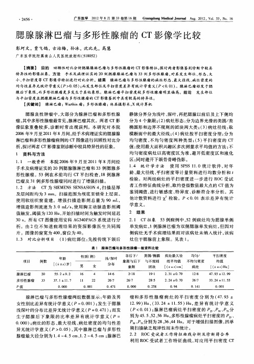
腺瘤最大径分别为 14~ . m、. 4 5c ; . 45e 12~ . m 腺淋 巴
别为 4 . 、 、6H , 5 5 5 5 u 多形性腺瘤病灶平扫密度 的 P 2 … P P , 分别 为 2 、6 4 u 83 、4H 。对于增强扫描 图像 , 因单 期扫描缺乏规律性而未作统计 。 2 2 R C受试者工作特征 曲线分析 及诊 断符 合率 . O 利用 R C受试 者工作 特征 曲线 , O 对应 用平 扫密 度 C T
均匀密度病 灶 以高密度 区为准 , 避开低密 度 区和液 化 区; 同时避开下颌骨骨嵴伪影 。 14 统 计 学 方 法 使用 S S 10统计 软 件 , 年 . P S1. 对 龄、 最大径线 、 平扫密度 等计量 资料进行 均数分 析和 t
检验。对两组病 灶 的平 扫密 度进一 步进行 R C受 试 O 者工作特征 曲线分析 , 取约登指数值最 大点 的 C T值为
【 关键词 】 腺淋 巴瘤 ; r i Wat n瘤;多形性腺 瘤;体层摄 影术 , h x线计算机
腮腺 良性肿瘤 中 , 部分为腺 淋 巴瘤和多形 性腺 大 瘤 , 中多形性腺瘤最常见 , 其 腺淋 巴瘤其次。两者 C T影
静脉分界分为浅叶 、 深叶 , 再把腮腺 以前后及上下侧均
分为 4个象限 ;2 病灶形态 : 为边界光滑的类圆/ () 分 类
女性别 比差异有统计学意义 ( 0 0 1 ; 生于腮腺 P= .0 ) 发 浅深 叶的分布 比差异无统计学 意义 ( 04 1 ; P= .7 ) 而发 生 于腮 腺 后 下 象 限 的 比率 差异 有 统 计 学 意 义 ( = P
000 ; . 0 ) 病灶 的形 态 、 大径线 、 最 病灶 密度 的均匀 性差 异无统计学 意义 ( P>00 ) 其 中腺淋 巴瘤 与多 形性 .5 ,
腮腺多形性腺瘤与Warthin瘤的临床对比分析
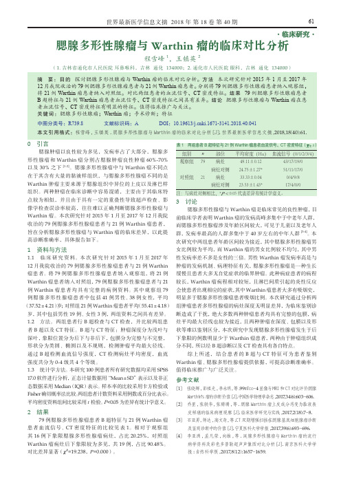
世界最新医学信息文摘 2018年 第18卷 第40期61·临床研究·腮腺多形性腺瘤与Warthin 瘤的临床对比分析程雪峰1,王镇英2(1.吉林省通化市人民医院 耳鼻喉科,吉林 通化 134000;2.通化市人民医院 眼科,吉林 通化 134000)0 引言腮腺肿瘤以良性较为多见,发病率占了大部分,腮腺多形性腺瘤和Warthin 瘤分别占腮腺肿瘤良性肿瘤60%-70%以及30%之下[1-2]。
腮腺多形性腺瘤中与Warthin 瘤不同点在于其含有大量的黏液样组织,与腮腺多形性腺瘤不同的是Warthin 肿瘤主要来源于腮腺组织中异位的上皮以及淋巴样组织。
两种肿瘤在临床诊断中容易混淆,主要由于其临床特点较为相似,并且由于具有一定的重叠性导致超声检查、影像学检查误诊率较高,往往难以正确判断腮腺多形性腺瘤与Warthin 瘤。
本次研究针对2015年1月至2017年12月我院收治的79例腮腺多形性腺瘤患者与21例Warthin 瘤患者,旨在分析腮腺多形性腺瘤与Warthin 瘤的临床差异,以此提高诊断准确率,具体报告如下。
1 资料与方法1.1 临床研究资料。
本次研究针对2015年1月至2017年12月我院收治的79例腮腺多形性腺瘤患者与21例Warthin 瘤患者。
将79例腮腺多形性腺瘤患者纳入观察组,将21例Warthin 瘤患者纳入对照组,79例腮腺多形性腺瘤患者与21例Warthin 瘤患者均具有完整的病例资料。
其中观察组79例腮腺多形性腺瘤患者中包括41例男性、38例女性,平均(37.52±4.21)岁;对照组21例Warthin 瘤患者平均(55.41±4.18)岁,其中包括男性19例、女性3例,两组资料之间具有差异。
1.2 方法。
两组患者行B 超检查与CT 检查,并比较两组患者B 超以及CT 特征。
B 超与CT 特征:肿瘤深度分为浅叶与深叶,象限位置分为后下与非后下,包膜分为完整与不完整,形状分为类圆、椭圆以及不规则,检测肿瘤平均最大径线。
腮腺基底细胞瘤与多形性腺瘤的临床和超声特征分析
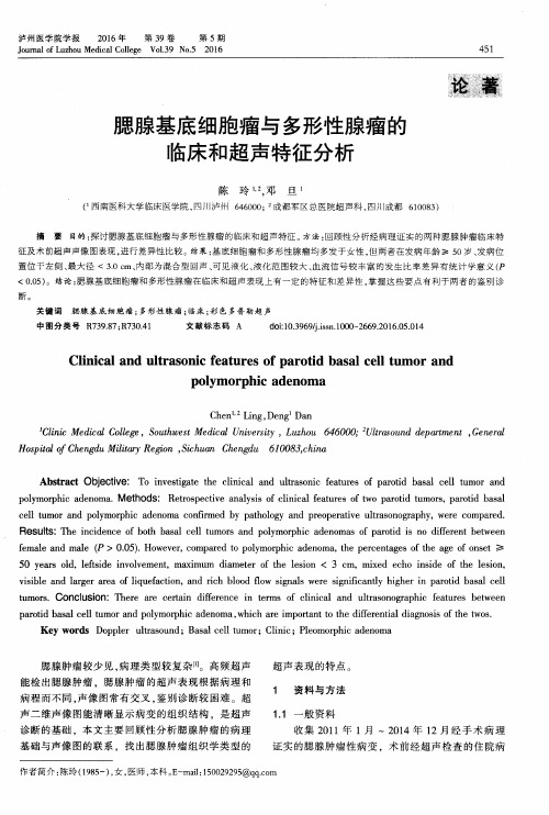
f e m a l e a n d m a l e( 尸>0 . 0 5 ) . Ho w e v e r , c o m p a r e d t o p o l y m o r p h i c a d e n o ma , t h e p e r c e n t a g e s o f t h e a g e o f o n s e t≥
Ho s p i t a l o f C h e n g d u Mi l i t a r y R e g i o n, S i c h u a n C h e n g d u 6 1 0 0 8 3 , c h i n a Ab s t r a c t Ob j e c t i v e :T o i n v e s t i g a t e t h e c l i n i c a l a n d u l t r a s o n i c f e a t u r e s o f p a r o t i d b a s a l c e l l t u mo r a n d
c e l l t u mo r a n d p o l y mo r p h i c a d e n o ma c o n f i r me d b y p a t h o l o g y a n d p r e o p e r a t i v e u h r a s o n o g r a p h y ,w e r e c o mp a r e d . Re s u l t s : T h e i n c i d e n c e o f b o t h b a s a l c e l l t u mo r s a n d p o l y mo r p h i c a d e n o ma s o f p a r o t i d i s n o d i f f e r e n t b e t w e e n
腮腺混合瘤和腺淋巴瘤的超声诊断及鉴别(腺淋巴瘤不是淋巴瘤)
腮腺混合瘤和腺淋巴瘤的超声诊断及鉴别(腺淋巴瘤不是淋巴瘤)腮腺种类很多,其中最常见的为混合瘤(多形性腺瘤)、腺淋巴瘤(Warthin瘤、淋巴乳头状囊腺瘤)。
腮腺腺淋巴瘤:又称为淋巴乳头状囊腺瘤或Warthin瘤,第2常见的腮腺肿瘤,约占腮腺肿瘤的6%~10%。
多发于老年男性(50-70岁),多有吸烟史,肿瘤大多生长缓慢,病程长短不一,肿块有消长史,绝大多数肿瘤位于腮腺后下极,表面光滑,质地较软,有时有弹性感,常呈多发性,约有12%病人为双侧腮腺肿瘤,也可单侧多发。
注意:腺淋巴瘤与淋巴瘤虽只差一字,但是其良恶性确截然不同,淋巴瘤为恶性肿瘤,腺淋巴瘤为良性肿瘤。
以往认为腺淋巴瘤与男性高龄吸烟者相关,本人认为主要与吸烟有关(与其发病机制有关),以往认为男性高发这应该是得益于男性吸烟比例比较多,对于“否认吸烟史”的,尤其是女性要注意询问其被动吸烟史。
其发生机制其中之一:腮腺导管上皮迷走于淋巴结并异常增殖形成,异位的导管闰管细胞和基底细胞可位于腮腺内或者腮腺周围淋巴结内,烟焦油中的化学物质作用于淋巴结中迷走的腮腺组织,导致这些组织化生。
由于腮腺后下极淋巴组织最为丰富,因此绝大多数腺淋巴瘤发生于腮腺组织后下限,该病基本不发生在无腺内淋巴结的其它涎腺,这是对该理论的有力支持。
超声表现:形态规则,边界清,圆形或椭圆形,无分叶,内部回声较混合瘤低,分三型:I型囊实间隔型:内见线状强回声分隔,呈网格状(特异性高,敏感性低);II型均匀实质型:均匀实性回声;III型囊性变为主型:表现为I型或者II型内部局限性无回声,囊实性成分境界清晰(蜂窝状无回声:特异性高,敏感性低的指标)。
CDFI:大部分肿块血流较丰富,比大部分腮腺肿瘤血供丰富,典型的呈淋巴结样的门型血流:呈分支状走行,从瘤体一极向内部树枝样散开,走行规则无扭曲。
男53岁,双侧腮腺内多发低回声,边界清,形态规则,内见多发小的无回声,CDFI:见较丰富分支状及边缘血流信号。
腮腺腺淋巴瘤与多形性腺瘤的MSCT征象对比分析
腮腺腺淋巴瘤与多形性腺瘤的MSCT征象对比分析王明杰,李旭丹,梁峰,陈淑艳(浙江省丽水市人民医院放射科浙江丽水323000)【摘要】目的回顾性对比分析腮腺腺淋巴瘤与多形性腺瘤的MSCT影像特征。
方法经手术及病理证实的24例腮腺腺淋巴瘤与35例多形性腺瘤,分析其临床及影像学资料,对肿瘤数量、发生部位、形态、边界、平扫密度、C T强化特征等进行对比分析。
结果腮腺腺淋巴瘤与多形性腺瘤的肿瘤数量、病灶形态、边界差异无统计学意义(I>0.05);病灶发生于后下象限、平扫密度较高、早期显著强化更常见于腮腺腺淋巴瘤,差异有统计学意义(I<0.05)。
3种阳性单一征象中,发生于后下象限敏感度最高(90. 3% ),早期显著强化特异度最高(92.4% );两种联合征象中,发生于后下象限+平扫密度较高敏感度最高(83.4%),发生于后下象限+早期显著强化特异度最高(94. 4% );3种征象联合时,诊断腺淋巴瘤的敏感度和特异度分别为63.1%、96.4%。
结论发生部位、平扫密度及早期显著强化在腮腺腺淋巴瘤与多形性腺瘤的MSCT影像鉴别中具有较高的特异性,多种征象联合应用大多数病灶术前可作出正确诊断。
【关键词】腮腺;腺淋巴瘤;多形性腺瘤;体层摄影术,X线计算机中图分类号:R739.8;R814.42 文献标识码:A 文章编号:10068011 (2018 & 05872184The comparative analysis on MSCT manifestation between parotid gland lymphoma and pleomorphic adenomaWANG M ingjie,LIANG Feng,LI Xudan,CHEN ShuyanDepartment o f Radiology,Lishui People's Hospital,Lishui323000,P. R. China【Abstract】Objective To discuss theMSCTfindings of parotid gland lymphomaand pleomorphic adenomaand its diagnostic value. Methods TheM SCTfindings of 24 cases parotid gland lymphomaand 35 cases of pleomorphic adenomabypathology were reviewed retrospectively,including their number,location,shape,border,scan d ensity,CT enhancemen and so on. Results Number,shape,border were no statistically significant differencc between parotid gland lymphoma and pleomorphic adenoma ( P >0. 05 ). Parotid gland lymphoma was on the posterior inferior quadrant,high cantly improved in enhanced arterial phase ( P<0.05). Among the relevant signs,the p osterior inferior qu sensitivity (90. 3% ),and significantly improved in enhanced arterial phase had the highest specificity (92. 4% ). Combining the posterior inferior quadrant and higher scan density achieved highest sensitivity (83. 4% ),and combining th quadrant and significantly improved in enhanced arterial phase achieved the highest specificity (94.4% ).combined,the sensitivity and specificity were 63. 1 % and 96. 4% respectively. Conclusion The posterior inferior quadrant,higher scan density a nd significantly improved in enhanced arterial phase had higher specificity in the MSCT diagnosis of parotid gland lymphoma and pleomorphic adenoma. The 3 signs combined use of most of the le atively.【Key words 】Parotid; Pleomorphic adenoma; Adenolymphoma; Tomography,spiral computed腮腺多形性腺瘤来源于腮腺上皮组织,是腮腺 最常见的良性肿瘤,约占腮腺良性肿瘤的80%。
腮腺多形性腺瘤与Warthin瘤99mTcO4^-唾液腺显像对比分析
[4] 邵晏清,熊然,张潇,等.改良 stoppa切口入路与腹直肌外侧入路
治疗合并骨盆骨折的髋臼骨折的疗效比较[J].医学综述,2016,22 (2):380-382. [5] 朱如里,郑闽前,马永平,等.Stoppa入路治疗骨盆、髋臼骨折的疗 效[J].临床骨科杂志,2016,19(1):46-48. [6] 苏以林,闵敏,冯耀华,等.Stoppa入路在骨盆与髋臼骨折治疗中 的应用研究进展[J].中华创伤骨科杂志,2015,17(8):687-691. [7] SomfordMP,DeVE,IjpmaFF.Theoriginsandcurrentapplicationsof classiceponymoustermsforpelvicandacetabularfractures:A historic review[J].JTraumaAcuteCareSurg,2017,82(4):802-809. [8] 李宝丰,陈蓓,李梅,等.应用改良 Stoppa入路手术治疗骨盆髋臼 骨折的疗效分析[J].中 国 骨 与 关 节 损 伤 杂 志,2016,31(10):1009 -1011. [9] HernefalkB,ErikssonN,BorgT,etal.Estimatingpre-traumaticqual ityoflifeinpatientswithsurgicallytreatedacetabularfracturesandpelvic ringinjuries:Doestimingmatter[J]?Injury,2016,47(2):389-394. [10] 韩飞,闫景龙.改良 Stoppa入路治疗骨盆髋臼骨折进展研究[J]. 创伤外科杂志,2016,20(2):123-125. [11] 刘巍,闫国富.改良 Stoppa入路在骨盆髋臼骨折前方手术中的应 用[J].中国药物与临床,2016,16(1):93-95. [12] 郭洪章.改良 Stoppa入路治疗骨盆髋臼骨折[J].临床骨科杂志, 2017,20(6):704-706. [13] 陈晓,马坤龙,徐海涛,等.改良 Stoppa入路与髂腹股沟入路治疗 骨盆、髋臼 骨 折 的 Meta分 析 [J].中 国 组 织 工 程 研 究,2017,21 (19):3108-3116. [14] 李民,李书奎,陈汉文,等.新型改良 Stoppa入路治疗髋臼并同侧 骨盆骨折效果[J].齐鲁医学杂志,2016,31(5):584-586. [15] 邹昌,方跃,屠重棋,等.改良 Stoppa入路联合髂窝入路治疗复杂 髋臼骨折[J].中华创伤骨科杂志,2015,17(8):669-675.
腮腺腺淋巴瘤与多形性腺瘤的超声诊断及病因分析
we e a a y e e r s e tv l c o d n o s r i a a d p t o o ia i d n s r n lz d r t o p c i ey a c r i g t u g c l n a h lg c l fn i g .Re u t sl s
【 图 分 类 号】 R7 9 8 中 3 . 【 献 标 志码 】 A 文
彩 超 检 查 对 腺 淋 巴瘤 及 多形 性 腺 瘤 有 重 要 的 鉴 别 诊 断 价 值 , 合 病 因 分 析 可 提 高 结
Eto o ia n l ssa ta ou a no i n a e o y p m a a e m o p ca n m a il gc la a y i nd Ulr s nd dig sso d n l m ho nd plo r hi de o YA N G e W U aixi ng W i。 H — a
Th a i f v r ia n Leabharlann r t o e t la d o c
h rz n a, y t h n ert bo d sg a ai r tt t a i e e t P< 0 0 P< 0 O ) Ag , e s kn o i tlc si c a g ai lo in lrt wee sai i ldf rn ( o c o。 o sc f . 5。 . 1 . e s x, mo ig a d oh rfco sweesait a i ee tP<O 0 P< O O ) C n lso s u ta o 0 r p i i au eu t o o n t e a tr r ttsi l f rn ( c df . 5, . 1 . o cu in lrs n g a hc s sf l meh df r
高频超声对腮腺非霍奇金淋巴瘤与多形性腺瘤的鉴别诊断价值

DOI :10.3969/j.issn.1672-9463.2020.07.019高频超声对腮腺非霍奇金淋巴瘤与多形性腺瘤的鉴别诊断价值李先晓 王艳清 张晓东 申发燕 黄婧颖作者单位:361000 福建厦门,厦门大学附属第一医院干部保健特诊部(李先晓),超声科(王艳清、张晓东),病理科(申发燕),质量管理部(黄婧颖)腮腺肿瘤是颌面部最常见的肿瘤之一,60%~…85%的唾液腺肿瘤发生在腮腺[1]。
不同病理来源的腮腺肿瘤治疗方法不同,尽早对腮腺肿瘤患者进行准确的诊断和有针对性的治疗对预后具有重要意义[2]。
腮腺占位性病变多为腮腺良性肿瘤,以多形性腺瘤最为常见(又称腮腺混合瘤)[3]。
腮腺淋巴瘤是属于淋巴造血系统的一组恶性肿瘤,其中以非霍奇金淋巴瘤(non-hodgkin…lymphoma,NHL )为多见[4]。
腮腺非霍奇金淋巴瘤与多形性腺瘤临床及影像学检查部分特征较相似,不易区分,在临床诊断上存在一定困难。
近年来,随着超声诊断技术的不断发展,高频超声检查在鉴别诊断腮腺肿瘤方面得到了广泛应用[4]。
此检查方法具有操作简单、对受检者的健康影响较小等优点[5]。
高频超声通过影像学反映出肿块形态、大小、质地、数量、内外部回声及前后方回声,从而对病变做出判断[6]。
本研究通过对腮腺NHL 和多形性腺瘤患者高频超声检查资料进行回顾性分析,观察二维声像图表现及彩色血流特征,结合临床资料特点,探讨高频超声对腮腺NHL 与多形性腺瘤的鉴别诊断价值。
1 材料与方法1.1研究对象 选择2015年5月~2020年1月于我院经手术病理证实腮腺NHL 患者(NHL 组)12例及多形性腺瘤患者(多形性腺瘤组)50例。
NHL 组纳入研究结节数22个,腮腺多形性腺瘤组纳入研究结节数55个。
1.2仪器与方法 患者术前行彩色高频超声检查,使用Philips…EPIQ…7、Philips…IU22型彩色多普勒超声诊断仪,高频线阵探头,频率5~12MHz,设置为浅表器官条件,彩色Scale5~6,彩色增益调至出现噪声之前为止。
腮腺多形性腺瘤与Warthin氏瘤的超声图像对比分析
Z h e j i a n g Un i v e r s i t y ,Ha n g z h o u,Zh e j i a n g 3 1 0 0 0 3 Ch i n a
Ab s t r a c t : Ob j e c t i v e T o d i s c u s s t h e ma n i f e s t s o f u l t r a s o u n d i n p a r o t i d g l a n d p l e o mo r p h i c a d e n o ma a n d Wa r t h i n s
( 1 1 . 6 % v s 0 . 0 ,P< 0 . 0 5 ) 。
结论
腮腺多形性腺瘤和 Wa r t h i n s 瘤 在 常 规 超 声 表 现 上 有 一 定 的 相 似 性 ,但 在 病 灶 呈 无 回声 且 伴 分 隔 时 ,应 首 先 考 虑 为
Wa r t h流
腮 腺 多形 性腺 瘤 与 W a r t h i n氏瘤 的超 声 图像 对 比分 析
朱 少明 陈剑 赵 齐 羽 蒋 天安
摘 要
目的 探 讨 腮 腺 多形 性 腺瘤 与 Wa r t h i n s 瘤的超声表现特点 。 方 法 回顾 性 分 析 经 病 理 证 实 的 4 3 例 腮 腺 多形 性 腺 瘤 ( 年龄 1 5  ̄7 2岁 ,平 均 4 1 . 5岁 ) 与 3 3 例 Wa r t h i n s 瘤 ( 年龄 2 3  ̄6 9 岁 ,平 均 5 1 . 6岁 ) 患 者 术 前 超 声 影 像 资 料 。 比较 两 者 在 肿 块 部 位 、形 态 、边 界 、包 膜 、 内 部 回声 均 匀 度 、 有 无 钙 化 、分 隔 及 血 供 分 布 上 的不 同 。 结果 多 形性 腺瘤 共 4 3个 病 灶 ,W a r t h i n s 瘤共 4 7 个 病 灶 ,两 者在 形态 、边 界 以 及 血 供 分 布 上 无 统 计 学 差 异 ,多 数 为 圆 形 或 类 圆形 ,边 界 清 晰 的 病 灶 。但 在 回声 强 度 、分 隔 和 钙 化 上 有 明显 差 异 ,多 形 性 腺 瘤 中低 回声 比例 高 于 Wa r t h i n s 瘤 ( 8 1 . 4 v s 3 6 . 2 ,P<0 . 0 5 ) ,分 隔 出 现 率 低 于 Wa r t h i n s 瘤 ( 7 . 0 v s 4 8 . 9 ,P< 0 . 0 5 ) ,钙 化 出 现 率 多 形 性 腺 瘤 高 于 Wa r t h i n s瘤
腮腺多形性腺瘤与腺淋巴瘤CT表现的对照分析
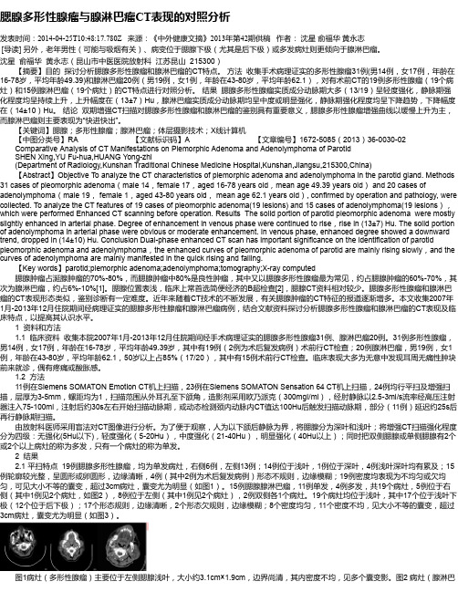
腮腺多形性腺瘤与腺淋巴瘤CT表现的对照分析发表时间:2014-04-25T10:48:17.780Z 来源:《中外健康文摘》2013年第42期供稿作者:沈星俞福华黄永志[导读] 另外,老年男性(可能与吸烟有关)、病变位于腮腺下极(尤其是后下极)或多发病灶则更倾向于腺淋巴瘤。
沈星俞福华黄永志(昆山市中医医院放射科江苏昆山 215300)【摘要】目的探讨分析腮腺多形性腺瘤和腺淋巴瘤的CT特点。
方法收集手术病理证实的多形性腺瘤31例(男14例,女17例,年龄在16-78岁,平均年龄49.39)和腺淋巴瘤20例(男19例,女1例,年龄在43-80岁,平均年龄62.1),对有术前CT的19例多形性腺瘤(19个病灶)和15例腺淋巴瘤(19个病灶)的CT特点进行对照分析。
结果腮腺多形性腺瘤实质成分动脉期大多(13/19)呈轻度强化,静脉期强化程度均呈持续上升,上升幅度在(13±7)Hu,腺淋巴瘤实质成分动脉期均呈中度或明显强化,静脉期强化程度均呈下降趋势,下降幅度在(14±10)Hu。
结论双期增强CT扫描对腮腺多形性腺瘤和腺淋巴瘤的鉴别具有重要意义,腮腺多形性腺瘤增强曲线以缓慢上升为主,而腺淋巴瘤则主要表现为“快进快出”。
【关键词】腮腺;多形性腺瘤;腺淋巴瘤;体层摄影技术;X线计算机【中图分类号】RA 【文献标识码】A 【文章编号】1672-5085(2013)36-0030-02 Comparative Analysis of CT Manifestations on Plemorphic Adenoma and Adenolymphoma of ParotidSHEN Xing,YU Fu-hua,HUANG Yong-zhi(Department of Radiology,Kunshan Traditional Chinese Medicine Hospital,Kunshan,Jiangsu,215300,China)【Abstract】Objective To analyze the CT characteristics of plemorphic adenoma and adenolymphoma in the parotid gland. Methods 31 cases of pleomorphic adenoma(male 14,female 17,aged 16-78 years old,mean age 49.39 years old) and 20 cases of adenolymphoma(male 19, female 1,aged 43-80 years old, mean age 62.1 years old), confirmed by operation and pathology, were collected. To analyze the CT features of 19 cases of pleomorphic adenoma(19 lesions) and 15 cases of adenolymphoma(19 lesions),which were performed Enhanced CT scanning before operation. Results The solid portion of parotid pleomorphic adenoma were mostly slightly enhanced in arterial phase. Degree of enhancement in venous phase were continued to rise,rise in (13±7) Hu. The solid portion of adenolymphoma in arterial phase were obvious or moderate enhancement. In venous phase, enhanced degree showed a downward trend, dropped in (14±10) Hu. Conclusion Dual-phase enhanced CT scan has important significance on the identification of parotid pleomorphic adenoma and adenolymphoma,the enhanced curves of pleomorphic adenoma of parotid are mainly rising slowly,and the curves of adenolymphoma are mainly manifested in the quick rising and falling.【Key words】parotid;plemorphic adenoma;adenolymphoma;tomography;X-ray computed腮腺肿瘤占涎腺肿瘤的70%-80%,而腮腺肿瘤中80%是良性肿瘤,其中又以腮腺多形性腺瘤最为常见,约占腮腺肿瘤的60%-70%,其次为腺淋巴瘤,约占6%-10%[1]。
腮腺多形性腺瘤与warthin瘤的ct和mri的影像学特征
left hepatectomy of a living donor for an adolescent in China[ J] . Chin
Med J ( Engl) ꎬ2019ꎬ132(2) :242 - 244.
程度保留功能肝体积ꎬ且可降低并发症发生风险ꎬ改善
预后ꎮ 三维可视化技术在评估、测量、观察方面虽已接
近人体真实情况ꎬ但计算机中虚拟进行肝切除操作体验
上与实际手术仍有较大差距ꎬ期待未来随计算机技术进
一步发展ꎬ可借助投影方式呈现重建器官三维模型ꎬ术
者可以直接通过投影假象进行虚拟手术操作以提高仿
真效果ꎮ
参考文献
the functional fascicular groups of along - segment peripheral nerve[ J] .
Neural Regen Resꎬ2018ꎬ13(8) :1465 - 1470.
王小明ꎬ 胡明华ꎬ 王冠男ꎬ 等. ICG - R15 联合三维可视化技术用
[2] Li Hꎬ Zheng Jꎬ Cai JYꎬ et al. Laparoscopic VS open hepatectomy for
hepatolithiasis: An updated systematicreview and meta - analysis [ J] .
World J Gastroenterolꎬ2017ꎬ23(43) :7791 - 7806.
除术的临床应用研究[ J] . 贵州医药ꎬ2019ꎬ43(7) :1066 - 1067.
腮腺腺淋巴瘤和多形性腺瘤的超声鉴别诊断研究
腮腺腺淋巴瘤和多形性腺瘤的超声鉴别诊断研究作者:杜忠实唐丽娜沈友洪来源:《中外医学研究》2018年第16期【摘要】目的:探究腮腺腺淋巴瘤和多形性腺瘤的超声鉴别诊断。
方法:回顾性分析笔者所在医院收治的330例腮腺良性肿瘤患者临床资料,均接受彩色多普勒超声诊断系统检查;以粗针穿刺活检(CNB)或术后病理学检查结果为依据分析超声检查结果的鉴别准确率;比较腮腺腺淋巴瘤患者及多形性腺瘤患者肿瘤形态、内部结构回声、钙化、主要血管分布、血流情况、液性暗区等超声影像特征差异。
结果:330例腮腺良性肿瘤患者经CNB或术后病理学检查确诊为腺淋巴瘤者68例(20.6%),确诊为多形性腺瘤者262例(79.4%)。
超声鉴别诊断腮腺腺淋巴瘤及多形性腺瘤的准确率为79.1%(261/330)。
与多形性腺瘤患者比较,腺淋巴瘤患者多见类椭圆状外形、内部结构回声不均匀、无钙化、主要血管分布于核心、血流信号丰富、存在液性暗区等超声影像特征,差异均有统计学意义(字2=25.732、46.703、9.516、41.645、55.116、42.702,P【关键词】腮腺;腺淋巴瘤;多形性腺瘤;超声;鉴别诊断doi:10.14033/ki.cfmr.2018.16.025 文献标识码 B 文章编号 1674-6805(2018)16-00-03Study on Ultrasonic Differential Diagnosis of Adenolymphoma in Parotid Gland and Pleomorphic Adenoma/DU Zhongshi,TANG Lina,SHEN Youhong,et al.//Chinese and Foreign Medical Research,2018,16(16):-57【Abstract】 Objective:To explore the ultrasonic differential diagnosis of adenolymphoma in parotid gland and pleomorphic adenoma.Method:The clinical data of 330 patients with benign tumor in parotid were analyzed retrospectively,and all the patients were given systematic examination with color Doppler ultrasound.The coarse needle biopsy(CNB) or postoperative pathological results were taken as the basis for identification accuracy of ultrasonic examination,and the characteristic differences of ultrasonic images in tumor morphology,internal structure,calcification,vascular distribution,blood flow and liquid dark area between parotid gland adenolymphoma patients and pleomorphic adenoma patients were compared.Result:After CNB or postoperative pathological examination,68 cases(20.6%) were diagnosed as adenolymphoma,and 262 cases(79.4%)were diagnosed as pleomorphic adenoma.The accuracy of ultrasonography in the diagnosis of parotid gland adenolymphoma and pleomorphic adenoma was 79.1%(261/330).Compared with the patients with pleomorphic adenoma,the patients with adenoma were characterized by elliptical shapes,uneven internal structure,none calcification,vascular distribution in core,abundant bloodflow signals,and fluid dark areas,the differences were statistically significant(字2=25.732,46.703,9.516,41.645,55.116,42.702,P【Key words】 Parotid gland; Adenolymphoma; Pleomorphic adenoma; Ultrasound;Differential diagnosisFirst-author’s address:Fujian Cancer Hospital,Fuzhou 350014,China作为涎腺常见的两类肿瘤类型,腺淋巴瘤及多形性腺瘤多发于腮腺与颌下腺。
常规超声联合剪切波弹性成像与MRI对腮腺多形性腺瘤与Warthin瘤的鉴别诊断价值
·临床研究·常规超声联合剪切波弹性成像与MRI对腮腺多形性腺瘤与Warthin瘤的鉴别诊断价值毛翠莲李奥胡彧马倩朱榴宁摘要目的探讨常规超声联合剪切波弹性成像(SWE)鉴别诊断腮腺多形性腺瘤与Warthin瘤的价值,并与MRI进行对比分析。
方法选取我院经手术病理证实的81例腮腺肿瘤患者,包括多形性腺瘤58例和Warthin瘤23例,比较两种病变的常规超声表现和SWE参数[杨氏模量最大值(Emax)、平均值(Emean)、最小值(Emin)、标准差(SD)]的差异。
分别建立基于常规超声表现、SWE参数、常规超声表现联合SWE参数鉴别诊断多形性腺瘤与Warthin瘤的Logistic回归模型,绘制受试者工作特征(ROC)曲线分析并比较不同模型、MRI鉴别诊断多形性腺瘤与Warthin瘤的效能。
结果多形性腺瘤与Warthin瘤肿块形态、内部强回声及无回声占比、主要血管分布、血流分级、Emax、Emean、SD比较,差异均有统计学意义(均P<0.05)。
建立了3个分别基于常规超声表现、SWE参数、常规超声表现联合SWE参数的Logistic回归模型(模型Ⅰ、模型Ⅱ、模型Ⅲ);ROC曲线分析显示,模型Ⅰ、模型Ⅱ、模型Ⅲ鉴别诊断多形性腺瘤与Warthin瘤的曲线下面积(AUC)分别为0.881、0.837、0.962,模型Ⅲ的AUC均高于模型Ⅰ和模型Ⅱ,差异均有统计学意义(均P<0.05)。
剔除MRI、常规超声及SWE误诊的病灶后,常规超声联合SWE(模型Ⅲ)鉴别诊断多形性腺瘤与Warthin瘤的灵敏度为85%,特异度为88%,准确率为86%,AUC为0.867;MRI的灵敏度为87%,特异度为84%,准确率为86%,AUC为0.856,二者AUC比较差异无统计学意义。
结论常规超声联合SWE在腮腺多形性腺瘤与Warthin瘤的鉴别诊断中具有较好的应用价值,且其诊断效能与MRI相当。
腮腺多形性腺瘤与Warthin瘤的临床对比分析
腮腺多形性腺瘤与Warthin瘤的临床对比分析赵金荣【摘要】Objective To compare and analysis clinical characteristics ,Dppler ultrasonography and CT imaging features of War‐thin′s tumor and pleomorphic adenoma of the parotid gland .Methods Thirty‐eight cases of Warthin′s tumor and107 cases of pleo‐morphic adenoma of the parotid gland in our hospital were selected and divided into Warthin′s tumor group and pleomorphic adeno‐ma group .Average age ,gender ,position of onset ,mean course of disease ,smoking rates and Dopplerultrasono graphy and CT ima‐ging features of 2 groups were compared and analyzed .Results Compared with pleomorphic adenoma group ,the average age ,per‐centage of males ,smoking rates were higher in Warthin′s tumor group but the mean course of disease was short ,whi ch showed sig‐nificant difference(P<0 .05) .The site of Warthin′s tumor mostly occurred in lower posterior polar of the parotid gland .Doppler re‐sults indicated more abundant blood supply in Warthin′s tumors group .CT imagine results also indicated higher density in Warthin′s tumors group than in pleomorphic adenomas group .Conclusion According to the group constituent ratio of disease ,average age , gender ,mean course of disease combined with Doppler ultrasonography and CT imaging features ,the tumor could be diagnosed be‐fore surgery ,which could be helpful to make the correct surgical plan .%目的:比较腮腺多形性腺瘤和Warthin瘤的超声、影像学特征及临床表现。
腮腺Warthin瘤与腮腺多形性腺瘤的超声对照分析
3李岭,张炽敏,李嘉,等.经阴道彩色多普勒超声对药物流产效果的早期预测[J ].中国实用妇科与产科杂志,2006,22(1):45-46.4陈智毅,梁伟翔,梁琨.经阴道彩色多普勒能量超声对早期妊娠黄体的临床研究[J ].实用妇产科杂志,2006,22(7):425-427.5徐丽.子宫位霞与药物流产效果的关系经验交流[J ].临床和实验医学杂志,2006,5(11):1815.6魏文锦.子宫位置与药物流产术成功率的关系[J ].实用临床医学,2006,7(5):104-106.7李蛉,张炽敏。
李嘉,等.经阴道彩色多普勒超声对早期妊娠子宫螺旋动脉血流的研究[J ].东南大学学报,2005,24(5):324-326.8罗毅,徐晓红.孕妇行超声检查对胚胎和胎儿安全性的研究进展[J ].广东医学院学报,2007,25(1):75-77.9Aardema MW ,Sam MC.Second trimester Doppler ultrasound screening of the uterine arteries differentiates between subsequent nor-mal and poor outcomes of hypertensive preg-nancy :two different pathophysiological enti-ties [J ].Clin Sci (Lond ),2004,106(5):377-382.10Agarwal N.Suneja A.Role of uterine artery velocimetry using color flow Doppler and elec-tromyography of uterus in prediction of pre-term labor [J ].J Obstet Gynaecol Res ,2004,30(6):402-408.11Papaqeorqhiou AT ,Yu CK.Assessment of risk for the development of pre -eclampsia by maternal characteristics an d uterine artery Doppler [J ].BJOG ,2005,112(6):703-706.12Makikallio K ,Jouppila P.First trimester uter-ine ,placental and yolk sac haemodynamicsin pre -eclampsia and preterm labour [J ].Hum Reprod ,2004,19(3):729-773.13Schwarze A ,Nelles.Doppler ultrasound of the uterine artery in the prediction of severe complications during low -risk pregnan cies [J ].Arch Gynecol Obstet ,2005,271(1):46-52.14段林.经阴道超声诊断异位妊娠的价值[J ].实用心脑肺血管病杂志,2010,18(3):345.15Duqof L ,Lynch AM.First trimester uterine artery Doppler abnormalities predict subse-quent intrauterine growth restriction [J ].Am J Obstet Gynecol ,2005,193(2):1208-1212.(收稿日期:2011-01-10;修回日期:2011-05-10)(本文编辑:刘莉)·短篇论著·腮腺Warthin 瘤与腮腺多形性腺瘤的超声对照分析莫清清作者单位:313000浙江省湖州市中心医院超声科【摘要】目的探讨腮腺Warthin 瘤与腮腺多形性腺瘤的超声鉴别要点。
超声检查在多形性腺瘤和Warthin瘤鉴别诊断中的应用
超声检查在多形性腺瘤和Warthin瘤鉴别诊断中的应用张春玲;潘丽霞;耿长辉;袁莉;董宁;罗玉菊【摘要】目的:回顾性分析腮腺多形性腺瘤与Warthin瘤的超声声像图,总结其声像图特征,为两者的鉴别提供参考。
方法:回顾经病理确诊的24例腮腺多形性腺瘤及18例Warthin瘤患者的超声检查结果,分析其声像图表现特征。
结果:大多数多形性腺瘤为低回声,回声均匀一致,或呈小分叶状,后方回声增强,周边型血流。
而Warthin瘤多呈椭圆形,内部常伴有囊性成分,为内在型血流。
结论:腮腺多形性腺瘤与 Warthin 瘤在声像图上有些相似,容易误诊。
超声检查在两者的鉴别诊断中有较高的临床价值,可为临床的治疗及手术方案提供鉴别依据。
%Objective: This paper aimed to make a retrospective analysis on the ultrasonographic features of common benign tumors of parotid gland (pleomorphic adenoma and Warthin's tumor). Methods:In 24 cases of parotid pleomorphic adenoma and 18 cases of Warthin tumor, the ultrasonographic characteristics were reviewed and compared. Results:It was found that most pleomorphic adenomas are hypoechoic, homogeneous and lobulated masses with acoustic enhancement and peripheral vascularization. Warthin's tumors were more likely to be ovals with cystic component and internal vascularization. Conclusion: Parotid pleomorphic adenoma and Warthin tumor share some similarities on sonograms. Ultrasound is highly valuable in the differential diagnosis of those lesions.【期刊名称】《口腔颌面外科杂志》【年(卷),期】2014(000)005【总页数】4页(P379-382)【关键词】多形性腺瘤;Warthin瘤;灰阶超声检查;彩色多普勒超声检查【作者】张春玲;潘丽霞;耿长辉;袁莉;董宁;罗玉菊【作者单位】大庆油田总医院超声科,黑龙江大庆 163001;大庆油田总医院超声科,黑龙江大庆 163001;大庆市人民医院普外科,黑龙江大庆 163001;大庆油田总医院超声科,黑龙江大庆 163001;大庆油田总医院超声科,黑龙江大庆163001;大庆油田总医院超声科,黑龙江大庆 163001【正文语种】中文【中图分类】R739.87诊断腮腺肿物,超声检查是无创的首选检查方法。
腮腺多形性腺瘤与Warthin瘤CT对照分析

腮腺多形性腺瘤与Warthin瘤CT对照分析白君;张朋;李亚军【期刊名称】《医学影像学杂志》【年(卷),期】2016(000)001【摘要】目的:探讨腮腺多形性腺瘤与腮腺Warthin瘤的CT表现,提高对两者的鉴别诊断水平。
方法回顾性分析经手术、病理证实的22例多形性腺瘤23个肿块、25例Warthin瘤35个肿块的CT表现。
结果58个肿块均表现为类圆形或椭圆形高密度,边缘清楚,两者的发病年龄、部位、平扫CT值及与同层咬肌的密度比较、增强后CT值、净强化CT值、与血管的密切程度、最大横纵径比均有统计学意义( P <0.05)。
结论在腮腺的良性肿瘤的鉴别诊断中,对于中老年男性,发生于后下象限,平扫与同侧咬肌密度相当或略高,中等或明显强化,与周围血管关系密切,纵椭圆形肿块提示War-thin瘤的诊断。
【总页数】3页(P25-27)【作者】白君;张朋;李亚军【作者单位】广东省深圳市龙岗区人民医院影像科广东深圳 518172; 中南大学湘雅二医院放射科湖南长沙 410011;中南大学湘雅二医院放射科湖南长沙 410011; 湖南省郴州市桂阳县第一人民医院放射科湖南郴州 424400;中南大学湘雅二医院放射科湖南长沙 410011【正文语种】中文【中图分类】R735.7;R814.42【相关文献】1.腮腺Warthin瘤与腮腺多形性腺瘤的超声对照分析 [J], 莫清清2.彩色多普勒超声鉴别诊断腮腺多形性腺瘤与Warthin瘤的临床价值分析 [J], 罗璐3.腮腺多形性腺瘤与Warthin瘤99m TcO4-唾液腺显像对比分析 [J], 周兴久;肖占森;池艳丽;杨吉刚;绳海燕4.腮腺多形性腺瘤与Warthin瘤的CT和MRI的影像学特征 [J], 王巍; 王超; 宋金丹5.腮腺多形性腺瘤与Warthin瘤CT和MRI的影像学特征 [J], 潘曰峰;刘青因版权原因,仅展示原文概要,查看原文内容请购买。
腮腺Warthin瘤的彩色多普勒超声特征分析
腮腺Warthin瘤的彩色多普勒超声特征分析吴晓倩;李国杰;朱向明;江峰【期刊名称】《皖南医学院学报》【年(卷),期】2015(34)1【摘要】目的:探讨腮腺Warthin瘤的彩色多普勒超声特征,以提高超声对腮腺肿瘤的诊断,为临床诊治提供参考。
方法:回顾性分析经病理证实的24例患者31个腮腺Warthin瘤的彩色多普勒超声图像,对病灶位置、形态、大小、边界、内部回声、血流分布等特征进行分析。
结果:31个病灶中①病灶多位于腮腺浅叶或下极;②形态为圆形或椭圆形,占80.65%(25/31);③大小多在2.0~3.5 cm之间,占90.32%(28/31);④边界清晰,占100%(31/31);⑤病灶内部回声低回声、多呈囊实性、或夹有网格状回声,占74.19%(23/31);⑥内部有不同程度血流信号存在,占77.42%(24/31);⑦本组半数患者伴颈部淋巴结肿大。
术前彩色多普勒超声诊断符合率为58.06%(18/31)。
结论:彩色多普勒超声对术前腮腺Warthin瘤的诊断具有一定的临床价值,但规范化分析腮腺病灶图像,掌握Warthin瘤的声像图特征,对腮腺Warthin瘤检出的准确性可以得到进一步提高。
%Objective:To examine the color Dopplerflow imaging( CDFI) properties of War thin′s tumor of parotid gland for improving the diagnostic accu-racy in this entity.Methods:The color Doppler flow imaging manifestations were retrospectively examined in 24of 31 cases confirmed pathologically as Warthin′s tumor of parotid gland regarding the lesion location,tumor size andmorphology,boundary,internal echo and blood flowfeatures.Results:Totally, 31 lesions were detected in the 24 cases.①The lesions were generally located at the superficial lobe or lower point of the parotid;②The lesions exhibited round or oval shape(80.65%;25/31);③ The tumor size ranged from 2.0 to 3.5 cm in diameter(90.32%;28/31);④The boundary was clearly defined in the total cases(100%;31/31);⑤ The internal echo was very low,and presented with cystic-solid or mesh-likee choes(74.19%;23/31);⑥Vascular flow was occasionally displayed on CDFI(77.42%;24/31);⑦50% of patients showed cervical lymph node enlargement,and 58.06% were consistent with the postoperative confirmation.Conclusion:Preoperative CDFI can be valuable in diagnosis of Warthin′s tumor of parotid gland.Nevertheless,careful examination of the characteristic images of the tumor may improve the diagnostic accuracy of this disease .【总页数】3页(P70-72)【作者】吴晓倩;李国杰;朱向明;江峰【作者单位】皖南医学院附属弋矶山医院超声医学科,安徽芜湖 241001;皖南医学院附属弋矶山医院超声医学科,安徽芜湖 241001;皖南医学院附属弋矶山医院超声医学科,安徽芜湖 241001;皖南医学院附属弋矶山医院超声医学科,安徽芜湖 241001【正文语种】中文【中图分类】R739.8【相关文献】1.腮腺Warthin瘤与腮腺多形性腺瘤的超声对照分析 [J], 莫清清2.彩色多普勒超声鉴别诊断腮腺多形性腺瘤与Warthin瘤的临床价值分析 [J], 罗璐3.腮腺Warthin瘤的CT诊断及误诊分析 [J], 霍敏华;王伟军4.DWI和动态对比增强MRI多参数鉴别腮腺Warthin瘤与多形性腺瘤 [J], 胡涛;方学文;刘琼;邹玉坚;姚兆友;高云;张坤林5.扩散峰度成像及动态增强MRI鉴别腮腺多形性腺瘤与Warthin瘤 [J], 胡涛;刘琼;邹玉坚;姚兆友;方学文因版权原因,仅展示原文概要,查看原文内容请购买。
- 1、下载文档前请自行甄别文档内容的完整性,平台不提供额外的编辑、内容补充、找答案等附加服务。
- 2、"仅部分预览"的文档,不可在线预览部分如存在完整性等问题,可反馈申请退款(可完整预览的文档不适用该条件!)。
- 3、如文档侵犯您的权益,请联系客服反馈,我们会尽快为您处理(人工客服工作时间:9:00-18:30)。
3李岭,张炽敏,李嘉,等.经阴道彩色多普勒超声对药物流产效果的早期预测[J ].中国实用妇科与产科杂志,2006,22(1):45-46.4陈智毅,梁伟翔,梁琨.经阴道彩色多普勒能量超声对早期妊娠黄体的临床研究[J ].实用妇产科杂志,2006,22(7):425-427.5徐丽.子宫位霞与药物流产效果的关系经验交流[J ].临床和实验医学杂志,2006,5(11):1815.6魏文锦.子宫位置与药物流产术成功率的关系[J ].实用临床医学,2006,7(5):104-106.7李蛉,张炽敏。
李嘉,等.经阴道彩色多普勒超声对早期妊娠子宫螺旋动脉血流的研究[J ].东南大学学报,2005,24(5):324-326.8罗毅,徐晓红.孕妇行超声检查对胚胎和胎儿安全性的研究进展[J ].广东医学院学报,2007,25(1):75-77.9Aardema MW ,Sam MC.Second trimester Doppler ultrasound screening of the uterine arteries differentiates between subsequent nor-mal and poor outcomes of hypertensive preg-nancy :two different pathophysiological enti-ties [J ].Clin Sci (Lond ),2004,106(5):377-382.10Agarwal N.Suneja A.Role of uterine artery velocimetry using color flow Doppler and elec-tromyography of uterus in prediction of pre-term labor [J ].J Obstet Gynaecol Res ,2004,30(6):402-408.11Papaqeorqhiou AT ,Yu CK.Assessment of risk for the development of pre -eclampsia by maternal characteristics an d uterine artery Doppler [J ].BJOG ,2005,112(6):703-706.12Makikallio K ,Jouppila P.First trimester uter-ine ,placental and yolk sac haemodynamicsin pre -eclampsia and preterm labour [J ].Hum Reprod ,2004,19(3):729-773.13Schwarze A ,Nelles.Doppler ultrasound of the uterine artery in the prediction of severe complications during low -risk pregnan cies [J ].Arch Gynecol Obstet ,2005,271(1):46-52.14段林.经阴道超声诊断异位妊娠的价值[J ].实用心脑肺血管病杂志,2010,18(3):345.15Duqof L ,Lynch AM.First trimester uterine artery Doppler abnormalities predict subse-quent intrauterine growth restriction [J ].Am J Obstet Gynecol ,2005,193(2):1208-1212.(收稿日期:2011-01-10;修回日期:2011-05-10)(本文编辑:刘莉)·短篇论著·腮腺Warthin 瘤与腮腺多形性腺瘤的超声对照分析莫清清作者单位:313000浙江省湖州市中心医院超声科【摘要】目的探讨腮腺Warthin 瘤与腮腺多形性腺瘤的超声鉴别要点。
方法对20例腮腺Warthin 瘤和24例腮腺多形性腺瘤在超声特征方面进行总结分析。
结果20例腮腺Warthin 瘤肿瘤个数共24个,2例双侧单发,1例一侧多发,一侧单发,包膜均完整;18例位于腮腺后下极,15例呈弱低回声;实质性回声6例,混合性回声14例。
24例腮腺多形性腺瘤均单侧单发,15例位于腮腺浅叶,9例位于腮腺深叶;包膜完整20例,不完整4例;9例呈分叶状,实性均匀低回声6例,实性不均匀低回声12例,混合性回声6例。
彩色多普勒示腮腺Warthin 瘤较多形性腺瘤血供丰富。
结论腮腺Warthin 瘤与多形性腺瘤在超声特征方面有各自的规律,根据它们的规律性并与临床特点相结合,可以帮助鉴别这两种腮腺肿瘤。
【关键词】腮腺Warthin 瘤;腮腺多形性腺瘤;超声检查,多普勒,彩色【中图分类号】R 781.7【文献标识码】B【文章编号】1007-9572(2011)06-2082-03Ultrasound Characteristics of Parotid Warthin's Tumor and Parotid Pleomorphic Adenoma MO Qing -qing.Ultra-sonography Department ,Huzhou Central Hospital ,Huzhou 313000,China【Abstract 】ObjectiveTo explore the ultrasound characteristics of parotid Warthin's tumor and parotid pleomorphicadenoma.MethodsUltrasound characteristics were analyzed in 20cases of parotid Warthin's tumor and 24cases of parotid ple-omorphic adenoma.ResultsOf the 20cases (24tumors )of parotid Warthin's Tumor ,considered ,2were with bilateral singletumors ,1was with unilateral multiple tumors ;integral capsules were found for all the 24tumors.tumors originated from the low-er posterior polar of the parotid gland in 18cases ;echoes turned out to be hypoechoic in 15cases ,while solid and mixed type echoes were detected in 6cases and 14cases respectively.All 24cases of pleomorphic adenomas turned out to be unilateral with·2802·a single tumor ,with 15of them originated from the superficial lobe of the parotid gland and the other 9located in the deep lobe ;integral capsules were noticed in 20of the 24cases ;the tumors turned out to be lobulated in 9cases ;solid homogeneous hypoe-choic echoes were detected in 6cases ,solid heterogeneous hypoechoic echoes were detected in 12cases ,the other 6cases were characterized by mixed echoes.Color Doppler results indicated more abundant blood supply in parotid Warthin's tumors than in pleomorphic adenomas.ConclusionParotid Warthin's tumor and parotid pleomorphic adenoma show different ultrasound char-acteristics ,which may assist the differentiation diagnosis between them.【Key words 】Warthin's tumor ;Parotid pleomorphic adenoma ;Ultrasonography ,Doppler ,color欍欍欍欍欍欍欍欍欍欍欍欍欍欍欍欍欍欍欍欍欍欍欍欍欍欍欍欍欍欍欍欍欍欍欍欍欍欍欍欍欍欍欍欍欍欍欍欍欍欍欍欍欍欍欍欍欍欍欍欍欍欍欍欍欍欍欍欍欍欍欍欍欍欍欍欍欍欍欍欍氥氥氥氥本文要点超声检查显示腮腺Warthin 瘤与多形性腺瘤的声像图特征有各自的规律,如下:1、90.0%(18/20)的腮腺Warthin瘤位于腮腺浅叶,62.5%(15/24)的腮腺多形性腺瘤位于腮腺浅叶。
2、15.0%(3/20)的腮腺Warthin 瘤为多发,而腮腺多形性腺瘤均为单发。
3、腮腺Warthin 瘤包膜均完整,16.7%(4/24)的腮腺多形性腺瘤包膜不完整。
4、腮腺Warthin 瘤以1、2级血流信号为主,而腮腺多形性腺瘤则以0、1级血流信号为主。
5、70.0%(14/20)的腮腺Warthin 瘤和25.0%(6/24)的腮腺多形性腺瘤为混合性回声,前者回声低于后者。
腮腺多形性腺瘤(混合瘤)、腮腺Warthin 瘤(腺淋巴瘤,乳头状淋巴囊腺瘤)是腮腺中排在前二位的良性肿瘤。
腮腺良性肿瘤较恶性肿瘤多,约占80%,多形性腺瘤占腮腺肿瘤的60% 70%[1],这类肿瘤含有肿瘤性上皮组织与黏液样组织或软骨样组织。
腮腺Warthin 瘤发病率仅次于多形性腺瘤,在腮腺肿瘤中占14% 30%,该肿瘤被认为来源于腮腺内异位起源的上皮及淋巴组织[2]。
本研究收集了我院2008—2009年20例腮腺War-thin 瘤和24例腮腺多形性腺瘤患者的临床资料,主要从超声检查特征方面进行总结分析,旨在提高二者的超声鉴别诊断水平。
