Microscope Spec(A005,A005+)
透射电子显微镜介绍

对于材料研究用的TEM试样大致有三种类型: 经悬浮分散的超细粉末颗粒。 用一定方法减薄的材料薄膜。 用复型方法将材料表面或断口形貌复制下来的复型膜。
对支持膜的要求:
➢ 要有相当好的机械强度,耐高能电子轰击; ➢ 应在高倍下不显示自身组织,本身颗粒度要小,以提高样品分辨率; ➢ 有较好的化学稳定性、导电性和导热性。
二、透射电子显微成像
使用透射电镜观察材料的组织、结构,需具备以下两个前提: 一是制备适合TEM观察的试样,厚度100-200nm,甚至更薄; 二是建立电子图像衬度理论 像衬度是指电子像图上不同区域间光强度的差别。 透射电镜的像衬度来源于样品对入射电子束的散射。可分为:
衍射衬度:晶体薄膜试样显微图像 质厚衬度 :非晶态试样图像
形貌+结构 空心结构
四、透射电镜得到的信息
晶格条纹+电子衍射
(1)量取两个晶面晶面之间的距离 (2)与标准卡片去比对,选择合适的面
四、透射电镜得到的信息
线扫 Line Scan 面扫 Mapping
EDS元素分析
四、透射电镜得到的信息
总
一般成像 模式
明场像 (BF) 暗场像 (DF)
微观形貌,厚度差异,尺寸大小 取向,分布,结构缺陷
在明场像情况下,原子序数较高或样品较厚的 区域在荧光屏上显示较暗的区域。在暗场像情 况下,与明场像相反。
质量厚度衬度:对于无定形或非晶体试样,电子图像的衬度是由于试样各 部分的密度ρ和厚度t不同形成的,简称质厚衬度。
成像的影响因素
➢ 电子数目越多,散射越厉害,透射电子就越少,从而图像就越暗 ➢ 样品厚度、原子序数、密度对衬度也有影响,一般有下列关系:
摄像头 PFMEA

5
40
A003(SMT加 元件贴错、贴反、漏 贴等不良 工)
影响后续生产
5
贴片位置不正确、装 错、装反物料、机器 程序角度错
3
对作业员进行培训、 首件要进行全检、物 料要核对好后上线
3
45
A004(装镜头 图像不正、不清晰、 影响图像效果、功能 不镜像 与要求不一样 、调焦距)
3
镜头装歪、焦距没有 调好、程序不对
严重度数起因机理q1来料检验没有核对好规格尺寸型号等w1物料仓储物料没有放好到指定货架划分区放置并且每个物料要做好物料标识卡a001领料a002程序烧入作业方式不当ic不良程序错乱放置不当写作业流程说明加强来料检验烧写前确认好程序按作业说明放置ic后进行元件贴错贴反漏贴等不良贴片位置不正确装错装反物料机器程序角度对作业员进行培训首件要进行全检物料要核对好后上线a004装镜头调焦距图像不正不清晰不镜影响图像效果功能与要求不一样镜头装歪焦距没有调好程序不对镜头要装到位图像调到最清晰确认好烧录的程序a005加工前盖后盖不防水端子线的护线套破损没有有防水功能易导致短漏装防水圈热风枪温度过高或吹的时间过长加强作业的培训作业前要检查热风枪的温度控制好作业时间a006装配灯板摄像机板焊接温度过高或过低焊定柱破损出现烫伤或焊点不良界面回路功能无法正常工作报废物料焊接方式不当电烙铁温度设定不当电批力度不对作业员进行培训使用恒温洛铁调节电批力度a007合壳前盖与后盖之间松动不防水螺丝漏打或没有打前后盖没有合到位密封胶没有打到位均匀作业员疏忽电批力度过大或打滑对作业员进行培训作业前调节好电批力度a008调试图像不清晰不感红外画面不同步或扭曲端子线接触不良镜头焦距没有装好光敏电阻没有焊好端子线接头处松动镜头焦距要调到位检查好焊点接头处a009老化老化电压过高或过低漏老化会烧掉一些部件易损坏产品无法检出先期老化元件电源供应调整不当作业疏漏加强作业的培训上电前核对好电压a010成品测试图像不清晰色彩度差不感红外画面不同步或扭曲端子线接触不良镜头焦距没有装好老化测试中器件损坏光敏电阻没有焊好端子线接头处松动镜头焦距要调到位检查好焊点接头处a011贴标签标签内容不对标签贴歪漏贴标签标签内容模糊不清来料检验作业员的疏忽加工不良加强来料检验加强作业员培训a012检验包支架松动外壳刮花包装盒破损来料检验作业员的疏忽加工不良加强来料检验加强作业员培训w2成品仓储成品没有归类且没有摆放在指定的区内成品数量不正确仓库员没有归类放好入库前没有清点成品数量加强仓库管理入库前核对成品数量d货物运输运输后产品出现不良货没有按时到达测试检验不到位地址没写清楚加强检验多次做振动自由落体实验rs返修作废漏作标识没有收到不良标签做多个标识标识不完整清晰标识位置不一致维修品没有给到流水线上重新测试检验后工序无法识别状态需重复确认影响维修作业可疑品呗使用不良标签丢失作业员重复
笔记本主板代工厂板号识别方法

笔记本主板代工厂板号识别方法1.笔记本主板代工厂家2.各个厂家的识别方法OEM(Original Equipment Manufacturer,初始设备制造商),OEM 代工厂不负责产品的研发,只管生产,不能将相关资料外泄,不得将产品提供给第三方。
举个例子:08年的时候,负责给SONY本本代工的富士康,内部员工泄露了当时SONY高端机M780的图纸,顿时网络上疯传。
SONY向富士康施加压力,富士康副总亲自负责处理这个事件,通过给各大维修网站发律师函,寻找图纸的泄露来源。
ODM(Original Design Manufacturer,原始设计制造商)。
ODM 是设备制造商自己拥有产品的知识产权,可以对产品进行全权处置,甚至可以提供成型产品,让品牌运营商看样订货。
所以ODM 会出现同一款机型为不同品牌运营商所采用的事情,比如方正T5800D、TCL C610、长城E2000 都是精英的G550。
当然,品牌商也可以买断某款机型,从而避免发生前面的问题。
目前主要从事代工的笔记本生产厂家有Quanta(广达)、Compal(仁宝)、Wistron(纬创)、Inventec(英业达)、Foxconn(富士康)、Clevo(蓝天)、ECS(精英)、FIC(大众)、Mitac(神基)、Twinhead(伦飞)、Uniwill(志和)、ARIMA(华宇)、TOPSTAR(顶星)等1.笔记本主板代工厂家广达(QUANTA)是一家老牌笔记本生产厂商,出货量目前第一,几乎与世界及国内各知名品牌都有联系的。
一般是英文字母和阿拉伯数字组成,例如ZR3 、SW7、GD1等等,有一部分是机种号前面带有DAO等字样的,例如DAOZO1、DAOSW7、DAOSW8等等,这个可能是分类料号系列<为hp dell lenovo acer 代工较多>仁宝(COMPAL)也是一家老牌笔记本生产厂商,出货量极大,几乎各知名品牌都有联系代工的主板一般都是可以找到LA号的,例如LA-4112P、LA-3001 等等,料号一般是前面三个英文字母后面加两位阿拉伯数字例如JAL80、HAU20、BTW20等,一般在内存槽处贴纸上<为hp dell lenovo acer 代工较多>纬创(WISTRON)前身是宏碁的系统整机制造部门,后独立成为纬创。
一文看懂各种型号的透射电子显微镜(图文详解)

电子显微神兵利器:各种型号的透射电子显微镜透射电子显微镜(Transmission Electron Microscopy,TEM)是通过穿透样品的电子束进行成像的放大设备。
电子束穿过样品以后,带有样品之中关于微结构及组成等方面的信息,将这些信息进行方法和处理,便可得到所需要的显微照片及多种图谱。
现在商业透射电镜最高的分辨率已经达到了0.8 Å,透射电镜作为一种极为重要的电子显微设备,在包括材料、生物、化学、物理等诸多领域发挥着不可替代的重要作用。
下面简单介绍一些不同品牌和型号的透射电镜。
世界上能生产透射电镜的厂家不多,主要是欧美日的大型电子公司,德国的蔡司(Zeiss),美国的FEI(电镜部门的前身是飞利浦的电子光学公司),日本的日本电子(JEOL)、日立(Hitachi)。
蔡司公司是德国老牌光学仪器公司,光学仪器,如光学显微镜、照相机、以及军事用途的光学瞄准器都是世界一流水平,二战时德国强大的坦克部队都是用的蔡司的瞄准系统,精确度相当的高!虽然蔡司涉足电子光学领域要晚于西门子和飞利浦(西门子和飞利浦分别于1939和1949年造出自己的第一台商业化透射电镜),但其强大的研发和生产能力使其很快在电子光学仪器领域占得了一席之地,下面介绍几款蔡司的产品。
Libra 120 (Libra是“天秤座”,蔡司的电镜型号无论透射扫描都是以星座的名字命名的)技术参数:LIBRA 120点分辨率:0.34nm能量分辨率:<1.5eV加速电压:(20)40-120kv放大倍率:8-630,000x电子枪:LaB6或W照明系统:Koehler(库勒)(平行束照明系统)真空系统:完全无油系统操作界面:基于Windows XP WinTEM此款电镜分辨率较低,加速电压最高仅120KV,比主流的200KV低了不少,看似性能一般。
Libra200技术参数:LIBRA 200 FE点分辨率:0.24nm能量分辨率:<0.7eV加速电压:200kv放大倍率:8-1,000,000x电子枪:热场发射电子枪照明系统:Koehler(库勒)(平行束照明系统)真空系统:完全无油系统操作界面:基于Windows XP WinTEM此款电镜带能量过滤器,可以使用能量损失谱对样品的微区进行元素分析。
Microscope阅读笔记

SEM:扫描电子显微镜(Scanning Electron Microscope)是1965年发明的较现代的细胞生物学研究工具,主要是利用二次电子信号成像来观察样品的表面形态,即用极狭窄的电子束去扫描样品,通过电子束与样品的相互作用产生各种效应,其中主要是样品的二次电子发射。
二次电子能够产生样品表面放大的形貌像,这个像是在样品被扫描时按时序建立起来的,即是使用逐点成像的方法获得放大像(黑白图)。
原理:扫描电子显微镜的制造依据是电子与物质的相互作用。
扫描电镜从原理上讲就是利用聚焦得非常细的高能电子束在试样上扫描,激发出各种物理信息。
通过对这些信息的接受、放大和显示成像,获得测试试样表面形貌的观察。
优点:扫描电镜(SEM)是介于透射电镜和光学显微镜之间的一种微观性貌观。
扫描电子显微镜观察手段,可直接利用样品表面材料的物质性能进行微观成像。
扫描电镜的优点是:①有较高的放大倍数,20-20万倍之间连续可调;②有很大的景深(可高达几毫米),视野大,成像富有立体感,可直接观察各种试样凹凸不平表面的细微结构;③试样制备简单。
目前的扫描电镜都配有X射线能谱仪装置,这样可以同时进行显微组织性貌的观察和微区成分分析,因此它是当今十分有用的科学研究仪器。
AFM:全称Atomic Force Microscope,即原子力显微镜,它是继扫描隧道显微镜(Scanning Tunneling Microscope)之后发明的一种具有原子级高分辨的新型仪器,可以在大气和液体环境下对各种材料和样品进行纳米区域的物理性质包括形貌进行探测,或者直接进行纳米操纵;现已广泛应用于半导体、纳米功能材料、生物、化工、食品、医药研究和科研院所各种纳米相关学科的研究实验等领域中,成为纳米科学研究的基本工具。
原理:当原子间距离减小到一定程度以后,原子间的作用力将迅速上升。
因此,由显微探针受力的大小就可以直接换算出样品表面的高度,从而获得样品表面形貌的信息。
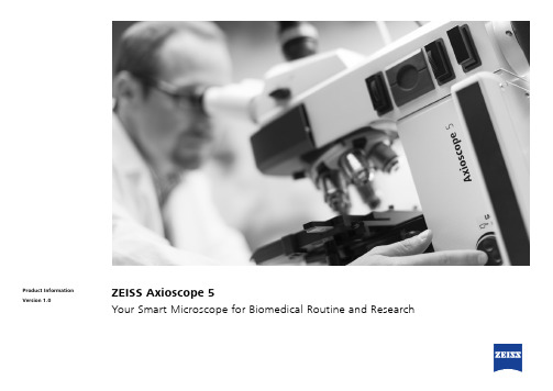
ZEISS Axioscope 5智能生物学显微镜商品说明书

ZEISS Axioscope 5Your Smart Microscope for Biomedical Routine and ResearchProduct InformationVersion 1.0In the past, documenting samples with multiple fluorescent labels in your routine lab could be time consuming. To get best image quality, you needed to manually switch filters, adjust illumination intensities and exposure times and to snap each single channel image. For three different channels, this could sum up to 15 steps and clicks. With Smart Microscopy from ZEISS, this is a thing of the past.Your Axioscope 5 with Axiocam 202 mono and Colibri 3 LED illumination takes this workload from you. You don't even need to move your hands from them icroscope stand anymore. All you have to do is focus and press Snap – and you're done! You can now concentrate on the essence of your job and let your Axioscope 5 work for you. You'll work more efficiently, save time and produce high contrast images with best image quality. What's more: this even works without any PC involved.Your Smart Microscope for Biomedical Routine and ResearchClick here to explore all features in an interactive infographic.J› In Brief › The Advantages › The Applications › The System› Technology and Details ›ServiceAnimationSimpler. More Intelligent. More Integrated.Capture Four Fluorescence Channelswith Just One ClickAcquiring fluorescent images has never been so easy. Combine Axioscope 5 with the high perfor-mance LED light source Colibri 3 and the sensitive, standalone microscope camera Axiocam 202 mono to have the perfect setup for easy multi-channel fluorescence d ocumentation. Switche ffortlessly between the channels for UV, blue, green and red excitation. Just select the relevant channels and press Snap. The system then takes over and automatically a djusts the exposure time, acquires the image, switches the channel and starts again. That's it: you get your overlayedm ultichannel fluorescence image including scale bar – even without a PC.Benefit from Smart LED IlluminationAxioscope 5 uses its transmitted white light LEDto provide powerful illumination with high colorfidelity. You will clearly see the subtle d ifferencesin your sample. And experience all the advantagesof LED illumination such as stable color tempera-ture, low energy consumption and long lifetime.Axioscope 5 comes with a light intensity managerthat produces uniform brightness at all magnifica-tions. Adjusting lamp brightness when you changemagnification is a thing of the past. That savesyou time and reduces eye fatigue, too.Smart Microscopy Makes Your DigitalD ocumentation FasterAxioscope 5 makes documenting your specimensvery efficient. The color impression shows up inthe camera image exactly the same as it appearsthrough the eyepieces. The smart Axioscope 5s ystem makes automatic adjustments for bright-ness and white balance to keep digital documen-tation easy. All you have to do is focus on yoursample, press the ergonomic Snap buttonon the microscope, and that's it. Acquiring highquality images with high color fidelity has neverbeen easier – and faster.› In Brief› The Advantages› The Applications› The System› Technology and Details ›ServiceExpand Your PossibilitiesUsed in combination with the microscope cameras Axiocam 202 mono or Axiocam 208 color, you have the full advantage of a smart standalonem icroscope solution.Camera settings such as white balance, contrast and exposure time are done automatically.W ithout needing additional imaging software or even a computer, you can:Stand-alone for Basic Routine Imaging ZEISS Axioscope 5 operates i ndependently of acomputer system.VZEISS Labscope for Advanced Routine Imaging Operating ZEISS Axioscope 5 with ZEISS Labscope imaging software is ideal for c onnected microscopyand standard multichannel fluorescence imaging.VZEISS ZEN for Research ApplicationsUse ZEN imaging software to perform advanced imaging tasks with ZEISS Axioscope 5.VVV• Snap images and record videos directly from your stand• Use mouse (and optionally keyboard) to control your c amera via OSD (on screen display)• Save settings• Store images with all metadata of the m icroscope and camera as well as scaling i nformation • Predefine the name or rename your imageThis is Smart Microscopy – Digital Documentation Made Easy › In Brief› The Advantages › The Applications › The System› Technology and Details › ServiceExpand Your PossibilitiesBoost your Efficiency – with Smart MicroscopyEfficiency and quality are key in your lab, but it can take a lot of time to acquire detail-rich, true-color images. You know the drill: place the sample, focus your region of interest, switch to the computer,a djust settings such as white balance, exposure time and gain, then acquire an image, insert a scale bar, switch back to the microscope … and so on. That's what a typical documentation workflowlooks like. Now, with the Axioscope 5 system, you can stay focused on your sample at all times, thanks to smart microscopy. Digital documentation is inherent in the system design. Just press thee rgonomic Snap button on the microscope and you're done. The procedure integrates perfectly with your established microscopy workflow and boosts your efficiency tremendously.› In Brief› The Advantages › The Applications › The System› Technology and Details › ServiceExpand Your PossibilitiesRat kidney, acquired in transmitted light brightfield,objective: Plan-Apochromat 20× / 0.8Rabbit muscle, acquired in DIC contrast,objective: Plan-Apochromat 63× / 1.4Trout cartilage acquired in phase c ontrast, objective: Plan-Apo-chromat 63× / 1.4Crystal, acquired in polarization c ontrast,objective: Plan-Neofluar 20×Whether unstained cells, histologically staineds ections, or other samples: transmitted light tech-niques continue to be the standard for manye xaminations.With Axioscope 5 you can use a sheer variety ofc ontrasting techniques for your applications:the classical methods of brightfield, darkfield,phase contrast, but also Differential I nterferenceContrast (DIC) and polarization c ontrast.A xioscope 5 can also be equipped with P lasDIC,the cost effective interference contrasting tech-nique.› In Brief› The Advantages› The Applications› The System› Technology and Details› ServiceExpand Your PossibilitiesMink Uterus Endometrium Epithelial Cells, vimentin – red, F-actin – green, nucleus – blue; acquired with ZEISS Axioscope 5, Colibri 3 andAxiocam 202 mono in stand-alone mode, objective: Plan-Apochromat 40× / 0.95ZEISS Colibri 3 LED IlluminationComplement your Axioscope 5 with the optional fluorescence LED illumination Colibri 3, and acquire brilliant fluorescence images with ease. Colibri 3 delivers the right wavelength and intensity to ex -cite fluorescent dyes and proteins in a gentle way.• Save time and money thanks to the long LED lifetime and adjustment-free operation. • Choose up to four configurable wavelengths to fit your needs. Upgrade anytime you need to.• Individually control and switch between c hannels for UV, blue, green and red excitation – or use selected wavelengths simultaneously.• With direct visual status feedback, you area lways sure which FL-LED is in use.• The integrated design saves space and makesfor easy and ergonomic operation.100 µm50 μmIndian muntiac, fibroblasts, F-actin – red, nucleus – green objective: Plan-Apochromat 20× / 0.8Mouse kidney in fluorescence, cryosection, AF 488 – WGA, AF 568 Phalloidin, DAPI, objective: Plan-Apochromat 20× / 0.8› In Brief› The Advantages › The Applications › The System› Technology and Details › ServiceHistological specimen, CDx immunohistological stain;Red: immunoreactive antigens in cytoplasm;Blue: nuclear counterstaining Ziehl-Neelsen-Färbung, objective: EC Plan-Neofluar 63× / 0.95 Korr.Chromosome specimen, Giemsa stain, objective: Plan-Apochromat 63× / 1.4 Renal tissue, Trichrome stain,objective: Plan-Apochromat 40× / 0.95Tailored Precisely to Your Applications› In Brief › The Advantages › The Applications › The System› Technology and Details › Service1235341 Microscope• ZEISS Axioscope 5, transmitted light, LED • ZEISS Axioscope 5, transmitted light, Hal 50• ZEISS Axioscope 5, fluorescence 2 Recommended Objectives • Plan-Apochromat • Plan-Neofluar • N-Achroplan5 Software • Stand-alone• Labscope imaging app • ZEN imaging softwareYour Flexible Choice of Components3 Illumination Transmitted light:• LED 10W, Hal 50, Hal 100Reflected light, fluorescence: • Colibri 3, HXP 120, and other4 Recommended Microscope Cameras • ZEISS Axiocam 202 mono • ZEISS Axiocam 208 color› In Brief › The Advantages › The Applications › The System› Technology and Details › ServiceSystem Overview› The Advantages› The Applications› The System› Technology and Details› ServiceSystem Overview› The Advantages› The Applications› The System› Technology and Details› ServiceTechnical Specifications› In Brief› The Advantages› The Applications› The System› Technology and Details› ServiceTechnical Specifications› The Advantages› The Applications› The System› Technology and Details› ServiceTechnical Specifications› The Advantages› The Applications› The System› Technology and Details› Service>> /microserviceBecause the ZEISS microscope system is one of your most important tools, we make sure it is always ready to perform. What’s more, we’ll see to it that you are employing all the options that get the best from your microscope. You can choose from a range of service products, each delivered by highly qualified ZEISS specialists who will support you long beyond the purchase of your system. Our aim is to enable you to experience those special moments that inspire your work.Repair. Maintain. Optimize.Attain maximum uptime with your microscope. A ZEISS Protect Service Agreement lets you budget for operating costs, all the while reducing costly downtime and achieving the best results through the improved performance of your system. Choose from service agreements designed to give you a range of options and control levels. We’ll work with you to select the service program that addresses your system needs and usage requirements, in line with your organization’s standard practices.Our service on-demand also brings you distinct advantages. ZEISS service staff will analyze issues at hand and resolve them – whether using remote maintenance software or working on site. Enhance Your Microscope System.Your ZEISS microscope system is designed for a variety of updates: open interfaces allow you to maintain a high technological level at all times. As a result you’ll work more efficiently now, while extending the productive lifetime of your microscope as new update possibilities come on stream.Profit from the optimized performance of your microscope system with services from ZEISS – now and for years to come.Count on Service in the True Sense of the Word› In Brief › The Advantages › The Applications › The System› Technology and Details › ServiceN o t a l l p r o d u c t s a r e a v a i l a b l e i n e v e r y c o u n t r y . U s e o f p r o d u c t s f o r m e d i c a l d i a g n o s t i c , t h e r a p e u t i c o r t r e a t m e n t p u r p o s e s m a y b e l i m i t e d b y l o c a l r e g u l a t i o n s .C o n t a c t y o u r l o c a l Z E I S S r e p r e s e n t a t i v e f o r m o r e i n f o r m a t i o n .E N _41_011_205 | C Z 05-2019 | D e s i g n , s c o p e o f d e l i v e r y , a n d t e c h n i c a l p r o g r e s s s u b j e c t t o c h a n g e w i t h o u t n o t i c e . | © C a r l Z e i s s M i c r o s c o p y G m b HCarl Zeiss Microscopy GmbH 07745 Jena, Germany ******************** /axioscope。
打包机故障代码

故障码编码说明
A X YY –Z
X :故障部位编码
X :0 综合故障
X :1 1#压盘小车
X :2 2#压盘小车
X :3 送线小车/升降台
X :4 防刮伤/放线架
X :5 1#打捆头及送线轮
X :6 2#打捆头及送线轮
X :7 3#打捆头及送线轮
X :8 4#打捆头及送线轮
X :9 液压站
YY :顺序号
Z - 故障级别编码
Z :1 一级故障,立即停液压站和所有的设备动作
Z :2 二级故障,立刻退出自动
Z :3 三级故障,立刻停止相关设备的动作,经过延时后退出自动
Z :4 四级故障,只作为设备动作的起动条件,不会立刻停止正在动作的设备
0 --- 综合故障
1 --- 1#压盘小车的故障
A120-4 1号压盘小车原位接近开关检测故障
2 --- 2#压盘小车的故障
3 --- 送线小车/升降台的故障
A351-4 导线小车原位接近开关检测故障
4 防划伤/放线架故障
51号打捆头及送线轮故障
A517-3 1号送线轮送线
62号打捆头及送线轮故障
A616-3 2号送线轮运行条件丢失A617-3 2号送线轮送线
73号打捆头及送线轮故障
A716-3 3号送线轮运行条件丢失A717-3 3号送线轮送线
84号打捆头及送线轮故障
A816-3 4号送线轮运行条件丢失A817-3 4号送线轮送线
9液压站故障。
星空彩妆系列产品价格表(修正)

81 82 83 84 85 86 87 88 89 90 91 92 93 94 95 96 97 98 99 100 101 102 103 104 105 106 107
星空幻影唇彩20# 3.5g 48 星空幻影唇彩21# 3.5g 48 星空幻影唇彩22# 3.5g 48 星空幻影唇彩23# 3.5g 48 星空纤长睫毛膏 4.5g 49 星空无瑕粉饼1# 9g 89 星空无瑕粉饼2# 9g 89 星空光采妆前乳 25g 89 星空水盈BB霜(滋润型) 28ml 68 星空水盈BB霜(清爽型) 28ml 68 星空保湿散粉 9g 79 星空深层净透卸妆液 120ml 78 星空多彩自由组合(红色) 88 星空多彩自由组合(蓝色) 88 星空迷幻彩妆套盒B(黄色) 0000 139 星空迷幻彩妆套盒B(黄绿色) 0000 139 星空迷幻彩妆套盒B(浅蓝色) 0000 139 星空迷幻彩妆套盒B(粉红色) 0000 139 星空持久倍护润唇膏(薄荷味感) 1.8g 28 星空持久倍护润唇膏(零味感) 1.8g 28 星空持久倍护润唇膏(草莓味感) 1.8g 28 星空持久倍护润唇膏(青苹味感) 1.8g 28 星空持久倍护润唇膏(香橙味感) 1.8g 28 星空多彩自由组合(红色)空盒 20元 星空多彩自由组合(蓝色)空盒 20元 星空多彩自由组合(红色)(空盒+眼影棒+胭脂刷) 30元 星空多彩自由组合(蓝色)(空盒+眼影棒+胭脂刷) 30元
Pageቤተ መጻሕፍቲ ባይዱ1
星空彩妆
27 28 29 30 31 32 33 34 35 36 37 38 39 40 41 42 43 44 45 46 47 48 49 50 51 52 53 54
星空多彩眼影(点金裸)A027 星空多彩眼影(闪炫粉)A028 星空多彩眼影(暖阳金)A029 星空多彩眼影(夜空金)A030 星空多彩眼影(淡粉蓝)A031 星空多彩眼影(幻影蓝)A032 星空多彩眼影(苹果蓝)A033 星空多彩眼影(炫彩蓝)A034 星空多彩眼影(迷金棕)A035 星空多彩眼影(纸醉棕)A036 星空多彩眼影(赤沙棕)A037 星空多彩眼影(闪赫棕)A038 星空多彩眼影(夕阳棕)A039 星空多彩眼影(墨翠棕)A040 星空多彩眼影(深眸棕)A041 星空多彩眼影(沉默棕)A042 星空多彩眼影(清新灰)A043 星空多彩眼影(迷烟灰)A044 星空多彩眼影(烟熏黑)A045 星空多彩眼影(自然黑)A046 星空多彩眼影(迷踪绿)A047 星空多彩眼影(墨踪绿)A048 星空多彩眼影(青春紫)A049 星空多彩眼影(魅力紫)A050 星空多彩眼影(深遂紫)A051 星空炫丽胭脂(蔷薇)B001 星空炫丽胭脂(海棠)B002 星空炫丽胭脂(桃花)B003
石英石收费说明

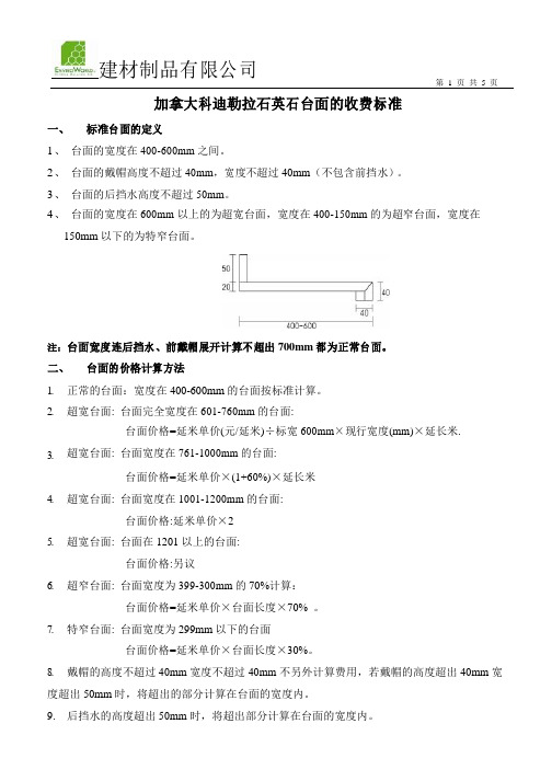
加拿大科迪勒拉石英石台面的收费标准一、标准台面的定义1、台面的宽度在400-600mm之间。
2、台面的戴帽高度不超过40mm,宽度不超过40mm(不包含前挡水)。
3、台面的后挡水高度不超过50mm。
4、台面的宽度在600mm 以上的为超宽台面,宽度在400-150mm的为超窄台面,宽度在150mm以下的为特窄台面。
注:台面宽度连后挡水、前戴帽展开计算不超出700mm都为正常台面。
二、台面的价格计算方法1.2.3.4.5.6.7.8.正常的台面:宽度在400-600mm的台面按标准计算。
超宽台面: 台面完全宽度在601-760mm的台面:台面价格=延米单价(元/延米)÷标宽600mm×现行宽度(mm)×延长米.超宽台面: 台面宽度在761-1000mm的台面:台面价格=延米单价×(1+60%)×延长米超宽台面: 台面宽度在1001-1200mm的台面:台面价格:延米单价×2超宽台面: 台面在1201 以上的台面:台面价格:另议超窄台面: 台面宽度为399-300mm的70%计算:台面价格=延米单价×台面长度×70% 。
特窄台面: 台面宽度为299mm以下的台面台面价格=延米单价×台面长度×30%。
戴帽的高度不超过40mm宽度不超过40mm不另外计算费用,若戴帽的高度超出40mm宽度超出50mm时,将超出的部分计算在台面的宽度内。
9.后挡水的高度超出50mm时,将超出部分计算在台面的宽度内。
10. 侧戴帽和侧挡水的尺寸应加在台面的长度上计费。
11. 如图的计价方法:600mm台面价格=延米单价×台面长度×[600+(130-50)+(100-40)]÷600 300mm台面价格=延米单价×台面长度×70%700mm台面价格=延米单价×台面长度×[700+130-50+(100-40)]÷600三、边型的收费费标准1、以下边型的款式不额外收费用:CO-A001 CO-A002 CO-A003CO-A004 CO-A005 CO-A006以下边型款式需加收200元/米:CO-B001 CO-B003 CO-B005CO-B006 CO-B007 2、其它款式的边型要延长工期的具体情况另议。
精微视达共聚焦参数

精微视达共聚焦参数英文回答:The confocal parameter in microscopy refers to the distance over which the microscope can maintain optimal focus. It is a crucial parameter that determines the depthof field and resolution of the microscope. In confocal microscopy, a pinhole is used to block out-of-focus light, allowing only the in-focus light to reach the detector.This results in high-resolution images with improvedcontrast and signal-to-noise ratio.The confocal parameter is influenced by several factors, including the numerical aperture (NA) of the objective lens, the wavelength of the light used, and the refractive indexof the medium. A higher NA and shorter wavelength willresult in a smaller confocal parameter, meaning that the microscope can maintain focus over a shorter distance. On the other hand, a higher refractive index will increase the confocal parameter, as the light will be more tightlyfocused.To illustrate the concept, let's consider an example. Imagine you are using a confocal microscope with a high NA objective lens and a short-wavelength light source. In this case, the confocal parameter will be small, allowing you to focus on a specific plane with high precision. This is particularly useful when imaging thin samples or when you want to isolate a specific layer within a thicker sample.Now let's switch to 中文回答:共聚焦显微镜中的共聚焦参数是指显微镜能够保持最佳焦距的距离。
金属显微组织检验方法美标英文

金属显微组织检验方法美标英文Title: Metal Microstructure Inspection Methods - American Standards (in English)Introduction:Metal microstructure plays a crucial role in determining the mechanical, physical, and chemical properties of metals.Accurate inspection and analysis of these microstructures are essential for ensuring the quality and performance of metal components.This document provides an overview of the commonly used metal microstructure inspection methods according to American standards, presenting them in English for international reference.1.Optical Microscopy (OM):Optical microscopy is one of the most traditional and widely used methods for metal microstructure examination.It involves the use of a light microscope to observe metallographic specimens that have been prepared by polishing and etching.The American Society for T esting and Materials (ASTM) provides standards such as ASTM E3-18, which offers guidelines for preparing and interpreting metallographic specimens.2.Scanning Electron Microscopy (SEM):Scanning Electron Microscopy is a powerful technique for examining the microstructure of metals at a higher resolution thanoptical microscopy.It utilizes an electron beam to scan the surface of a specimen and generate detailed images.ASTM E1587-19 provides guidelines for the use of SEM in microstructural analysis.3.Transmission Electron Microscopy (TEM):Transmission Electron Microscopy is used for observing the internal microstructure of metals at an ultra-high resolution.It involves passing a beam of electrons through an ultra-thin specimen.The ASTM E283-18 standard provides the practices for the determination of grain size in metals using TEM.4.X-Ray Diffraction (XRD):X-Ray Diffraction is a non-destructive method used to identify the crystallographic structure and phase composition of metals.It is particularly useful for qualitative and quantitative analysis of the phases present in a material.The ASTM E975-16 standard outlines the procedures for XRD analysis of metals.5.hardness Testing:Hardness testing is a common method for indirectly assessing the microstructure of metals.Different hardness testing methods, such as Rockwell, Brinell, and Vickers, are standardized by ASTM.For instance, ASTM E18 provides the guidelines for Rockwell hardness testing, which can provide insights into the microhardness of metal samples.6.Eddy Current Testing (ECT):Eddy current testing is an electromagnetic technique used for detecting surface and near-surface defects in conductive materials.It can also provide information about microstructural changes.The ASTME309-14 standard covers the practice for using eddy current methods for crack detection in metals.Conclusion:The above-mentioned metal microstructure inspection methods adhere to the American standards provided by recognized organizations such as ASTM.These standards ensure that the examination and analysis of metal microstructures are consistent, reliable, and of high quality.By utilizing these methods, professionals can accurately assess the microstructure of metals, leading to improved product performance and safety in various industries.。
南京建成生物工程研究所价目表

南京建成生物工程研究所价目表南京建成生物工程研究所价目表二○○四年七月一、抗氧化检测试剂及科研试剂:(注:**为新试剂盒)序号产品名称规格单位检测方法售价厂家A001 超氧化物歧化酶(SOD)测试盒100T 盒羟胺法240.00 建成50T 盒羟胺法130.00 建成A003 丙二醛(MDA)测试盒100T 盒TBA法150.00 建成50T 盒TBA法90.00 建成A002 黄嘌呤氧化酶(XOD)测试盒50T 盒比色法130.00 建成A015 总抗氧化能力(T-AOC)测试盒50T 盒比色法120.00 建成25T 盒比色法70.00 建成A018 羟自由基测试盒50T 盒比色法150.00 建成A052 超氧阴离子自由基(O )测试盒50T 盒比色法130.00 建成A044 髓过氧化物酶(MPO)测试盒50T 盒比色法160.00 建成A007 过氧化氢酶(CAT)测试盒紫外分光光度法100T 盒紫外比色法100.00 建成可见光分光光度法100T 盒钼酸铵法100.00 建成A064 过氧化氢(H2O2)测试盒50T 盒比色法50.00 建成A004 谷胱甘肽—S转移酶(GSH—ST)测试盒80T 盒比色法200.00 建成A005 谷胱甘肽—过氧化物酶(GSH—PX)测试盒50T 盒比色法200.00 建成A062 谷胱甘肽-还原酶(GR)测试盒50T 盒比色法200.00 建成A006 还原型谷胱甘肽(GSH)测试盒50T 盒比色法180.00 建成A061 氧化型谷胱甘肽(GSSG) 测试盒50T 盒比色法180.00 建成A063 巯基(-SH)测试盒50T 盒比色法180.00 建成A034 单胺氧化酶(MAO)测试盒50T 盒紫外比色法150.00 建成A012一氧化氮(NO)测试盒(硝酸还原酶法) 50T 盒比色法300.00 建成25T 盒比色法180.00 建成A013 一氧化氮(NO)测试盒(化学法测NO2—)100T 盒比色法150.00 建成A014 -1 一氧化氮合成酶(NOS)测试盒[(T-NOS、iNOS、cNOS)三型同时出结果] 50T 盒比色法480.00 建成25T 盒比色法260.00 建成A014-2 一氧化氮合成酶(NOS)测试盒[总NOS(T-NOS)] 50T 盒比色法350.00 建成25T 盒比色法200.00 建成A019-1 乳酸(LD)测试盒(测全血)50T 盒紫外比色法240.00 建成50T 盒可见光比色法240.00 建成A019-2 乳酸(LD)测试盒(测血清、组织)50T 盒比色法200.00 建成A020 乳酸脱氢酶(LDH)测试盒100T 盒比色法240.00 建成50T 盒比色法130.00 建成A016-1 A TP酶测试盒(组织及细胞膜需高速离心)(可测Na+K+、Ca2+Mg2+ ATPase) 100T 盒比色法240.00 建成50T 盒比色法150.00 建成A016-2 A TP酶测试盒(组织及细胞膜不需高速离心)(可测Na+K+、Ca2+Mg2+ ATPase) 100T 盒比色法300.00 建成50T 盒比色法180.00 建成A057 Cu-A TP酶(Cu-A TPase)测试盒100T 盒比色法300.00 建成50T 盒比色法180.00 建成序号产品名称规格单位检测方法售价厂家A069 氢-钾A TP酶(H+-A TPase)测试盒100T 盒比色法300.00 建成50T 盒比色法180.00 建成A070-1 超微量A TP酶测试盒(可测Na+K+、Ca2+Mg2+ A TPase、总ATPase)200T 盒比色法390.00 建成100T 盒比色法230.00 建成A070-2 超微量A TP酶测试盒(可测总A TPase)100T 盒比色法300.00 建成50T 盒比色法180.00 建成A021 苹果酸脱氢酶测试盒50T 盒紫外比色法面议建成A022 琥珀酸脱氢酶测试盒50T 盒比色法240.00 建成A032 肌酸激酶(CK)测试盒50T 盒比色法180.00 建成A023 胆碱酯酶(CHE)测试盒50T 盒比色法70.00 建成A024 乙酰胆碱酯酶(T-CHE)测试盒50T 盒比色法150.00 建成A025 丁酰胆碱酯酶测试盒50T 盒比色法150.00 建成A027 葡萄糖-6-磷酸脱氢酶(G-6-PD)测试盒50T 盒比色法100.00 建成A031 b-N-乙酰氨基葡萄糖苷酶(NAG)测试盒50T 盒比色法260.00 建成A053 β-葡萄糖醛酸苷酶测试盒50T 盒比色法200.00 建成A055** 红细胞NADH高铁血红蛋白还原酶测试盒50T 盒比色法260.00 建成A059 碱性磷酸酶(AKP)测试盒50T 盒比色法100.00 建成A060 酸性磷酸酶(ACP)测试盒50T 盒比色法130.00 建成A049 前列腺酸性磷酸酶(PACP)测试盒50T 盒比色法180.00 建成A058 抗酒石酸酸性磷酸酶(StrACP)测试盒50T 盒比色法180.00 建成A068 钙调神经磷酸酶(CaN)测试盒25T 盒比色法200.00 建成A072 全血2,3-二磷酸甘油酸(2,3-DPG)测试盒50T 盒比色法面议建成A047 谷氨酰胺合成酶(GS)测试盒50T 盒比色法300.00 建成A048 腺苷脱氨酶(ADA)测试盒50T 盒比色法130.00 建成A073** 谷氨酰胺测试盒50T 盒比色法200.00 建成A074** 谷氨酸测试盒50T 盒比色法260.00 建成A075** 谷氨酰胺、谷氨酸(两种均可测)50T 盒比色法480.00 建成A076** 丙酮酸激酶(PK)测试盒50T 盒比色法面议建成A077** 己糖激酶(HK)测试盒50T 盒比色法面议建成A036 唾液酸(SA)测试盒(带SA标准) 50T 盒比色法200.00 建成A017-1 细胞凋亡测试盒(TdT酶、稀释液、POD)10片盒TUNEL法1450.00 建成A017-2 细胞凋亡测试盒(TdT介导的原位末端切口平移双标记法)(全套)10片盒TUNEL法1850.00 建成A008 维生素E(VE)测试盒50T 盒比色法130.00 建成A009 维生素C(VC)测试盒50T 盒比色法150.00 建成A010 维生素B1(V B1)测试盒50T 盒比色法面议建成A011 维生素B2(V B2)测试盒50T 盒比色法面议建成A026 总氨基酸(T—AA)测试盒80T 盒比色法100.00 建成A030-1 羟脯氨酸测试前处理试剂消化液50T 瓶比色法100.00 建成调PH试剂50T 瓶比色法70.00 建成A030-2 羟脯氨酸(Hyp)测试盒(可测血清、培养液、尿液、心肌、皮肤、软骨、肝脏等等)50T 盒比色法180.00 建成A041 5’-核苷酸酶(5’-NT)测试盒50T 盒比色法150.00 建成A035 D—木糖测试盒50T 盒比色法100.00 建成A056 糖化血红蛋白(GHb)测试盒15T 盒比色法55.00 建成A037 糖化血清蛋白(GSP)测试盒(果糖胺法)(带标准)50T 盒比色法150.00 建成25T 盒比色法80.00 建成A043 肝/肌糖元测试盒50T 盒比色法160.00 建成A038 脂蛋白(a)[LP(a)]测试盒96T 盒ELISA法380.00 建成A054 脂肪酶测试盒50T 盒比色法200.00 建成A067 总脂酶【脂蛋白脂酶(LPL)/肝脂酶(HL)】测试盒100T 盒比色法300.00 建成50T 盒比色法160.00 建成A042 游离脂肪酸(NEFA)测试盒50T 盒比色法160.00 建成A033 脂褐质测试盒50T 盒荧光比色法100.00 建成A039 血清铁测试盒50T 盒比色法100.00 建成A040 总铁结合力(TIBC)测试盒50T 盒比色法180.00 建成A029 铜兰蛋白(CP)测试盒50T 盒比色法150.00 建成A071 微量游离血红蛋白测试盒50T 盒比色法80.00 建成A028 白蛋白测试盒(溴甲酚绿法)100T 盒比色法30.00 建成A045-1 总蛋白定量测试盒(双缩脲法)100T 盒比色法30.00 建成A045-2 总蛋白定量测试盒(考马斯亮兰法)100T 盒比色法30.00 建成A046 蛋白标准品(总蛋白、白蛋白) 1ml 支7.00 建成A050-1 溶菌酶(LZM)测试盒(带标准)30T 盒试管比浊法60.00 建成A050-2 溶菌酶(LZM)测试盒(带标准)30T 盒平板法60.00 建成A050-3 溶菌酶标准品2mg 支6.00 建成A050-4 微球菌粉30mg 支40.00 建成A065 解脲(溶脲)支原体快速培养基(UU培养基)每人份支培养基10.00 建成A066 人型支原体快速培养基每人份支培养基10.00 建成A051-1 肝素溶液10ml 支20.00 建成A051-2 肝素抗凝管10ml 支2.00 建成二、不孕症系列酶免疫试剂:序号产品名称规格单位检测方法售价厂家B001 抗心磷脂抗体测试盒(ACL)IgG类48T 盒ELISA 160.00 建成抗心磷脂抗体测试盒(ACL)IgA类48T 盒ELISA 160.00 建成抗心磷脂抗体测试盒(ACL)IgM类48T 盒ELISA 160.00 建成B002 抗精子抗体测试盒(AsAb)IgG类48T 盒ELISA 160.00 建成抗精子抗体测试盒(AsAb)IgA类48T 盒ELISA 160.00 建成抗精子抗体测试盒(AsAb)IgM类48T 盒ELISA 160.00 建成B003 抗子宫内膜抗体测试盒(EmAb)IgG类48T 盒ELISA 260.00 建成抗子宫内膜抗体测试盒(EmAb)IgA类48T 盒ELISA 260.00 建成抗子宫内膜抗体测试盒(EmAb)IgM类48T 盒ELISA 260.00 建成B004 抗卵巢抗体测试盒(AoAb)IgG类48T 盒ELISA 260.00 建成抗卵巢抗体测试盒(AoAb)IgA类48T 盒ELISA 260.00 建成抗卵巢抗体测试盒(AoAb)IgM类48T 盒ELISA 260.00 建成三、常用试剂:序号产品名称规格单位检测方法售价厂家C001 钾(K)测试盒(带标准)30T 盒比色法(上机)45.00 建成30T 盒比浊法(手工)35.00 建成C002 钠(Na)测试盒(带标准)30T 盒比色法(上机)45.00 建成30T 盒比浊法(手工)35.00 建成C003 氯(Cl)测试盒(带标准)30T 盒比色法(上机)45.00 建成30T 盒比色法(手工)28.00 建成C004 钙(Ca)测试盒(带标准)30T 盒比色法(上机)40.00 建成30T 盒比色法(手工)30.00 建成C006 磷(Pi)测试盒(带标准)100T 套孔雀绿显色39.00 建成A059 碱性磷酸酶(AKP)测试盒50T 盒比色法100.00 建成A060 酸性磷酸酶(ACP)测试盒50T 盒比色法130.00 建成C009 谷丙转氨酶(SGPT)测试盒100T 盒比色法30.00 建成C010 谷草转氨酶(SGOT)测试盒100T 盒比色法30.00 建成C018 总胆红素、直接胆红素测试盒80T 套咖啡因比色法65.00 建成80T 套一步法65.00 建成C017 Υ—谷氨酰转移酶(Υ—GT)测试盒100T 套比色法88.00 建成C020 尿胆红素测试盒50T 套哈里森氏法25.00 建成C033 尿胆原测试盒50T 套25.00 建成C013 尿素氮(BUN)测试盒100T 套脲酶比色法36.00 建成100T 套二乙酰-肟比色法27.00 建成C016 淀粉酶(AMS)测试盒100T 套淀粉一碘比色法39.00 建成C012 尿酸测试盒50T 套比色法40.00 建成C011-1 肌酐(Cr)测试盒(苦味酸法)100T 套比色法(除蛋白)60.00 建成C011-2 肌酐(Cr)测试盒(苦味酸法)100T 套比色法(不除蛋白)40.00 建成C021 血红蛋白稀释液(测试液)5ml 支HICN比色法4.50 建成C022 血红蛋白标准品5ml×4支套比色法12.00 建成C023 血红蛋白校正液250ml 瓶比色法35.00 建成100ml 瓶比色法14.00 建成C024 血小板稀释液(计数液)100ml 瓶8.00 建成C025 白细胞稀释液(计数液)100ml 瓶8.00 建成C037 红细胞稀释液(计数液)100ml 瓶8.00 建成C026 血细胞仪清洗液250ml 瓶25.00 建成C028 CO2结合力测试盒2ml×3×10 套滴定法40.00 建成C034 脑脊液蛋白测试盒50T 盒磺基水杨酸法50.00 建成C038 血沉测试液100ml 瓶10.00 建成C039 嗜酸粒细胞直接计数液40ml×1 瓶50.00 建成C040 网织红细胞计数液8 ml×2 套试管法30.00 建成10 ml×1 瓶玻片法20.00 建成C035-1 尿蛋白定性测试盒100T 盒磺基水杨酸法30.00 建成C035-2 尿蛋白定量测试盒100T 盒CBB法30.00 建成C041 尿糖试剂100ml 瓶班氏法40.00 建成C027 尿粪隐血测试盒100T 套联苯胺法15.00 建成100T 套邻联甲苯胺法15.00 建成A051-1 肝素溶液10ml 支20.00 建成A051-2 肝素抗凝管10ml 支2.00 建成四、染液类序号产品名称规格单位检测方法售价厂家D001 碱性磷酸酶染液50片套染色50.00 建成D002 酸性磷酸酶染液50片套染色50.00 建成D003-1 酸性酯酶染液5.0ml×4 盒染色50.00 建成D003-2 中性酯酶染液5.0ml×4 盒染色50.00 建成D003-3 碱性酯酶染液5.0ml×4 盒染色50.00 建成D003-4 双重酯酶染液5.0ml´4 盒染色60.00 建成D009 快速血细胞染液50ml×2 套染色40.00 建成D004 糖元染液10ml×4 盒染色40.00 建成D005 苏木素染液50ml×1 瓶染色40.00 建成D019 伊红染液100ml×1 套染色50.00 建成D006 H-E染液套染色55.00 建成D007 瑞氏染液50ml×2 套染色40.00 建成D010 瑞氏一姬姆萨复合染液50ml×2 套染色40.00 建成D011 姬姆萨染液套染色30.00 建成D015 巴氏染液(脱落细胞染色)套染色50.00 建成D017 快速H-E组织细胞及脱落细胞染液10ml×4 染色20.00 建成D008 革兰氏染液50ml×4 套染色50.00 建成D012 抗酸染液50ml×2 套染色40.00 建成D013 溴酚兰溶液1.0ml 支染色10.00 建成D014 溴乙淀溶液1.0ml 支染色20.00 建成D016 细胞活性鉴别染液20ml 瓶伊文斯蓝法20.00 建成D018 血管通透性测试染液20ml 瓶美蓝法20.00 建成D020** 过氧化物酶染液5ml 套染色20.00 建成五、各种浓度及PH值的缓冲液:(注: 以下缓冲液PH值、摩尔浓度按客户要求配制。
美乐视显微镜产品概览说明书

– Scope of delivery
– optional optional optional
–
P10
F2.8 f = 4,7 – 75,2 mm
16x / 8x 405 x 303 Auto / manual Auto / manual removable horizontal / vertical
integrated preview monitor » 16X optical zoom for presenting even the smallest details » Optional mini board: painting, marking and writing – wirelessly in the whole room » Saving images, videos or group rks directly and easily on a SD(HC) card or on a PC
external 4,7 kg 376 x 549 x 482 376 x 181 x 482
Scope of delivery Scope of delivery
– Scope of delivery
optional Scope of delivery
– – optional optional –
General
Power Supply Weight Dimensions
AC 100 V to 240V, 50/60 Hz
W x H x D max. (mm) W x H x D min. (mm)
Accessories
Remote control VGA cable DVI cable USB cable RS-232C cable Software Win / Mac Bag Microscope adapter SD memory Card Tablet CRA-1 Lighting box
1国内外电子显微镜产品介绍

国内外电子显微镜产品介绍世界上第一台电子显微镜1932年诞生于德国柏林,当时它的放大倍数仪12倍,1940年美国RCA公司制成第一台商品磁式电镜,以后各国科学技术工作者为获取高分辨本领的电镜而作出了不懈的努力,l974年科学家海勒和朗勃将消像散器用于电镜而使电镜的分辨本领达到1nm,实现了突破。
至50年代,英、美、德、荷、前苏联等国把透射式电镜(电子穿透样品而成像的电镜)投入批量生产。
透射电镜也随蕾科学技术发展和科研生产需要而实现电镜性能的高中低系列化。
高档的商品透射电镜其分辨本领一般为0.2-0.3nm。
由于一般加速电压为100kV的透射电镜,只随时厚度小于0.1μm的样品进行观察,为了观察数微米以上的样品和活的生物样品,出现了加速电压为350万伏的超高压电镜。
电子显微镜的另一分支是扫描电镜,它用间接成像的方法来获得被校样品的放大像,由于它有较大的聚焦深度,可以获得较清晰的“立体”像,同时它能观察较大的、表面粗糙的样品(因此样品制备简单)而得到广泛应用。
但真正高品性的扫描电镜是于1965年由英国检桥仪器公司生产的,它用的是二次电子成像技术。
当时分辨本领为25μm。
目前生产扫描电镜的国家主富有日、英、莫和我国,其生产规模已超过透射电镜,商品高性能扫描电镜.其分辨本领约为6-7nm。
扫描电镜首次采用场效发射枪是在1968年美国芝加哥大学,获得了零点几nm的分辨本领。
从现有的资料看,近两年来,世界各国的透射电镜的分辨本领,照明系统,成像系统,图像接收和处理系统.真空系统,微区分析性能等方面虽都有改进,但没有突破性进展,有新特点的透射电镜出现也不多,例如荷兰飞利浦公司的CM-120型透射电镜,晶格分辨本领0.34nm,点分辨本领0.49nm,加速电压为20-120KV,放大倍数为20-370000倍,带有EDAX能谱仪,专为生命科学而设计,其主要特点是有高衬度和高的成分分析灵敏度,因此可以快速完成点成分分析和x射线成分分布图的分析.试样室可用于冷湿工作状态。
银耳、木耳多糖清除羟自由基的比较实验

银耳、木耳多糖清除羟自由基的比较实验张磊;徐皓;孟超;殷晓蕾;黄超;李明明;宫晓婷【摘要】目的研究银耳多糖羟自由基的清除作用.方法采用酶解法提取银耳多糖粗品,蒽酮-硫酸比色法测定银耳多糖的含量,并稀释成三个浓度梯度,与过氧化氢-亚铁离子-水杨酸反应体系中的羟自由基进行反应以测定羟基清除率,以黑木耳为对照.结果银耳多糖清除羟自由基的效果随着浓度增加而增强:在浓度为0.5 mg/mL、1.5 mg/mL、3 mg/mL时其清除率分别为10.36%、18.04%、35.74%.结论银耳多糖与木耳多糖对羟自由基均有清除作用.3 mg/mL的银耳多糖对羟自由基清除率最高为35.74%,木耳多糖在同等浓度范围内为52%,银耳多糖的清除率小于同浓度下黑木耳多糖对自由基的清除率.【期刊名称】《泰山医学院学报》【年(卷),期】2019(040)007【总页数】4页(P493-496)【关键词】多糖;羟自由基;清除率【作者】张磊;徐皓;孟超;殷晓蕾;黄超;李明明;宫晓婷【作者单位】生命科学学院,山东第一医科大学(山东省医学科学院),山东泰安271016;生命科学学院,山东第一医科大学(山东省医学科学院),山东泰安 271016;生命科学学院,山东第一医科大学(山东省医学科学院),山东泰安 271016;生命科学学院,山东第一医科大学(山东省医学科学院),山东泰安 271016;生命科学学院,山东第一医科大学(山东省医学科学院),山东泰安 271016;生命科学学院,山东第一医科大学(山东省医学科学院),山东泰安 271016;生命科学学院,山东第一医科大学(山东省医学科学院),山东泰安 271016【正文语种】中文【中图分类】Q-331银耳(Tremella)又名白木耳、雪耳,属于隔担子菌亚门银耳科。
银耳在我国具有悠久的食用和药用历史,含有碳水化合物、蛋白质、维生素和多种氨基酸等。
目前研究最多的是银耳多糖。
银耳多糖(Tremella polysaccharides,TP)是重要的生物活性物质,可以通过激活免疫细胞来提高机体的免疫功能,使吞噬细胞吞噬能力提升,增加淋巴细胞活性从而提高其功能[1-3],抗肿瘤[4]、预防肠道疾病[5-7]等功效。
RamanMicroscope:拉曼显微镜

Micro-Raman Spectroscope顯微拉曼光譜儀儀器中文名稱:顯微拉曼光譜儀儀器英文名稱:Micro-Raman Spectroscope一、儀器資訊●購置年月: 2011年7 月●廠牌:WITec Confocal Raman Microscope Alpha300 R●重要規格:雷射光源:HeNe Laser 633 nm (35 mW) / Nd:YAG Laser 532 nm (50 mW) 可見光鏡頭:100X (NA 0.9, working distance 0.31 nm)50X (NA 0.4, working distance 1.1 nm)20X (NA 0.7, working distance 3.0 nm)工作範圍(400~900 nm)Detector:CCD 工作範圍(200~1050 nm) 解析度(1024*127 pixel)光柵(Grating):1800 lines/mm工作範圍(300~700 nm)600 lines/mm工作範圍(700~1500 nm)XY軸stage: Y piezo-platform for continuous scan range up to 200 μm in x-, y-direction, 20 μm in z-direction二、服務項目拉曼光譜量測,映像分析三、申請服務辦法1.自行操作訓練之申請方法1-1. 本系每實驗室申請自行操作之人數上限為2 人。
1-2. 完成繳費程序,再與儀器聯絡人確認訓練事宜。
1-3. 申請者需經3小時的上機操作訓練後方可申請考核。
1-4. 使用者若連續3個月未使用此機器,需重新確認使用資格。
1-5. 使用者需事先上網登記使用時段,方可上機使用。
2.申請委託服務之方法2-1. 與儀器聯絡人確認後填寫委託操作申請表。
2-2. 申請人須預約時段前2天,將樣品送交儀器聯絡人。
- 1、下载文档前请自行甄别文档内容的完整性,平台不提供额外的编辑、内容补充、找答案等附加服务。
- 2、"仅部分预览"的文档,不可在线预览部分如存在完整性等问题,可反馈申请退款(可完整预览的文档不适用该条件!)。
- 3、如文档侵犯您的权益,请联系客服反馈,我们会尽快为您处理(人工客服工作时间:9:00-18:30)。
Portable Digital Microscope A005 A005+
Applications
Skin inspection
Hair root inspection
Industry inspection, i.e. PCB , precision mechanism
Printing inspection
Textile inspection
Biology observation
Cultural relic jewelry identification
Mark and physic evidence reconnaissance
Others
Specifications:
Output Model Magnification Resolution Focus Measurement Light Video
Frame
A005 1-300X 1600*1200 Manual+Auto-focus Length,Arc
LED 15-30FPS USB2.0
length,Angle
LED 15-30FPS USB2.0 A005+ 1-500X 2592*1944 Manual+Auto-focus Length,Arc
length,Angle
Working platforms:
PC system requested
Windows 98SE/Me/2000/XP,Win98/98SE/VISTA
Pentium 233MHz processor or senior than it.
256MB SDRAM internal storage
USB2.0 interface
600MB available HD space
Component Parts:
1. Digital Microscope body
2. CD: Product descriptions and Software included
3. Microscope stand
Hardware Installing and Using
1.Connect USB to PC interface
2.Microscope stand is used for fixing lens-barrel, keep lens aims at the object to observe.
3. Choose suitable observation height by adjusting the stand.Central focus to promote the microscope image slider to adjust the focus clear.
4.Revolve the button on USB wire to adjust brightness.
5.Fix the microscope in needed place firstly when operating by hands,
then adjust focal distance .
Warn and notice!
1. Don’t touch the lens and LED with your hand as it may be dangerous or destroy the lens.
2. Don’t dismantle the product which can lead to malfunction or electric shock accident.
3. Don’t pull out the plug or other interface with damp hand .
4. Don’t link other plug to the product which may lead to accidents.
5. Don’t clear the product with alcohol or organic dissolvent.
6. If the lens or viseur becomes dirty, you’d better use dry non-gunny cloth or special lens paper to clear. Don’t touch the lens with hands which may produce scraping marks on its surface. Don’t apply force when clear it.
7. This product is designed for indoor use please avoid exposing in the open air. Lens damage can come from over-high temperature and dampness.
8. favorable use environment temperature 0°C ~ 40°C relative dampness 45% ~ 85%
9. If foreign matter or water permeated into endoscope or pull out the USB immediately and send to maintain center, no dry it with hair dryer.
10.When using the USB ware, avoiding people’s being stumbled or endoscope’s falling by USB.
11. To avoid possible electric shock accidents, break off the power before remove the computer or display.
Solution of malfunction
No
Malfunction
eg
Possible reasons Solution
1
Image black
screen Lighting malfunction
Didn’t open the preview
Choose wrong device
Didn’t connect well with
PC
Turn on the light
Open the preview
Choose suitable device
Reconnect USB interface
2 mage being
Vague
discolor low
frame rate
Dirty lens
Wrong focal distance
Didn’t connect well with
PC
Lower PC configuration
Click the lens
Adjust focal distance
Reconnect US interface
Change to another PC
3
Image
distortion Didn’t connect well with
PC
microscope malfunction
Reconnect USB interface
Return to factory to maintain
4
Image
flashing Wrong frame rate
Choose the same frame rate
with ur residence town
5
Can’t be
recognized Didn’t connect well with
PC
microscope malfunction
Add dampness of pointx
Return to factory to maintain
6
Pointx: slow
reaction no Climate factor
microscope malfunction
Straighten the tube
Return to factory to maintain
reaction Certifications
Disclaimer
This product’s manufacturer and retailer won’t be responsible for any products damage results from improper use. This instruction book’s contents are synchronous with current products. Manufactured by D&F Corp. we won’t be responsible for any error in contents because products update. Meanwhile we don’t undertake the obligation of update text content and information.。
