神经解剖学英文课件-脑干内部结构解剖英语课件
合集下载
系统解剖 英文版 神经系统 脑干Brain stem
Pons ➢ Attached to 5th to 8th cranial n. ➢ Divided into the basilar part and tegmentum
Midbrain ➢ Attached to the 3rd and 4th cranial n. ➢ Divided into the cerebral peduncle and tectum
Brachium of inferior colliculus 下丘臂
Fourth ventricle 第四脑室
Central canal →fourth ventricle →mesencephalic aqueduct→third ventricle
Crus cerebri 大脑脚底 Interpeduncular fossa 脚间窝
Oculomotor nerve emerges on the medial side of crus cerebri Posterior perforated substance 后穿质
★ Dorsal Surface of Midbrain
Superior colliculus 上丘 Constitute the centers for visual reflexes
Inferior colliculus 下丘 Associated with auditory pathway
Brachium of superior colliculus 上丘臂
➢ Inferior cerebellar peduncle 小脑下脚
➢ Obex 闩
Upper portion: forms the lower half of rhomboid fossa 菱形窝
脑干内部结构解剖学教学PPT课件

21
3. 一般内脏运动柱:
general visceral motor column(GVM)
位于躯体运动柱的外侧,靠近界沟, 自上而下有4个核团:
涎核(superior salivatory nucleus) 下泌涎核(inferior salivatory nucleus) 迷走神经背核(dorsal nucleus of vagus nerve)。
10
●功能相同的脑神经核,在脑干内有规 律地排列成纵行的细胞柱,称脑神经核 机能柱。 ●同一机能柱内的脑神经核多是不连续 的。 ●一般和特殊内脏感觉核实为一个机能 柱,均为孤束核。 ●故每侧脑干实为6个脑神经功能柱。
11
●它们在脑干中有一定的排列关系: 感觉柱位于界沟的外侧 运动柱位于界沟的内侧 内脏相关的机能柱靠近界沟 躯体相关的机能柱远离界沟
25
5. 一般躯体感觉柱:
general somatic afferent column(GSA)
位于内脏感觉柱的腹外侧。自上 而下为3个核团:
三叉神经中脑核
(mesencephalic nucleus of trigeminal nerve)
17
(1)三叉神经运动核: 位于脑桥中部,发出纤维行向腹
外侧,构成三叉神经运动根,加入下 颌神经,支配咀嚼肌、二腹肌前腹、 下颌舌骨肌、腭帆张肌、鼓膜张肌。
18
(2)面神经核: 位于脑桥下部,上橄榄核背外侧。
面神经核发出的纤维行向背内侧,从内 侧绕展神经核背方,行向腹外侧,经延 髓脑桥沟出脑。支配面肌、颈阔肌、茎 突舌骨肌和镫骨肌和二腹肌后腹。
14
(1)动眼神经核:位于中脑上丘阶段, 中脑水管腹侧。 支配下直肌、内直肌、 下斜肌、上直肌。动眼神经核发出的纤 维经大脑脚底内侧出脑,组成动眼神经。
神经解剖学英文课件-面部和头皮解剖英语课件

i muscle
上皱眉肌
降眉间肌
Orbicularis oculi muscle
frontalis
Muscles of the mouth
• orbicularis oris m. • buccinator m.
• Depressor anguli oris m. • Zygomatic major/minor m. • Levator labii superioris m.
Vessels of face
The lymphatic vessels of the face
• Lymph from the deep parotid nodes drains into deep cervical lymph nodes.
•Lymph from the upper lip and lateral parts of the lower lip drain into the submandibular lymph nodes
The retromandibular vein
• the union of the superificial temporal vein and maxillary veins, and descends within parotid gland
Facial vein
• begins at the angular vein
cavernous plexus
②Deep facial v→ pteygoid plexus→cavernous sinus
• deep facial vein which links facial vein to the pterygoid plexus, so, appearing “danger area of the face”— from root of nose to angles of the mouth
神经解剖学英文课件-头骨解剖英文课件
Fractures of the Calvaria
incisive fossa greater & lesser palatine foramina
• Mandibular fossae • Occipital condyles • Jugular foramen • Carotid canal • Jugular foramen. • Stylomastoid foramen
Internal Aspect of Base of Skull
is formed by frontal bone anteriorly, ethmoid bone in middle, body & lesser wings of sphenoid posteriorly. Its greater part is formed by ridged orbital plates of frontal bone, which support frontal lobes of brain and form roofs of orbits.
foramen spinosum • sulcus for ICA
is formed largely by occipital bone. Broad grooves in this fossa are formed by transverse and sigmoid sinuses.
Posterior cranial fossa
Pterion
Infratemporal fossa
Posterior aspect of skull
External occipital protuberance Superior nuchal line
人体解剖学脑干-PPT
等处发出的纤维,后经小脑下脚终止于小脑皮质。在小 脑对运动的控制特别是对运动的学习和记忆起重要作用。
下页
系统解剖:脑干(brain stem)
内部结构
脑干外形
脑干内部结构
(3)脑桥核: 居脑桥基底部的纤维束之间,由若干群细胞构成, 为大脑皮质与小脑皮质之间的中继站。 (4)红核:
脑干功能 居上丘平面的被盖部,呈圆柱状。主要接受来自小 脑及大脑皮质的传入纤维,并发出红核脊髓束。与 躯体运动(屈肌的运动)有关。
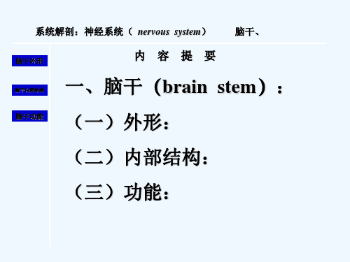
系统解剖:神经系统( nervous system) 脑干、
脑干外形
内容提要
脑干内部结构 一、脑干(brain stem):
脑干功能 (一)外形:
(二)内部结构:
(三)功能:
脑的分部
脑 端脑
的 区
间脑
分 小脑
中脑
脑干 脑桥
延髓
一、脑干的位置和外形
位于颅后窝,脊髓 和间脑之间,小脑腹侧。
系统解剖:脑干(brain stem)
脑干内部结构
脑干功能
内部结构
返回
系统解剖:脑干(brain stem)
脑干外形
脑干内部结构
脑干功能
内部结构
蜗神经核
返回
系统解剖:脑干(brain stem)
脑干外形
脑干内部结构
脑干功能
内部结构
下图
返回
系统解剖:脑干(brain stem)
脑干外形
脑干内部结构
脑干功能
内部结构
下图
返回
系统解剖:脑干(brain stem)
与脑相连的脑神经部位
脑干外形
脑干内部结构
脑干功能
端脑—Ⅰ 间脑—Ⅱ 中脑 —Ⅲ、Ⅳ 脑桥—Ⅴ、Ⅵ、Ⅶ、Ⅷ 延髓—Ⅸ、Ⅹ、Ⅺ、Ⅻ
神经解剖学英文课件-大脑半球英文课件

边缘叶
边缘系统
• Limbic lobe is a part of the
limbic system.
• Cingulate gyrus扣带回 Hippocampus gyrus海马回
Uncus
Hippocampus海马
• Mammillary bodies
• Amygdala杏仁核
• Olfactory bulb嗅球
The postcentral gyrus (+posterior portion of paracentral lobule) is somatosensory area, which receives impulses from contralateral somatic sensory organs.--thalamocortic tract
投影区面积与肌的灵活性成正变关系
*The motor area of one side generally supplies opposite muscles of the body.一般交叉性管理
*The motor area gives off pyramidal tract. The lower one third of the precentral gyrus gives off the corticonuclear tract. The corticospinal tract arises from the upper two thirds of precentral gyrus and anterior part of the paracentral lobule.
movements
(opposite)
2. Character:
神经解剖学英文课件-间脑和小脑解剖英文课件

External features of diencephalon
position
• It is located between the midbrain and cerebral hemisphere • It is almost entirely surrounded by cerebral hemispheres. • Only a small area (ventral portion of hypothalamus can be seen
Habenular
•Pineal body
•posterior commissure
epithalamus metathalamus
Epithalamus Metathalamus
Stria terminalis separate the caudate nucleus
stria medullaris lies in the junction between the superior and medial aspect of the thalamus & connect the pineal body
• metathalamus
lateral and medial geniculate nuclei
Internal structures of thalamus
• A Y-shaped internal medullary lamina divides it into three grey masses.
links the pituitory gland
hypothalamus
hypothalamus
Internal structures of thalamus, hypothalamus and metathalamus
脑干解剖学习PPT课件

②神经滑三车角神的经深核面,发出的 纤维在位锥于体中与脑橄下榄丘之水间平离, 大脑脑,水称管 舌腹 下侧 神。 经该 ,核 支发配出同 纤侧维的向舌后 内绕 、中 外脑 肌水 。管周围 灰质,于前髓帆内交叉,
VII 面神经核
IX
X XI 舌下神经核
在脑干背面离脑,构成滑
疑核
车神经,支配上斜肌- 。
副神经核
丝共同组成马尾。
-
10
脑N核六个功能柱模式图
躯体运动柱 特殊内脏运动柱 一般内脏运动柱 一般内脏感觉柱 界沟 特殊内脏感觉柱 一般躯体感觉柱 特殊躯体感觉柱
内耳
头面皮肤 牙粘膜
味觉 内脏 内脏
-
11
1)the general somatic motor nuclei:
①③ 展动神眼经神核经核
III
3)一般内脏运动核:支配头、颈、胸、腹部的平滑肌、 心肌和腺体。
4)一般内脏感觉核:接受脏器和心血管的感觉纤维。 5)特殊内脏感觉核:接受初级味觉纤维。 6)一般躯体感觉核:接受头面部皮肤,眼及口、鼻腔
粘膜的感觉纤维。
7)特殊躯体感觉核:接受内耳的听觉和平衡觉纤维。
-
7
脊椎动物神经管的形成:神经管有两个主要的轴线:背腹轴和前后(头尾)轴。 前后轴将神经系统分成前脑、中脑、后脑和脊髓,还将这些区域细分为更加特 殊的神经结构。在背腹轴上,不同的区域也有不同的神经细胞种类。在有些部 位,还有左右轴,即左右两侧分布不同的神经细胞。外周神经系统来源于与神 经板相邻的神经脊,后者是外胚层中一群特殊的细胞,从发源地迁移到胚胎多 个部位,形成包括外周神经系统在内的多种组织。即脊髓平面的神经系统及其 周围组织,背侧在上,腹侧在下。
展神经核 V
脑干内部结构脑神经非脑神经核团课堂PPT
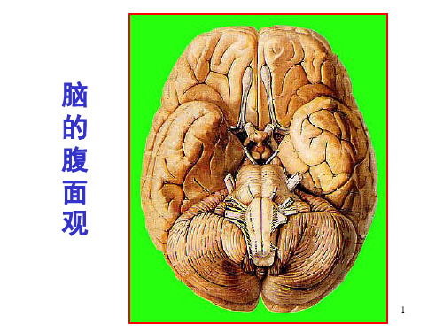
脑 的 腹 面 观
.
1
脑干 腹面观
中脑 脑桥
大脑脚 crus cerebri 脚间窝 基底沟 小脑中脚
延髓 延髓脑桥沟
锥体 pyramid
橄榄 olive
锥体交叉decussation of p.
.
2
脑干 背面观
中脑 脑桥
延髓
上丘 superior colliculus
下丘 IV
小脑上脚 菱形窝上半
1)下丘(中央核): 位于中脑下丘的深方, 接受蜗神经核的纤维,
发出纤维至内侧膝状体, 传导听觉冲动,完成听反射。
中央核central nucleus
2)上丘(核): 指中脑上丘深方呈灰白质交替成层排列的结构,
接受视束、下丘和脊髓的传入纤维, 发出纤维至脊髓和脑干,完成视听反射。
上丘灰质层
.
41
5)黑质substantia nigra:位于 4)红核red nucleus :
接受头面部皮肤粘膜和牙齿 的痛、温觉和触觉传入
.
三叉神经中脑核 mesencephalic nuc. of trigeminal n.
三叉神经脑桥核 pontine nuc. of trigeminal n.
三叉神经脊束核 spinal tract nuc. of trigeminal n.
32
7特殊躯体感觉柱:
大脑的脚底与被盖之间,由多巴
位于中脑的上丘水平被盖部,
胺能神经元组成。与纹状体有往 返联系。参与运动调节,损伤引 起震颤麻痹。
接受小脑和大脑皮质纤维, 发出纤维到脊髓、下橄榄核, 参与控制运动。
红核red nucleus
.
42
(三)白质
1、上行纤维束 1)内侧丘系medial lemniscus: 薄束核与楔束核发出的纤维在中央管腹侧交叉 称为内侧丘系交叉,折向上行为内侧丘系。
.
1
脑干 腹面观
中脑 脑桥
大脑脚 crus cerebri 脚间窝 基底沟 小脑中脚
延髓 延髓脑桥沟
锥体 pyramid
橄榄 olive
锥体交叉decussation of p.
.
2
脑干 背面观
中脑 脑桥
延髓
上丘 superior colliculus
下丘 IV
小脑上脚 菱形窝上半
1)下丘(中央核): 位于中脑下丘的深方, 接受蜗神经核的纤维,
发出纤维至内侧膝状体, 传导听觉冲动,完成听反射。
中央核central nucleus
2)上丘(核): 指中脑上丘深方呈灰白质交替成层排列的结构,
接受视束、下丘和脊髓的传入纤维, 发出纤维至脊髓和脑干,完成视听反射。
上丘灰质层
.
41
5)黑质substantia nigra:位于 4)红核red nucleus :
接受头面部皮肤粘膜和牙齿 的痛、温觉和触觉传入
.
三叉神经中脑核 mesencephalic nuc. of trigeminal n.
三叉神经脑桥核 pontine nuc. of trigeminal n.
三叉神经脊束核 spinal tract nuc. of trigeminal n.
32
7特殊躯体感觉柱:
大脑的脚底与被盖之间,由多巴
位于中脑的上丘水平被盖部,
胺能神经元组成。与纹状体有往 返联系。参与运动调节,损伤引 起震颤麻痹。
接受小脑和大脑皮质纤维, 发出纤维到脊髓、下橄榄核, 参与控制运动。
红核red nucleus
.
42
(三)白质
1、上行纤维束 1)内侧丘系medial lemniscus: 薄束核与楔束核发出的纤维在中央管腹侧交叉 称为内侧丘系交叉,折向上行为内侧丘系。
神经解剖学英文课件-迷走神经英文课件

the soft palate, pharynx, and larynx.
Parasympathetic fibers in the vagus nerve
Parasympathetic fibers originate from the dorsal motor nucleus of vagus. They are distributed widely throughout the respiratory,gastrointestin al system and cardiovascular. GVM
vagus nerve
vagus nerve
• CN X • Start:brainstem
(medulla oblongata) • Distribute :
in the neck, thorax, and abdomen • Mixed fiba →jugular foramen → carotid sheath → on the side of mediastinum → esophgeal plexus → esophgeal hiatus → anterior and posterior vagal trunks
motor fibers in the vagus nerve innervates to the muscles of larynx and pharynx through the superior
laryngeal nerves and the recurrent nerves SVM
function
• This nerve supplies motor and sensory parasympathetic fibres to pretty much everything from the neck down to the first third of the transverse colon.
- 1、下载文档前请自行甄别文档内容的完整性,平台不提供额外的编辑、内容补充、找答案等附加服务。
- 2、"仅部分预览"的文档,不可在线预览部分如存在完整性等问题,可反馈申请退款(可完整预览的文档不适用该条件!)。
- 3、如文档侵犯您的权益,请联系客服反馈,我们会尽快为您处理(人工客服工作时间:9:00-18:30)。
Cranial nerve nuclei and their CN
Cranial nerve nuclei and their CN
relay nuclei of ascending & descending tract ( NON cranial nuclei )
•Nucleus Gracilis & Nucleus Cuneatus •Inferior olivary nucleus •Pontine nucleus •Superior & inferior collicus nuclei •Red nucleus •Substantia nigra
Development of spinal cord & brain stem
Development of spinal cord & brain stem
Asecending tracts: Fasciculus gracilis & cuneatus Spinothalamic Descending tracts: Corticospinal tract Intersegmental tract ❖ Fasciculus proprius Central canal
three nulei column Anterior,posterior,and lateral or intermediolateral horn
Motor Column 1. GSE 2. SVE 3. GVE
Sensory Column 4. GVA 5. SVA 6. GSA 7. SSA
❖ contains important reflex centers associated with the control of respiration and the cardiovascular system and with the control of consciousness
❖ contains the important nuclei of cranial nerves III through XII
sensory nuclei of CN
General & special visceral sensory nuclei ----Solitary nucleus
sensory nuclei of CN
General somatic sensory nuclei ❖ Trigeminal nuclei : Chief sensory ❖ Mesencephlic ❖ Spinal
Internal structures of brain stem
Dept. of Anatomy Zhou Hong Ying
grey matter white matter reticular formation
Functions of brainቤተ መጻሕፍቲ ባይዱstem
❖ conduit for the ascending tracts and descending tracts connecting the spinal cord to the different parts of the higher centers in the forebrain
Sulucus limitans
Components of Cranial Nerves
Sulucus limitans
General Visceral Afferent Special Visceral Afferent
Internal structures in brain stem
➢ Central canal –expend to form Ⅳventricle ➢ 7 nulei column: Somatic sensory Somatic motor General visceral motor General visceral sensory Special visceral sensory Special somatic sensory Special visceral motor
Grey matter : Cranial nerve nuclei
somatic motor nuclei
❖ Oculomotor nuclei ❖ Trochlear nuclei ❖ Abducens nuclei ❖ Hypoglossal nuclei
Ⅲ, Ⅳ, Ⅵ, Ⅻ cranial nerve nuclei near the median plane.
Special somatic sensory nuclei
Vestibular nuclei from midpons to rostral medulla just lie beneath the vestibular area.
Cochlear nuclei between the pons and medulla just lie beneath the lateral recess of rhomboid fossa.
Special visceral motor nuclei
❖ Trigeminal motor nuclei ❖ Facial nuclei ❖ Accessory spinal nuclei ❖ Ambiguus nuclei
General visceral motor nuclei
❖ Accessory oculomotor nuclei ❖ Superior & inferior salivatory nuclei ❖ Dorsal motor vagus nuclei
Nuclei CN connected to cranial nerves
Grey matter
❖Nuclei CN connected to cranial nerves
❖ relay nuclei of ascending & descending tract ( NON cranial nuclei )- Nucleus of ascending and descending tract at different level of the brain stem