【医学英文课件】 peripheral nervous system
nervous system

Sensory projection system
• specific projection system • 1 The pathways from the specific sensory relay nucleus,including associated nucleus of thalamus to mainly in layer 4 of the cerebral cortex. • 2 projection region: specific area ( point to point ), axo-body synapse 3 the function role: difinite sensation and initiates impulses .
Thalamus
Thalamus
1. Specific sensory relay nucleus 2. Associated nucleus 3. Nonspecific projection nucleus
1.
Specofic sensory relay nuclei receive direct sensory projection, and then project to the specific area in cortex.
Pain, temperature (lateral); Crude touch, pressure (anterior) • 1st order neuron enters spinal cord through dorsal root • 2nd order neuron crosses over in spinal cord; ascends to thalamus • 3rd order neuron projects from thalamus to somatosensory cortex
神经系统(英文版)课件

CENTRAL NERVOUS SYSTEM 中枢神经系统 脑 brain 脊髓 spinal cord
PERIPHERAL NERVOUS SYSTEM 周围神经系统 脑神经 cranial nerves 脊神经 spinal nerves 内脏神经 autonomic nervous system
内脏神经 autonomic nervous system 分布于心肌、平滑肌和 腺体,不受主观意识控 制,又称自主神经或植 物神经。又分为交感神 经sympathetic nerve 和付交感神经
parasympathetic
nerve
fundamental tissue of the nervous system:
Nissl bodies尼氏体
神经元纤 维 neurofibril
Nissl body Neurofibrils
树突棘 dendrite spine contacted by different types of synaptic terminals
types of neurons(1): morphologic types of neurons: classed by the configuration of their processes as unipolar, bipolar,or multipolar
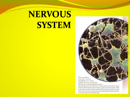
the nervous tissue is made up of two classes of cells, the neurons and neurologia.
神经元
1.构造structure of neuቤተ መጻሕፍቲ ባይዱons:
胞体cell body
轴突axon 树突dendrites (Fig1:
ervoussystem神经系统

For study purposes, the nervous system may be
divided into the central nervous system (CNS), consisting of the brain and spinal cord脊髓, and
3.Structure of a common neuron dendrites(树突): receive stimuli or message from other
cells, and transmit the message to the cell body
cell body(细胞体): contains nucleus, mitochondria 线粒体 and other organelles
axon(轴突):conduct message away from the cell body
Synapse(突触)即轴突的末端:the connection between a
nerve cell axon and target cells靶细胞, which may be
other nerve cells, muscle cells, or gland cells. At the
external changes and transmit these
signals to CNS motor nerve fibers运动神经纤维 : carry signals to
skeletal muscles to produce action
人体系统解剖学:周围神经系统-PPT课件

nervous system
第十五章 周围神经系统
Peripheral nervous system
-1-
【目的和要求】
1、掌握脊神经的构成、分区。 2、掌握颈丛的组成、位置及各主要皮支的浅出部
位和分布,掌握膈神经的行径及分布。 3、掌握臂丛的组成和位置,掌握胸长、胸背神经
行径及分布,掌握正中、尺、桡、肌皮、腋神 经的行程、主要分支和分布。 4、掌握胸神经前支在胸腹壁的节段性分布。 5、掌握腰丛、骶丛组成和位置。股神经、闭孔神 经的行经和分布。 6、掌握坐骨、胫、腓总、腓浅、腓深神经的行径 和分布。
-27-
-28-
4、 腰丛
组成:
T12一部分 ,L1—3,L4一部分
位置: 腰大肌深方 分支:
①髂腹下神经 ②髂腹股沟神经 ③生殖股神经 提睾肌 ④股外侧皮神经 ⑤股神经 ⑥闭孔神经
-29-
髂腹下神经 髂腹股沟神经
生殖股神经 股外侧皮神经
-30-
股神经 闭孔神经
股神经Femoral nerve
2)手掌:
侧半除外)
返支: 鱼际肌(拇收肌除外)、
1、2蚓状肌
皮支: 掌心、鱼际、桡侧三个半手指的掌 面及其中、远节手指的背面
-20-
(3)损伤表现
前臂不能旋前,屈 腕力减弱,拇指不 能屈曲,拇指不能 对掌,出现“猿 手”,拇、示、中 指远节皮肤感觉障 碍明显
-21-
7.尺神经Ulnar nerve
组成: 腰骶干(L4一部分,L5), S1—5, Co
位置: 骶骨与梨状肌的前面、髂内动脉的后方
-32-
• 分支 – 臀上神经 – 臀下神经 – 股后皮神经 – 阴部神经 – 坐骨神经
NERVOUS SYSTEM(神经组织) PPT

3. Axon – a long cell process of the neuron. It conducts action potentials away or towards the CNS.
Types of neurons:
1. Multipolar – several dendrites and one axon. Location is motor and CNS neuron.
4. The autonomic nervous system is divided into sympathetic, parasympathetic and enteric portions.
Neuron or Nerve Cell
Receive stimuli and transmit action potentials to other neurons or effector organs. Parts include;
Ref lexes:
It is an involuntary reaction in response to a stimulus applied to the periphery and transmitted to the CNS. Reflex arc is the neuronal pathway by which a reflex occur. The reflex arc is the basic functional unit of the nervous system because it is the smallest, simplest pathway capable of receiving a stimulus and yielding a response. Components of reflex arc: (1) Sensory receptor, (2) Sensory neuron, (3) Interneurons, (4) Motor neuron and (5) Effector organ.
医学英语,chapter11 Nervous System

Chapter 11Nervous SystemAnatomy and Physiology of the Nervous SystemThe nervous system is responsible for coordinating all the activity of the body. To do this it first receives information from both external and internal sensory receptors and then uses that information to adjust the activity of muscles and glands to match the needs of the body.The nervous system can be subdivided into the central nervous system (CNS) and the peripheral nervous system (PNS). The CNS consists of the brain and spinal cord. Sensory information comes into the CNS, where it is processed. Motor messages then exit the CNS carrying commands to muscles and glands. The PNS contains the cranial nerves and spinal nerves. Sensory nerves carry information to the CNS and motor nerves carry commands away from the CNS. All portions of the nervous system are composed of nervous tissue.Nervous TissueNervous tissue consists of two basic types of cells: neurons and neuroglial cells. Neurons are individual nerve cells. These are the cells that are capable of conducting electrical impulses in response to a stimulus. Neurons have three basic parts: dendrites, a nerve cell body, and an axon. Dendrites are highly branched projections that receive impulses. The nerve cell body contains the nucleus and many of the other organelles of the cell. A neuron has only a single axon, a projection from the nerve cell body that conducts the electrical impulse toward its destination. The point at which the axon of one neuron meets the dendrite of the next neuron is called a synapse. Electrical impulses cannot pass directly across the gap between two neurons, called the synaptic cleft. They instead require the help of a chemical messenger, called a neurotransmitter. Figure 11.1A variety of neuroglial cells are found in nervous tissue. Each has a different support function for the neurons. For example, some neuroglial cells produce myelin, a fatty substance that acts as insulation for many axons. They do not conduct electrical impulses.Central Nervous SystemBecause the central nervous system (CNS) is a combination of the brain and spinal cord, it is able to receive impulses from all over the body, process this information, and then respond with an action. This system consists of both gray and white matter. Gray mater is comprised of unsheathed or uncovered cell bodies and dendrites. White matter is myelinated nerve fibers. Bundles of nerve fibers interconnecting different parts of the CNS are called tracts. The CNS is encased and protected by three membranes known as the meninges.Figure 11. 1 NeuronThe BrainThe brain is one of the largest organs in the body and coordinates most body activities. It is the center for all thought, memory, judgment, and emotion. Each part of the brain is responsible for controlling different body functions, such as temperature regulation and breathing. There are four sections to the brain: cerebrum,cerebellum, diencephalon, and brain stem.Figure 11.2 The BrainThe largest section of the brain is the cerebrum. It is located in the upper portion of the brain and is the area that processes thoughts, judgment, memory, association skills, and the ability to discriminate between items. The outer layer of the cerebrum is the cerebral cortex, which is composed of folds of gray matter. The elevated portions of the cerebrum, or convolutions, are called gyri and are separated by fissures, or valleys, called sulci. The cerebrum is subdivided into left and right haves called cerebral hemispheres. Each hemisphere has four lobes. The lobes and their locations and functions are as follows:1.Frontal lobe: Most anterior portion of the cerebrum; controls motor function,personality, and speech.2.Parietal lobe: The most superior portion of the cerebrum; receives and interpretsnerve impulses from sensory receptors and interprets language.3.Occipital lobe: The most posterior portion of the cerebrum; controls vision.4.Temporal lobe: The left and right lateral portion of the cerebrum; controls hearingand smell.Figure 11.3 Lobes of the CerebrumThe diencephalon, located below the cerebrum, contains two of the most critical areas of the brain, the thalamus and the hypothalamus. The thalamus is composed of gray matter and acts as a center for relaying impulses from the eyes, ears, and skin to the cerebrum. Our pain perception is controlled by the thalamus. The hypothalamus, lying just below the thalamus, controls body temperature, appetite, sleep, sexual desire, and emotions such as fear. The hypothalamus is actually responsible for controlling the autonomic nervous system, cardiovascular system, the gastrointestinal system, and the release of hormones from the pituitary gland.The cerebellum, the second largest portion of the brain, is located beneath the posterior part of the cerebrum. This part of the brain aids in coordinating voluntary body movements and maintaining balance and equilibrium. The cerebellum refines the muscular movement that is initiated in the cerebrum.The final portion of the brain is the brain stem. This area has three components: midbrain, pons, and medulla oblongata. The midbrain acts as a pathway for impulses to be conducted between the brain and the spinal cord. The pons—a term that means bridge—connects the cerebellum to the rest of the brain. The medulla oblongata is the most inferior positioned portion of the brain; it connects the brain to the spinal cord. However, this vital area contains the centers that control respiration, heart rate, temperature, and blood pressure. Additionally this is the site where nerve tracts cross from one side of the brain to control functions and movement on the other side of the body.Figure 11.4 Brain StemThe brain has four interconnected cavities called ventricles: one in each cerebral hemisphere, one in the thalamus, and one in front of the cerebellum. These contain cerebrospinal fluid, which is the watery, clear fluid that provides protection from shock or sudden motion to the brain.Spinal CordThe function of the spinal cord is to provide a pathway for impulses traveling to and from the brain. The spinal cord is actually a column of nervous tissue that extends from the medulla oblongata of the brain down to the level of the second lumbar vertebra within the vertebral column. The vertebral column consists of the 33 vertebrae of the backbone. They line up to form a continuous canal for the spinal cord called the spinal cavity or vertebral canal.Similar to the brain, the spinal cord is also protected by cerebrospinal fluid. The inner core of the spinal cord contains gray matter. This inner core consists of cell bodies and dendrites of peripheral nerves. The outer portion of the spinal cord is myelinated white matter. The white matter is either ascending tracts carrying sensory information up to the brain or descending tracts carrying motor commands down fromthe brain to a peripheral nerve.MeningesThe meninges are three layers of connective tissue membranes that surround the brain and spinal cord. Moving from external to internal, the meninges are1.Dura mater: The name means tough mother; it forms a tough, fibrous sac aroundthe CNS.2.Subdural space: The actual space between the dura mater and arachnoid layers.3.Arachnoid layer: The name means spider-like, it is a thin, delicate layer attached tothe pia mater by web-like filaments.4.Subarachnoid space: The space between the arachnoid layer and the pia mater; itcontains cerebrospinal fluid.5.Pia mater: The name means soft mother; it is the innermost membrane layer and isapplied directly to the surface of the brain.Figure 11.5 MeningesPeripheral Nervous SystemThe peripheral nervous system (PNS) includes both the 12 pairs of cranial nerves and the 31 pairs of spinal nerves. A nerve is a group or bundle of axon fibers located outside the central nervous system that carries messages between the CNS and the various parts of the body. Whether a nerve is cranial or spinal is determined by where the nerve originates. Cranial nerves arise from the brain, mainly at the medulla oblongata. Spinal nerves split off from the spinal cord, and one pair ( a left and a right ) exits between each pair of vertebrae. The point where either type of nerve is attached to the CNS is called the nerve root. The names of most nerves reflect either the organ the nerve serves or the portion of the body the nerve is traveling through. The entire list of cranial nerves is found in Table 11.1. Figure 11.7 illustrates some of the major spinal nerves in the human body.Table11.1 Cranial NervesNumber Name Function1 Olfactory Transports impulses for sense of smell2 Optic Carries impulses for sense of sight3 Oculomotor Motor impulses for eye muscle movement and thepupil of eye4 Trochlear Controls oblique muscle of eye on each side5 Trigeminal Carries sensory facial impulses and controlsmuscles for chewing; branches into eyes,forehead,upper and lower jaw6 Abducens Controls an eyeball muscle to turn eye to side7 Facial Controls facial muscles for expression, salivation,and taste on two-third of tongue8 Vestibulocochlear Responsible for impulses of equilibrium andhearing; also called auditory nerve9 Glossopharyngeal Carries sensory impulses from pharynx(swallowing) and taste on one-third of tongue10 Vagus Supplies most organs in abdominal and thoraciccavities11 Accessory Controls the neck and shoulder muscles12 Hypoglossal Controls tongue musclesFigure 11. 6 Cranial NervesFigure 11.7 Major spinal nervesAlthough most nerves carry information to and from the CNS, individual neurons carry information in only one direction. Afferent neurons, also called sensory neurons, carry sensory information from a sensory receptor to the CNS. Efferent neurons, also called motor neurons, carry activity instructions from the CNS to muscles or glands out in the body. The nerve cell bodies of the neurons forming the nerve are grouped together in a knot-like mass, called a ganglion, located outside the CNS.The nerves of the PNS are subdivided into two divisions, the autonomic nervous system and somatic nerves, each serving a different area of the body.Autonomic Nervous SystemThe autonomic nervous system (ANS) is involved with the control of involuntary or unconscious bodily functions. It may increase or decrease the activity of the smooth muscle found in viscera and blood vessels, the cardiac muscle of the heart, and glands. The ANS is divided into two branches: sympathetic branch and parasympathetic branch. The sympathetic nerves stimulate the body in times of stress and crisis. These nerves increase heart rate, dilate airways, increase blood pressure, inhibit digestion and stimulate the production of adrenaline during a crisis. The parasympathetic nerves serve as a counterbalance for the sympathetic nerves. Therefore, they cause heart rate to slow down, lower blood pressure, and stimulate digestion.Somatic NervesSomatic nerves serve the skin and skeletal muscles and are mainly involved with the conscious and voluntary activities of the body. The large variety of sensory receptors found in the dermis layer of the skin use somatic nerves to send their information, such as touch, temperature, pressure, and pain, to the brain. These are also the nerves that carry motor commands to skeletal muscles.Combining Forms Commonly Used in Nervous SystemCombining Form Meaning Examplecephal/o head cephalalgiacerebell/o cerebellum cerebellar cerebellitiis cerebr/o cerebrum cerebral cerebrospinal encephal/o brain encephalomalaciagli/o glue glioneuromamedull/o medulla medulloadrenalmening/o meninges meningitismyelomeningocele meningi/o meninges meningiomamyel/o spinal cord poliomyelitisnarc/o stupor narcohypnosisneur/o nerve neurectomy neuromaphas/o speech aphasiapoli/o gray matter polioencephalopathypont/o pons pontocerebellarradicul/o nerve root radiculoneuritisthalam/o thalamus thalamocorticalventricul/o ventricle ventriculographySuffixes Commonly Used in Nervous SystemSuffix Meaning Example-algesia sensitivity to pain analgesia-esthesia feeling, sensation anesthesia-kinesia movement bradykinesia-lepsy seizure narcolepsy-paresis weakness hemiparesis-phasia speech dysphasia-plegia paralysis paraplegia-sthenia strength myasthenia-taxia muscle coordination ataxiaDiagnostic Procedures Relating to This SystemBabinski’s reflex Reflex test developed by Joseph Babinski , a Frenchneurologist, to determine lesions andabnormalities in the nervous system. The Babinskireflex is present if the great toe extends instead offlexes when the lateral sole of the foot is stroked.The normal response to this stimulation is flexion ofthe toe.Brain scan Injection of radioactive isotopes into the circulationto determine the function and abnormality of thebrain.Cerebral angiography X-ray of the blood vessels of the brain after theinjection of a radiopaque dye.Cerebralspinal fluid analysis Laboratory examination of the clear, watery, colorlessfluid from within the brain and spinal cord.Infections and the abnormal presence of blood canbe detected in this test.Echoencephalography Recording of the ultrasonic echoes of the brain. Usefulin determining abnormal patterns of shifting in thebrain.Electroencephalography Recording the electrical activity of the brain byplacing electrodes at various positions on the scalp.Also used in sleep studies to determine if there is anormal pattern of activity during sleep. Electromyography(EMG)Recording of the contraction of muscles as a result ofreceiving electrical stimulation.Lumbar puncture (LP)Puncture with a needle into the lumbar area (usuallythe fourth intervertebral space) to withdraw fluid forexamination and for the injection of anesthesia. Alsocalled spinal puncture or spinal tapMyelography Injection of a radiopaque dye into the spinal canal. AnX-ray is then taken to examine the normal andabnormal outlines made by the dye. Pneumoencephalography X-ray examination of the brain following withdrawalof cerebrospinal fluid and injection of air or gas viaspinal puncture.Positron emission tomography Use of positive radionuclides to reconstruct brainsections. Measurement can be taken of oxygen andglucose uptake, cerebral blood flow, and bloodvolume. The amount of glucose the brain usesindicates how metabolically active the tissue is. Romberg’s test Text developed by Moritz Romberg, a Germanphysician, that is used to establish neurologicalfunction; the person is asked to close his or her eyesand place the feet together. This test for body balanceis positive if the patient sways when the eyes areclosed.Pathology Relating to the Nervous SystemConcussionDefinitionConcussions range in severity from mild to severe, but they all share one common factor — they temporarily interfere with the way your brain works. They can affect memory, judgment, reflexes, speech, balance and coordination.Usually caused by a blow to the head, concussions don't always involve a loss of consciousness. In fact, most people who have concussions never black out. Many people have had concussions and not realized it.Concussions are common, particularly if you play a contact sport like football. But every concussion, no matter how mild, injures your brain. This injury needs time and rest to heal properly. Luckily, most concussions are mild and people usually recover fully.SymptomsThe signs and symptoms of a concussion can be subtle and may not appear immediately. Symptoms can last for days, weeks or longer.The two most common concussion symptoms are confusion and amnesia. The amnesia, which may or may not be preceded by a loss of consciousness, almost always involves the loss of memory of the impact that caused the concussion.Other immediate signs and symptoms of a concussion may include:∙Headache∙Dizziness∙Ringing in the ears∙Nausea or vomiting∙Slurred speechSome symptoms of concussions don't appear until hours or days later. They include:∙Mood and cognitive disturbances∙Sensitivity to light and noise∙Sleep disturbancesHead trauma is very common in young children. But concussions can be difficult to recognize in infants and toddlers because they can't readily communicate how they feel. Nonverbal clues of a concussion may include:∙Listlessness, tiring easily∙Irritability, crankiness∙Change in eating or sleeping patterns∙Lack of interest in favorite toys∙Loss of balance, unsteady walkingCausesYour brain has the consistency of gelatin. It's cushioned from everyday jolts and bumps by the cerebrospinal fluid that it floats in, inside your skull. A violent blow to your head can cause your brain to slide forcefully against the inner wall of your skull. Even the sudden stop of a car crash can bounce your brain off the inside of your skull. This can result in bleeding in or around your brain and the tearing of nerve fibers.Tests and diagnosisDiagnosing a concussion is usually straightforward. If a blow to your head has knocked you out or left you dazed, you've had a concussion. It's more difficult, however, to determine whether the blow has caused potentially serious bleeding or swelling in your skull. Signs and symptoms of these injuries may not appear until hours or days after the injury.Your doctor may start your evaluation with questions about the accident, then proceed to a neurological exam. This exam includes checking your memory and concentration, vision, hearing, balance, coordination and reflexes.The standard test to assess post-concussion damage is a computerized tomography (CT) scan. A CT scanner takes multiple cross-sectional X-rays and combines all the resulting images to produce detailed, two-dimensional images of your skull and brain. During the procedure, you lie still on a table that slides through a large, doughnut-shaped X-ray machine. The scan is painless and generally takes less than 10 minutes.Not every concussion requires a CT scan, but the test is usually done as a precaution if there's a chance your injury is more severe than your immediate condition suggests. You're more likely to need a scan if you:∙Are younger than 16 or older than 64∙Fell from a height of more than three feet∙Had a motor vehicle accident∙Are under the influence of alcohol or drugs∙Are unable to recall the accident for more than 30 minutes after it occurred∙Have persistent trouble with short-term memory — that is, retaining new information — after you've completely regained consciousness∙Vomited∙Had a seizure∙Suffered bruises, scrapes or cuts on your head and neck∙Fractured your skullYou may need to be hospitalized overnight for observation after a concussion. If your doctor says it's OK for you to be observed at home, someone should check on you periodically for at least 24 hours. You may need to be awakened every two hours to make sure you can be roused to normal consciousness.How you feel painJabbing, throbbing, burning or stinging — any way you describe pain, you want it to stop. Finding out how your brain processes pain signals can help.Your experience of pain is part biology, but it's also influenced by psychological and cultural factors. Despite years of research, questions linger about exactly what happens between the moment you stub your toe and the moment you say "ouch" — or some other choice word.How pain messages travelPain results from a series of exchanges among three major components of your nervous system:∙Your peripheral nerves. These nerves extend from your spinal cord to your skin, muscles and internal organs. Some peripheral nerve fibers end with receptors that respond to touch, pressure, vibration, cold and warmth. Other types of nerve fibers end with nociceptors (no-sih-SEP-turs) — which are receptors that detect actual or potential tissue damage.Nociceptors are most concentrated in areas prone to injury, such as your fingers and toes. When nociceptors detect a harmful stimulus — such as the hard surface that stubbed your toe — they relay pain messages in the form of electricalimpulses along a peripheral nerve to your spinal cord and brain. Sensations ofsevere pain are transmitted almost instantaneously.∙Your spinal cord. The nerve fibers that transmit pain messages — such as the throbbing pain from that stubbed toe — enter the spinal cord in an area called the dorsal horn. There, they release chemicals (neurotransmitters) that activate othernerve cells in the spinal cord, which process the information and then transmit it up to the brain.∙Your brain. When news of your stubbed toe travels up the spinal cord, it arrives at the thalamus — a sorting and switching station deep inside your brain.The thalamus forwards the message simultaneously to three specialized regions of the brain: the physical sensation region (somatosensory cortex), the emotionalfeeling region (limbic system) and the thinking region (frontal cortex). Your brain responds to pain by sending messages that moderate the pain in the spinal cord.The location of your pain can affect how you perceive it. A headache that interferes with work or concentration may be more bothersome — and therefore receive a stronger response — than arthritic pain in your knee or a cut to your finger.How you react to pain messagesCurrent understanding of pain is based on gate-control theory, which grew out of observations of World War II veterans and their reactions to different types of injuries. The central concepts of gate-control theory are:∙Pain messages don't travel directly from your pain receptors to your brain.When pain messages reach your spinal cord, they meet up with specialized nerve cells that act as gatekeepers, which filter the pain messages on their way to your brain. For severe pain that's linked to bodily harm, such as when you touch a hot stove, the "gate" is wide open, and the messages take an express route to yourbrain. Weak pain messages, however, may be filtered or blocked out by the gate.Nerve fibers that transmit touch also affect gatekeeper cells. This explains whyrubbing a sore area — such as the site of a stubbed toe — makes it feel better. The signals of touch from the rubbing actually decrease the transmission of painsignals.∙Messages can change within your peripheral nerves and spinal cord. Nerve cells in your spinal cord may release chemicals that intensify the pain, increasing the strength of the pain signal that reaches your brain. This is called wind-up orsensitization. At the same time, inflammation at the site of injury may add to your pain.∙Messages from your brain also affect the gate. Rather than just reacting to pain, your brain actually sends messages that influence your perception of pain.Your brain may signal nerve cells to release natural painkillers, such asendorphins (en-DOR-fins) or enkephalins (en-KEF-uh-lins), which diminish the pain messages.This last idea explains how your brain — and its psychological and emotional processes — can affect your experience of pain. In fact, how you interpret pain messages and tolerate pain can be affected by your:∙Emotional and psychological state∙Memories of past pain experiences∙Upbringing∙Attitude∙Expectations∙Beliefs and values∙Age∙Sex∙Social and cultural influencesFor example, a minor sensation that would barely register as pain, such as a dentist's probe, can actually produce exaggerated pain for a child who's never been to the dentist and who's heard horror stories about what it's like.But your emotional state can also work in your favor. Athletes can condition themselves to endure pain that would incapacitate others. And, if you were raised in a home or culture that taught you to "Grin and bear it" or to "Bite the bullet," you may experience less discomfort than do people who focus on their pain or who are more prone to complain.Vocabulariesabducens 【✌♌♎◆♦☜⏹】外展神经afferent 【 ✌♐☜❒☜⏹♦】 传入的arachnoid 【☜❒✌⏹♓♎】 蛛网状的axon 【 ✌♦⏹】 轴突cerebellum 【 ♦♏❒♓♌♏●☜❍】小脑cerebrum 【 ♦♏❒♓♌❒☜❍】 大脑cleft 【 ●♏♐♦】 裂缝concussion 【 ☜⏹✈☞☎☜✆⏹】脑震荡convolution 【 ⏹☜●◆☞☎☜✆⏹】脑回cortex 【 ♦♏♦】 皮质cumulative 【 ◆❍◆●☜♦♓】积累的dendrites 【 ♎♏⏹♎❒♋♓♦】 树突diencephalon 【 ♎♋♓♏⏹♦♏♐☜●⏹】间脑dura mater 【♎☺☜❒☜ ❍♏✋♦☜☎❒✆】硬脑膜efferent 【 ♏♐☜❒☜⏹♦】 传出的epilepsy 【 ♏☐♓●♏☐♦♓】 癫痫症equilibrium 【 ♓♦♓●♓♌❒♓☜❍】均衡filament 【 ♐♓●☜❍☜⏹♦】 细丝ganglion 【 ♈✌☠●✋☜⏹】 神经节gelatin 【 ♎✞♏●☜♦♓⏹】 凝胶gyri 【♊♎✞♋✋❒♋✋】 脑回(复数)jolts 【✞☜◆●♦】 摇晃meninges 【❍♓⏹♓⏹♎✞♓】脑膜myelin 【 ❍♋♓☜●♓☎✆⏹】髓鞘质myelinated 【♊❍♋✋☜●✋⏹♏✋♦✋♎】有髓鞘的neuroglial 【⏹◆❒♈●♓☜●】神经胶质的neurotransmitter 【 ⏹◆☜❒☜♦❒✌⏹♦❍♓♦☜】神经递质nociceptors 【 ⏹☜◆♦♓♦♏☐♦☜】伤害感受器oblongata 【 ♌●☠♈♦☜】延髓oculomotor 【 ◆●☜◆❍☜◆♦☜】眼球运动的olfactory 【 ●♐✌♦☜❒♓】 嗅觉的peripheral 【☐☜❒♓♐☜❒☜●】 周围的pia 【 ☐♋♓☜】 软脑膜pons 【☐⏹】 脑桥sulcus 【 ♦✈●☜♦】 沟(复数为sulci) synapse 【 ♦♋✋⏹✌☐♦】 突触thalamus 【 ✌●☜❍☜♦】 丘脑trigeminal 【♦❒♋♓♎✞♏❍♓⏹●】三叉神经的trochlear 【 ♦❒●♓☜】 滑车的unsheathe 【 ✈⏹☞♓❆】 拔出鞘vagus 【 ♏♓♈☜♦】 迷走神经vestibulocochlear 前庭耳蜗的ExercisesA. Complete the following statements1. The study of the nervous system is called _________.2. The organs of the nervous system are the _______,_______, and ______.3. The two divisions of the nervous system are the ________ and _______.4. The neurons that carry impulses away from the brain and spinal cord are called _________ neurons.5. The neurons that carry impulses to the brain and spinal cord are called _____neurons.6. The largest portion of the brain is the __________.7. The second largest portion of the brain is the _______.8. The occipital lobe controls _________.9. The temporal lobe controls _______ and ________.10. The two divisions of the autonomic nervous system are the ______ and ______.B. State the described terms using the combining forms provided.The combining from neur/o refers to the nerve. Use it to write a term that means1. inflammation of the nerve ________2. specialist in nerves __________3. pain in the nerve _________4. inflammation of many nerves _____________5. excision of a nerve ______________6. surgical repair of a nerve _____________7. incision into a nerve __________8. suture of a nerve ______________The combining form mening/o refers to the meninges or membranes. Use it to write a term that means9. inflammation of the meninges _________10. protrusion of the meninges ______11. Protrusion of the spinal cord and the meninges ________The combining form encephal/o refers to the brain. Use it to write a term that means12. X-ray examination of the brain ___________13. disease of the brain _______________14. inflammation of the brain _______15. protrusion of the brain ________16. inflammation of brain and spinal cord __________The combining form cerebr/o refers to the cerebrum. Use it to write a term that means17. pertaining to the cerebrum and spinal cord __________18. hardening of the cerebrum ____________19. any disease of the cerebrum ___________20. inflammation of the cerebrum and meninges ______21. pertaining to the cerebrum ___________C. Match the cranial nerves in column A with the functions they control in column B.A B1. _____olfactory a. carries facial sensory impulses2. _____optic b. turn eye to side3. _____oculomotor c. controls tongue muscles4. _____trochlear d. eye muscles and controls pupils5. _____trigeminal e. swallowing6. _____abducens f. controls facial muscles7. _____facial g. eye muscle movement。
周围神经疾病diseaseofperipheralnervoussystem

•
0.6~0.8g/D; 其次 苯妥英钠;再则 氯硝基安定
• 2 氯苯氨丁酸 可试用,30~40mg/D
• 3 大剂量维生素 报道有效,机理不清
• 4 哌咪清
• 物理疗法 无水酒精或甘油封闭神经分支半月神经节
•
经皮半月神经节射频电凝疗法
• 手术治疗 三叉神经感觉根部分切断术
医学ppt
11
•
三叉神经微血管减压术
• 2 三叉神经的一支或两支分布区发作性剧痛,
• 第二、三支最常见,多为单侧,存在扳机点
• 3 严重者可伴有面肌反射性抽搐
• 4 周期性发作,频度、持续时间不等
• 5 神经系统检查一般无阳性体征
医学ppt
9
三鉴别诊断
• 继发性三叉神经痛 常合并其他颅神经麻痹
医学ppt
6
三叉神经痛 trigeminal neuralgia
一种原因不明的三
叉神经分布区短暂而反
复发作的剧痛。
医学ppt
7
一 病因
• 确切病因和发病机制目前尚不清楚 • 可能为三叉神经脱髓鞘而产生异位
冲动或伪突触传递所致 • 也可能因为颅后窝小的异常血管团
压迫三叉神经根或延髓外侧面所致
医学ppt
• 射性疼痛
• 2 行走,活动或牵拉坐骨神经可诱发或加剧疼痛
• 3 沿坐骨神经径路上可有压痛点
• 体征:Lasegue 征(+)
•
患侧臀肌松弛,小腿萎缩;
•
小腿及足背外侧感觉减退;
•
踝反射减退或消失;
•
压颈静脉实验医可学p激pt 发或加剧下肢疼痛19
三 治疗
• 病因治疗
• 腰椎间盘突出引起的坐骨神经痛
The Peripheral Nervous System (PNS)外周神经系统

Motor
• General somatic motor • Signals contraction of skeletal muscles • Under our voluntary control • Visceral motor • Makes up autonomic nervous system (ANS) • Regulates the contraction of smooth and cardiac muscle, controls function of visceral organs • ANS has two divisions • Parasympathetic • Sympathetic
ht”
• Catabolic (expend energy) • Mass activation prepares for
intense activity. • Heart rate (HR) increases. • Bronchioles dilate. • Blood [glucose] increases. • Adrenaline
Summary
• Decreases HR. • Dilates visceral blood ves
•
sels. Increases digestive activit y.
Company name
7
Dual innervation of many organs — having a brake and an accelerator provides more control
Somatic Nerves
1 2
9
Sensory inputs
MotorSomatic Nerves
【系解精品】9.The Visceral Nervous System

Peripheral part:周围部
1. sympathetic trunks 交感干 paravertebral ganglia节后纤维 Interganglionic branches节间支 2. sympathetic ganglia 交感神经节 paravertebral ganglia 椎旁节 prevertebral ganglia 椎前节 3. sympathetic plexuses 交感丛 4. sympathetic nerves 交感神经 5. communicating branches 交通支 gray ramus 灰交通支 white ramus 白交通支
Fibers
Thick myelinated Preganglionic: thin myelinated Postganglionic: unmyelinated
Distributive form Nerve trunk
Nerve plexuses
Control
Voluntary (consciousness)
From lower center Single neuron to effect require
Kind of fibers
One
Two neurons: preganglionic neuron (fiber) and postganglionic neuron (fiber)
Two: sympathetic and parasympathetic
Conserve and restore body energy (rest and relaxation)
- 1、下载文档前请自行甄别文档内容的完整性,平台不提供额外的编辑、内容补充、找答案等附加服务。
- 2、"仅部分预览"的文档,不可在线预览部分如存在完整性等问题,可反馈申请退款(可完整预览的文档不适用该条件!)。
- 3、如文档侵犯您的权益,请联系客服反馈,我们会尽快为您处理(人工客服工作时间:9:00-18:30)。
Figure 48.5 Schwann cells
Figure 48.1 Overview of a vertebrate nervous system
Figure 48.3 The knee-jerk reflex
Figure 48.6 Measuring membrane potentials
Figure 48.16x Spinal cord
Figure 48.18 The main roles of the parasympathetic and sympathetic nerves in regulating internal body functions
Figure 48.20x1 Cerebral cortex, gray and white matter
Figure 48.19 Embryonic development of the brain
Figure 48.20 The main parts of the human brain
Figure 48.20x2 Cerebral cortex
Figure 48.24 Structure and functional areas of the cerebrum
Figure 48.29 Neural progenitor cell
Figure 48.0x1 Aplysia neuron
Figure 48.0x2 Frog neuron
Figure 48.2x Neurons
Figure 48.0 A neuron on a microprocessor
Figure 48.14 Summation of postsynaptic potentials
Table 48.1 The Major Known Neurotransmitters
Figure 48.15 Diversity in nervous systems
Figure 48.16 Thte
Figure 48.21 The reticular formation
Fugure 48.22a Electroencephalogram (EEG) electrodes
Figure 48.22b-d Brain waves recorded by an electroencephalogram (EEG)
Figure 48.23 Activity rhythms in a nocturnal mammal, the northern flying squirrel
Figure 48.28 How do developing axons know which way to go?
Figure 48.28x1 Brain MRI
Figure 48.26 Mapping language areas of the cerebral cortex
Figure 48.25 Primary motor and somatosensory areas of the human cerebral cortex
Figure 48.27 The limbic system
Figure 48.9 The role of voltage-gated ion channels in the action potential (Layer 4)
Figure 48.9 The role of voltage-gated ion channels in the action potential (Layer 5)
Figure 48.10 Propagation of the action potential
Figure 48.11 Saltatory conduction
Figure 48.12 A chemical synapse
Figure 48.13 Integration of multiple synaptic inputs
Figure 48.7 The basis of the membrane potential
Figure 48.8 Graded potentials and the action potential in a neuron
Figure 48.9 The role of voltage-gated ion channels in the action potential (Layer 1)
Figure 48.9 The role of voltage-gated ion channels in the action potential (Layer 2)
Figure 48.9 The role of voltage-gated ion channels in the action potential (Layer 3)
