新城疫病毒M蛋白R42A突变影响M蛋白细胞核定位并降低病毒复制与致病性
新城疫病毒(NDV)基质(M)蛋白在体身对炎症因子诱导的作用
I 一2 TN — L 1 、 F a等促炎 症 细胞 因子 , 些 非 免疫 细 某 胞, 如血 管 内皮 细胞 , 能 产生 这些 细 胞 因子 , 是 也 只 程度不 同 。对各 种病毒 感染 的最初应 答包括 诱导 固
有 性 干 扰 素 (F a l I N 了 。 在 适 当 刺 激 下 , I N—/ 、F 一 ) 3 任
维普资讯
1 4
中 国兽 医 杂 志 2 0 0 8年 ( 4 第 4卷 ) d期 第
新 城 疫 病 毒 ( DV) 质 ( ) 白在 体 外 N 基 M 蛋 对 炎 症 因 子 诱 导 的 作 用
王 蕾 ,陈福 勇 ,郑世 军 ,索 勋
( 中圉 农 业 大学 动物 医学 院 ,北 京 海淀 1 0 9 0 1 3)
TNF d S g ma公 司 ) — ( ia 。
病毒 易于诱 导不 同类 型 的固有和获 得性免 疫应 答 , 因 是 在 感 染 的 宿 主 细 胞 的 胞 浆 内 复 制 并 产 生 原
蛋 白性 质 的 产 物 。 固 有 免 疫 细 胞 能 产 生 I 一 、I 6 I 1 I 、 一
中图 分 类 号 : 8 8 3 j 3 ¥ 5 . 1 .
文 献标 识 码 : B
文 章 编 号 :5 96 0 ( 0 8 0 —0 4 0 0 2 —0 5 2 0 ) 40 内最 严 重 的家 禽 传 染 病 。为 了更好 的预 防新 城疫 , 要 对病 毒 的致 病 性 需 以及 对 宿 主 的 免 疫 反 应 等 分 子 机 制 进 行 更 好 的
的病毒 的独特 的分子途 径激 活或者抑 制受感 染细胞
的特 殊 细 胞 因 子 的产 生 一 。
NDV 的 M ( ti r ti ) 白全 长 12 1 p, ma rx p o en 蛋 4b
新城疫病毒HN蛋白的表达及免疫原性检测的开题报告
新城疫病毒HN蛋白的表达及免疫原性检测的开题报告
一、课题研究背景及意义
新城疫病毒是一种动物源性RNA病毒,是家禽和野生鸟类感染的病原体,也可通过与家禽接触等途径传播给人类。
该病毒感染人类会引起严重的肺炎和急性呼吸系统症状,目前没有特效治疗方法。
因此,针对新城疫病毒的研究非常重要。
新城疫病毒HN蛋白是该病毒的重要组成部分,具有免疫原性。
因此,对HN蛋白的表达及免疫原性检测研究具有重要意义,可为新城疫病毒的诊断和防治提供基础研究支持。
二、研究内容及方法
本研究旨在构建新城疫病毒HN蛋白的表达载体,通过细胞转染获得表达的重组蛋白,并进行免疫原性检测,以评估该蛋白的免疫原性。
具体实验步骤如下:
1.构建新城疫病毒HN蛋白的表达载体。
首先,利用PCR扩增HN基因序列并克隆到相关表达载体中,然后通过序列分析确保正确性。
2.细胞转染获得表达的重组蛋白。
将构建好的表达载体转染到目标细胞中,然后进行诱导表达,通过Western blot 等方法检测表达的重组蛋白。
3.进行免疫原性检测。
利用Western blot、ELISA和免疫荧光染色等常用方法,检测表达的重组蛋白的免疫原性。
三、预期研究结果及意义
预期本研究将成功构建新城疫病毒HN蛋白的表达载体,并成功获得表达的重组蛋白,同时确定其在表达过程中的免疫原性。
这将为新城疫病毒的诊断和防治提供基础研究支持,为该病的防治工作提供重要的科学依据。
刘秀梵我国新城疫的防控实践和基因VII型新城疫疫苗的研发116-2
② 与早期病毒F48株(基因IX型)和Hert33株(基因IV型)相比, 在体内的复制能力强,呼吸道和肠道的载毒量高,排毒时间长。
LaSota免疫鸡群感染VIId亚型NDV与F48E8后的排毒测定(阳性数/样品数)
组别
HI效价 (Log2)
攻毒株
3日
喉头 泄殖腔
5日
喉头 泄殖腔
7日
喉头 泄殖腔
6
F48E8 5/10* 0/10
V4 (I型)
Y YD
E
GV S T I G
LaSota (II型)
Y YD
E
GV S T I G
分离株(VIId型) H
H
N
E/G/K/ D
D
E
N
I
V
R
流行株HN蛋白上的5个抗原区(193-201位、345353位、513-521位、494位及569位氨基酸区域)及邻 近的氨基酸与疫苗株存在明显的不同。
Immune efficacy of NDV/A-VII in chickens
Frequency of isolation of challenge virus in commercial chickens
HI效价(log2)
10
8
A-VII-k
LaSota-k
6
4
2
0 7天
14天
21天
免疫后不同时间抗体HI效价上升曲线 ﹡均以自身免疫毒株作为4单位病毒
效 检 试 验 结 果 显 示 , 致 弱 株 A-VII 免 疫 原 性 强 , 其 灭 活 苗 以 较 低 剂 量 (20ul)免疫SPF鸡时即可产生高滴度的HI抗体,且其诱导抗体产生的速 度较LaSota快。
利用M基因鉴别新城疫病毒中强毒株与弱毒株RT-nPCR方法的建立
利用M基因鉴别新城疫病毒中强毒株与弱毒株RT-nPCR方法的建立朱小甫;吴旭锦【摘要】根据GenBank上公开的新城疫病毒(Newcastle disease virus,NDV)基因序列,针对M基因差异位点设计了3对引物,通过优化反应体系与条件,建立了能够区分NDV中强毒株与弱毒株的RT-nPCR检测方法.特异性试验显示,NDV中强毒株扩增出543、375 bp两个条带,弱毒株仅出现375 bp一个条带,其他常见禽病毒为阴性.灵敏度试验结果表明,该方法检测cDNA含量极限为6.15×10⁻⁴ ng/μL.临床样品检测发现鸡群NDV弱毒带毒率高,中强毒株带毒率较低.结果表明,本研究成功建立了一种基于M基因的快速鉴别诊断NDV中强毒株与弱毒株的RT-nPCR 方法.%According to the Newcastle disease virus(NDV) gene sequence published in GenBank, three pairs of primers were designed for M gene difference sites of NDV, we changed reaction system and conditions until PCR reaction was optimal. The results of sensitivity test showed that the limit of cDNA content was 6.15×10⁻⁴ ng/μL. Specificity tests showed that 543 bp and 375 bp bands were found in the velogenic NDV strains, and only 375 bp bands were found in the lentogenic strains. Other common avian viruses were negative. The clinical samples showed that the lentogenic NDV infection rate of the chickens was high and that of the velogenic strains was low. A rapid RT-nPCR method for the rapid differential diagnosis of velogenic and lentogenic NDV strains based on M gene was successfully established, which provided a technical means for rapid clinical differential diagnosis.【期刊名称】《中国动物传染病学报》【年(卷),期】2017(025)005【总页数】5页(P21-25)【关键词】新城疫病毒;中强毒株;弱毒株;M基因;鉴别诊断【作者】朱小甫;吴旭锦【作者单位】咸阳职业技术学院畜牧兽医研究所动物疫病分子生物学诊断实验室,咸阳 712000;咸阳职业技术学院畜牧兽医研究所动物疫病分子生物学诊断实验室,咸阳 712000【正文语种】中文【中图分类】S852.659.5新城疫(newcastle disease,ND)是一种禽类烈性传染病,感染率高,死亡率高,对多种家禽和野禽均有致病性,广泛分布于世界各地[1]。
核衣壳蛋白及“六碱基规则”在新城疫病毒复制中的作用
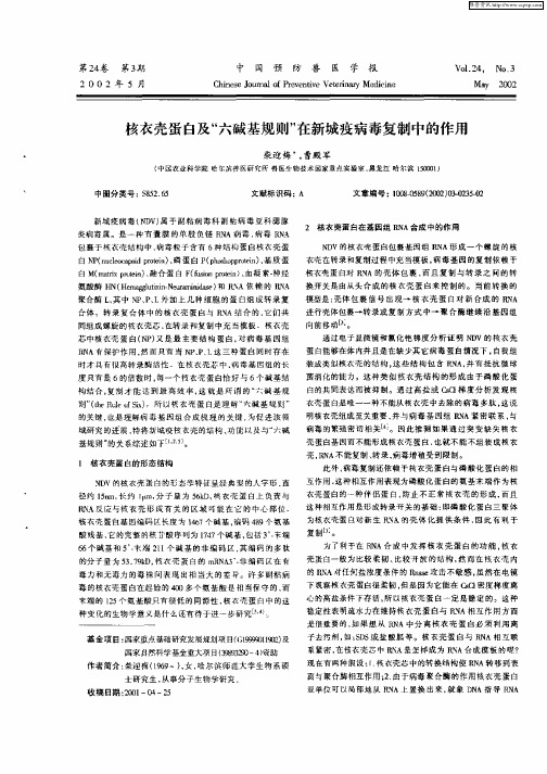
壳 , A不能复制 、 录, 硎 转 病毒增殖受 到限制。 1 核衣壳蛋 白的形态结构
N V 的核 衣 壳 蛋 白 的形 态 学 特 征 呈 经 典 型 的人 字 形 , D 直 径 约 1r 长 约 1 , 子 量 为 5k , 衣 壳 蛋 白 上 负 责 与 5m, i 山n 分 6D 棱 Ps dA反 应 与 核 衣 壳 形 成 有 关 的 区 域 可 能 在 占的 中心 部 位 : ' 核 衣 壳 蛋 白 基 因 编 码 区 长 度 为 16 47个 碱 基 . 编码 49个 氨 基 3 ,
壳 蛋 白基 因 而 不 能形 成 核衣 壳 蛋 白 , 就 不 能 不 组 装 成 核 衣 也
的关键 也是理解病 毒 基因组 合成以 理的关 键 , 为促进该领
域研 究的进展 , 特将新城疫桉衣壳的结构 、 能以致与“ 碱 功 六
基规 则 ” 的关 系综 述 如 下 NL ,5。 J
则 ” t u f 。 所 核 衣 壳 蛋 白是 理 解 “ 碱 基 规 则 ” ( eR l o s ) h e 走
菌消化的能力 。这种 类似核 衣壳 结构 的形 成 由于磷 酸化 蛋 白的共同表达而被抑 制。通过高盐 或 a 梯 度分析发 现 核 衣壳蛋 白是唯一一种不能从核衣壳 中去脒的病毒多 肽 . 这说 明核衣壳 组成 至关重要 , 并与病毒 基因组 R A紧密联 系 与 N 病毒 的繁殖密切 相关¨ 。因此推 测 如果通过 突变缺 失核 衣
装成 类似 核 衣 壳 的结 构 , 些 结 构 包 含 R A. 有 抵 抗 徽 球 这 N . 并
硎 A有保护作用 , 而只有 当 N 、 、 然 P P L这三种蛋 白同时存在 时才具 有很 高转 录酶活性 在榜衣壳芯 中 , 病毒基 固组的长 度 只有 是 6的倍 数时 , 每一个桉衣壳 蛋白惜好与 6个碱基结 构结台 , 复制才 能达 到最 高 效率 , 就是 所谓 的 “ 这 六碱 基规
鹅源新城疫病毒部分生物学特性鉴定及其囊膜糖蛋白基因序列分析

病毒的变异: 基因突变、重 组、插入、缺 失等
ቤተ መጻሕፍቲ ባይዱ
病毒的进化趋 势:适应环境 变化、增强毒 力、提高传播 能力等
流行病学特征: 病毒的传播途 径、感染率、 死亡率等
防控措施:疫 苗研发、药物 治疗、隔离防 护等
0
0
0
0
1
2
3
4
疫苗研制:针对鹅源新城疫病毒,研制有效的疫苗
病毒滴度:通过 鸡胚或细胞培养 测定病毒滴度
病毒稳定性:病 毒在不同温度、 pH值和保存时间 下的稳定性
鹅源新城疫病毒是一种高 度传染性的病毒,对鹅和 其他禽类具有很高的致病
性。
病毒在感染鹅和其他禽类 后,会引起免疫系统的强 烈反应,导致炎症和组织
损伤,进而引发死亡。
病毒可以通过呼吸道、消 化道和接触等多种途径传 播,导致鹅和其他禽类出 现呼吸道症状、腹泻、神
免疫程序制定:根据鹅的生理特点和病毒特性,制定合理的免疫程序
疫苗效果评估:通过实验,评估疫苗的保护效果和免疫持久性 疫苗推广应用:将研制成功的疫苗和免疫程序推广应用到实际生产中,提高鹅的健康水平和生 产性能
抗病毒药物的作用机制
抗病毒药物的筛选方法
抗病毒药物的研究进展
抗病毒药物的应用和效果 评估
加强养殖场的 管理,提高养 殖场的生物安 全水平
抗原表位的应用 :疫苗研发、免 疫诊断等
抗原变异:鹅源新城疫病毒抗原变异的机制和特点 变异类型:抗原变异的类型和影响因素 变异频率:鹅源新城疫病毒抗原变异的频率和规律 变异后果:抗原变异对鹅源新城疫病毒免疫反应的影响和后果
目的:评估鹅源新 城疫病毒疫苗的免 疫保护效果
试验设计:选择适 当的动物模型,如 鸡或鸭,进行免疫 接种和攻毒试验
219457073_新城疫病毒M_蛋白247_位赖氨酸对其功能的影响
Chinese Journal of Animal Infectious Diseases中国动物传染病学报收稿日期:2020-12-14基金项目:中央级科研院所基本科研业务费专项(2019JB09);上海市兽医生物技术重点实验室开放基金(shklab202001)作者简介:彭听雨,女,硕士研究生,预防兽医学专业通信作者:仇旭升,E-mail:******************.cn2023,31(2):22-27·研究论文·摘 要:新城疫病毒(NDV )基质蛋白(M )作为病毒核糖核蛋白复合体与糖蛋白之间的桥梁,协调了病毒粒子的组装过程以驱动病毒的出芽。
类似于大多数副黏病毒M 蛋白,NDV 的M 蛋白具有核质穿梭特点,其出入核依赖于自身携带的出核信号(NES )和入核信号(NLS )。
通常NLS 是一簇多数由碱性氨基酸组成的序列。
经对比发现,NDV Herts/33强毒株与La Sota 疫苗株 M 蛋白 NLS 氨基酸组成存在差异,即247 位氨基酸存在赖氨酸(K )/精氨酸(R )替换。
为了探究两种NLS 对于M 蛋白功能有何影响,本试验构建了p3XFLAG-Herts/33-WT-M 和p3XFLAG-Herts/33-K247R 重组表达真核质粒,并利用Western blot 及间接免疫荧光实验检验两种信号在M 蛋白入核、出核以及出芽方面的差异。
结果显示,R247K 突变改变了M 蛋白的核质分布,使位于胞质中的M 蛋白含量增高,从而提高了病毒样颗粒(VLP )的出芽效率。
本研究为M 蛋白在NDV 出芽中的信号调节机制研究奠定了基础。
关键词:新城疫病毒;基质蛋白;点突变;病毒样颗粒中图分类号:S852.65文献标志码:A文章编号:1674-6422(2023)02-0022-06新城疫病毒M 蛋白247位赖氨酸对其功能的影响彭听雨1,杨冰欢1,谭 磊1,2,孙英杰1,宋翠萍1,刘炜玮1,廖 瑛1,丁 铲1,仇旭升1,2(1.中国农业科学院上海兽医研究所,上海200241;2.上海市兽医生物技术重点实验室,上海 200240)Abstract: The matrix protein (M) of Newcastle disease virus (NDV) acts as a bridge between viral ribonucleoprotein complex and glycoproteins, which coordinates the assembly process of virus particles to drive the virus budding. Similar to most Paramyxovirus M proteins, NDV M protein has the characteristics of nucleocytoplasmic shuttle, and its access to the nucleus depends on its own nuclear exit signal (NES) and nuclear entry signal (NLS). NLS is usually a cluster of basic amino acids. Previous studies have found that the NLS amino acid composition of M protein of NDV Herts/33 virulent strain is different from that of La Sota vaccine strain, i.e. lysine(k) /arginine(R) substitution at 247 amino acid. In order to explore the effect of two NLS on the function of M protein, the recombinant eukaryotic plasmids of p3XFLAG-Herts/33-WT-M and p3XFLAG-Herts/33-K247R were constructed, and the differences of the two signals in the aspects of M protein nucleation and budding were detected via Western blot and indirect immunofluorescence assay. The results showed that R247K mutation changed the ability of M protein to enter the nucleus, increased the content of M protein in cytoplasm, and promote the budding of virus-like particles (VLP). This study laid a foundation for the signal regulation of M protein in NDV budding.Key words: Newcastle disease virus; matrix protein; site-directed mutation; virus-like particlesEffect of 247 Lysine Substitution in Newcastle Disease Virus Matrix Protein on ItsFunctionsPENG Tingyu 1, YANG Binghuan 1, TAN Lei 1,2, SUN Yingjie 1, SONG Cuiping 1, LIU Weiwei 1,LIAO Ying 1, DING Chan 1, QIU Xusheng 1,2(1. Shanghai Veterinary Research Institute, CAAS, Shanghai 200241, China; 2. Shanghai Key Laboratory of Veterinary Biotechnology,Shanghai 200240, China)· 23 ·彭听雨等:新城疫病毒M 蛋白247位赖氨酸对其功能的影响第31卷第2期新城疫病毒(Newcastle disease virus, NDV)是世界动物卫生组织(World Organisation for Animal Health, OIE)列出的高度接触性、烈性动物传染病病原之一,造成养禽业巨大损失。
新城疫病毒通过影响细胞微丝骨架对宫颈癌HeLa细胞增殖、迁移和侵袭的抑制作用
天津医药2021年1月第49卷第1期新城疫病毒通过影响细胞微丝骨架对宫颈癌HeLa 细胞增殖、迁移和侵袭的抑制作用金燕,王静,庞淼,张晓雪,刘开扬△摘要:目的探讨新城疫病毒(NDV )在感染宫颈癌HeLa 细胞过程中,是否通过影响细胞微丝骨架对细胞增殖、迁移和侵袭具有抑制作用。
方法将宫颈癌HeLa 细胞分为对照组、新城疫病毒感染组(NDV F3组)和细胞松弛素D组(Cytochalasin D 组)。
通过光学倒置显微镜观察NDV F3感染后的细胞形态变化及微丝骨架结构变化;综合利用CCK-8、TUNEL 凋亡检测及Hoechst33258染色检测细胞的增殖和凋亡情况;划痕实验和Transwell 实验分别观察NDV F3对细胞的迁移和侵袭能力的影响;Western blot 检测NDV F3作用细胞后微丝骨架相关蛋白F-肌动蛋白(F-actin )、RhoA 和Rho 激酶(ROCK )2的表达情况。
结果NDV F3作用细胞后,细胞失去正常形态,部分细胞有融合现象,随着感染时间的增加,细胞变圆、脱落、细胞破裂,贴壁细胞剩余较少;CCK-8结果显示细胞增殖率下降;TUNEL检测和Hoechst33258染色显示细胞在NDV F3的作用下发生凋亡,并且在感染复数(MOI )为0.1时凋亡更为显著;划痕实验和Transwell 实验显示NDV F3对细胞的迁移和侵袭能力均有阻碍作用,划痕实验在24h 与48h 时,NDV F3组、Cytochalasin D 组与对照组相比迁移率均下降,在48h 时最为显著(P <0.01),Transwell 实验显示细胞侵袭数NDV F3组和Cytochalasin D 组与对照组相比数量明显减少(P <0.01);Western blot 结果显示F-actin 、RhoA 和ROCK2在作用36h 和48h 时表达量较对照组降低(P <0.01)。
结论NDV F3对HeLa 细胞的增殖、侵袭、迁移均有抑制作用,其机制可能与微丝骨架相关的RhoA/ROCK 信号通路有关。
新城疫病毒W蛋白的细胞定位差异及其影响机制
新城疫病毒W 蛋白的细胞定位差异及其影响机制新城疫病毒(NDV )严重危害全球养禽业。
该病原为有囊膜的单股负链RNA 病毒,属于副黏病毒科、Orthoavulavirus 属。
NDV 含有6个结构基因分别编码6个主要的结构蛋白NP 、P 、M 、F 、HN 和L 。
此外,P 基因转录过程中还能通过特定位点的“RNA 编辑”使读码框发生移码产生非结构蛋白V 和W 。
P 、V 和W 具有相同的N 端和不同的C 端,近年研究表明V 蛋白独特的C 端可拮抗IFN 产生、抑制细胞凋亡、促进病毒增殖,在NDV 致病过程中发挥重要作用。
而W 蛋白在NDV 感染中的作用尚不清楚。
近期发表于《Journal of Virology 》的研究论文,对NDV W 蛋白的细胞定位机制及其在病毒复制和致病过程中的作用进行了研究,并取得重要成果。
作者前期研究证实NDV 感染过程中会产生W 蛋白,不同毒株W 蛋白长度不同且细胞定位存在差异。
基因VII 型毒株SG10的W 蛋白(227aa )定位在细胞质而基因II 型毒株La Sota 的W 蛋白(179aa )定位于细胞核。
为阐明W 蛋白定位差异是否影响NDV 的毒力、复制能力和致病性,作者首先研究了W 蛋白的定位差异机制。
通过序列分析、截短或定点突变和免疫染色发现W 蛋白定位差异与其长度有关,180aa~227aa 是定位差异的关键区域,首次揭示W 蛋白211aa~224aa 存在保守的核输出信号(NES )。
对转染后不同时间W 蛋白的定位分析发现胞质定位的W 蛋白存在核质穿梭过程,缺失NES 的W 蛋白入核后滞留在细胞核内。
作者通过Co-IP 揭示W 蛋白通过核输入信号(NLS )与KPNA1、KPNA2、KP-NA6互作被转运入核,通过LMB 抑制试验证实W 蛋白以非CRM1依赖的方式被转运出核。
进一步研究发现胞质中的W 蛋白与Tom20存在明显的共定位现象,揭示W 蛋白靶向线粒体定位。
HMGB1蛋白在鸡新城疫病毒引起炎症反应中的作用机制研究
HMGB1蛋白在鸡新城疫病毒引起炎症反应中的作用机制研究新城疫是由新城疫病毒感染引起的一种高致病性的家禽病毒性疾病。
因为新城疫具有较高的感染率和致死率,且在全球范围内都可以传播,所以威胁到全球养禽业。
新城疫病毒感染禽类能激活宿主天然免疫反应,引起大量炎性因子表达,形成炎症因子风暴,而不同毒株NDV感染后诱导细胞因子表达的水平存在差异。
在被强毒NDV感染的禽类体内,其呼吸道和消化道黏膜等会产生出血等渗出性炎症病理现象。
作为一种晚期炎症因子,由感染引起、被主动释放至细胞外的高迁移率组蛋白B(High mobility group box-1,HMGB1)具有促炎因子样作用,是一种重要的炎症调控因子,关于鸡HMGB1蛋白的功能研究鲜有报道,鸡HMGB1蛋白在新城疫病毒引起炎症反应中所发挥的作用值得研究。
为了更好的控制临床上新城疫病毒感染引起的炎症反应,降低由炎症导致的感染禽类死亡提供理论指导,我们进行了以下的研究。
(1)本研究通过从CEF细胞和HeLa细胞中扩增出鸡HMGB1和人HMGB1基因序列,并将鸡HMGB1和人HMGB1蛋白序列进行比对,发现两者在A盒和B盒区域相似性较高,在羧基端有较大差异。
通过构建pEGFP-N1-chHMGB1重组表达质粒,转染至HeLa细胞后发现鸡HMGB1定位于细胞核区域。
通过半定量RT-PCR实验,对成年SFP鸡各组织脏器中HMGB1含量进行分析,发现鸡HMGB1蛋白广泛分布于鸡体的各种组织中,其中法氏囊和肠道丰度较高,而肌肉和内脏器官丰度相对较低。
(2)本研究通过构建pET-28a-chHMGB1原核表达载体,原核表达鸡HMGB1重组蛋白,并免疫小鼠,五次免疫后取小鼠全血制备多克隆抗体,再通过融合免疫小鼠脾细胞和骨髓瘤细胞,经四次亚克隆后,获得三株针对重组鸡HMGB1蛋白的单克隆抗体。
经过实验验证,所制备的多克隆抗体和三株单克隆抗体均具有ELISA、Western Blot和IFA效价。
流感病毒M1蛋白与M2蛋白相互作用以及其在病毒出芽中的作用
流感病毒M1蛋白与M2蛋白相互作用以及其在病毒出芽中的作用侯天龙;包旦奇;李泽君;刘芹防;刘焕奇【摘要】流感病毒为囊膜病毒,以出芽的方式释放病毒。
M1蛋白是流感病毒的基质蛋白,在维持病毒颗粒形态与病毒的致病性中发挥重要作用。
M2蛋白是离子通道蛋白,在病毒出芽过程中发挥重要作用。
研究显示M1蛋白与M2蛋白存在相互作用,但是这种互作在病毒出芽中的功能尚不清楚。
根据M1蛋白的结构,M1蛋白分成N端、C端和中间区域3个区域,与M2蛋白相互作用的区域也不清楚。
本文利用双分子荧光互补实验与病毒出芽实验方法研究了M1蛋白与M2蛋白互作的区域及这种互作在病毒出芽中的作用。
研究结果显示,M2蛋白通过与M1蛋白的N端1~160个氨基酸相互作用,从而帮助M1蛋白出芽;M1蛋白的1~20氨基酸与140~160氨基酸在决定M1蛋白的稳定性与功能发挥中具有重要作用,缺失这2个氨基酸序列可导致M1蛋白N端1~160个氨基酸蛋白不稳定,并不能与M2蛋白互作。
本研究揭示了3H1N1流感病毒M1蛋白与M2蛋白互作功能区域、以及他们之间相互作用在流感病毒出芽中发挥的重要作用。
%Influenza virus is an enveloped virus and released from infected cells by budding. M1 protein, a matrix protein of influenza virus, plays an important role in the pathogenicity of the virus and maintenance of the viral morphology. M2 protein, an ion channel protein, plays an important role in virus budding process. Previous studies have shown that M1 protein interacts with M2 protein but the function of this interaction in virus budding remains unclear and the interacting regions of M1 and M2 is unknown. M1 is divided into three regions, N terminal, C end and middleregion according to its structure. In the present study, BiFC( biomolecular fluorescence complementation) and VLP(virus-like particle) experiments were conducted to investigate the interaction regions of M1 and M2 and functions of such interactions in virus budding. The results showed that M2 protein interacted with M1 protein through N terminal 1-160 amino acids of M1 protein and M2 protein facilitated budding of M1 protein by interacting with this region of M1 protein. The regions consisting of 1-20 and 140-160 amino acids of M1 protein determined the stability and function of this protein. This study revealed the interacting regions between M1 protein and M2 protein and the function of this interaction in virus budding of influenza virus.【期刊名称】《中国动物传染病学报》【年(卷),期】2016(000)006【总页数】5页(P1-5)【关键词】流感病毒;M1蛋白;M2蛋白;出芽【作者】侯天龙;包旦奇;李泽君;刘芹防;刘焕奇【作者单位】青岛农业大学动物科技学院,青岛266109; 中国农业科学院上海兽医研究所,上海200241;内蒙古农业大学,呼和浩特010018;中国农业科学院上海兽医研究所,上海200241;中国农业科学院上海兽医研究所,上海200241;青岛农业大学动物科技学院,青岛266109【正文语种】中文【中图分类】S852.659.5流感病毒为正黏病毒科、流感病毒属的有囊膜病毒,基因组分为分节段的、单股负链的RNA(SsRNA)[1]。
新城疫病毒抑制肝星状细胞活化的初步研究
新城疫病毒抑制肝星状细胞活化的初步研究李亚琳渭南师范学院化学与生命科学学院陕西渭南 714099摘要:目的探讨新城疫病毒(Newcastle disease virus, NDV)在活化肝星状细胞(Hepatic stellate cell, HSC)中的复制对HSC的活化增殖和细胞生物学功能的阻碍。
方式用不同滴度的NDV溶瘤株Italien(NDV-Italien)感染经transforming growth factor-β1 (TGF-β1)刺激活化的人肝星状细胞系LX-2,MTT法检测NDV 对细胞增殖率的阻碍。
免疫荧光显微镜技术检测活化标志物α-SMA 的表达转变。
结果NDV 的感染抑制了LX-2细胞的增殖,细胞增殖率和病毒滴度呈负相关。
与未刺激组相较,NDV在TGF-β1刺激的LX-2细胞中的增殖抑制率提高,LX-2细胞的活化标志物α-SMA的表达显著性下调。
结论NDV在活化LX-2细胞中的复制具有抑制细胞增殖和HSC活化的作用。
关键词:新城疫病毒;肝星状细胞; TGF-β1A preliminary study of Newcastle disease virus repressingthe avtivation of hepatic stellate cellYalin LiCollege of Chemistry and Life Science, Weinan Teacher University, Weinan, Shaanxi, China, 714099Abstract: Aim To explore the influence of NDV replication on LX-2 cells on the cell viability and behavior of cellular biology. Methods Transforming growth factor-β1 (TGF-β1)-stimulated LX-2 cells were infected by different titres of oncolytic NDV-Italien, the proliferation rate of LX-2 cells was detected by MTT assay. Immunofluorescent technique was used to detect the expression of activation markers α-SMA in LX-2 cells. Results NDV infection in LX-2 cells repressed the cell proliferation in a dose-dependent manner. Compared with control, the growth inhibition rate of activated LX-2 cells was increased, the expression of activation markersα-SMA of LX-2 cells were down-regulated . Conclusion The replication ofNDV in activated LX-2 cells represses the cell proliferation and attenuated the activation of LX-2 cells.Keywords: Newcastle disease virus; hepatic stellate cell; TGF-β1基金项目:陕西省教育厅资助项目(新城疫病毒抑制肝星状细胞活化的体内外研究),(项目编号:009JK427);渭南师范学院基金资助项目(新城疫病毒抑制小鼠肝纤维化的机制研究),(项目编号:10YKZ054)作者单位:714000 陕西省渭南市,渭南师范学院作者简介: 李亚琳,女,博士,副教授,要紧从事肿瘤生物医治研究Email;通信地址:陕西省渭南市朝阳路西段渭南师范学院化学与生命科学学院,联系:2133096,电话:肝星状细胞(HSC)活化是肝纤维化发生的要紧缘故,而肝纤维化是肝癌前病变的重要病理时期。
水禽源新城疫病毒经鸡传代后HN蛋白变异的研究的开题报告
水禽源新城疫病毒经鸡传代后HN蛋白变异的研究的
开题报告
【摘要】
禽流感病毒是一种极具传染性和致死性的禽类疾病,对禽类养殖业造成了巨大的损失和威胁。
本研究旨在探究水禽源新城疫病毒经鸡传代后HN蛋白变异的机制及其对病毒致病性的影响,为禽流感疫苗研究及防控提供参考。
【研究背景】
禽流感病毒是一种常见的高致病性病原体,不仅造成了禽类死亡和生产力下降,还可能造成人类感染和疫情的爆发。
当前,禽流感疫苗的研发主要以血凝抑制试验为依据,但该方法存在一定的局限性和不足。
【研究方法】
本文将通过鸡传代的方式,将水禽源新城疫病毒不断传代,观察其HN蛋白的变异情况,并通过序列分析、免疫学检测等方法检测变异后病毒对细胞的侵染能力、毒力及其对疫苗的抵抗力等指标进行测试分析。
【预期结果】
本研究预期得到在鸡传代过程中,水禽源新城疫病毒HN蛋白的变异可能存在一定的规律性,并可能导致病毒的特异性变化及对宿主的侵染能力、毒力、抵抗力等生物学特性的影响。
同时本研究结果可能为禽流感疫苗研制提供一定的启示和参考。
【结论】
本研究将对水禽源新城疫病毒经鸡传代后HN蛋白变异的机制及其对病毒致病性的影响进行探究,为禽流感疫苗的开发及控制提供理论指导和实践支持,具有重要的意义。
新城疫病毒溶瘤作用原理
新城疫病毒抗肿瘤机理研究及改造新城疫病毒(Newcastle DiseaseV irus, NDV)可在肿瘤细胞内增殖, 并选择性地杀伤、裂解肿瘤细胞, 而对正常细胞却没有杀伤作用。
新城疫病毒基因组长约为15~16 kb,编码6个结构蛋白:核衣壳蛋白(NP)、磷蛋白(P)、基质蛋白(M)、融合蛋白(F)、血凝素神经氨酸酶蛋白(HN) 和大聚合酶蛋白(L)。
编码这些蛋白的基因按3′-NP-P-M-F-HN-L-5′的顺序依次排列,每个基因之间包含由非转录的基因序列构成的接合区域,这些接合区由3 部分组成:基因起始序列(GS)、基因间序列(IG)、基因终止序列(GE)。
新城疫病毒作为溶瘤病毒的科学性:细胞对NDV 病毒的敏感性与抗病毒基因RIG-I、IRF3、IRF7和IFN-β的基础表达呈负相关,这些基因在正常细胞中表达水平远高于肿瘤细胞。
此外,研究表明多数肿瘤细胞大量表达NDV 的受体——唾液酸,NDV 通过与人或鼠的肿瘤细胞表面所分布的大量唾液酸残基结合而进入细胞,因此可广泛杀伤各种癌症细胞。
应用siRNA 筛选方法在肿瘤细胞中寻找导致肿瘤细胞对病毒易感的基因,结果发现鸟苷三磷酸酶Rac1 作为致癌蛋白对NDV 的复制及肿瘤的锚定非依赖性生长至关重要。
Rac1 蛋白的表达可以使NDV 在非肿瘤细胞复制并发挥溶瘤作用。
根据报道,NDV 病毒在人类癌细胞内复制是在正常细胞中复制的10 000 倍,与其他具有内源性溶瘤能力的病毒相比,NDV没有神经嗜性(Nonneurotropic),对人类更加安全。
有临床实验报道NDV感染后,在人群中除有轻微流感症状和轻度结膜炎外,没有其他病理学反应。
同时,NDV 可有选择性地在肿瘤细胞中增殖并起到特异杀伤作用,被认为可作为临床使用的溶瘤试剂。
这些研究结果阐明了NDV的作用机制,为安全有效地治疗癌症奠定了基础。
目前,NDV抗肿瘤的研究方向主要有以下两类:HN作为生物制剂替代NDV全病毒用于抗肿瘤治疗随着对NDV抗肿瘤机制研究的不断深入, 发现NDV的包膜糖蛋白血凝素- 神经氨酸酶(Hemagglutininneuram inidase, HN)是NDV 抗肿瘤的主要功能性蛋白, NDV在抗肿瘤作用中, HN起着重要作用, 是NDV抗肿瘤作用的主要分子基础。
影响新城疫病毒抗原性和分子变异的因素分析
影响新城疫病毒抗原性和分子变异的因素分析
秦卓明
【期刊名称】《中国家禽》
【年(卷),期】2010(32)22
【摘要】1新城疫病毒的变异
选择与免疫原性密切相关的新城疫病毒(NDV)HN基因,对不同基因型(F基因型为Ⅰ~Ⅶ)的NDV参考株在HN基因ORF编码区内,进行了同义替代(Ks)和非同义替代(Ka)的比较,
【总页数】2页(P34-35)
【作者】秦卓明
【作者单位】山东农业科学院家禽研究所
【正文语种】中文
【中图分类】S852.659.5
【相关文献】
1.新城疫病毒F基因在大肠埃希菌中的表达及其抗原性分析
2.新城疫病毒HN基因分子遗传变异及其功能性研究进展
3.H_3N_2流感病毒变异株抗原性的分子学研究
4.乙肝病毒表面抗原‘a’决定簇变异对其抗原性的影响
5.影响新城疫病毒分子变异的因素分析
因版权原因,仅展示原文概要,查看原文内容请购买。
- 1、下载文档前请自行甄别文档内容的完整性,平台不提供额外的编辑、内容补充、找答案等附加服务。
- 2、"仅部分预览"的文档,不可在线预览部分如存在完整性等问题,可反馈申请退款(可完整预览的文档不适用该条件!)。
- 3、如文档侵犯您的权益,请联系客服反馈,我们会尽快为您处理(人工客服工作时间:9:00-18:30)。
Short Communication A single amino acid mutation,R42A,in the Newcastle disease virus matrix protein abrogates its nuclear localization and attenuates viral replication and pathogenicityZhiqiang Duan,1Juan Li,1Jie Zhu,1Jian Chen,1Haixu Xu,1Yuyang Wang,1Huimou Liu,1Shunlin Hu1,2,3and Xiufan Liu1,2,3CorrespondenceXiufan Liuxfliu@Received20December2013 Accepted3March20141College of Veterinary Medicine,Yangzhou University,Yangzhou225009,PR China2Jiangsu Co-innovation Center for Prevention and Control of Important Animal Infectious Diseases and Zoonoses,Yangzhou University,Yangzhou225009,PR China3Ministry of Educational Key Lab for Avian Preventive Medicine,Yangzhou University,Yangzhou 225009,PR ChinaThe Newcastle disease virus(NDV)matrix(M)protein is a highly basic and nucleocytoplasmic shuttling viral protein.Previous study has demonstrated that the N-terminal100aa of NDV M protein are somewhat acidic overall,but the remainder of the polypeptide is strongly basic.In this study,we investigated the role of the N-terminal basic residues in the subcellular localization of M protein and in the replication and pathogenicity of NDV.We found that mutation of the basic residue arginine(R)to alanine(A)at position42disrupted M’s nuclear localization.Moreover,a recombinant virus with R42A mutation in the M protein reduced viral replication in DF-1cells and attenuated the virulence and pathogenicity of the virus in chickens.This is the first report to show that a basic residue mutation in the NDV M protein abrogates its nuclear localization and attenuates viral replication and pathogenicity.Newcastle disease virus(NDV)is an important avian pathogen and causes substantial economic losses to the poultry industry worldwide(Yusoff&Tan,2001).It is classi-fied in the genus Avulavirus of the family Paramyxoviridae, which is an enveloped virus with a negative-strand RNA genome that encodes at least six viral proteins(Alexander, 2000).The envelope of NDV contains two viral surface glycoproteins,the haemagglutinin-neuraminidase(HN) and fusion(F)proteins.The HN protein is involved in attachment to host-cell receptors and release of virions,and the F protein mediates fusion of the viral envelope with the host-cell plasma membrane(Mayo,2002).In addition,the inner surface of the viral envelope is coated with the soluble matrix(M)protein(Klenk et al.,1977),which is considered as the third NDV envelope protein.Like other paramyxo-virus M proteins,the NDV M protein is demonstrated to be a nucleocytoplasmic shuttling protein(Harrison et al., 2010).In addition to functioning for the assembly and budding of viral particles in the cytoplasm and at the cell membrane later in infection(Pantua et al.,2006),the NDV M protein is observed to localize in the nucleus early in infection and becomes associated with nucleoli and remains in this structure throughout infection(Duan et al.,2013; Peeples et al.,1992).This nuclear-nucleolar localization of the M protein is thought to regulate the balance between viral replication and transcription,which is analogous to the measles virus M protein(Iwasaki et al.,2009),and also inhibit host protein synthesis similar to the vesicular stomatitis virus M protein(Rajani et al.,2012).These studies support the notion that NDV M protein is an essential multifunctional viral protein that plays important roles in the virus life cycle.The M proteins of most paramyxoviruses,including NDV, are reported to have the highly basic,hydrophobic but not membrane-spanning properties(Bellini et al.,1986; Blumberg et al.,1984;Chambers et al.,1986).This is consistent with the known peripheral attachment of the paramyxovirus M proteins to the viral membrane.The basic residues,however,are not distributed uniformly throughout the NDV M protein sequence.Analysis of the charge distribution in M protein showed that the N-terminal100aa are somewhat acidic overall,but the remainder of the polypeptide is strongly basic(Chambers et al.,1986).Recent studies have reported that a majority of cellular or viral proteins possess basic residue(arginine or lysine)-rich peptides to mediate their nuclear or nucleolar localization(Nair et al.,2003;Scott et al.,2010).MutationsOne supplementary table is available with the online version of thispaper.Journal of General Virology(2014),95,1067–1073DOI10.1099/vir.0.062992-0 062992G2014The Authors Printed in Great Britain1067of one or more basic amino acids abrogate the nuclear or nucleolar localization of these proteins.A good example is that mutation of lysine to alanine at position 258in Nipah virus M protein disrupts its nuclear localization and reduces virus replication (Wang et al.,2010).Therefore,this study was undertaken to examine the role of the basic residues at the N-terminal 100aa in the subcellular localization of NDV M protein.We have also evaluated the effect of basic residue mutation on the replication and pathogenicity of NDV.In the present study,the M gene of NDV strain Goose/China /ZJ1/2000(GenBank accession number AF431744;Huang et al.,2004)was amplified and cloned into vector pEGFP-C1(Clontech)to create pEGFP-M.The GFP-M is validated as a suitable system for studying the subcellular localization of M protein (Duan et al.,2013).As expected,the GFP-M protein in transfected BSR cells was localized mainly in the nucleus and nucleolus,with less fluorescence in the cytoplasm (Fig.1a,lower panels).In contrast,the GFP alone was found in the whole cells (Fig.1a,upper(d)Anti-GFPHNG FPG F P -M/R 42K G F P -M /R 42AG FP -M NPAnti-HNAnti-NPGFP(a)GFP-MMerge/DAPI(b)GFP-MGFP-MR5A R42AK34A K35A R45AK53AK83AR42KMerge/DAPIMerge/DAPIM wtR42AR42KMerge/DAPIAnti-Flag (c)Fig.1.Subcellular localization of NDV M protein and its mutants fused with GFP (a,b)or Flag tag (c).At 24h post-transfection,BSR cells were fixed and observed directly under fluorescence microscope,or were analysed by indirect immunofluorescent assay.DAPI was used to stain nuclei.(d)Co-immunoprecipitation assays were used to detect the interaction of M or its mutants with HN and NP proteins.(e)The matrix protein sequences of 13viruses from different genera within the family Paramyxoviridae were aligned using CLUSTAL W (version 1.83).(f)Green fluorescence in DF-1cells infected with the indicated viruses was examined at 12and 24h post-infection (p.i.)(magnification,¾100).Cells were also fixed at 8h p.i.and prepared for the examination of subcellular localization of M proteins.DAPI was used to stain nuclei (magnification,¾200).Z.Duan and others 1068Journal of General Virology 95panels).When mutation of the N-terminal basic residues arginine (R)or lysine (K)to alanine (A)in the GFP-M protein was investigated,only GFP-M with the R42A mutation exhibited the cytoplasmic localization (Fig.1b).Sequence analysis of 187complete M protein amino acid sequences obtained from GenBank revealed that theresidue of some NDV strains at position 42was K (data not shown),but R to K substitution did not alter M’s nuclear localization (Fig.1b).Meanwhile,WT M (M wt )or its mutants fused with a small Flag tag showed identical localizations (Fig.1c),demonstrating that the localization change of M mutants was not relevant to the fused tag.InGenus Matrix protein282925242428242424373628424847514542424242425554544641453936364036363649484836Avulavirus RubulavirusRespirovirusMorbillivirus HenipavirusHendra virusNipah virus Newcastle disease virus Mumps virus M Tioman virus Simian virus 41Human parainfluenza virus 1Human parainfluenza virus 3Measles virus Rinderpest virusCanine distemper virus Tupaia paramyxo virus Sendai virusUnassigned Species(e)12 h p.i.24 h p.i.rZJ1GFPGFP Anti-M Merge/DAPIrZJ1GFP rZJ1GFP-M/R42A rZJ1GFP-M/R45A rZJ1rZJ1GFP-M/R42ArZJ1GFP-M/R45A(f)Characterization of NDV M mutation R42A1069addition,co-immunoprecipitation assays revealed that M carrying either the R42A or the R42K mutation did not affect its interaction with viral HN protein and nucleo-capsid(NP)protein(Fig.1d).We next aligned the M protein sequences of13viruses from different genera within the family Paramyxoviridae.Interestingly,there are 11R and two K residues at this position(Fig.1e).These results suggested that K42could potentially compromise functions similar to the residue R42.Therefore,we confirmed that this R residue in most of paramyxovirus M proteins was conserved and might serve important roles in virus replication.To further analyse whether disruption of M protein nuclear localization affects NDV replication and pathogenicity,a cDNA clone of NDV strain ZJ1expressing GFP(pNDV/ ZJ1GFP)(Hu et al.,2007)was used to introduce individual amino acid substitution in the M protein(M/R42A or M/ R45A).The point mutagenesis was performed as previously described(Duan et al.,2014).Results of the mutant viruses rescue showed that the haemagglutinin(HA)-positive allantoic fluid was detected in the rescued virus rZJ1GFP and rZJ1GFP-M/R45A,but two extra egg passages were required for rZJ1GFP-M/R42A to be detected by HA test (data not shown),indicating that the R42A mutant virus grew slowly in chicken eggs.In addition,DF-1cells(a chicken embryo fibroblast cell line)infected with serial10-fold dilutions of the viruses were examined.As expected, cells infected with rZJ1GFP,rZJ1GFP-M/R42A or rZJ1GFP-M/R45A exhibited green fluorescence at12and24h post-infection(p.i.)in comparison with rZJ1(Fig.1f,upper panels),demonstrating the successful rescue of the viruses. Moreover,indirect immunofluorescence assay using anti-M polyclonal antibody obtained the same localizations as plasmids expressing GFP-tagged M wt or its mutants(Fig.1f, lower panels).To further determine the stability of each M gene mutant,the recovered virus was plaque purified and passaged five times in9-day-old specific pathogen free(SPF) chicken eggs.Sequence analysis of the M gene in the mutant virus after five passages showed that the introduced mutation was unaltered,and no compensatory mutations in other viral genes were observed(data not shown).We then evaluated the biological characteristics of the parental and mutant viruses.Three internationally accepted pathogenicity tests,the mean death time(MDT),the intracerebral pathogenicity index(ICPI)and the intraven-ous pathogenicity index(IVPI),were performed according to the standard procedures(Alexander,1989).The MDT result showed a significant increase(P,0.01)in the time required by the M/R42A mutant virus(115h)to kill9-day-old chicken embryos when compared with that required for rZJ1GFP(52h)and rZJ1GFP-M/R45A(58h)(Table1). The results of ICPI and IVPI values revealed that both of the values of rZJ1GFP-M/R42A were lower than that for rZJ1GFP and rZJ1GFP-M/R45A(Table1).In addition,the growth ability of the mutant virus in the embryonated eggs and cell cultures measured by50%egg infectious dose (EID50)and TCID50test also showed a significant decrease in virus titres(P,0.01)(Table1).Together,these results demonstrated that disruption of M protein nuclear localization caused by M/R42A mutation significantly attenuated the virus. To compare the in vitro growth characteristics of the mutant viruses with the parental virus,we performed multicycle growth kinetics in DF-1cells.The results analysed using the independent samples t-test showed that the virus titres of rZJ1GFP-M/R42A from12to60h p.i.were significantly lower than that of rZJ1GFP and rZJ1GFP-M/ R45A(P,0.05)(Fig.2a).And the cytopathic effect and expression of GFP in rZJ1GFP-M/R42A-infected cells were much slighter than rZJ1GFP and rZJ1GFP-M/R45A at36h p.i.(Fig.2b).Moreover,plaque assays were used to evaluate the pathogenicity of the mutant viruses as previously described(Hu et al.,2011).The plaques produced by the viruses were measured using the GNU image manipulation program version2.8()(Negovetich& Webster,2010).As shown in Fig.2c,DF-1cells infected with rZJ1GFP or rZJ1GFP-M/R45A developed large plaques with a mean size of2.78±0.40mm and2.55±0.30mm,respect-ively.While cells infected with rZJ1GFP-M/R42A developed much smaller plaques with a mean size of0.85±0.33mm. These data suggested that the R42A mutation reduced the replication and pathogenicity of NDV in avian cells.To further investigate the in vivo pathogenesis assessment in chickens,364-week-old SPF White Leghorn chickens were randomly allocated in three experimental groups, consisting of rZJ1GFP-(n512),rZJ1GFP-M/R42A-(n512) and mock-(PBS,n512)infected groups.Birds were inoculated intranasally with106.0TICD50of each virus in a volume of0.1ml.The subsequent experiments were performed as described previously(Susta et al.,2010,2013). Results showed that birds inoculated with the parental virus rZJ1GFP presented the typical clinical symp-toms,such as head twitch,hemiparesis and paralysis atTable1.Biological characteristics of the parental and mutant virusesVirus Pathogenicity Virus titreMDT(h)ICPI IVPI EID50ml”1TCID50ml”1HArZJ1GFP52 1.91 2.80109.25108.907log2rZJ1GFP-M/R42A115 1.64 2.03107.17107.55log2rZJ1GFP-M/R45A58 1.89 2.74109.14108.837log2Z.Duan and others1070Journal of General Virology95Spleen HE(e)(i)(ii)(iii)(v)(vi)(iv)(viii)(ix)(vii)(xi)(xii)(x)(xiv)(xv)(xiii)(xvii)(xviii)(xvi)P B Sr Z J 1G F Pr Z J 1G F P -M /R 42ASpleen IHC Thymus HE Thymus IHC Bursa HEBursa IHC(b)rZJ1GFPGFPMergerZJ1GFP-M/R45ArZJ1GFP-M/R42A(c)rZJ1GFP rZJ1GFP-M/R45ArZJ1GFP-M/R42A V i r u s t i t r e [l o g 10 T C I D 50 (g t i s s u e )–1](d)rZJ1GFPrZJ1GFP-M/R42A************TissusH e ar tL iv erL un gK i d ne yB ra i nT hy mu sB ur s aP an c r ea sS pl ee n6428V i r u s t i t r e (l o g 10T C I D 50 m l –1)864210rZJ1GFP(a)rZJ1GFP-M/R45ArZJ1GFP-M/R42A 0Time p.i. (h)6604836241272Characterization of NDV M mutation R42A 10715days p.i.,and were humanely euthanized.Whereas birdsinoculated with the mutant virus rZJ1GFP-M/R42A pre-sented delayed and mild clinical signs,which were characterized by the observation that three birds hadneurological signs at9days p.i.and two of them died at 10days p.i.,and there were no deaths in the subsequent days(Table S1,available in the online Supplementary Material). To evaluate the replication of rZJ1GFP-M/R42A in chickentissues at5days p.i.,viral load in visceral organs was assessed by titration[log10TCID50(g tissue)21]in DF-1cells using the method of Reed and Muench(Reed&Muench,1938).Results showed that the mutant virus rZJ1GFP-M/R42A only replicated in spleen,lung,thymus and bursa ofFabricius,and only to a small extent;by contrast,the parental virus rZJ1GFP replicated in multiple tissues and had relatively higher virus titres in the lymphoid organs (spleen,thymus and bursa of Fabricius)(Fig.2d).Moreover, the results of histopathology and immunohistochemistry detection of the lymphoid organs showed that birds inoculated with rZJ1GFP-M/R42A displayed much milder histological changes and much weaker immunostaining intensity than that for rZJ1GFP,while mock-infected birds did not present any microscopic lesions and immunoreacti-vity(Fig.2e,Table S1).Furthermore,birds inoculated with rZJ1GFP shed the highest amount of virus in oral secretions at2days p.i.(mean virus titre103.35TCID500.1ml21).But rZJ1GFP-M/R42A shed the highest amount of virus(101.75 TCID500.1ml21)in oral secretions at7days p.i.,indicating more delayed and much lower virus replication in the shedding of the mutant virus.In summary,we investigated the role of R42in thesubcellular localization of M protein and in the replication and pathogenicity of NDV.Previous study showed that NDV M protein is localized in the nucleus via a bipartite nuclear localization signal(NLS)independent of other viral proteins(Coleman&Peeples,1993;Peeples et al.,1992). Mutations of the key basic residues in NLS disrupt the nuclear localization of M protein(Coleman&Peeples, 1993),but the effect of disruption of M protein nuclear localization on NDV replication is not known.Several studies have indicated that the nuclear localization of NDV M protein is to regulate the replication and transcription of the viral genome and inhibit host cell functions,which is similar to the functions of some paramyxovirus M proteins (Iwasaki et al.,2009;Rajani et al.,2012).A recent report only showed that NDV replication is inhibited by RNA interference targeting the M gene in chicken embryo fibroblasts(Yin et al.,2010).However,little is known about the importance of the M protein’s nuclear localization in the replication and pathogenicity of NDV. In this study,we found that the basic residue R42A mutation in the N terminus of M protein could also abrogate its nuclear localization.And a recombinant virus with R42A mutation greatly reduced viral replication in DF-1cells and attenuated the virulence and pathogenicity of the virus in chickens.It is demonstrated that the virulence of NDV is mainly determined by the cleavage of the F protein and by both the stem region and the globular head of the HN protein(Nagai et al.,1976;Panda et al., 2004;Peeters et al.,1999).In recent years,an increasing number of researchers have focused on the role of F and HN proteins in NDV pathogenesis(Cornax et al.,2013; Dortmans et al.,2009;Estevez et al.,2011;Samal et al., 2011,2013).In the present study,R42A mutation in the M protein similarly attenuated NDV replication and patho-genicity,indicating that the M protein may also influence the virulence of NDV.Taken together,this is the first report to show that a basic amino acid mutation in the NDV M protein abrogates its nuclear localization and attenuates viral replication and pathogenicity,providing us with new insights into further investigating the role of M protein in the pathogenicity and pathogenesis of NDV. AcknowledgementsWe would like to thank Dr Chan Ding(Shanghai Veterinary Research Institute,China)for providing mouse anti-NDV NP monoclonal antibody(3F5).This work was supported by the National Natural Science Foundation of China(31172338),a project funded by the Excellent Doctoral Dissertation of Yangzhou University,the Earmarked Fund for Modern Agro-industry Technology Research System(nycytx-41-G07),the Priority Academic Program Development of Jiangsu Higher Education Institutions and the Chinese Special Fund for Agro-scientific Research in the Public Interest(201303033). ReferencesAlexander,D.J.(1989).Newcastle disease.In A Laboratory Manual for the Isolation and Identification of Avian Pathogens,3rd edn,pp.parison of in vitro growth characteristics and in vivo pathogenesis of mutant viruses and parental virus.(a,b) Growth kinetics curves and the cytopathic effect of the parental and mutant viruses in DF-1cells.Cells were infected with the viruses at an m.o.i.of0.1.Values represent the mean and SD of three independent tests.The cytopathic effect and expression of GFP in cells infected with the indicated viruses were observed at36h p.i.(c)Plaque assay of the parental and mutant viruses in DF-1cells.The mean plaque size of10representative plaques for each virus is indicated.(d)Viral load in visceral organs of chickens infected with the parental and mutant viruses at5days p.i.Virus titres of the collected tissues were assessed in DF-1 cells and are presented as log10TCID50(g tissue)”1.Values represent the mean and SD of three independent experiments.The statistical significance was determined using the independent-samples t-test(***P,0.001,**P,0.01and*P,0.05).(e) Decreased severity of the mutant virus-induced microscopic lesions in chickens at5days p.i.Photomicrographs illustrating haematoxylin and eosin(HE)staining(first,third and fifth columns)and immunohistochemistry(IHC;second,fourth and sixth columns)on sections of spleen,thymus and bursa of Fabricius.The histopathological changes and immunohistochemical detection of HN protein are indicated with arrows.Bars,100m m(i–iii,vii–ix,xiii–xv);50m m(iv–vi,x–xii,xvi–xviii).Z.Duan and others1072Journal of General Virology95114–120.Edited by H.G.Purchase,L.H.Arp,C.H.Domermuth& J.E.Pearson.Kennett Square,PA:American Association for Avian Pathologists,Inc.Alexander, D.J.(2000).Newcastle disease and other avian paramyxoviruses.Rev Sci Tech19,443–462.Bellini,W.J.,Englund,G.,Richardson, C. D.,Rozenblatt,S.& Lazzarini,R.A.(1986).Matrix genes of measles virus and canine distemper virus:cloning,nucleotide sequences,and deduced amino acid sequences.J Virol58,408–416.Blumberg,B.M.,Rose,K.,Simona,M.G.,Roux,L.,Giorgi,C.& Kolakofsky, D.(1984).Analysis of the Sendai virus M gene and protein.J Virol52,656–663.Chambers,P.,Millar,N.S.,Platt,S.G.&Emmerson,P.T.(1986). Nucleotide sequence of the gene encoding the matrix protein of Newcastle disease virus.Nucleic Acids Res14,9051–9061. Coleman,N. A.&Peeples,M. E.(1993).The matrix protein of Newcastle disease virus localizes to the nucleus via a bipartite nuclear localization signal.Virology195,596–607.Cornax,I.,Diel,D.G.,Rue,C.A.,Estevez,C.,Yu,Q.,Miller,P.J.& Afonso,C.L.(2013).Newcastle disease virus fusion and haemagglu-tinin-neuraminidase proteins contribute to its macrophage host range.J Gen Virol94,1189–1194.Dortmans,J.C.,Koch,G.,Rottier,P.J.&Peeters,B.P.(2009). Virulence of pigeon paramyxovirus type1does not always correlate with the cleavability of its fusion protein.J Gen Virol90,2746–2750. Duan,Z.,Li,Q.,He,L.,Zhao,G.,Chen,J.,Hu,S.&Liu,X.(2013). Application of green fluorescent protein-labeled assay for the study of subcellular localization of Newcastle disease virus matrix protein. J Virol Methods194,118–122.Duan,Z.,Hu,Z.,Zhu,J.,Xu,H.,Chen,J.,Liu,H.,Hu,S.&Liu,X. (2014).Mutations in the FPIV motif of Newcastle disease virus matrix protein attenuate virus replication and reduce virus budding.Arch Virol159.Estevez,C.,King,D.J.,Luo,M.&Yu,Q.(2011).A single amino acid substitution in the haemagglutinin-neuraminidase protein of Newcastle disease virus results in increased fusion promotion and decreased neuraminidase activities without changes in virus patho-type.J Gen Virol92,544–551.Harrison,M.S.,Sakaguchi,T.&Schmitt, A.P.(2010).Para-myxovirus assembly and budding:building particles that transmit infections.Int J Biochem Cell Biol42,1416–1429.Hu,S.-L.,Sun,Q.,Wang,Q.-Z.,Liu,Y.-L.,Wu,Y.-T.&Liu,X.-F.(2007). Rescue and preliminary application of a recombinant Newcastle disease virus expressing green fluorescent protein gene.Virol Sin22, 34–40.Hu,Z.,Hu,S.,Meng,C.,Wang,X.,Zhu,J.&Liu,X.(2011).Generation of a genotype VII Newcastle disease virus vaccine candidate with high yield in embryonated chicken eggs.Avian Dis55,391–397. Huang,Y.,Wan,H.Q.,Liu,H.Q.,Wu,Y.T.&Liu,X.F.(2004). Genomic sequence of an isolate of Newcastle disease virus isolated from an outbreak in geese:a novel six nucleotide insertion in the non-coding region of the nucleoprotein gene.Brief Report.Arch Virol149, 1445–1457.Iwasaki,M.,Takeda,M.,Shirogane,Y.,Nakatsu,Y.,Nakamura,T.& Yanagi,Y.(2009).The matrix protein of measles virus regulates viral RNA synthesis and assembly by interacting with the nucleocapsid protein.J Virol83,10374–10383.Klenk,H.D.,Nagai,Y.,Rott,R.&Nicolau,C.(1977).The struc-ture and function of paramyxovirus glycoproteins.Med Microbiol Immunol(Berl)164,35–47.Mayo,M. A.(2002).A summary of taxonomic changes recently approved by ICTV.Arch Virol147,1655–1663.Nagai,Y.,Klenk,H.D.&Rott,R.(1976).Proteolytic cleavage of the viral glycoproteins and its significance for the virulence of Newcastle disease virus.Virology72,494–508.Nair,R.,Carter,P.&Rost,B.(2003).NLSdb:database of nuclear localization signals.Nucleic Acids Res31,397–399.Negovetich,N.J.&Webster,R.G.(2010).Thermostability of subpopulations of H2N3influenza virus isolates from mallard ducks. J Virol84,9369–9376.Panda,A.,Huang,Z.,Elankumaran,S.,Rockemann,D.D.&Samal, S.K.(2004).Role of fusion protein cleavage site in the virulence of Newcastle disease virus.Microb Pathog36,1–10.Pantua,H.D.,McGinnes,L.W.,Peeples,M.E.&Morrison,T.G. (2006).Requirements for the assembly and release of Newcastle disease virus-like particles.J Virol80,11062–11073.Peeples,M.E.,Wang,C.,Gupta,K.C.&Coleman,N.(1992).Nuclear entry and nucleolar localization of the Newcastle disease virus(NDV) matrix protein occur early in infection and do not require other NDV proteins.J Virol66,3263–3269.Peeters,B.P.,de Leeuw,O.S.,Koch,G.&Gielkens,A.L.(1999). Rescue of Newcastle disease virus from cloned cDNA:evidence that cleavability of the fusion protein is a major determinant for virulence. J Virol73,5001–5009.Rajani,K.R.,Pettit Kneller,E.L.,McKenzie,M.O.,Horita,D.A., Chou,J.W.&Lyles,D.S.(2012).Complexes of vesicular stomatitis virus matrix protein with host Rae1and Nup98involved in inhibition of host transcription.PLoS Pathog8,e1002929.Reed,L.J.&Muench,H.(1938).A simple method of estimating fifty percent endpoints.Am J Hyg27,493–497.Samal,S.,Kumar,S.,Khattar,S.K.&Samal,S.K.(2011).A single amino acid change,Q114R,in the cleavage-site sequence of Newcastle disease virus fusion protein attenuates viral replication and patho-genicity.J Gen Virol92,2333–2338.Samal,S.,Khattar,S.K.,Paldurai, A.,Palaniyandi,S.,Zhu,X., Collins,P.L.&Samal,S.K.(2013).Mutations in the cytoplasmic domain of the Newcastle disease virus fusion protein confer hyperfusogenic phenotypes modulating viral replication and patho-genicity.J Virol87,10083–10093.Scott,M.S.,Boisvert,F.M.,McDowall,M.D.,Lamond,A.I.&Barton, G.J.(2010).Characterization and prediction of protein nucleolar localization sequences.Nucleic Acids Res38,7388–7399.Susta,L.,Miller,P.J.,Afonso,C.L.,Estevez,C.,Yu,Q.,Zhang,J.& Brown,C.C.(2010).Pathogenicity evaluation of different Newcastle disease virus chimeras in4-week-old chickens.Trop Anim Health Prod 42,1785–1795.Susta,L.,Cornax,I.,Diel,D.G.,Garcia,S.C.,Miller,P.J.,Liu,X.,Hu, S.,Brown,C.C.&Afonso,C.L.(2013).Expression of interferon gamma by a highly virulent strain of Newcastle disease virus decreases its pathogenicity in chickens.Microb Pathog61-62,73–83.Wang,Y.E.,Park,A.,Lake,M.,Pentecost,M.,Torres,B.,Yun,T.E., Wolf,M. C.,Holbrook,M.R.,Freiberg, A.N.&Lee, B.(2010). Ubiquitin-regulated nuclear-cytoplasmic trafficking of the Nipah virus matrix protein is important for viral budding.PLoS Pathog6, e1001186.Yin,R.,Ding,Z.,Liu,X.,Mu,L.,Cong,Y.&Stoeger,T.(2010). Inhibition of Newcastle disease virus replication by RNA interference targeting the matrix protein gene in chicken embryo fibroblasts. J Virol Methods167,107–111.Yusoff,K.&Tan,W.S.(2001).Newcastle disease virus:macro-molecules and opportunities.Avian Pathol30,439–455.Characterization of NDV M mutation R42A1073。
